A Recessive Gene for Primary Vesicoureteral Reflux
Maps to Chromosome 12p11-q13
Patricia L. Weng,*†Simone Sanna-Cherchi,* Terry Hensle,‡Ellen Shapiro,§
Alan Werzberger,储Gianluca Caridi,¶Claudia Izzi,** Anita Konka,* Adam C. Reese,* Rong Cheng,††Samuel Werzberger,* Richard N. Schlussel,‡Robert D. Burk,‡‡ Joseph H. Lee,§§Roberto Ravazzolo,储储Francesco Scolari,** Gian Marco Ghiggeri,¶ Kenneth Glassberg,‡and Ali G. Gharavi*
*Department of Medicine, Division of Nephrology, Columbia University College of Physicians and Surgeons, New York, New York;†Department of Pediatrics, Division of Pediatric Nephrology, Mount Sinai School of Medicine, New York, New York;‡Department of Urology, Columbia University College of Physicians and Surgeons, New York, New York;§Department of Urology, New York University School of Medicine, New York, New York;储Department of Pediatrics, Columbia University College of Physicians and Surgeons, New York, New York;¶Laboratory on Pathophysiology of Uremia, G. Gaslini Institute, Genoa, Italy; **Division of Nephrology, Hospital of Montichiari, Italy,††Taub Institute for Research on Alzheimer’s Disease and the Aging Brain, College of Physicians and Surgeons, Columbia University,‡‡Department of Pediatrics/Genetics, Albert Einstein College of Medicine of Yeshiva University,§§Department of Epidemiology, Columbia University Mailman School of Public Health;储储Laboratory of Molecular Genetics, G. Gaslini Institute, Genoa, Italy
ABSTRACT
Primary vesicoureteral reflux (pVUR) is one of the most common causes of pediatric kidney failure. Linkage scans suggest that pVUR is genetically heterogeneous with two loci on chromosomes 1p13 and 2q37 under autosomal dominant inheritance. Absence of pVUR in parents of affected individuals raises the possibility of a recessive contribution to pVUR. We performed a genome-wide linkage scan in 12 large families segregating pVUR, comprising 72 affected individuals. To avoid potential misspecification of the trait locus, we performed a parametric linkage analysis using both dominant and recessive models. Analysis under the dominant model yielded no signals across the entire genome. In contrast, we identified a unique linkage peak under the recessive model on chromosome 12p11-q13 (D12S1048), which we confirmed by fine mapping. This interval achieved a peak heterogeneity LOD score of 3.6 with 60% of families linked. This heterogeneity LOD score improved to 4.5 with exclusion of two high-density pedigrees that failed to link across the entire genome. The linkage signal on chromosome 12p11-q13 originated from pedigrees of varying ethnicity, suggesting that recessive inheritance of a high frequency risk allele occurs in pVUR kindreds from many different populations. In conclusion, this study identifies a major new locus for pVUR and suggests that in addition to genetic heterogeneity, recessive contributions should be considered in all pVUR genome scans.
J Am Soc Nephrol 20: 1633–1640, 2009. doi: 10.1681/ASN.2008111199
Vesicoureteral reflux (VUR; OMIM no. 193000) is the retrograde flow of urine from the bladder to the ureters and the kidneys during micturation. Uncor-rected, VUR can lead to repeated urinary tract in-fections, renal scarring and reflux nephropathy, ac-counting for up to 25% of pediatric end stage renal disease.1,2VUR is commonly seen as an isolated disor-der (primary VUR; pVUR), but it can also present in association with complex congenital abnormalities of the kidney and urinary tract or with specific syndromic disorders, such as renal-coloboma and branchio-oto-renal syndromes.3– 8
pVUR has a strong hereditary component, with monozygotic twin concordance rates of
Received November 21, 2008. Accepted February 10, 2009. Published online ahead of print. Publication date available at www.jasn.org.
P.L.W. and S.S.C. contributed equally to this paper
Correspondence: Dr. Ali G. Gharavi, Division of Nephrology, Columbia University, 1150 Street Nicholas Avenue, Russ Berrie Pavilion #302, New York, NY 10032. Phone: 212-851-5556; Fax: 212-851-5520; E-mail: [email protected]
80%.9 –12 Sibling recurrence rates of 30% to 65% have suggested segregation of a single gene or oligogenes with large effects.9,12–14 Interestingly however, the three pub-lished genome-wide linkage scans of pVUR have strongly suggested multifactorial determination.15–17 Two pVUR loci have been identified with genome-wide significance on chromosomes 1p13 and 2q37 under an autosomal dom-inant transmission with locus heterogeneity.15,16 Multiple suggestive signals have also been reported, but remarkably, these studies show little overlap.15–17 These data suggest that pVUR may be extremely heterogeneous, with muta-tions in different genes each accounting for a fraction of cases. The genes underlying pVUR loci have not yet
been identified, but two recent studies have reported segregating mutations in the ROBO2 gene in up to 5% of pVUR families.18,19
Despite evidence for genetic hetero-geneity and different subtypes of disease, genetic studies have all modeled pVUR as an autosomal dominant trait.15–17,20 Recessive inheritance has generally not been considered because the absence of affected parents can be explained by spontaneous resolution of pVUR with older age. However, many pVUR cohorts are composed of affected sibships or pedigrees compatible with autosomal re-cessive transmission, suggesting the po-tential for alternative modes of inheri-tance.9 –12,16,17,20 –22 Systematic family screening to clarify the mode of inheri-tance is not feasible for pVUR because the standard diagnostic tool, the voiding cystourethrogram (VCUG), is invasive and would expose participants to radia-tion. Formal assessment of a recessive contribution in sporadic pVUR has also been difficult because studies have been conducted in populations with low con-sanguinity rates.9 –12,16,17,20 –22 However, recent studies have identified an unex-pected recessive contribution to several complex traits such as ductus arteriosus or autism.23,24 Thus, in addition to ge-netic heterogeneity, genes with alterna-tive modes of transmission may
segre-gate among pVUR families, and
misspecification of the inheritance model may complicate mapping studies of this trait.
Several approaches can be considered to address the difficulties imposed by com-plex inheritance, variable penetrance, and genetic heterogeneity. Studying large, well characterized cohorts with newer single-nucleotide polymorphism (SNP)-based technologies can maximize in-heritance information across the genome and increase the power of linkage studies.25 In addition, in the setting of locus heterogeneity and uncertainty about the mode of transmission, analysis under a dominant and a recessive model has greater power compared with nonparametric methods and more often results in detection of the correct mode of transmission without incurring a significant pen-alty for multiple testing.26 –29 We combined these ap-proaches in this study and successfully localized a major gene for VUR, which unexpectedly demonstrates autosomal recessive transmission.
Figure 1. Pedigree structure of the 16 families studied. Asterisks (*) mark the individ-uals from whom DNA was available for the study. Patients with other urinary tract (UT) anomalies are indicated by a blackened rectangle within the symbol.
RESULTS
We identified 16 large Caucasian pedigrees from the United States (n⫽ 8) and Italy (n ⫽ 8) ascertained through an index case with pVUR documented by positive VCUG and obtained DNA from 200 individuals (Figure 1 and Supplemental Table S1). Of the United States kindreds, six were of Hasidic Jewish origin (K117, K120, K122, K138, K144, K146), and two were of Irish American origin (K118, K119). Among the 184 relatives of the probands, 56 individuals (30%) had pVUR based on a positive VCUG without other renal or urologic defects, and 11
individuals (6%) had urinary tract abnormalities other than pVUR (Supplemental Table S1). The prevalence of pVUR and other urinary tract abnormalities among relatives was approx-imately 30-fold and 60-fold higher, respectively, than the re-ported prevalence in the general population.14,30These data are consistent with the known familial aggregation of pVUR and other urinary tract defects, indicating a strong genetic effect on these traits. The remaining 117 individuals were considered as phenotype unknown. Based on our primary phenotype of pVUR without other urologic abnormalities, 12 families were informative for linkage (72 affected individuals, 22 males and 50 females). If individuals with non pVUR urinary tract defects were also considered as affected, all 16 families were informa-tive (broad pVUR phenotype, 83 affected individuals, 28 males and 55 females).
Interpretation of modes of inheritance in pVUR is compli-cated as a result of incomplete penetrance of the trait as well as spontaneous resolution of disease with increasing age. Exami-nation of the pedigree structure revealed that seven families (44%) demonstrated parent-child transmission, suggestive of autosomal dominant inheritance. Absence of parent-child transmission in the remaining families was compatible either with dominant transmission with incomplete penetrance or recessive transmission of a high frequency gene. In the setting of genetic heterogeneity and uncertain mode of inheritance, parametric, LOD-based analysis under two simple models (dominant and recessive) is more powerful than allele sharing linkage methods.26 –28Therefore, our primary analysis was per-formed by computing heterogeneity LOD scores under both dominant and recessive models, using pVUR as our primary phenotype (12 informative families: K108, K110, K113, K116, K117, K118, K119, K120, K122, K138, K144, K146).
As with previous pVUR genome scans, analysis under ge-netic homogeneity did not identify any significant linkages un-der the dominant or recessive models.15–17Under genetic het-erogeneity and dominant transmission, the highest genome-wide heterogeneity LOD (HLOD) was on chromosome 8 (multipoint HLOD ⫽ 1.7, ␣ ⫽ 0.6, nonparametric linkage [NPL]⫽ 1.3, P ⫽ 0.05). However, after saturating this locus with 16 microsatellite markers, the HLOD decreased to zero. Combining microsatellite with SNP data has been shown to increase information content and improve the resolution of genome scans.25Therefore, to exclude low marker informa-tiveness as a cause of false negative results, we performed ge-nome-wide SNP genotyping (Affymetrix 10K arrays) in 95 in-dividuals in the most informative families. This analysis did not reveal novel loci across the genome, indicating that the absence of linkage under the dominant model is not due to lack of marker informativeness. Post hoc analysis of the seven ped-igrees with parent-child transmission also did not reveal any promising signals. Hence, we found no significant or sugges-tive loci across the genome under the dominant model (sup-plemental Figure S1).
In contrast, heterogeneity analysis under the recessive model identified a single promising signal on chromosome 12 Table 1. Pairwise HLOD scores for Chr12 p11-q13
Marker Mb pVUR Phenotype Broad pVUR Phenotype rs2054436 21.5 0.9 0.7 rs725124 23.7 0.7 0.4 D12S1591 24.0 2.2 1.2 TSC54265 25.1 0.5 0.8 D12S1596 25.8 1.5 1.2 rs2343866 26.2 0.2 0.0 rs1388659 27.1 0.4 0.3 TA27A06P 27.5 0.6 0.2 D12S1643 29.2 1.4 0.7 rs958478 29.3 0.1 0.0 D12S1681 30.4 0.9 0.3 D12S1584 31.6 0.7 0.1 D12S345 33.0 2.6 1.9 D12S2080 33.3 0.9 0.4 rs1352123 37.4 0.6 0.9 rs721709 39.0 1.6 1.4 D12S1048 39.3 1.8 1.5 rs2215456 40.6 1.6 0.9 D12S1589 40.7 1.6 1.5 D12S291 41.7 2.8 2.8 rs721483 42.0 0.2 0.3 TA91H06M 42.4 0.8 0.5 D12S1687 43.0 1.3 0.7 rs1377002 43.2 0.3 0.2 D12S85 45.6 0.3 0.2 rs215389 46.1 1.3 1.4 rs2254210 46.6 0.3 0.3 D12S2196 47.0 1.3 1.2 rs953673 47.4 1.4 1.1 D12S1627 48.1 1.9 1.7 rs4133070 48.4 1.6 1.8 rs1316607 49.3 0.9 1.2 D12S361 49.8 1.6 2.2 D12S1712 50.7 2.0 1.9 UT5029 50.9 1.3 1.4 rs686339 51.3 0.1 0.1 D12S398 51.5 3.8 3.1 D12S1586 52.4 1.6 1.6 D12S1707 53.3 1.0 1.2 rs2371455 55.1 0.5 0.6 D12S1644 55.8 1.0 0.4
Bolded numbers indicate HLOD scores⬎2. Broad phenotype includes individuals with pVUR or other urinary tract abnormalities.
with an HLOD of 1.4 on pairwise analysis (D12S297, ␣ ⫽ 0.64). Multipoint analysis augmented the HLOD to 2.7 across this interval (␣ ⫽ 0.65, peak at D12S1301). Importantly, this was the only peak with multipoint HLODⱖ2 across the entire genome under either the dominant or recessive model (Sup-plemental Figures S1 and S2). To confirm this finding, we per-formed high resolution mapping with 22 additional
microsat-ellite markers and combined these with the SNP data, resulting in a mean intermarker distance of 0.77 Mb across the chromosome 12p11-q13 re-gion. We observed positive pairwise HLODs across all markers within this interval, including one marker with genome-wide significance (D12S398, HLOD ⫽ 3.8,␣ ⫽ 0.82, Table 1).31 Next, multipoint analysis of linkage confirmed these results (multipoint HLOD 3.6 at marker D12S1048, ␣ ⫽ 0.6, Figure 2A). This result ex-ceeds traditional genome-wide significance thresholds.31,32Furthermore, based on 1000 sim-ulated genome-scans of the pedigrees under the assumption of no linkage, the empiric genome-wide significance thresholds for testing two mod-els under genetic heterogeneity were 2.7 and 3.3 at P⫽ 0.05 and 0.01, respectively. The HLOD of 3.6 therefore corresponds to an empiric genome-wide P value of 0.005, confirming genome-genome-wide significance of findings by empiric criteria. The LOD-1 interval spans 22.7 Mb between markers rs1388659 and D12S361 and includes 161 genes. We next performed comparison of haplotypes across the chromosome 12 linkage regions to ex-plore the possibility of a founder mutation, but found no evi-dence of shared segments among Hasidic or among Italian pedigrees (Supplemental Table S2). However, since these ped-igrees were not closely related, the shared segments may be below the resolution of our typed markers.
Alternative analyses were also performed to determine whether varying model parameters, analytic algorithm, or pheno-type assignment impacted the chromosome 12 linkage results. Examination of pedigree LOD scores revealed that both Hasidic and nonHasidic pedigrees contributed to the linkage signal on chromosome 12. In addition, multipoint analysis in the six Ha-sidic Jewish pedigrees using marker allele frequencies from eth-nicity matched controls revealed a peak HLOD of 2.6 (marker D12S291,␣ ⫽ 0.6). This further confirmed that most, but not all of the linkage signal originated from this subgroup (Table 2). Varying gene frequency (0.01 to 0.1) had negligible effects on overall linkage findings, with the best LOD scores achieved by modeling a high frequency risk allele (Table 3). Nonparametric analyses were also conducted and yielded a NPL score of 4.0 and P⫽ 1 ⫻ 10⫺4(at D12S1301), which falls just below the genome-wide significance threshold. Finally, to determine the impact of a broader phenotype assignment, we repeated the linkage analysis after including as affected all individuals with other renal and uro-Table 2. Peak multipoint LOD scores for each individual
pedigree at the chromosome 12p11-q13
Pedigree Ethnicity LOD pVUR Phenotype LOD Broad Phenotype 120 Ashkenazi Jewish 1.9 1.9 138 Ashkenazi Jewish 1.5 1.5 119 Irish-American 1.0 1.0 108 Italian 0.8 0.8 116 Italian 0.5 -1.0 146 Ashkenazi Jewish 0.5 0.5 144 Ashkenazi Jewish 0.1 0.1 110 Italian -0.5 -0.5 113 Italian -0.6 -0.6 118 Irish-American -0.7 -0.7 117 Ashkenazi Jewish -1.2 -1.2 122 Ashkenazi Jewish -1.4 -1.6 143 Italian N.I. 0.6 140 Italian N.I. 0.1 141 Italian N.I. -0.1 102 Italian N.I. -0.4
Broad phenotype includes individuals with pVUR or other urinary tract abnormalities. N.I., pedigree is not informative for pVUR phenotype.
Table 3. Peak multipoint HLOD scores at chromosome 12p11-q13 with varying disease gene frequency
Disease Gene Frequency 0.01 0.05 0.10
HLOD (␣), pVUR phenotype 3.4 (0.7) 3.6 (0.6) 3.4 (0.65) HLOD (␣), broad pVUR phenotype 3.2 (0.6) 3.4 (0.5) 3.0 (0.5)
Broad pVUR phenotype includes individuals with pVUR or other urinary tract abnormalities. The␣ value indicates the percent of families linked.
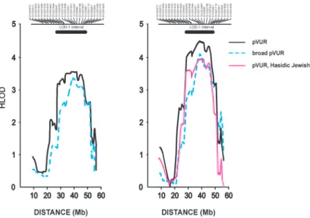
Figure 2. (A) HLOD plot of chromosome 12p11-q13 locus in the full cohort. The multipoint HLOD scores for the pVUR and broad pVUR phenotypes are shown on the y-axis. The x-axis denotes Mb distance based on the NCBI human physical map build 36.3. The location of the microsatellite markers genotyped is shown above the graph. The LOD-1 interval is indicated by the horizontal bar above the HLOD curve. (B) HLOD plot after post hoc exclusion of K117 and K122.
logic clinical disorders. This expanded the cohort to 16 pedigrees and 83 affected individuals. Despite the broader phenotype, link-age to chromosome 12p11-q13 was confirmed, with a peak HLOD score of 3.4 (␣ ⫽ 0.5, Figure 2A). This locus remained the only suggestive or significant signal across the genome under ei-ther phenotype assignment scheme, with the next best multipoint HLOD score being 1.0 on chromosome 8. Altogether, these data demonstrate that the chromosome 12 linkage results were robust to varying analytic parameters and phenotype assignment criteria. Finally, we scrutinized the genome in two large families (K117 and K122) that did not demonstrate linkage to chromo-some 12 and were each large enough to achieve genome-wide significance (LOD⬎3). Remarkably, there was no evidence for linkage across the entire genome in either kindred under both dominant and recessive inheritance, even after incorporation of genome-wide SNPs into microsatellite data. The best signals were multipoint LOD scores of 1.3 with the dominant model on chromosome 8 (D8S592) for K117 and 1.2 with the reces-sive model for K122 on chromosomes 2 (SraP) and 10 (D10S1412), which are all below the suggestive significance threshold. Given the large size of these pedigrees and the com-prehensive analyses performed across the genome, the absence of any linkage signals cannot be attributed to genetic heteroge-neity or low power. These data suggest that problems such as complex structure or bilineal inheritance likely confound the analysis of linkage in these pedigrees. Because these two pedi-grees did not map anywhere across the genome and conse-quently could only obscure linkage signals in our study, we performed post hoc analysis of the chromosome 12 locus after exclusion of these two complex kindreds (Figure 2B). This re-sulted in HLOD scores of 4.5 (␣ ⫽ 0.8) and 4.1 (␣ ⫽ 0.7) on chromosome 12p11-q13 for the pVUR and broad pVUR phe-notypes, respectively (Figure 2B). Significantly, the remaining four Hasidic Jewish pedigrees demonstrate a LOD score of 3.9 with genetic homogeneity across the same region, indicating that most of the linkage signal originates from this subgroup (Figure 2B).
DISCUSSION
In the present study, we combined multiple approaches to overcome problems such as complex inheritance, incomplete penetrance, and genetic heterogeneity to localize pVUR sus-ceptibility loci. Instead of studying sib pairs with nonparamet-ric methods, we ascertained uniquely large families and ana-lyzed the genome under both dominant and recessive transmission, because this approach avoids potential misspeci-fication of the genetic model and maximizes power in the anal-ysis of complex traits.26,29The genome scan under the domi-nant model provided no signals across the entire genome. On the other hand, analysis under a recessive model localized a major susceptibility gene to an approximately 22-Mb interval on chromosome 12p11-q13, with a peak HLOD score of 3.6 (␣ ⫽ 0.6, NPL ⫽ 4.0, P ⫽ 1 ⫻ 10⫺4). This linkage peak was the
only positive signal across the entire genome, exceeding ge-nome-wide and empiric significance thresholds, and was not significantly changed by alternative analytic models or incor-poration of a broader phenotype. These data emphasize the utility of parametric models for detection of major genes un-derlying complex traits.
The localization of a major gene with the recessive model may seem surprising because the literature has primarily fo-cused on VUR segregating as a dominant trait.15,16,19,33–35The absence of disease transmission across multiple generations has usually been attributed to incomplete penetrance or unde-tectable VUR due to spontaneous resolution in older individ-uals. However, given that many pVUR families consist of af-fected sib pairs, and considering the high degree of genetic heterogeneity of this trait, it is likely that genes with different modes of inheritance segregate among different pVUR pedi-grees. The lack of parent-offspring transmission may therefore represent true recessive inheritance in some kindreds. Low penetrance recessive alleles imparting large effects have been implicated in several other complex traits such as patent duc-tus arteriosus, Hirschsprung disease, or autism.23,24,36For these traits, gene localization was achieved by homozygosity map-ping in consanguineous populations that were enriched for recessive disorders.23,24,36,37 Although we did not set out to study consanguineous populations, our cohort included six Hasidic Jewish kindreds, which contributed a significant pro-portion (but not all) of the linkage signal on chromosome 12 (Figure 2B). This population has many characteristics of a clas-sic isolate, such as a limited set of founders, high endogamy, and recent expansion.38,39In such a population, a common predisposing allele can achieve a high frequency due to founder effects, selection, or drift, enhancing the probability of reces-sive disorders.39,40However, because several other pedigrees also contributed to the chromosome 12 linkage signal, these data suggest that recessive transmission applies to pVUR fam-ilies of varying ethnicity.
The complexity of pVUR is further demonstrated by the lack of compelling signals across the genome in two high-den-sity Hasidic Jewish kindreds (K117 and K122). These kindreds contained a total of 21 pVUR cases and were each predicted to be large enough to exceed LOD⬎3 under dominant (K117 and K122) or recessive (K117) models. Thus, the absence of linkage cannot be attributed to low power and genetic heterogeneity. Moreover, phenotyping error is unlikely, since all affecteds had VCUG-documented VUR. Such high-density pedigrees are commonly enriched for intrafamilial heterogeneity (the situa-tion where affected individuals in a family have different risk alleles in the same gene or different genetic forms of disease).41 Therefore, the most likely alternative explanation is that these pedigrees have a complex hidden genealogical structure, such that risk alleles segregate across multiple lines of descent. This is a common phenomenon in population isolates, and the presence of all affected sibships in K117 would further support this possibility. Although intrafamilial heterogeneity reduces power in linkage analysis of complex traits, it does not produce
false positive signals nor necessarily lead to false exclusion of linkage.41The effects of such confounders can be mitigated by studying large cohorts and applying systematic analytic ap-proaches, as demonstrated by the successful localization of a pVUR gene in our study.
There are 161 genes in the conservative LOD-1 interval on the chromosome 12 p11-q13 locus (National Center for Bio-technology Information [NCBI] Map Viewer). Among these, 19 genes have been implicated in human traits (OMIM). Based on a recently published study, 83 positional candidates have murine homologs and detectable expression in the murine metanephric mesenchyme and ureteric bud tip and stalk (Sup-plemental Table S3).42 These positional candidates may be pursued by systematic sequence analysis; however, it would be preferable to achieve further reduction of this locus through an interval-specific association study.43– 45Since the post hoc anal-ysis indicated a strong linkage signal with homogeneity among the Hasidic Jewish families, association mapping may be espe-cially suitable in this population because of its limited number of founders.38,39,46,47Furthermore, one would expect enrich-ment for homozygous segenrich-ments surrounding susceptibility genes among pVUR cases from this population. We did not detect regions of homozygosity within the chromosome 12 lo-cus in the Hasidic Jewish patients, but this may be due to the lack of close consanguinity among families studied. Therefore, a higher resolution analysis may be required to detect autozy-gous segments in these kindreds.
These findings have many important implications for fu-ture genetic studies of pVUR. Because the chromosome 12 signal originated from pedigrees of varying ethnicity, recessive transmission may be applicable to pVUR kindreds from many different populations. Thus, in addition to genetic heterogene-ity, variable modes of transmission should be considered in all pVUR linkage scans, and analysis under recessive transmission is henceforth warranted. It is also worth noting that sporadic disease cannot be differentiated from recessive transmission in the absence of an affected family member or consanguinity. Consequently, the recessive contribution to pVUR may have been underestimated in nonfamilial cases as well. If recessive transmission accounts for a significant fraction of sporadic pVUR, this offers a potentially powerful setting for a genome-wide association study. As demonstrated by recent studies of age-related macular degeneration, modest sized case/control cohorts may be quite successful in such situations.48Thus, as-sociation scans in sporadic pVUR may offer an additional promising approach for resolving the genetic basis of this trait.
CONCISE METHODS Patients and Phenotypes
All families were ascertained through an index case diagnosed with primary VUR by a VCUG. Index cases and family members had no evidence of secondary causes of VUR or syndromic abnormalities. In addition, we conducted extensive family history interviews and
searched medical records to identify family members diagnosed with pVUR documented by VCUG or any clinical or urologic abnormali-ties that may be pathogenically related to pVUR (e.g., other urologic anomalies or a diagnosis of ESRD in absence of any obvious cause such as glomerulonephritis and/or diabetes mellitus). This led to the identification of 56 relatives with pVUR and 11 relatives with other renal or urologic abnormalities (Supplemental Table S1). For linkage analysis, we considered all individuals with pVUR diagnosed with VCUG as affected, leaving all others as phenotype unknown. Subse-quently, we performed linkage analysis by also including the 11 addi-tional family members with clinically related abnormalities in the affected cohort. Because VUR is known to resolve with age, we classi-fied individuals that did not undergo a VCUG and those with a neg-ative VCUG as phenotype unknown in all analyses (affected only analysis). All individuals gave informed consent, and the study pro-tocol adhered to the Declaration of Helsinki and was approved by the Western Institutional Review Board for Columbia University and the ethics committees at the University of Brescia and at the Gaslini In-stitute.
Genotyping
Total genomic DNA was isolated from peripheral white blood cells of the patients and relatives using standard procedures. We performed genome-wide scans using both microsatellites and SNPs. The micro-satellite scan was performed with 393 micromicro-satellites (intermarker distance approximately 10 cM) genotyped across the genome in all 200 individuals (Marshfield Mammalian Genotyping Service). To maximize inheritance information across the genome, we also typed 95 individuals (65 affecteds) in the most complex families with 10,204 SNPs using the GeneChips Mapping 10K 2.0 Arrays (Affymetrix, Santa Clara, California). DNA processing and gene-chip hybridiza-tion were performed as suggested by the manufacturer. We fine-mapped two loci suggestive of linkage on chromosome 8 and chro-mosome 12 (with 16 and 22 polymorphic microsatellite markers, respectively). Integration of the most informative SNPs from 10K GeneChip data (minor allele frequency [MAF]ⱖ0.2) with the fine mapping microsatellites on the chromosome 12 locus yielded an av-erage marker spacing of 0.77 Mb and avav-erage information content of 0.9 (standard deviation⫽ 0.1).
Analysis of Linkage
We performed pairwise and multipoint analyses of linkage, using FASTLINK4.1,49and SimWalk2 2.90,50respectively. Since pVUR is
known to be genetically heterogeneous, we computed parametric LOD scores under a dominant and a recessive model, with disease gene frequencies of 0.01 and 0.05, respectively. We used penetrance of 75% and phenocopy of 0.01 for both models (since affected only analysis was performed, penetrance parameters did not affect LOD statistics). For comparison, we concurrently computed nonparamet-ric statistics with the SimWalk2 program (NPLpairsscore and
associ-ated exact P value). We calculassoci-ated allele frequencies on the basis of the frequencies observed in the dataset for the genome-wide microsatel-lites, whereas for the SNP data, we based frequencies on Caucasian allele frequencies provided by Affymetrix. In the fine mapping exper-iments, we obtained control allele frequencies from 40 Ashkenazi
Jew-ish individuals. We used publJew-ished thresholds for significant linkage under heterogeneity (LOD⫽ 3.3).31,32Moreover, we estimated
em-piric thresholds of significance by performing 1000 genome scans with microsatellites spaced every 10 cM across the genome under the hypothesis of no linkage, using the same structure as our pedigrees, and performing pairwise genome scans under the dominant and re-cessive models described above.
ACKNOWLEDGMENTS
We thank all of the patients and their families for participating in the study. This study was supported by National Institutes of Health 1R21 DK073903– 01. Genome-wide STR genotyping was performed by the Mammalian Genotyping Service at the Marshfield clinic (NO1-HV-48141). P.L.W. is supported by a National Kidney Foundation Clini-cal Research Fellowship grant and National Institute of Child Health and Human Development HD052890. S.S.C. is supported by the Telethon Grant GFP05012. We are grateful to J.D. Terwilliger and David Greenberg for their insightful comments. We would also like to thank Jonathan Barasch and Kai Schmidt-Ott for the mouse gene expression data. This study was presented in part at the 2008 annual meeting of the American Society of Nephrology.
The URLs for data presented herein are as follows:
NCBI Map Viewer, http://www.ncbi.nlm.nih.gov/mapview (build 36.3).
Online Mendelian Inheritance in Man (OMIM), http://www. ncbi.nlm.nih.gov/Omim (vesicoureteral reflux).
NCBI HomoloGene, http://www.ncbi.nlm.nih.gov/homologene (release 63)
DISCLOSURES None.
REFERENCES
1. Ardissino G, Dacco V, Testa S, Bonaudo R, Claris-Appiani A, Taioli E, Marra G, Edefonti A, Sereni F: Epidemiology of chronic renal failure in children: data from the ItalKid project. Pediatrics 111: e382– e387, 2003
2. Smith JM, Stablein DM, Munoz R, Hebert D, McDonald RA: Contribu-tions of the Transplant Registry: The 2006 Annual Report of the North American Pediatric Renal Trials and Collaborative Studies (NAPRTCS). Pediatr Transplant 11: 366-373, 2007
3. Abdelhak S, Kalatzis V, Heilig R, Compain S, Samson D, Vincent C, Weil D, Cruaud C, Sahly I, Leibovici M, Bitner-Glindzicz M, Francis M, Lacombe D, Vigneron J, Charachon R, Boven K, Bedbeder P, Van Regemorter N, Weissenbach J, Petit C: A human homologue of the Drosophila eyes absent gene underlies branchio-oto-renal (BOR) syn-drome and identifies a novel gene family. Nat Genet 15: 157-164, 1997
4. Hoskins BE, Cramer CH, Silvius D, Zou D, Raymond RM, Orten DJ, Kimberling WJ, Smith RJ, Weil D, Petit C, Otto EA, Xu PX, Hildebrandt F: Transcription factor SIX5 is mutated in patients with branchio-oto-renal syndrome. Am J Hum Genet 80: 800-804, 2007
5. Ruf RG, Xu PX, Silvius D, Otto EA, Beekmann F, Muerb UT, Kumar S,
Neuhaus TJ, Kemper MJ, Raymond RM, Jr, Brophy PD, Berkman J, Gattas M, Hyland V, Ruf EM, Schwartz C, Chang EH, Smith RJ, Stratakis CA, Weil D, Petit C, Hildebrandt F: SIX1 mutations cause branchio-oto-renal syndrome by disruption of EYA1-SIX1-DNA com-plexes. Proc Natl Acad Sci U S A 101: 8090-8095, 2004
6. Sanyanusin P, Schimmenti LA, McNoe LA, Ward TA, Pierpont ME, Sullivan MJ, Dobyns WB, Eccles MR: Mutation of the PAX2 gene in a family with optic nerve colobomas, renal anomalies and vesicoureteral reflux. Nat Genet 9: 358-364, 1995
7. Hinchliffe SA, Chan YF, Jones H, Chan N, Kreczy A, van Velzen D: Renal hypoplasia and postnatally acquired cortical loss in children with vesicoureteral reflux. Pediatr Nephrol 6: 439-444, 1992
8. Karnak I, Woo LL, Shah SN, Sirajuddin A, Kay R, Ross JH: Prenatally detected ureteropelvic junction obstruction: clinical features and as-sociated urologic abnormalities. Pediatr Surg Int 24: 395-402, 2008 9. Kenda RB, Fettich JJ: Vesicoureteric reflux and renal scars in
asymp-tomatic siblings of children with reflux. Arch Dis Child 67: 506-508, 1992
10. Noe HN: The long-term results of prospective sibling reflux screening. J Urol 148: 1739-1742, 1992
11. Tobenkin MI: Hereditary vesicoureteral reflux. South Med J 57: 139-147, 1964
12. Kaefer M, Curran M, Treves ST, Bauer S, Hendren WH, Peters CA, Atala A, Diamond D, Retik A: Sibling vesicoureteral reflux in multiple gestation births. Pediatrics 105: 800-804, 2000
13. Noe HN, Wyatt RJ, Peeden JN, Jr, Rivas ML: The transmission of vesicoureteral reflux from parent to child. J Urol 148: 1869-1871, 1992 14. Scott JE, Swallow V, Coulthard MG, Lambert HJ, Lee RE: Screening of newborn babies for familial ureteric reflux. Lancet 350: 396-400, 1997 15. Feather SA, Malcolm S, Woolf AS, Wright V, Blaydon D, Reid CJ, Flinter FA, Proesmans W, Devriendt K, Carter J, Warwicker P, Good-ship TH, GoodGood-ship JA: Primary, nonsyndromic vesicoureteric reflux and its nephropathy is genetically heterogeneous, with a locus on chromosome 1. Am J Hum Genet 66: 1420-1425, 2000
16. Kelly H, Molony CM, Darlow JM, Pirker ME, Yoneda A, Green AJ, Puri P, Barton DE: A genome-wide scan for genes involved in primary vesicoureteric reflux. J Med Genet 44: 710-717, 2007
17. Conte ML, Bertoli-Avella AM, de Graaf BM, Punzo F, Lama G, La Manna A, Grassia C, Rambaldi PF, Oostra BA, Perrotta S: A genome search for primary vesicoureteral reflux shows further evidence for genetic heterogeneity. Pediatr Nephrol 23: 587-595, 2008
18. Lu W, van Eerde AM, Fan X, Quintero-Rivera F, Kulkarni S, Ferguson H, Kim HG, Fan Y, Xi Q, Li QG, Sanlaville D, Andrews W, Sundaresan V, Bi W, Yan J, Giltay JC, Wijmenga C, de Jong TP, Feather SA, Woolf AS, Rao Y, Lupski JR, Eccles MR, Quade BJ, Gusella JF, Morton CC, Maas RL: Disruption of ROBO2 is associated with urinary tract anom-alies and confers risk of vesicoureteral reflux. Am J Hum Genet 80: 616-632, 2007
19. Bertoli-Avella AM, Conte ML, Punzo F, de Graaf BM, Lama G, La Manna A, Polito C, Grassia C, Nobili B, Rambaldi PF, Oostra BA, Perrotta S: ROBO2 gene variants are associated with familial vesi-coureteral reflux. J Am Soc Nephrol 19: 825-831, 2008
20. Chapman CJ, Bailey RR, Janus ED, Abbott, G.D, Lynn, K.L: Vesi-coureteric reflux: segregation analysis. Am J Med Genet 20: 577-584, 1985
21. de Vargas A, Evans K, Ransley P, Rosenberg AR, Rothwell D, Sher-wood T, Williams DI, Barratt TM, Carter CO: A family study of vesi-coureteric reflux. J Med Genet 15: 85-96, 1978
22. Fried K, Yuval E, Eidelman, A, Beer S: Familial primary vesicoureteral reflux. Clin Genet 7: 144-147, 1975
23. Mani A, Meraji SM, Houshyar R, Radhakrishnan J, Ahangar M, Rezaie TM, Taghavinejad MA, Broumand B, Zhao H, Nelson-Williams C, Lifton RP: Finding genetic contributions to sporadic disease: a reces-sive locus at 12q24 commonly contributes to patent ductus arteriosus. Proc Natl Acad Sci U S A 99: 15054-15059, 2002
Balkhy S, Gascon G, Hashmi A, Al-Saad S, Ware J, Joseph RM, Greenblatt R, Gleason D, Ertelt JA, Apse KA, Bodell A, Partlow JN, Barry B, Yao H, Markianos K, Ferland RJ, Greenberg ME, Walsh CA: Identifying autism loci and genes by tracing recent shared ancestry. Science 321: 218-223, 2008
25. Schaid DJ, Guenther JC, Christensen GB, Hebbring S, Rosenow C, Hilker CA, McDonnell SK, Cunningham JM, Slager SL, Blute ML, Thibodeau SN: Comparison of microsatellites versus single-nucleotide polymorphisms in a genome linkage screen for prostate cancer-sus-ceptibility Loci. Am J Hum Genet 75: 948-965, 2004
26. Hodge SE, Abreu PC, Greenberg DA: Magnitude of type I error when single-locus linkage analysis is maximized over models: a simulation study. Am J Hum Genet 60: 217-227, 1997
27. Greenberg DA, Hodge SE: Linkage analysis under ‘random‘ and ‘ge-netic‘ reduced penetrance. Genet Epidemiol 6: 259-264, 1989 28. Greenberg DA: Linkage analysis assuming a single-locus mode of
inheritance for traits determined by two loci: inferring mode of inher-itance and estimating penetrance. Genet Epidemiol 7: 467-479, 1990 29. Abreu PC, Greenberg DA, Hodge SE: Direct power comparisons between simple LOD scores and NPL scores for linkage analysis in complex diseases. Am J Hum Genet 65: 847-857, 1999
30. Yoshida J, Tsuchiya M, Tatsuma N, Murakami M: Mass screening for early detection of congenital kidney and urinary tract abnormalities in infancy. Pediatr Int 45: 142-149, 2003
31. Lander E, Kruglyak L: Genetic dissection of complex traits: guidelines for interpreting and reporting linkage results. Nat Genet 11: 241-247, 1995 32. Faraway JJ: Distribution of the admixture test for the detection of
linkage under heterogeneity. Genet Epidemiol 10: 75-83, 1993 33. Devriendt K, Groenen P, Van Esch H, van Dijck M, Van de Ven W,
Fryns JP, Proesmans W: Vesico-ureteral reflux: a genetic condition? Eur J Pediatr 157: 265-271, 1998
34. Lewy PR, Belman AB: Familial occurrence of nonobstructive, nonin-fectious vesicoureteral reflux with renal scarring. J Pediatr 86: 851-856, 1975
35. Sanna-Cherchi S, Reese A, Hensle T, Caridi G, Izzi C, Kim YY, Konka A, Murer L, Scolari F, Ravazzolo R, Ghiggeri GM, Gharavi AG: Familial vesicoureteral reflux: testing replication of linkage in seven new mul-tigenerational kindreds. J Am Soc Nephrol 16: 1781-1787, 2005 36. Puffenberger EG, Kauffman ER, Bolk S, Matise TC, Washington SS,
Angrist M, Weissenbach J, Garver KL, Mascari M, Ladda R, Slaugen-haupt SA, Chakravarti A: Identity-by-descent and association mapping of a recessive gene for Hirschsprung disease on human chromosome 13q22. Hum Mol Genet 3: 1217-1225, 1994
37. Lander ES, Botstein D: Homozygosity mapping: a way to map human recessive traits with the DNA of inbred children. Science 236: 1567-1570, 1987
38. Hammer MF, Redd AJ, Wood ET, Bonner MR, Jarjanazi H, Karafet T, Santachiara-Benerecetti S, Oppenheim A, Jobling MA, Jenkins T, Ostrer H, Bonne-Tamir B: Jewish and Middle Eastern non-Jewish populations share a common pool of Y-chromosome biallelic haplo-types. Proc Natl Acad Sci U S A 97: 6769-6774, 2000
39. Ostrer H: A genetic profile of contemporary Jewish populations. Nat Rev Genet 2: 891-898, 2001
40. Risch N, Tang H, Katzenstein H, Ekstein J: Geographic distribution of disease mutations in the Ashkenazi Jewish population supports ge-netic drift over selection. Am J Hum Genet 72: 812-822, 2003 41. Durner M, Greenberg DA, Hodge SE: Inter- and intrafamilial
hetero-geneity: effective sampling strategies and comparison of analysis methods. Am J Hum Genet 51: 859-870, 1992
42. Schmidt-Ott KM, Yang J, Chen X, Wang H, Paragas N, Mori K, Li JY, Lu B, Costantini F, Schiffer M, Bottinger E, Barasch J: Novel regulators of kidney development from the tips of the ureteric bud. J Am Soc Nephrol 16: 1993-2002, 2005
43. Edwards AO, Ritter R, 3rd, Abel KJ, Manning A, Panhuysen C, Farrer LA: Complement factor H polymorphism and age-related macular degeneration. Science 308: 421-424, 2005
44. Haines JL, Hauser MA, Schmidt S, Scott WK, Olson LM, Gallins P, Spencer KL, Kwan SY, Noureddine M, Gilbert JR, Schnetz-Boutaud N, Agarwal A, Postel EA, Pericak-Vance MA: Complement factor H vari-ant increases the risk of age-related macular degeneration. Science 308: 419-421, 2005
45. Grant SF, Thorleifsson G, Reynisdottir I, Benediktsson R, Manolescu A, Sainz J, Helgason A, Stefansson H, Emilsson V, Helgadottir A, Styrkars-dottir U, Magnusson KP, Walters GB, PalsStyrkars-dottir E, JonsStyrkars-dottir T, Gud-mundsdottir T, Gylfason A, SaeGud-mundsdottir J, Wilensky RL, Reilly MP, Rader DJ, Bagger Y, Christiansen C, Gudnason V, Sigurdsson G, Thorsteinsdottir U, Gulcher JR, Kong A, Stefansson K: Variant of transcription factor 7-like 2 (TCF7L2) gene confers risk of type 2 diabetes. Nat Genet 2006
46. Bronstein M, Pisante A, Yakir B, Darvasi A: Type 2 diabetes suscepti-bility loci in the Ashkenazi Jewish population. Hum Genet 124: 101-104, 2008
47. Friedrichsen DM, Stanford JL, Isaacs SD, Janer M, Chang BL, Deutsch K, Gillanders E, Kolb S, Wiley KE, Badzioch MD, Zheng SL, Walsh PC, Jarvik GP, Hood L, Trent JM, Isaacs WB, Ostrander EA, Xu J: Identi-fication of a prostate cancer susceptibility locus on chromosome 7q11-21 in Jewish families. Proc Natl Acad Sci U S A 101: 1939-1944, 2004
48. Klein RJ, Zeiss C, Chew EY, Tsai JY, Sackler RS, Haynes C, Henning AK, SanGiovanni JP, Mane SM, Mayne ST, Bracken MB, Ferris FL, Ott J, Barnstable C, Hoh J: Complement factor H polymorphism in age-related macular degeneration. Science 308: 385-389, 2005 49. Cottingham RW, Jr, Idury RM, Schaffer AA: Faster sequential genetic
linkage computations. Am J Hum Genet 53: 252-263, 1993 50. Sobel E, Lange K: Descent graphs in pedigree analysis: applications to
haplotyping, location scores, and marker-sharing statistics. Am J Hum Genet 58: 1323-1337, 1996
Supplemental information for this article is avialable online at http://www. jasn.org/.