Contents lists available atScienceDirect
Brain, Behavior, and Immunity
journal homepage:www.elsevier.com/locate/ybrbiShort Communication
Antibodies to neuronal surface proteins in Tourette Syndrome: Lack of
evidence in a European paediatric cohort
V. Baglioni
a,b, E. Coutinho
a, D.A. Menassa
a, M.P. Giannoccaro
a, L. Jacobson
a, M. Buttiglione
c,
O. Petruzzelli
c, F. Cardona
b, A. Vincent
a, the EMTICS collaborative group
aNuffield Department of Clinical Neurosciences, John Radcliffe Hospital, University of Oxford, Oxford OX3 9DU, UK bDepartment of Human Neurosciences, Sapienza University of Rome, 00185 Rome, Italy
cDepartment of Biomedical Sciences and Human Oncology, University of Bari, Bari, Italy
A B S T R A C T
In Tourette Syndrome (TS) a role for autoantibodies directed against neuronal proteins has long been suspected, but so far results are still inconsistent. The aim of this study was to look for antibodies to specific or undefined neuronal proteins that could be involved in the aetiology of the disease.
Sera from children with Tourette Syndrome or another chronic tic disorder (TS/TD), collected as part of the longitudinal European Multicenter Tics in Children Study, were investigated. Participants included 30 siblings of patients with TS/TD prior to developing tics (preclinical stage) and the same children after the first tic onset (onset), and 158 patients in the chronic phase undergoing an acute relapse (exacerbation). Presence of antibodies binding to rodent brain tissue was assessed by immunohistology on rat brain sections and by immunofluorescent staining of live hippocampal neurons. Live cell-based assays were used to screen for antibodies to NMDAR, CASPR2, LGI1, AMPAR and GABAAR.
Immunohistology indicated evidence of antibodies reactive with brain tissue, binding mainly to the hippocampus, the basal ganglia or the cerebellum in 26/218 (12%), with 8% of the preclinical or onset sera binding to the dentate gyrus/CA3 region or cerebellum. Only two individuals (one pre-clinical, one chronic) had antibodies binding the NMDAR and the binding was only weakly positive. No other specific antibodies were detected.
Despite some immunoreactivity towards neuronal antigens on brain tissue, this was not mirrored by antibodies binding to live neurons, suggesting the presence of non-specific antibodies or those that bind non-pathogenic intracellular epitopes. NMDAR or the other neuronal surface antibodies tested were very infrequent in these patients. The evidence for pathogenic antibodies that could be causative of TS is weak.
1. Introduction
Tourette Syndrome (TS) is a chronic tic disorder (TD) with an es-timated paediatric prevalence of 3–8/1000 (6–18 age range) (Leckman, 2002). Tics usually fluctuate in severity, peaking around puberty and persist into adulthood in 30–40% of cases (Martino et al., 2015). A multifactorial model was proposed for the aetiology of TDs in which genetic and environmental factors could play a synergistic role (Hoekstra et al., 2012). In particular, a dysfunctional neural-immune cross-talk in TS/TD has been proposed in line with the concept of an alteration in the immune response linked with an inflammatory status (Frick and Pittenger, 2016; Martino et al., 2015). This hypothesis is supported by findings of increased levels of the proinflammatory cy-tokines Tumor Necrosis Factor-alpha and Interleukin-2 (Leckman et al., 2005), decreased numbers of regulatory T cells, and raised intrathecal
immunoglobulin synthesis, all suggesting an ongoing inflammatory response or predisposition to autoimmunity in these patients (Wenzel et al., 2011; Kawikova et al., 2007).
The association of specific autoantibodies with TS has frequently been suggested (Yeh et al., 2012; Morris-Berry et al., 2013), mainly based on the similarity between chronic tic disorders and other post-streptococcal conditions such as Sydenham’s Chorea, Paediatric Auto-immune Neuropsychiatric Disorders Associated With Streptococcal In-fections (PANDAS), or the broader PANS (Paediatric Acute-onset Neu-ropsychiatric Syndrome), which have been linked to molecular mimicry between β-haemolytic streptococcus (GABHS) and host neural antigens (Leckman, 2002). Since dysregulation of dopamine transmission could underlie these conditions, antibodies to the dopamine D2 receptor (DRD2) have been investigated and reported to be present in some patients (Singer et al., 2015; Dale et al., 2012). However, none of the
https://doi.org/10.1016/j.bbi.2019.08.008
Received 27 June 2019; Received in revised form 12 August 2019; Accepted 14 August 2019
Abbreviations: NMDAR, N-methyl D-aspartate receptors; VGKCs, Voltage-gated potassium channels; CASPR2, contactin-associated protein-2; LGI1, leucine-rich,
glioma inactivated 1; GABA A, gamma-aminobutyric acid receptor A; AMPA, α-amino-3-hydroxy-5-methyl-4-isoxazolepropionic acid receptor; CBAs, cell-based assays; DG, dentate gyrus; CA3, Cornu Ammonis region 3; RT, room temperature; PFA, paraformaldehyde; TBS, tris-buffered saline; PBS, Phosphate-buffered saline; BSA, Bovine serum albumin; MRI, Magnetic resonance imaging; TD, Tic Disorders; TS, Tourette Syndrome; TNF-alpha, Tumor necrosis factor-alpha; IL-2, Interleukin 2
Available online 16 August 2019
0889-1591/ © 2019 Elsevier Inc. All rights reserved.
antibodies reported have been found in a substantial proportion of patients with TD, or shown to relate clearly to treatment-responses (Singer et al., 2015). This may be partly because some positivity has been found using antibody-detection techniques (i.e. ELISA, western blotting) that are not always the most appropriate for demonstrating antibodies to neuronal surface proteins, denaturing the natural epitopes conformation (Singer et al., 2015; Yeh et al., 2012).
There have been recent reports of neuronal-surface antibodies such as NMDAR, and the voltage-gated potassium channels (VGKC), LGI1 and CASPR2-antibodies (Poot, 2015; Sühs et al., 2015). Generally, those antibodies lead to neurological disorders including idiopathic limbic encephalitis and Morvan’s syndrome (Irani and Vincent, 2016), but they may also have a role in several neurodevelopmental conditions. Prenatal exposure to CASPR2 antibodies, in particular, has been asso-ciated with autism and other developmental disorders (Brimberg et al., 2016; Coutinho et al., 2017) and exposure during childhood with ADHD and Obsessive-compulsive disorder (OCD) (Poot, 2015). Inter-estingly, a complex chromosomal insertion/translocation with an in-terruption of the CNTNAP2 gene, encoding for CASPR2 was described in the TS members of a family (Verkerk et al., 2003). It was suggested that the involuntary movements were related to potassium channel dysfunction. Potassium channels are known to associate with CASPR2 in the nervous system.
Here we used procedures (cell based assay CBA; Immunohistochemistry, IHC; neuronal cultures) employed to discover new neuronal surface antibodies (e.g. Lancaster and Dalmau, 2012) hoping to identify evidence for a specific antibody that might be useful in the diagnosis and management of these challenging disorders.
2. Methods
2.1. Paediatric cohort
The samples were obtained as part of the EMTICS study, a long-itudinal observational European multicenter project, with 16 partici-pating clinical centers, enrolling paediatric participants aged 3–16 years (Schrag et al., 2018). A total of 218 samples from 188 participants were available for analysis, divided into three subgroups on the basis of disease stage. The samples were obtained from three subgroups: pre-clinical siblings of patients with TS/TD (Preclinic n = 30), taken before the presentation of clinical tic manifestations (mean time 1 year); samples from the same children during the acute phase when they had developed tics for the first time (Onset n = 30); and samples taken at the time of an exacerbation of tics in patients with TS/TD (Exacerbation n = 158) who were followed over the course of 20 months (Table 1). This study was designed to detect potential anti-bodies at times when they might be most evident (i.e. at Onset and Exacerbation) and to compare with the time at which they were less likely (Preclinic).
The clinical variables described were age (mean age 9 years; range 3–16 years), gender (M: F = 4:1), the presence of GABHS infections (positive swab throat in the 9% of the cohort), and tic severity using the Yale Global Tic Severity Scale (YGTSS) in the two symptomatic groups (mean Total YGTSS score 30; observed range 6–40).
2.2. Antibody screening
We looked for those antibodies that had already been described in autoimmune neurological diseases (NMDAR; CASPR2; LGI1, AMPAR, GABAAR) and aimed to search for evidence of possible unknown anti-bodies in TS/TD. The antibody testing was conducted in the Nuffield Department of Clinical Neurosciences of the Oxford University. 2.3. Immunohistology
All samples were screened for binding to unknown antigenic targets
using immunohistochemistry (IHC) staining on rat brain sections, as described (Gastaldi et al., 2017). Sprague Dawley rat brains were im-mersed for 1 h in 4% paraformaldehyde (PFA), cryoprotected in 40% sucrose and snap-frozen in dry-ice cooled isopentane. Twelve-µm sa-gittal brain sections were incubated with a solution of tris-buffered saline (TBS)-0.3% H2O2for 15 min at room temperature (RT), washed and blocked in 10% normal goat serum (NGS) for 1 h before incubation with patients’ serum (1:100 in 5% NGS) overnight at 4 °C. The next day slides were washed and incubated with secondary antibodies (biotiny-lated goat anti-human IgG antibody, Vector lab, 1:1000) for 2 h, wa-shed, and further incubated with the avidin-biotin complex (ABC, Vectastain Elite ABC Kit Standard, 1:100, 1 h). The reaction was de-veloped using brown 3,3′-diaminobenzidine (ImmPACT DAB perox-idase (HRP) Substrate, Vector lab) as per manufacturer’s instructions. Slides were dehydrated by immersion in progressive concentration of ethanol solutions followed by p-Xylene and mounted with DPX moun-tant for histology (Sigma-Aldrich®). Sections were viewed using a light Microscope (Nikon Eclipse E400) and images taken using the Aperio ScanScope (Leica Biosystem).
2.4. Primary neuronal cultures
Because of the higher possibility of finding specific antibodies in the sera during the acute clinical phases of the disorder, only the 30 Onset and 60 Exacerbation samples were tested for binding to rat dissociated hippocampal neuronal cultures. These were prepared following an es-tablished protocol (Kaech and Banker, 2006) and antibody testing was performed as described in previous reports (eg.Irani et al., 2010). After 12 days in culture, the live neurons were incubated with the sera (1:100) in neurobasal (NBS) medium. Coverslips were washed, fixed with PFA 3% in PBS (10 min, RT) and incubated with Alexa Fluor® Goat anti-human IgG H&L 488 secondary antibody (1:1000 in complete NBS-1% BSA) before washing and mounting with DAPI. Coverslips were visualized using a fluorescence microscope (Leica DM 2500™). 2.5. Cell based assays
Antibodies to specific antigens were investigated on human em-bryonic kidney (HEK) 293 T cells after transfection with the plasmid of interest, as performed in the Oxford laboratory protocol for clinical diagnoses. The specific extracellular antibodies measured in all 218 samples were NMDAR, CASPR2 and LGI1. GABAAand AMPA receptors antibodies were investigated only in those samples showing binding to brain tissue by immunohistochemistry. Live cells were incubated with subjects’ serum (1:20 or 1:100) in DMEM supplemented with HEPES Table 1
Summary of results.
Immunohistochemistry on sagittal rat brain sections (n = 218) Preclinical Group (n = 30) Tic Onset Group (n = 30) Tic Exacerbation Group (n = 158)
Cell-based assays for neuronal surface antibodies
Total positive sera (12%) 3 (10%) 5 (17%) 18 (11%) Basal ganglia/striatum (7.8%) 2 (7%) 4 (14%) 11 (7%) Hippocampus (10%) 3 (10%) 5 (17%) 14 (9%)
•
DG/CA3 (5%) 2 (7%) 3 (10%) 5 (3%) Cerebellum/ molecular layer(10.5%) 2 (7%) 4 (14%) 17 (10,7%)
Live cell-based assays
NMDAR (n = 218) (1%) 1 0 1 CASPR2 (n = 218) 0 0 0 LGI1 (n = 218) 0 0 0 AMPAR (n = 26) 0 0 0 GABAAR (n = 26) 0 0 0 Binding to hippocampal
and 1% Bovine serum albumin (BSA) for 1 h at RT. Coverslips were then washed and fixed in 4% PFA. After further washes, they were incubated with secondary antibodies (1:1000; Alexa Fluor™ 568 anti-human IgG H &L chain rises in goat; 1 h, RT) washed and mounted onto glass mi-croscope slides with DAPI. Antibody binding to the expressed antigen was observed using a fluorescence microscope (Leica DM 2500™). 2.6. Statistical analysis
Statistical analysis was performed using IBM SPSS Statistics 21 and GraphPad Prism 6.0. Relationships between evidence of im-munoreactivity and clinical features were analysed, using the chi-square test, the Fisher’s exact test and linear regression (p = 0.05), depending on number of groups and data distribution.
3. Results
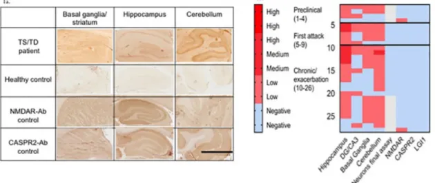
Using immunohistology on sagittal rat brain sections, 26/218 (12%) sera showed mild to moderate IgG binding (seeFig. 1a for examples). Binding was scored as moderate in 6/26 of these samples and as weak in 20/26 (Table 1). There were no differences in binding percentages between the three groups (p = 0.753). In 16 of the 26 sera, the binding was not clearly regionally focussed, with similar intensity binding to the hippocampus, basal ganglia and cerebellum. In particular, there were no samples binding more strongly to the basal ganglia than to the hippocampus and cerebellum. Ten of the 26 sera, however, bound to the dentate gyrus and CA3 subfields of the hippocampus, which have been implicated previously in TS/TD, but neither these nor any other sera bound to the live primary hippocampal neurons. Only two sera bound to the NMDAR; none bound to CASPR2, LGI1, AMPAR or GABAA R. The results are summarised inTable 1and the heatmap inFig. 1b illustrates the results of all 28 sera that showed some evidence of im-munoreactivity.
There were no relationships between these results and age (p = 0.690), gender (p = 0.631), tic severity as measured by the YGTSS (p = 0.955) or throat swab status (p = 0.343). The DG and CA3 im-munoreactivity was more frequent (8%) in Preclinical or Onset patients than in those in the Exacerbation group (3%), but this difference did not
reach significance (p = 0.1).
4. Discussion
Taking advantage of samples collected on a large longitudinal European observational study, 218 sera from 188 TS/TD paediatric participants were investigated for the possible existence of antibodies to neuronal receptors or other proteins that might be involved in the pa-thogenesis of the disease. Of particular importance, the samples ana-lysed were from different stages of the disease (preclinic, acute onset, and exacerbation during the chronic phases) designed to identify pu-tative antibodies in TS/TD. Although there was some evidence of neuronal antibody reactivity to hippocampus, basal ganglia and cere-bellum, there was no apparent association between the presence of this reactivity and the symptom stage or the reported clinical variables. Only two sera presented a weak positivity for NMDAR-antibodies and this prevalence is well in line with the general population rating (about 1–3%) (Ando et al, 2016; Steiner et al, 2013). Thus, the findings of this study did not support the frequent presence of neuronal antibodies in TS/TD children.
A potentially interesting result was the binding of a proportion of serum IgG antibodies to the dentate gyrus (DG) and CA3 subfields of the hippocampus, which have been suggested to be important in the pa-thogenesis of TS. The hypothesis is that these areas, receiving projec-tions from the prefrontal cortex, then modulate dopaminergic input to the ventral striatum (Albin, 2018; Peterson et al., 2007). Indeed, a brain Magnetic Resonance Imaging-MRI cross-sectional study reported an enlargement of the DG and CA3 subfields in children with TS, and re-duced volumes in the same hippocampal areas in adult patients (Peterson et al., 2007). However, the DG/CA3 subfield antibody binding was only found in 10/218 (5%) of patients we examined. Al-though it was a little more frequent in the 5/60 (8%) preclinical and acute onset patients compared to those with a chronic disease 5/158 (3%), this was not statistically significant.
Our results are in line with those of a recent study on a group of adult participants with TS (n = 51); an anti-nuclear pattern of munoreactivity was observed in 14% (7 out 51) of samples by im-munofluorescence (IF) on hippocampus and cerebellum sections (Sühs
Fig. 1. Examples of immunohistology on rodent brain tissue sections. 1a. Binding of antibodies by immunohistology. An example of a TS patient’s serum showing binding to basal ganglia, to the dentate gyrus DG and CA3 area of hippocampus and to the molecular layer of the cerebellum. None showed regionally specific and intense binding. IHC scalebar = 1 mm 1b. A heatmap representing results of the 28/218 (12.8%) sera that showed any neuronal immunoreactivity (on brain sections, live hippocampal cultures or cell-based assay). The results of each individual serum are represented horizontally. In each patient category (Preclinical, Onset, Exacerbation) there were sera that bound to all regions of the brain. Note that binding to the basal ganglia was less common than to other regions, binding was mainly moderate rather than strong, none of these were positive for specific antibodies but two additional patients had low titre NMDAR-Abs. Scoring scale: Blue indicates no binding detected. Pink to red colours denote the strength of the immunohistological or immunofluorescent signal from weakly positive to strongly positive. (For interpretation of the references to colour in this figure legend, the reader is referred to the web version of this article.)
et al., 2015), but no antibodies against specific neuronal surface anti-gens were detected by CBAs, or against well-defined intracellular target antigens. Thus collectively, there is a lack of evidence of antibody re-activity in either adult or paediatric TS/TD patients.
Previous studies have been inconclusive, perhaps biased by testing sera sampled at different time points of a disease which is characterised by a waxing and waning course of tic disorders (Brimberg et al., 2012), and the use of less suitable detection techniques (i.e. western blotting; ELISA) where both intracellular and extracellular epitopes are exposed and the antigens are not in their native membrane conformation (see Martino et al., 2015for a review). In addition, the clinical character-istics of the enrolled participants have not necessarily been restricted to TS, with some also presenting with other neurodevelopmental disorders (i.e. OCD/ADHD/Autism) (Cavanna, 2018).
Nevertheless, there has been support for immune dysregulation in patients with TS/TD. A study on cerebrospinal fluid (CSF) of adult patients with TS identified IgG oligoclonal bands in 38%, consistent with an autoimmune process in at least a subgroup of TS, and clearly different from the 3% found in the healthy population (Wenzel et al., 2011). The same group, using the CBAs and SY5Y neuronal stem-cells and astrocytes cultures, did not detect antibodies to any of the antigens in the CSF, even in those samples where there were oligoclonal bands. Other potential reactivity with brain tissue was not examined but these results do suggest immune activation within the cerebrospinal com-partment which could be either primary (i.e. causative) or secondary (Baumgärtel et al., 2016).
A main limitation of our study was the absence of a matched control group of healthy children for comparison with the TS sera results. Also, only a limited number of potential specific antigens were screened and there could be others involved. Nevertheless, the strengths of this study are in the large paediatric cohort, the comparison of samples from both before and at tic onset in 30 participants, and the approaches used to look for evidence of cell-surface autoantibodies.
In conclusion, specific neuronal surface antibodies were not de-tected in our cohort of children with TS/TD, either by CBAs for NMDAR and other specific antibodies. There was neuronal reactivity towards rodent brain tissue sections in a limited number of samples, but this was not supported by binding to live neurons in culture. Thus, our results fail to support the involvement of a specific antibody in the patho-genesis of childhood TD/TS.
Acknowledgements
The authors are deeply grateful to all children and their parents who willingly participated to make this research possible. The EMTICS project has received funding from the European Union’s Seventh Framework Programme for research, technological development and demonstration under Grant agreement No. 278367.
Appendix
The EMTICS group members are
Zacharias Anastasiou1, Alan Apter2, Valentina Baglioni3, Erika Bartolini4, Noa Benaroya-Milshtein2, Benjamin Bodmer5, Emese Bognar6, Bianka Burger7,8, Maura Buttiglione9, Francesco Cardona3, Marta Correa Vela10, Roberta Creti11, Andrea Dietrich12, Nanette M. Debes13, Androulla Efstratiou14, Maria Cristina Ferro15, Carolin Fremer16, Blanca Garcia-Delgar17, Maria Gariup18,19, Marianthi Georgitsi20,21, Mariangela Gulisano15, Annelieke Hagen22,23, Julie Hagstrøm24, Tammy J. Hedderly25, Isobel Heyman26, Pieter J. Hoekstra12, Chaim Huyser22,23, Monica Imperi11, Iordanis Karagiannidis20, Giovanni Laviola27, Simone Macri27, Marcos Madruga-Garrido28, Immaculada Margarit4, Anna Marotta29, Davide Martino30, Ute C. Meier31, Pablo Mir10, Natalie Moll33, Astrid Morer18,34,35, Kirsten Müller-Vahl17, Alexander Münchau35, Peter Nagy6, Valeria Neri3, Thaïra J.C. Openneer12, Graziella Orefici36, Peristera Paschou37,
Alessandra Pellico15, Onofrio Petruzzelli9, Cesare Porcelli29, Marina Redondo17, Renata Rizzo15, Paolo Roazzi39, Veit Roessner5, Daphna Ruhrman2, Jaana M.L. Schnell7, Anette Schrag40, Gregor A. Schütze32, Markus J. Schwarz32, Paola Rosaria Silvestri3, Liselotte Skov41, Tamar Steinberg2, Sara Stöber41, Marco Tallon42, Zsanett Tarnok6
1 1 Department of Statistical Science, University College London, London, UK
2 Child and Adolescent Psychiatry Department, Schneider Children’s Medical Center of Israel, affiliated to Sackler Faculty of Medicine, Tel Aviv University, Petah-Tikva, Israel
3 University La Sapienza of Rome, Department of Human Neurosciences, Rome, Italy
4 GSK, Siena, Italy
5 Department of Child and Adolescent Psychiatry, Faculty of Medicine of the TU Dresden, Dresden, Germany
6 Vadaskert Child and Adolescent Psychiatric Hospital, Budapest, Hungary
7 Department of Psychiatry and Psychotherapy, University Hospital, LMU Munich, Munich, Germany
8 Marion von Tessin Memory-Zentrum gGmbH, Munich, Germany 9 University of Bari “Aldo Moro”, Medical School, Department of
Biological Sciences and Human Oncology, Bari, Italy
10 Unidad de Trastornos del Movimiento, Servicio de Neurología y Neurofisiología Clinica, Instituto de Biomedicina de Sevilla (IBiS), Hospital Universitario Virgen del Rocio/CSIC/Universidad de Sevilla, Seville, Spain
11 Department of Infectious Diseases, Istituto Superiore di Sanità, Rome, Italy
12 University of Groningen, University Medical Center Groningen, Department of Child and Adolescent Psychiatry, Groningen, the Netherlands
13 Paediatric Department, Herlev University Hospital, Herlev, Denmark
14 WHO Global Collaborating Centre for Reference and Research on Diphtheria and Streptococcal Infections; Reference Microbiology, Directorate National Infection Service, Public Health England, London, UK
15 Child Neuropsychiatry Section, Department of Clinical and Experimental Medicine, School of Medicine, Catania University, Catania, Italy
16 Clinic of Psychiatry, Socialpsychiatry and Psychotherapy, Hannover Medical School, Hannover, Germany
17 Department of Child and Adolescent Psychiatry and Psychology, Institute of Neurosciences, Hospital Clinic Universitari, Barcelona, Spain
18 University of Barcelona, Barcelona, Spain
19 Copenhagen Psychiatric Center, Intensive Inpatient Unit, Copenhagen, Denmark
20 Department of Molecular Biology and Genetics, Democritus University of Thrace, Alexandroupoli, Greece
21 Department of Medicine, Aristotle University of Thessaloniki, Thessaloniki, Greece
22 De Bascule, Academic Center for Child and Adolescent Psychiatry, Amsterdam, the Netherlands
23 Academic Medical Center, Department of Child and Adolescent Psychiatry, Amsterdam, the Netherlands
24 Child and Adolescent Mental Health Center, Mental Health Services, Capital Region of Denmark and University of Copenhagen, Copenhagen, Denmark
25 Evelina London Children’s Hospital GSTT, Kings Health Partners AHSC, London, UK
26 Great Ormond Street Hospital for Children, and UCL Institute of Child Health, London, UK
27 Reference Centre for Behavioural Sciences and Mental Health, Istituto Superiore di Sanità, Roma, Italy
28 Sección de Neuropediatría, Instituto de Biomedicina de Sevilla (IBiS), Hospital Universitario Virgen del Rocío/CSIC/Universidad de Sevilla, Seville, Spain
29 Azienda Sanitaria Locale di Bari, Mental Health Department, Child and Adolescent Service of Bari Metropolitan Area, Bari, Italy 30 Department of Clinical Neurosciences, University of Calgary,
Calgary, Canada
31 Blizard Institute, Queen Mary University of London, London, UK 32 Institute of Laboratory Medicine, University Hospital, LMU Munich,
Munich, Germany
33 Institut d’Investigacions Biomediques August Pi i Sunyer (IDIBAPS), Barcelona, Spain
34 Centro de Investigacion en Red de Salud Mental (CIBERSAM), Instituto Carlos III, Spain
35 Institute of Neurogenetics, University of Lübeck, Lübeck, Germany 36 formerly Department of Infectious Diseases, Istituto Superiore di
Sanità, Rome, Italy
37 Department of Biological Sciences, Purdue University, West Lafayette, USA
38 Service of Child and Adolescent Psychiatry, Department of Psychiatry, University Medical Center, University of Lausanne, Lausanne, Switzerland
39 Health Technology Assessment Centre, Istituto Superiore di Sanità, Rome, Italy
40 Department of Clinical Neuroscience, UCL Institute of Neurology, University College London, London, UK
41 Concentris research management GmbH, Fürstenfeldbruck, Germany
42 IT Service, Istituto Superiore di Sanità, Rome, Italy Laboratory centres and research management: 1 Advanced Practical Diagnostics (ApDia), Belgium
Appendix A. Supplementary data
Supplementary data to this article can be found online athttps:// doi.org/10.1016/j.bbi.2019.08.008.
References
Albin, R.L., 2018. Tourette syndrome: a disorder of the social decision-making network. Brain 141 (2), 332–347.https://doi.org/10.1093/brain/awx204.
Ando, Y., Shimazaki, H., Shiota, K., et al., 2016. Prevalence of elevated serum anti-N-methyl-D-aspartate receptor antibody titers in patients presenting exclusively with psychiatric symptoms: a comparative follow-up study. BMC Psychiatry 16, 226.
https://doi.org/10.1186/s12888-016-0948-9.
Baumgärtel, C., Sühs, K.W., Müller-Vahl, K.R., 2016. Immunity in Gilles de la Tourette-Syndrome: results from a cerebrospinal fluid study. Poster Presentation in ESSTS Congress.
Brimberg, L., Benhar, I., Mascaro-Blanco, A., et al., 2012. Behavioral, pharmacological, and immunological abnormalities after streptococcal exposure: a novel rat model of Sydenham chorea and related neuropsychiatric disorders.
Neuropsychopharmacology 37, 2076–2087.
Brimberg, L., Mader, S., Jeganathan, V., et al., 2016. Caspr2-reactive antibody cloned from a mother of an ASD child mediates an ASD-like phenotype in mice. Mol. Psychiatry.https://doi.org/10.1038/mp.2016.165.
Cavanna, A.E., 2018. Gilles de la Tourette syndrome as a paradigmatic neuropsychiatric disorder. CNS Spectr. 23 (3), 213–218.https://doi.org/10.1017/
S1092852918000834.Epub 2018 May 21 PMID: 29781408.
Coutinho, E., Jacobson, L., Pedersen, M.G., et al., 2017. CASPR2 autoantibodies are raised during pregnancy in mothers of children with mental retardation and disorders of psychological development but not autism. J. Neurol. Neurosurg. Psychiatry 88 (9), 718–721.https://doi.org/10.1136/jnnp-2016-315251.
Dale, R.C., Merheb, V., Pillai, S., et al., 2012. Antibodies to surface dopamine-2 receptor in autoimmune movement and psychiatric disorders. Brain 135 (Pt 11), 3453–3468.
https://doi.org/10.1093/brain/aws256.
Frick, L., Pittenger, C., 2016. Microglial dysregulation in OCD, Tourette Syndrome, and PANDAS. J. Immunol. Res.https://doi.org/10.1155/2016/8606057.
Gastaldi, M., Waters, P., Vincent, A., 2017. Detection of NMDARs antibodies in en-cephalitis. Methods Mol. Biol. 1677, 117–126. https://doi.org/10.1007/978-1-4939-7321-7_4.
Hoekstra, P.J., Dietrich, A., Edwards, M.J., Elamin, I., Martino, D., 2012. Environmental factors in Tourette syndrome. Neurosci. Biobehav. Rev. 37, 1040–1049.
Kaech, S., Banker, G., 2006. Culturing hippocampal neurons. Nat. Protoc. 1 (5), 2406–2415.
Kawikova, I., Leckman, J.F., Kronig, H., et al., 2007. Decreased numbers of regulatory T cells suggest impaired immune tolerance in children with Tourette’s syndrome: a preliminary study. Biol. Psychiatry 61, 273–278.
Irani, S.R., Alexander, S., Waters, P., et al., 2010. Antibodies to Kv1 potassium channel-complex proteins leucine-rich, glioma inactivated 1 protein and contactin-associated protein-2 in limbic encephalitis, Morvan's syndrome and acquired neuromyotonia. Brain 133 (9), 2734–2748.https://doi.org/10.1093/brain/awq213.Epub 2010 Jul 27.
Irani, S.R., Vincent, A., 2016. Voltage-gated potassium channel-complex autoimmunity and associated clinical syndromes. Handb. Clin. Neurol. 133, 185–197.https://doi. org/10.1016/B978-0-444-63432-0.00011-6.Review. PMID: 27112678.
Lancaster, E., Dalmau, J., 2012. Neuronal autoantigens–pathogenesis, associated dis-orders and antibody testing. Nat. Rev. Neurol. 8 (7), 380–390.https://doi.org/10. 1038/nrneurol.2012.99.
Leckman, J.F., 2002. Tourette's syndrome. Lancet 360 (9345), 1577–1586.
Leckman, J.F., Katsovich, L., Kawikova, I., et al., 2005. Increased serum levels of tumour necrosis factor-alpha and IL-12 in Tourette’s syndrome. Biol. Psychiatry 57, 667–673.
Martino, D., Zisa, P., Buttiglione, M., 2015. The role of immune mechanisms in Tourette syndrome. Brain Res. 1617, 126–143.
Morris-Berry, C.M., Pollard, M., Gao, S., et al., 2013. Anti-streptococcal, tubulin, and dopamine receptor 2 antibodies in children with PANDAS and Tourette syndrome: single-point and longitudinal assessments. J. Neuroimmunol. 264, 106–113.
Peterson, B.S., Choi, H.A., Hao, X., et al., 2007. Morphologic features of the amygdala and hippocampus in children and adults with Tourette syndrome. Arch. Gen. Psychiatry 64, 1281–1291.
Poot, M., 2015. Connecting the CNTNAP2 networks with neurodevelopmental disorders. Mol. Syndromol. 6 (1), 7–22.https://doi.org/10.1159/000371594.
Schrag, A., Martino, D., EMTICS Collaborative Group, 2018. European Multicentre Tics in Children Studies (EMTICS): protocol for two cohort studies to assess risk factors for tic onset and exacerbation in children and adolescents. Eur. Child Adolesc. Psychiatry.https://doi.org/10.1007/s00787-018-1190-4.
Singer, H.S., Mascaro-Blanco, A., Alvarez, K., et al., 2015. Neuronal antibody biomarkers for Sydenham's chorea identify a new group of children with chronic recurrent epi-sodic acute exacerbations of tic and obsessive compulsive symptoms following a streptococcal infection. PLoS One 10 (3), e0120499.https://doi.org/10.1371/ journal.pone.0120499.eCollection 2015.
Steiner, J., Walter, M., Glanz, W., et al., 2013. Increased prevalence of diverse N-methyl-D-aspartate glutamate receptor antibodies in patients with an initial diagnosis of schizophrenia: specific relevance of IgG NR1a antibodies for distinction from N-me-thyl-D-aspartate glutamate receptor encephalitis. JAMA Psychiatry 70 (3), 271–278. Sühs, K.W., Skripuletz, T., Pul, R., et al., 2015. Gilles de la Tourette syndrome is not
linked to contactin-associated protein receptor 2 antibodies. Mol. Brain 8 (1), 62.
https://doi.org/10.1186/s13041-015-0154-6.
Verkerk, A.J., Mathews, C.A., Joosse, M., et al., 2003. CNTNAP2 is disrupted in a family with Gilles de la Tourette syndrome and obsessive compulsive disorder. Genomics 82, 1–9.
Wenzel, C., Wurster, U., Muller-Vahl, K.R., 2011. Oligoclonal bands in cerebrospinal fluid in patients with Tourette’s syndrome. Mov. Disord. 26, 343–346.
Yeh, C.B., Shui, H.A., Chu, T.H., et al., 2012. Hyperpolarisation-activated cyclic nucleo-tide channel 4 (HCN4) involvement in Tourette’s syndrome autoimmunity. J. Neuroimmunol. 250, 18–26.