DOTTORATO DI RICERCA
Metodologie di Ricerca nelle Malattie Vascolari
Ciclo XXII
Settore scientifico-disciplinare di afferenza: MED22 – MED05
VASCULAR WALL STEM CELLS. SELECTION
AND CONDITIONING OF PROGENITORS
USEFUL FOR CELL THERAPY. A
PATHOLOGICAL CASE STUDY
Tesi di Dottorato
Coordinatore Dottorato Presentata da:
Chiar.mo Prof. Andrea Stella Dott.ssa Sabrina Valente
Relatore
Chiar.mo Prof. Gianandrea Pasquinelli
I
TABLE OF CONTENETS
INTRODUCTION
1. VASCULAR SYSTEM 2
1.1 Blood vessels: histology and classification 3
2. STEM CELLS 8
2.1 Definition and classification 8
2.2 Embryonic Stem Cells 11
2.3 Adult Stem Cells or Somatic Stem Cells 13
2.4 Mesenchymal Stem Cells 18
3. STEM CELLS NICHE 26
3.1 Hematopoietic stem cells niche 28
3.2 Epidermal stem cells niche 30
3.3 Intestinal stem cells niche 32
3.4 Neural stem cells niche 33
3.5 Vascular niche 35
4. VASCULAR WALL RESIDENT STEM CELLS 38
5. HOMOGRAFT 43
EXPERIMENTAL DESIGN
6. AIM OF THE WORK 487. MATHERIALS AND METHODS 50
7.1 Organ culture 50
7.1.1 Organ culture procedure 50
7.1.2 Histological analysis 51
7.1.3 Vascular wall remodeling:immunohistochemical analysi 52 7.1.4 Ultrastructural analysis: TEM 54
7.2 Pathological cases: human homograft arterial 54
II
7.4 “Vasculogenic zone”: in situ immunohistochemical analysis 55
7.5 Isolation and cell culture 56
7.6 Immunophenotyping: flow cytometry analysis 57
7.7 Immunofluorescence analysis 58
7.8 Ultrastuctural analysis: transmission electron microscopy 59
7.9 Stem cell gene expression: RT-PCR 60
7.10 Proliferation assay: Alamar Blue 63
7.11 In vitro multilineage capacity differentiation 64
7.12 Angiogenic differentiation 64
7.12.1 In vitro angiogenesis assay: Matrigel 65
7.12.2 Flow cytometry assay 65
7.12.3 Immunofluorescence staining 66
7.12.4 Ultrastructural analysis: TEM 66
7.13 Adipogenic differentiation 67
6.13.1 oil Red O staining 67
7.13.1 Ultrastructural analysis: TEM 68
7.14 Osteogenic differentiation 68
7.14.1 von Kossa staining 69
7.14.2 Ultrastructural analysis: TEM 69
7.15 Chondrogenic differentiation 70
7.15.1 Alcian blue staining 70
7.15.2 Type II collagen: immunohistochemical analysis 71
7.15.3 Ultrastructural analysis: TEM 71
7.16 Leiomyogenic differentiation 72
7.16.1 Immunofluorescence analysis 72
7.16.2 Semiquantitative analysis: RT-PCR 73
7.16.3 Ultrastructural analysis: TEM 74
8. RESULTS 75
III
8.1.1 Organ culture histological analysis 75
8.1.2 Vascular wall remodeling: immunohistochemical analys 77 8.1.3 Ultrastructural analysis: TEM 80
8.2 Histological analysis of human vascular wall homograft 82
8.3 “Vasculogenic zone”: in situ immunohistochemical analysis 83 8.4 Isolation and cell culture 86
8.5 Immunophenotyping: flow cytometry analysis 87
8.6 Immunofluorescence analysis 89
8.7 Ultrastuctural analysis: transmission electron microscopy 90
8.8 Stem cell gene expression: RT-PCR 92
8.9 Proliferation assay: Alamar Blue 93
8.10 Angiogenic differentiation 95
8.10.1 In vitro angiogenesis assay: Matrigel 95
8.10.2 Flow cytometry assay 96
8.10.3 Immunofluorescence staining 97
8.10.4 Ultrastructural analysis: TEM 97
8.11 Adipogenic differentiation 98
8.11.1 oil Red O staining 98
8.11.2 Ultrastructural analysis: TEM 99
8.12 Osteogenic differentiation 100
8.12.1 von Kossa staining 101
8.12.2 Ultrastructural analysis: TEM 102
8.13 Chondrogenic differentiation 103
8.13.1 Alcian blue staining 104
8.13.2 Type II collagen: immunohistochemical analysis 104
8.13.3 Ultrastructural analysis: TEM 105
8.14 Leiomyogenic differentiation 106
8.14.1 Immunofluorescence analysi 107
IV
8.14.3 Ultrastructural analysis: TEM 108
9. DISCUSSION 110
10. REFERENCES 112
V
Ab: Alcian blue Abs: Antibodies
ALCAM :activated leucocyte adhesion molecule APC: Allophycocyanin
ASCs: Adult Stem Cells
ASMA: Alpha Smooth Muscle Actin
BCRP1: Breast Cancer Resistance Protein
BDMA: Benzyldimetylammina
BDNF: Brain-Derived Neurotrophic Factor bFGF : basic Fibroblast Growth Factor
BM-HSCs: Bone Marrow- Hematopoietic Stem Cells BMI-1: B lymphoma Mo-MLV insertion region 1
BM-MSCs: Bone Marrow derived Mesenchymal Stem Cells BMP: Bone Morphogenetic Protein
BSA: Bovine Serum Albumine CD: Cluster of differentiation
CEPs: Circulating endothelial progenitors CFU-f: Colony forming unit-fibroblasts CVCs: Calcifying vascular cells
DAB: Diaminobenzidine
DAPI: 4‟, 6-diamidino-2-phenyl indole
DMEM: Dulbecco‟s Modified Eagle‟s Medium EBs: Embryonic Bodies
ECM: Extracellulaire Matrix ECs: Endothelial Cells
EPCs: Endothelial Progenitors Cells ESCs: Embryonic Stem Cells
FACS: Fluorescence activated cell sorting FBS: Fetal Bovine Serum
VI
FGF: Fibroblast Growth Factor FITC: Fluorescein isothiocyanate flk1: Fetal liver kinase 1
GDNF: Glial cell line-Derived Neurotrophic Factor GPI: Glycophosphatidylinositol
GVD: Graft-Vs-host Disease H&E: Hematoxylin and Eosin
hESCs: human Embryonic Stem Cells HFSCs: Hair Follicle Stem Cells
HITA: Human Internal Thoracic Aortas HLA: Human Leukocyte Antigen
H-MSCs: Homograft-Mesenchymal Stem Cells HPCs: Hematopoietic Progenitor Cells
HSCs: Hematopoietic Stem Cells
HUVEC: Human Umbilical Vein Endothelial Cells hVPCs: human Vascular Progenitor Cells
IBMX: IsoButhyl-Methyl Xanthine ICM: Inner Cell Mass
IGF: Insulin-like Growth Factor IHC: Immunohistochemistry IL-8: Interleukin 8
iPSCS: induced Pluripotent Stem Cells ISCs: Intestinal Stem Cells
IVF: In Vitro Fertilization
KDR: Kinase insert domain receptor LM: Light microscopy
LV: Lateral Ventricle
moAbs: Monoclonal Antibodies MEFs: Mouse Embryonic Fibroblsts
VII
mESCs: mouse Embryonic Stem Cells MHC: Major Histocompatibility Complex MSC: Marrow Stromal Cell
MSCs: Mesenchymal stem cells NSCs: Neuronal Stem Cells o.n.: over night
OCT4: Octamer-4 P: Passage
pAbs: polyclonal Antibodies PBS: Phosphate Buffered Saline PC5: Phycoerythrin-Cyanin 5.1 PCR: Polymerase Chain Reaction
PDGF-BB: Platelet-Derived Growth Factor beta PE: PhycoErythrin
PECAM-1: Platelet Endothelial Cell Adhesion Molecule-1 RT: Reverse Transcriptase
rER: rough endoplasmic reticulum rt: room temperature
RT-PCR: Reverse Transcriptase- Polymerase Chain Reaction Sca1: Stem Cell Antigen-1
SGZ: SubGranular Zone SH2: Src Homology 2 SH3: Src Homology 3 SH4: Src homology 4 SMCs: Smooth muscle cells
SmGM2: Smooth muscle Growth Medium SOX2: SRY (sex determining region Y)-box 2 SP: Side Population
VIII
SSEA-1: Stage-Specific Emrbyonic Antigen-1 SSEA-3: Stage-Specific Embryonic Antigen-3 SSEA-4: Stage-Specific Embryonic Antigen-4
STAT 3: Signal Transducer and Activator of Transcription 3 STAT 5: Signal Transducer and Activator of Transcription 5 SVZ: SubVentricular Zone
TA: Transit Amplyfing TBS: Tris Baffered Saline
Tel: Transcription factor translocation Ets leukemia TEM: Transmission Electron Microscopy
TGF-α: Transforming Growth Factor, alpha TGF-β: Transforming Growth Factor βeta
TGF-β1: Transforming Growth Factor βeta class 1 TGF-β3: Transforming Growth Factor βeta class 3
tie2: Tyrosine kinase with immunoglobulin-like and EGF-like domains 2 UV: UltraViolet
VEGF: Vascular endothelial growth factor
VEGF-R1 or Flt1: Vascular endothelial growth factor receptor 1 or
fms-like tyrosine-kinase 1
VEGF-R2 or KDR: Vascular endothelial growth factor receptor 2 or
kinase-insert domain receptor
VWCs: Vascular Wall Cells
VW-EPCs: Vascular Wall- Endothelial Progenitor Cells vWF: von Willebrand Factor
- 2 -
1. VASCULAR SYSTEM
Vascular system is a close, double and complete system that carry the blood in every human body district; it contain the heart, the central organ and propeller of blood circulation, and blood vessels, divided into artery, vein, capillary in which the blood flow. Vascular system begins with the aorta, the main and largest artery, that through several forks give origin to minor arteries with a decreasing diameter (from 25 mm in aorta to 0,2 mm in arterioles) that reach organs and tissues. Depending on the caliber and on the quantity in the thickness of the vessel wall of elastic fibers and muscle fibrous cells we can distingue elastic and muscular arteries and arterioles and they are a transporter of oxygenated blood from the heart to all the peripheral tissues of the organism. Arterioles ramify in lower caliber arterial vessels becoming thinner near organs and forming nets know as capillary plexus that is regarded as a bridge between arteries and veins, which are involved in the exchange of substances between blood and tissues. Thanks to their small diameter (5-10 µm), their thin wall with a single layer of endothelial cells and the low hydrostatic pressure with which blood flow into, capillaries can easily exchange breathing gases, nutrients, enzymes, hormones and waste products. After the release of the necessary substances and building-up of deoxygenated blood rich of carbon dioxide the blood pass from the capillaries to very small veins (venules) that carry this blood from periphery to the heart. Those small veins merge each other forming a decreasing number of veins with an increasing caliber till they flow in the cave vein that reaches the heart. Vein‟s characteristics are the quite thin wall, they are not really elastic comparing to same caliber artery even if they have a similar structural organization. Major caliber veins contain special valves, known as swallow nest that impede the blood ebb, contributing to the regulation of blood flow in a centripetal way.
- 3 -
a b
Arteries and veins are classified mainly by their anatomic position and divided in 3 main classes: Resistance vessels (arteries and arterioles); Exchange vessels (capillaries, sinusoid and venules) and at the end capacity vessels (veins) (Stranding S 2009).
1.1 BLOOD VESSELS: HISTOLOGY and CLASSFICATION
All arterial system wall is constituted by 3 concentric laminas: Internal laminae (intimal), Medial Laminae (mesoartery) and External Laminae (adventitia). Between those laminae, as bounds, there are 2 other laminae of elastic tissue, called inner elastic and outer laminae. (Fig.1)
Fig.1: The three concentric laminas of artery wall: intima, media, and adventitial layer. a) Schematic representation; b) Histology (H&E)
- 4 -
Intimal Tunica
The Intimal tunica is the most internal layer, in close contact with the flowing blood; it is constituted by an endothelial and a sub-endothelial layer. The endothelial layer presents a single line of endothelial cells that are overlooking to the longitudinal axis of the vessel. Those cells are present in the whole vascular system and in the heart. The endothelial cell leans on a meagre layer of loose connective tissue, or sub-endothelial laminae, with mesenchymal cell interpose in it, fibroblast end rare smooth muscle cells. The intimal tunica acts as an interface with the blood and guarantee the regolation of the transport of material and blood through tissues.
Medial Tunica
The medial Tunica is the intermedial layer between the intimal and adventitia, separated by the internal and external elastic laminae. In the large is the thickest tunica, that mainly presents the vessel, and permit a taxonomic placement. Medial tunica contains in essence smooth muscle cells in a matrix rich in elastic fibers, collagen and proteoglican; those components are always present in the vessel wall, the amount of each component is different according to the area in which they are. Medial tunica smooth muscle cells are roundly arranged in the vessel lumen.
The activation of the sympathetic permit both the twitch of the vessel, reducing his diameter, (vasoconstriction) and the relax of the vessel, increasing his diameter, (vasodilatation); those actions leads to changing in both blood pressure and bloodstream through the vessel. Medial tunica separate from the adventitial tunica by an aggregate of elastic fibers called external elastic laminae. The role of the medial tunica is both to bestow elasticity to the vessel (in big caliber arteries there are abundant elastic
- 5 -
fibers and few contractive ones) and contraction (in muscular arteries there are more muscular than elastic components).
Adventitial Tunica
The adventitial tunica is placed externally to the external elastic laminae, it is a loose connective tissue, rich in collagen fibers sprawling orientated, few smooth muscle and inflammatory cells, and adipocytes. It can be considered a connective sheath, with a containing role; it glides into the surrounding connective tissue securing steadily blood vessels. In large and medium caliber vessels the adventitial tunica can be well-developed to hold in his thickness both vasa vasorum (small vessels drizzling and feeding vascular wall) and nerva nervorum (vegetative sympathetic fibers controlling mainly the smooth muscle fibers of the medial tunica).
From the heart to peripheral capillaries blood flow through a network of arteries with a decreasing diameter; by their size and structural characteristics arteries are distinguished in large caliber elastic artery, medium caliber muscular artery and small caliber artery different by contractility and elasticity. Different composition gives also different characteristic in the conduction and distribution suitable with the anatomic area in which they are. Both kind of artery present the same wall structure with the 3 layers, the difference between elastic and muscular arteries depend on the amount of elastic tissue in the medial tunica (Fig.2).
- 6 - b a
Fig.2: Differences between elastic and muscular arteries. a) Schematic representation; b) Modulation of elastic and muscular components depending on the parietal thickness
Large arteries or elastic arteries or conduction arteries: large vessels that
carry the blood from the heart to the muscular arteries. Examples of elastic arteries are pulmonary trunks and aortic arch and their principal branches (such as pulmonary artery, common carotyd, subclavian and iliac common).The vessel wall, characterized by numerous elastic fibers, is thin and presents an ample lumen with a diameter over 2,5 cm. Medial tunica is rich of elastic fibers (40-70 concentric elastic laminae) extremely extending, able to accumulate energy impressed on the haematic mass by ventricular systols and release it slowly during diastole to the blood flowing toward the periphery. The aim is convert the intermittent haematic flow deriving from the heart into a continuous (laminar) flow, necessary to the peripheral district (capillary) in which there will be the exchanges.
Medium caliber arteries or muscular arteries or distribution arteries:
- 7 -
skeletal muscles and internal organs too. Classic examples of muscular arteries are external carotid artery, bronchial arteries, femoral arteries and mesentheric arteries. Those arteries present variable diameters, from 2,5 to 7 mm, with a large lumen and a strong not elastic wall, that modify itself to become mainly a leiomuscular component, in fact they must be able to help with its contractions the blood to flow where heart force is not sufficient to let the blood going on. Medial tunica presents 40-70 layers of concentric smooth muscle cells that, wrapping in a concentric way as opposed to the vessel axis, form a kind of muff around it. There are no elastic lamellae, with the exception of the elastic layers, the internal and the external one.
Small caliber arteries or arterioles: are artery branches that arrive to the
capillary, its diameter is less than 0,5mm. Rich in muscular tissue, those small arteries are characterized by a gradual loss of elastic tissue, elastic internal and external laminae appear as fragmented, and it is difficult to discern all layers. They present also a small lumen and a thick and contractile vessel wall that regulates and control flow resistance (Martini
- 8 -
2. STEM CELLS
Stem cell research is one of the most fascinating areas of contemporary biology since the cells are "parent" of each organ, tissue and organism cell type, due to their, innate properties to renew themselves for an indefinite period, known as “immortal” because they are able to develop into specialized cells of various body tissues in early life and growth. These specific characteristics make cells stem very promising for the treatment of severely debilitating degenerative diseases and could be interesting for many applications in tissue engineering, cells therapy and drug screening. The biology of stem cells is important in understanding the ontogeny but also to maintain homeostasis by replacing damage cells in adult organism.
2.1 DEFINITION and CLASSIFICATION
Stem cells differ from other cells type in the body and depending on their origin; they can be distinguished in three important characteristics (Fig.3) (National Institute of Health 2002; Ulloa-Montoya et al., 2005):
Stem cells are capable to self-dividing and self-renew: they are able to
renew themselves through numerous cycles of cell division for long periods maintaining the undifferentiated state. During early development, stem cell division is symmetrical i.e. each cell divides itself to give rise to two unspecialized daughter cells, like the parent, each one with the same potential. In the later development, the cell divides itself asymmetrically with of the daughter cells will maintain a stem characteristic and the second will be a more differentiated cell.
- 9 -
Stem cells are unspecialized: they haven‟t acquired yet any tissue-specific
structures that allow them to perform specialized functions like the ability to become any cell type.
Stem cells are able to give rise to specialized cells: under special
conditions, unspecialized cells begin to "differentiate" into specialized cell type and to develop into specific tissues and organs. Each step of the differentiation is trigger by internal signals (cell‟s genes) and external signals such as cytokines and chemokines secreted by other cells, physical contact with adjacent cells and other molecules in the microenvironment.
Fig.3: Definition of “stem cells”
Depending on differentiation potential, stem cells are classified in: totipotent, pluripotent, multipotent and unipotent. All human beings start their lives from a single cell called a “zygote”, produced from the fusion of an egg and a sperm cell. The zygote is defined the totipotent stem cell and can differentiate into embryonic and extra-embryonic cell types constructing a complete, viable, organism. The zygote, after a series of multiple successive divisions, in about 5 days, gives rise to cave spherical agglomerate known as blastocyst. Embryonic Stem Cells, originate as inner mass cells within a blastocyst, are capable to differentiate into cells
- 10 -
from all three germinal layers: endoderm, mesoderm and ectoderm and to generate all tissues present into adult organism thank to their characteristic
pluripotency. The Adult Stem Cells or Somatic Stem Cells, typically
programmed to give rise to different cell types of the tissue of derivating, are called multipotent cell and can give rise only to certain cell types. The main role of adult stem cells in a living organism is to maintain and to repair the tissue in which they are found. The unipotent stem cells have the capacity to differentiate into only one type of cell or tissue, which is a lower potential. Despite their limited differentiation potential, unipotent cells still have therapeutic potential to treat injuries and diseases. We can assert that a stem cell with its characteristic open a new research for its possible therapeutic for degenerative diseases like Alzheimer disease, Parkinson disease, stroke, cardiopathy, diabete and cancer (Fig.4).
- 11 - 2.2 EMBRYONIC STEM CELLS
Embryonic Stem Cells (ESCs) derived from totipotent cells of the early mammalian embryo and are capable of unlimited and undifferentiated proliferation in vitro. These stem cells obtained from the epiblast tissue of the inner cell mass (ICM) of a blastocysts, of a 4-5 days old embryos, consisting of 50-150 cells (Fox news) (Fig.5).
Fig.5: Schematic representation to obtain ESCs
ESCs are the best characterized of all the stem cells; they are pluripotent thank of the ability to differentiate into cells deriving from the three main germ lines: ectoderm (neurons, skin, etc), mesoderm (muscle and bone) and endoderm (Hepatocytes, pancreatic beta cells) and not contributing to the extra-embryonic membranes (Fig.6). We can assert that sufficiently and necessarily stimulate for a specific cell type, they can develop into almost all cell types of the adult body. ESCs are immortal, have been maintained in culture for several doublings, and are able to maintain a normal karyotype (Amit et al., 2003).
- 12 -
Fig.6: Pluripotency of ESCs
The mouse embryos ESCs have been isolated and cultured since the 1980 by various groups of researchers (Evans et al., 1981, Axelrod 1984, Wobus
et al, 1984, Doetschman et al., 1985). These pioneers established that
murine embryonic stem cells line could differentiate into several different cell types (Thompson et al., 1995, 1998) and when injected into a pre-implantation embryo, can produce functional differentiated progeny in all tissue and organ (Smith et al., 2001).
In 1995 and 1998, ESCs were isolated from primate and human (Amit et
al., 2003). human Embryonic Stem Cells (hESCs) should be able to give
the same results of mouse Embryonic Stem Cells (mESCs), but for ethical reasons it cannot be demonstrated. ESCs, as their name suggests, derive from embryos produced with in vitro fertilization (IVF) for clinical purpose and then donated for research purposes with informed consent of the donors. ESCs were defined by the presence of several transcription factors and cell surface protein further by cellular morphology. The transcription factors Oct-4 (Octamer-4), Nanog and Sox2 (Sex determining region Y-box2) form the core regulatory network that ensures the suppression of gene that lead to differentiation and the maintance of pluripotency (Boyer
- 13 -
et al., 2005). The cell surface antigens commonly used to identify mESCs
are Stage-Specific Embryonic Antigen-1 (SSEA-1), the glycolipids SSEA3, SSEA4 (Henderson et al., 2002) and the keratan sulfate antigens Tra-1-60 and Tra-1-81. The undifferentiated state of hESCs was maintained in culture condition, they grown on a feeder layer of mouse embryonic fibroblasts (MEFs) and require the presence of basic Fibroblast Growth Factor (bFGF or FGF-2)(National Institutes of Health). The differentiation potential was tested in vitro by suspension culturing that form three-dimensional mature cells aggregate derived by ectodermal, endodermal e mesodermal line called embryoid bodies (EBs)(Shamblott et al., 1998); their following injection into immunodeficient mice, develop teratomas that typically contain a mixture of many, differentiated or partly differentiated, cell types of the ectoderm, mesoderm and endoderm layers (Wobus et al.,
1984; Reubinoff et al., 2000) this could be a disadvantage for cell therapy.
ESCs remain a potentially source for regenerative medicine and tissue replacement after injury or disease; but considering the ethical issue, the possible development of teratomas and highly immunogenic potential has prompted researchers to study the Adult Stem Cells or Somatic Stem Cells.
2.3 ADULT STEM CELLS or SOMATIC STEM CELLS
The term Adult Stem Cells (ASCs), also known as Somatic Stem Cells (SSCs), refers to any stem cell which is found in a developed organism, in children as well as adults. ASCs seems to be an undifferentiated cell that can renew itself and differentiate in almost the total of the major specialized cell types related to the tissue or organ (Gardner, 2002); some of them are indeed lineage-restricted (multipotent) and are generally referred to their tissutal origin (i.e. adipose-derived stem cell,
- 14 -
etc).(Barrilleaux et al., 2006, Gimble et al., 2007). The primary roles of ASCs in a living organism are to maintain and to repair the tissue in which they reside; they are also presents in several different tissues including bone marrow, blood, brain, peripheral blood, blood vessels, skin, teeth, heart, liver, skeletal muscle, testis and ovarian epithelium and those findings led us to believe that ASCs can be used for transplantation-based therapies. In fact, the use of ASCs in research and therapy is not as controversial as ESCs, because the production of these cells does not require the destruction of an embryo.
ASCs research of began in 1960, when some researchers discovered that the bone marrow contains two population of stem cells: HEMATOPOIETIC
STEM CELLS (HSCs) forming all types of blood cells in the body (Islam, 1985) and MESENCHYMAL STEM CELLS (MSCs) discovered
subsequently and they can generate bone, cartilage, fat and cell that support the formation of blood and fibrous connective tissue. (Friedenstein et al.,
1974) (Fig.7)
- 15 -
The HSCs are the best characterized multipotent stem cells that give rise to all the blood cell types including myeloid and lymphoid lineages and contain cells with long-term and short-term regeneration capacities and committed multipotent, oligopotent, and unipotent progenitors. The HSCs were isolated from bone marrow in the mouse (Spangrude et al., 1988) and in murine transplantation experiments it has been demonstrated that one single HSCs reconstitute the entire mouse blood tissue (Smith et al., 1991). Subsequently, HSCs were found and isolated also in adult human tissues like bone marrow, umbilical cord blood, placenta and peripheral blood by the expression of cell surface marker CD34+/CD38-. The role of HSCs is to differentiate continuously into multiple lineage of different blood cell type and replicate themselves thanks to their self-renewal ability to prevent the depletion of the stem cells pool in bone marrow (Huang et al., 2007).
HSCs have been studied by scientists for many years, and they were the first stem cells from the bone marrow to be used successfully in therapies. In fact BM-HSCs have been used for decades to treat blood cancer ( i.e. leukemia) and other blood disorders such as aplastic anemia, thalassemia, etc (Ulloa-Montoya et al., 2005) and more recently, to treat breast cancer and coronary artery disease. The HSCs are mostly quiescent cells and are about the ≈ 0.05% of the bone marrow components and through intrinsic and extrinsic signals, they proliferate and differentiate into progenitor cells which eventually give arise to terminally differentiate cells in the peripheral blood. External environmental signals must integrate with intrinsic molecular mechanisms to control HSCs fate. In particular, several transcription factors are implicated in the regulation of self renewal such as transcription factor translocation Ets leukemia (tel )(Hock et al., 2004), Hox4 (Sauvageau et al., 2004), signal transducer and activator of transcription 5 (Stat5) (Kato et al., 2005), Stat3 (Chung et al., 2006). In addition, it was demonstrated that several proteins involved in the
- 16 -
modulation of gene expression are necessary to regulate HSCs self-renewal such as B lymphoma Mo-MLV insertion region 1 (BMI-1),which together leading to the repression of transcriptional activity through the maintaining of epigenetic memory (Iwama et al., 2004). On the other hand, the environment signals implicated in the regulation of HSCs and self-renewal are the transduction pathways of Notch (plays an important role in cell fate for maintaining HSCs in the undifferentiated state (Varnum-Finney et al.,
2000, Calvi et al., 2003), WNT (Duncan et al., 2005) and bone
morphogenetic protein (BMP).
A number of other adult stem cells have been studied even if they weren‟t as well characterized as HSC. Neural Stem Cells (NSCs) that give arise to neurons, astrocytes and oligodendrocytes (Gage 2000). Mesenchymal Stem Cells (MSCs) that differentiate into fibroblasts, osteoblasts, chondrablasts, adipocytes and skeletal muscle. (Pittenger et al., 1999, Prockop 1997,
Friedestein 1982). Other stem cells have been identified, including
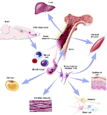
gastrointestinal stem cells (Potten 1998), epidermal stem cells (Watt 1998) and hepatic stem cells(also called oval cells)(Alison et al., 1998). The idea that stem cell are committed cells has been recently challenged by several bizarre and unexpected findings. Several experiments have suggested that certain adult stem cells types are pluripotent and can transdifferentiate into different cell types not only of the origin tissue but also other tissue i.e. hematopoietic stem cells may differentiate into three major types of brain cells (neurons, oligodendrocytes and astrocytes) (Krause et al., 2001); bone marrow stromal stem cells into cardiac muscle cells (Ferrari et al.,1998) and endothelium (Rafii et al., 1994; Asahara et al., 1997), and brain stem cells into blood cells (Bjornson et al., 1999) and dental pulp stem cells into neural tissue (Shen et al., 2003). This reported phenomenon is called
- 17 -
Fig 8: Plasticity of Adult Stem Cells
It has also recently demonstrated that certain adult cell types can be “reprogrammed” into other cell types using an in vivo well-controlled genetic modification process. For example, pancreatic beta cells, the insulin-producing cells that are lost or damaged in diabetes, could possibly be created by other reprogramming pancreatic cells (Zhou et al., 2008). In addition, beside the reprogram of the cell to become a specific cell type, it is now possible to reprogram adult somatic cells, through the introduction of embryonic genes, to become like embryonic stem cells a process called induced pluripotent stem cells (iPSCs).
Adult stem cells offer a hope in the future for cell therapy to treat diseases like Parkinson, Alzheimer, stroke, heart disease, diabetes and rheumatoid arthritis. In addition, using patient's own cells it can be overcome the immunological compatibility issue. But ASCs are rare in mature tissue and it‟s still a challenge and expanding methods in culture, have not yet been worked out. This is an important problem because for stem cell
- 18 -
replacement therapies it‟s necessary a large numbers of cells. However, ESCs have been found to be better for both differentiation potential and ability to divide themselves in culture, but their production meets strong resistance and limitations of ethical issues.
2.4 MESENCHYMAL STEM CELL
The adult bone marrow contains not only the hematopoietic stem cells, but also MSCs that represent an archetype of multipotent adult stem cell capable of giving arise to a number differentiated mesodermal cells of various type, including chondrocytes, osteocytes, adipocytes, myocytes and bone marrow stromal cells (Deans et al., 2000, Pittenger et al., 1999); that can be promising for their application in regenerative medicine. Interest in MSCs began over 130 years ago when Cohnheim, a German pathologist, suggested that the bone marrow gave rise to fibroblast-like cells during the repair process (Ross et al., 1970, Petrakis et al., 1961). Later in 1976, Friedenstein described first the fibroblast precursor from bone marrows capable of osteogenesis. Since their original description, these bone marrow multipotent progenitors were known with different names. The original term “colony forming unit-fibroblast (CFU-F)” or “marrow stromal fibroblasts (MSF)” (Castro-Malaspina et al., 1980, Piersma et al.,
1985, Kuznetsov et al., 1997) has been gradually abandoned and replaced
by different and still indistinct denominations like “marrow stromal cells (MSCs)” (Prockop et al., 1997), “mesenchymal stem cells (MSCs)” (Caplan et al., 1994), or mesenchymal progenitor cells (Conget et al.,
1999). An attempt to clarify the nomenclature for MSCs has recently been
proposed by the International Society for Cellular Therapy and discussed at several international meetings such as Adult Mesenchymal Stem Cells in
- 19 -
Regenerative medicine (MSC 2007, http://www.msc2007.net). Although MSCs were originally isolated from bone marrows (Friedenstein et al.,
1966, Pittenger et al., 1999), similar populations reside in a different
numbers of adult and fetal (in ’t Anker et al., 2003) tissues, including the spleen, amniotic fluid, cartilage, muscle, tendons, peripheral blood (Zvaifler et al., 2000, Kuznetsov et al., 2001) and tissue adipose (Zuk et al.,
2001, Alhadlaq et al., 2004) and more recently from deciduous tooth, fetal
membrane (Zhang et al., 2004) and umbilical cord (Bieback et al., 2004,
Kogler et al., 2004). Initially, peripheral blood was the first sources studied
for its easy availability. Only one research‟s group isolated mononuclear cells population by adherence, morphology, phenotype and differentiation potential typical characteristics of MSCs; these cells were isolated from over 100 samples of peripheral blood of healthy volunteers, but these data haven‟t never been confirmed (Zvaifler et al., 2000). Alternative sources are the fetal membrane a fetal discarded tissue and above all they don‟t generate ethical conflicts. Moreover, MSCs have high efficient recovery with no intrusive procedures (Alviano et al., 2007); the umbilical cord blood is rich in MSCs, because during embryonic development the embryonic hematopoiesis change site, from the yolk sac, the initial site, to liver and then to bone marrow with a consequent migration of HSCs and MSCs; but an interesting source could be the adult dental pulp and adipose tissue, very accessible tissue research. Many studies showed the presence of stem cells in dental pulp (Gronthos et al., 2000, Pierdomenico et al.,
2005) and adipose tissue (De Ugarte et al., 2003, Zuk et al., 2002)
possessing stem cell-like qualities, including a good self-renewal and multilineage differentiation (adipogenic and osteogenic) abilities. This source could still be a fascinating subject of study and a valuable therapeutic tool.
- 20 -
In bone marrow, MSCs are important components of the HSCs niche. In fact, all niche‟s components like stroma, stromal cells (endothelial cells, adipocytes, macrophages, reticular cells, fibroblasts and osteoprogenitors) and cellular microenvironment, where MSCs are presumed to exist, ensure the survival and growth of HSCs (Koller et al., 1997, Strobel et al., 1986,
Tavassoli et al., 1982). MSCs have been defined by their plastic adherent
growth and subsequent expansion under specific culture conditions, by a panel of non-specific surface antigens and by their in vitro e in vivo differentiation potential (Javazon et al., 2004). The gold standard assay utilized to identify MSC is CFU-F assay which identifies adherent, spindle-shaped cells that proliferate to form colonies (Friedenstein et al., 1970). MSCs, with their fibroblast-like morphology, were initially isolated as the plastic adherent fraction of bone marrow (Friedenstein et al., 1970). A Percoll density gradient was used to eliminate unwanted cell types, present in the bone marrow aspirate, and the present MSCs were a small percentage (estimated at about 0.001-0.01%). The general procedure used to isolate mononuclear cells is a gradient centrifugation and the following seeding on culture plates in medium with fetal bovine serum (FBS). After attachment of the adherent cell fraction, the medium is removed to eliminate non-adherents cell, the non-adherents cell are so expanded in a limited number of passages (Ulloa-Montoya et al., 2005). Although there aren‟t specific markers MSCs, many attempts have been made to develop a cell-surface antigen profile to improve the purification and identification of MSCs. Minimal criteria, to define human MSCs proposed by the Mesenchymal and Tissue Stem Cell Committee of the International Society for Cellular Therapy (Dominici et al., 2006), are the positivity for the following antigens:
- 21 -
CD105 this antibody SH2 identifies an epitope of endoglin (CD105), the Transforming growth factor beta (TGFβ) receptor III present on endothelial cells, erythroblasts, monocytes, and connective tissue stromal cells and facilitates enrichment of stromal progenitors from bone marrow (Short et al., 2003);
CD73 a glycoprotein, identified by monoclonal antibody SH3 and SH4, involved in B-cell activation (Short et al., 2003). It is expressed by lymphocytes and endothelial cells;
CD90 or Thy1: a 25–37 kDa heavily N-glycosylated, glycophosphatidylinositol (GPI) anchored conserved cell surface protein, originally discovered as a thymocyte antigen. Thy-1 can be used as a marker for a variety of stem cells and for the axonal processes of mature neurons. Structural study of Thy-1 lead to the foundation of the Immunoglobulin superfamily.
In contrast, the MSCs were negative for other markers of the hematopoietic lineage including CD34(a transmembrane protein that defined ˜ 1% of normal bone marrow mononuclear cells including hematopoietic precursors/stem cells and normal endothelial cells and its is considered a primitive HSCs marker); CD31(glycoprotein also designed platelet endothelial cell adhesion molecule-1 (PECAM-1) that is normally expressed on endothelial cells, circulating and tissutal hematopoietic cells including platelets, monocytes/macrophages, granulocytes and B-cells); CD45( recognizes a family of proteins known as the leukocyte common antigen exclusively expressed on the surface of almost all haemato-lymphoid cells and their progenitors) and CD14 or CD11b (an immune cell marker).
- 22 -
The identification of a definitive marker that allows the isolation of MSCs from fresh tissue could be very important. Stro-1 is the best-know MSC marker because the cell population negative for Stro-1 is not capable to form colonies. Stro-1 positive cells can become HSC-supporting fibroblasts, smooth muscle cells, adipocytes, osteoblasts and chondrocytes (Dennis et al., 2002) which reinforce the functional role of MSCs. In addition, the expression of Stro-1 distinguishes two cultured population of MSCs with different homing and HSC-supportive capacities (Bensidhoum
et al., 2004). However, Stro-1 is not specific for these cells and its
expression in MSCs is gradually lost during culture expansion (Gronthos et
al., 2003), so the Stro-1 labeling to isolate and/or identificate MSCs it‟s
possible only during early passages. In some cases, other cell surface markers have been empirically used to isolate the human bone marrow mononuclear cells by fluorescence-activated cell sorting (FACS) or magnetic bead cell sorting based both on the expression of several markers including, CD49b, CD146, CD130, CD200, CD44, CD166.
Since that were first discovered, several studies demonstrated the multilineage differentiation potential of MSCs populations showing their capacity to develop into terminally differentiated mesenchymal phenotypes including bone (Bruder et al., 1997), cartilage (Kadiyala et al., 1997), tendon (Young et al., 1998), muscle (Ferrari et al., 1998), adipose tissue (Dennis et al., 1999) and hematopoietic-supporting stroma (Prockop et al.,
1997) (Fig.9) and showing also a high degree of plasticity (D’Ippolito et al., 2004, Zhao et al., 2002).
- 23 -
Fig.9: The mesengenic process of MSCs
Furthermore, MSCs can commit to a particular differentiation pathway by their self-renewal and proliferation abilities and the micro-environmental in which they are. The lineage-committed cell progresses through several stages of maturation process a terminal differentiation, which is characterized by the cessation of proliferative capacity and the synthesis of tissue-specific markers, including components of the extracellular matrix (ECM)(Baksh et al., 2004).
Individual colonies derived from single MSC precursor have also been reported to be heterogeneous for their multilineage differentiation potential (Friedenstein et al., 1970). In the 1999, Pittenger et al reported that only one-third of the initial adherent BM-MSCs clones are pluripotent (osteo/chondro/adipo) (Pittenger et al., 1999). Furthermore, non-immortalized cell clones have been used to investigate the nature and properties of committed progenitors present in culture of BM-MSCs. This study demonstrated that 30% of all clones exhibited a tri-lineage(osteo/chondro/adipo) differentiation potential, while the remainder displayed a bi-lineage (osteo/chondro) or uni-lineage (osteo)(Muraglia et
- 24 -
al., 2000). The heterogeneity, both in vivo and in vitro studies, could be
explained by the notion that MSCs in the bone marrow are a pool of cells that include MSCs and different subpopulations at different state of differentiation. During in vitro culture, were isolated all or a subset of these MSCs. During differentiation, the proliferative potential of these different MSCs decreases and, depending on the initial state of differentiation, both proliferative and multilineage potential become limited (Baksh et al., 2004) (Fig.10).
Fig.10: Models of MSCs differentiation; A) capacity to differentiate into all
connective tissue cell types; B) MSCs population that with different differentiation potentials (Baksh et al., 2004)
For the immunological profile, MSCs express intermediate levels of human leukocyte antigen (HLA) class I molecules major histocompatibility complex (MHC) and low levels of class II HLA and Fas ligand; they do not express the costimulatory molecules B7-1, B7-2, CD40, or CD40L. The immunosuppressive nature of MSCs is of clinical relevance in allogeneic transplantation since it could reduce the incidence and severity of graft-vs-host disease (GVD) (Le Blanc et al., 2003 a, b). Clinically, the easy of isolation, expansion potential, migratory capacity and immunosuppressive capability of MSCs has made them a popular cell type for investigating
- 25 -
regenerative medicine, gene therapy and tissue engineering (Fig.11). Several studies based on animal transplantation, shown that ex-vivo expanded MSCs were able to differentiate into cells of the residing tissue, to repair damaged tissue and to restore partially its normal function, generating promising results for the treatments of several illness, including bone (Mauney et al., 2005), cardiovascular (Zimmet et al., 2005) and brain disease (Zimmet et al., 2005). Recent studies demonstrated that the therapeutic contribution of MSCs transplantation could be caused not only by direct differentiation but also by paracrine activities which supply with large amounts of cytokines and growth factors. These bioactive factors suppress the local immune system, inhibit apoptosis, and enhance angiogenesis (Caplan et al., 2006). MSCs are promising also for tissue engineering. Bioengineered structures with a defined shape made with biomaterials like collagen type I, fibronectin, alginate, polylactic acid and alginate can be combinated with MSCs, culturing in bioreactors, it could be possible to obtain tissue and organs (Stock et al., 2001) as reported in pre-clinic animal model studies for the treatment of a large bone defects (Kon
et al., 2000).
Fig.11: Potential uses of Adult Stem Cells in regenerative medicine, cells
- 26 -
3.
STEM CELLS NICHE
It is well know that self renewal and pluripotentiallity are specific skills of adult stem cells, but the idea that extremely specialized external stimuli and microenvironment can affect the regulation of the specific stem characteristic brought several team to go in for it. Microenvironment are constituted by adult tissue-specific stem cell but also other kind of cells characterized by the present of adhesive molecules and extracellular matrix proteins, that have an important role in the space localization and organization, in fact it has also regulating functions to determinate stem cell differentiation into various kind of mature cells, replacing cells lost due to natural cell death (apoptosis) or injury, safeguards against excessive stem cell production that could lead to cancer and to preserve a sufficient stock of stem cell for the future, infact, stem cells must periodically activate to produce progenitor or transit amplifying (TA) cells that are committed to produce mature cell lineages (Fig.12).
- 27 -
The niche concept was introduced in 1978 by Schofield studies (Schofield
1978); his team proved that microenvironment cells has a role in
maintaining in a quiescent condition the hematopoietic stem cell, and suggest the presence of a “stem cell niche”. The idea of specialistic areas in the microenvironment seems to be supported by the production of grown factor in outline compartment (Gordon MY 2008). Stomal cells releases locally grown factors that bind extracellular matrix structures, in this way target cells recognized them by specific receptor (Gordon MY 2008). This mechanism permit to localize high concentration of specific grown factors in specific microenvironment areas. Several increasing studies had shown the present of a wide range of humoral factors, cytokines, chemokines and adhesive factors supporting adult stem cells. A recent hypothesis is that the regulating marrow stimulus is placed in a 3D organization to establish the “stem niches”. Niche can be defined as a spatial microstructure in which houses adult stem cells, which start self renewal activity interacting with external stimuli. Niche characteristics are here resumed: the number of stem cells in a niche is well-regulated; stem status depends on the interaction with other histologically different cells; stimuli produced by niches supply the molecular base for the interaction between cells, and allow the transduction of activator and inhibitor signals, presiding over the expansion of stem compartment and into a niche also a not stem cell can reach pseudo-stem characteristics.
All those results suggest that stem niche not have only a structural role, but seems to have the intrinsic potential to lead the destiny of the cells in it. This hypothesis was supported by several researchers that during several studies identified different niches in different anatomic district.
- 28 -
3.1 HEMATOPOIETIC STEM CELLS NICHE
The bone marrow contains multiple stem cell types, including HSCs and mesenchymal MSCs which have the ability to self-renew and to differentiate into cartilage, bone and adipose tissues at the single cell level (Pittenger et al., 1999). HSCs are the best characterized adult stem cell population. Single HSCs are multipotent, highly self-renewing, and cycle with slow kinetics. In adult bone marrow, a part of these HSCs are known to reside in two different niches, an “endosteal” niche and a “perivascular” niche. The endosteal niche it seems to maintain HSC in a long term quiescence whereas perivascular niche it seems to maintain the quiescence of HSCs for a shorter period, supporting HSC‟s proliferation, favoring myeloid and megakaryocytic lineage differentiation and mediating HSC‟s circulation (Perry et al., 2007). In the endosteal niche, HSCs are associated with a subset of osteoblasts that line the inner surface of trabecular bone cavities and giving arise to progenitors that migrate to blood vessels in the middle of bone marrow cavity where they mature and differentiate (Nilsson
et al., 2001, Gong 1978, Heissig et al., 2002) (Fig.13a). Studies have
shown that the position of osteoblast cells is a for the begin of the process as previously described (Zhang et al., 2003).The role of osteoblasts a support for HSC growth was suggested through in vitro coculture experiments (Taichman et al. 1994, 2000), and simultaneously using genetic mutant mouse models (Zhang et al., 2003, Calvi et al., 2003, Arai
et al., 2004) was identified osteoblastic cells as a key component of the
HSC niche. Endosteal osteoblasts are thought to provide a several factors that regulate HSC number and function (Zhang et al., 2003, Calvi et al.,
2003). A possible mechanism by which osteoblasts would regulate the
number of HSCs is through secretion of osteopontin that is a bone matrix glycoprotein that seems to maintain HSC in quiescence form and to
- 29 -
regulate HSC proliferation in a negative way. An increasing number of molecular studies point to the existence of a complex paracrine signaling network at the interface between the niche osteoblast and the adjacent HSCs; Kit ligand is expressed by osteoblasts and is able to activate Kit on HSCs surface. Notch signaling plays an important role in cell fate to maintain HSCs in an undifferentiated state (Calvi et al., 2003, Duncan et
al., 2005, Varnum-Finney et al., 2000). Another important interaction is
between the ligand angiopoietin-1 a the osteoblast surface and the receptor Tie-2 (Tyrosine kinase with immunoglobulin-like and EGF-like domains 2) expressed on HSCs, which has been shown to modulate HSC quiescence (Arai et al., 2004) (Fig.13b).
Fig.13: The Hematopoietic Stem Cells (HSCs) niche in the bone marrow.
a) Schematic diagram of HSCs and niche cellular components in the bone marrow; b) Extrinsic signaling pathways that regulate proliferation and differentiation of HSCs (Li et al., 2005)
a
- 30 - 3.2 EPIDERMAL STEM CELLS NICHE
Skin epidermis, with its appendix hair follicle structure, is a regenerating organ with a well-organized architecture. Each hair follicle is composed of a permanent portion, which includes sebaceous glands and the underlying bulge area, and a dynamic renewing portion which give arise two stem cells population within the hair follicle and interfollicular regions. The first population, the epidermal stem cell is located in the basal layer of the skin clustered in epidermal proliferation units (Potten 1981), normally gives rise to stratified skin layers. The second, hair follicle stem cells (HFSCs), resides in a region of the outer root sheath called the “bulge”, and it is responsible for the regeneration of hair and sebaceous glands, restore the epidermis after wounding (Taylor et al., 2000, Rendl et al., 2005) and that can be activated during the hair cycle in response to injury (Fig.14a).
The bulge area act as a niche where HFSCs (Niemann et al., 2002) are located, maintained (Cotsarelis et al. 1990, Sun et al. 1991) and also responsible for the long-term replenishment of the interfollicular epidermis. Bulge stem cells are generally quiescent, multipotent and, after their activation, giving rise to daughter cells; the daughter cells retained in the bulge remain as stem cells while other daughter cells migrate down to become hair-matrix progenitors responsible for hair regeneration (Cotsarelis et al., 1990, Niemann et al., 2002,Oshima et al., 2001, Taylor et
al., 2000). The multipotentiality of single HFSCs has been shown by using
cells expanded in vitro. An in situ tracking method has shown that progenitors in the hair follicle contribute to single lineages and possess limited self-renewal potential, suggesting that it may be possible to measure various lineage potentials rigorously when and how they segregate after HFSCs activation (Legue et al., 2005).
- 31 -
The molecular analysis epithelial stem cells has revealed the following features: 1) the expression of adhesion molecules known to be involved in stem cell-niche interaction, 2) the presence of growth inhibition factors such as TGFβ/BMP molecules and cell cycle inhibitors, and 3) Wnt pathway including receptors and inhibitors such as Dkk, sFRP, and WIF is important in the hair follicle niche. Taken together, these molecular features indicate that the epithelial stem cell niche is a growth- and differentiation-restricted environment (Tumbar et al. 2004) (Fig.14b).
Fig.14: The Epidermal Stem Cells (ESCs) niche. a) Schematic diagram of the major types and spatial orientations of HSCs that make up the hair follicle; b) Interactive signaling pathways that mediate HFSC proliferation (Li et al., 2005)
a
b
- 32 - 3.3 INTESTINAL STEM CELLS NICHE
The intestinal epithelium can be divided into two regions, a region containing pericryptal fibroblasts and mesenchyme functional differentiated cells (villa), and a proliferative region (crypt Lieberkühn) which represents the stem cell niche. Intestinal regeneration begins with intestinal stem cells (ISCs), which give rise to four different types of epithelial lineages: columnar enterocytes, mucin-producing goblet cells, Paneth cells, and enteroendocrine cells (Bjerknes et al., 1999, et al., 1995,
Winton et al., 2000) (Fig.15a).
ISCs are generally proposed to be located at the fourth or fifth position from the crypt bottom, above the Paneth cells (Booth et al., 2000, He et al.
2004, Sancho et al. 2004). The crypt is a contiguous pocket of epithelial
cells at the base of the villus. ISCs and TA cells within the crypt regenerate the entire villus every 3 to 5 days (Potten et al., 1990). Genetic marker shows that crypts derive from an individual or few ISCs and that each villus is the product of cells the coming from several adjacent crypts (Gordon et al., 1992). There are four to six ISCs per crypt that are located in ring diameters of about four cells from the crypt bottom. Progeny of activated ISCs migrate upwards to become TA cells. When they reach the top of the crypt, TA cells stop proliferating and assume their appropriate positions within the villus structure.
During postnatal intestinal regeneration, mesenchymal cells subjacent to epithelial cells play a role in epithelial cell proliferation, differentiation, and apoptosis; BMP4, expressed in the ISC-adjacent mesenchymal cells, is one of the putative niche signals (He et al. 2004); endothelial cells provide ISCs with survival signals such as fibroblast growth factor (FGF) (Paris et
al. 2001); myofibroblasts surrounding epithelial cells, supported ISCs
- 33 -
Molecular analysis showed that signal Wnt plays a positive role in the promotion of ISC activation/self-renewal; in contrast, BMP signaling restricts ISC activation/self-renewal and crypt cell fate (Haramis et al.
2004, He et al. 2004) (Fig.15b).
Fig.15: The Intestinal Stem Cells (ISCs) niche. A) Schematic diagram of
the major types and spatial orientations of cells found within the crypt niche and the villus. B) Interactive signaling pathways that mediate ISC proliferation (Li et al., 200)5
3.4 NEURAL STEM CELLS NICHE
In adult life, neurogenesis is possible in specific brain area where were identified and characterized neuronal niches.
Neural stem cells (NSC) were identified in 1990 and then isolated from various regions in the adult brain and peripheral nervous system
(Alvarez-Buylla et al., 1990).The subventricular zone (SVZ) and the subgranular
a
- 34 -
zone (SGZ) of the hippocampus region are the primary and
well-characterized germinal regions in which NSCs reside and support neurogenesis in the adult brain (Doetsch et al. 1999, 2003, Lois et al. 1993)
SUBVENTRICULAR ZONE (SVZ) is a single layer of multi-ciliated
ependymal cells separates the SVZ from the lateral ventricle (LV). There are four main cell types in the SVZ: neuroblasts (Type A cells), SVZ astrocytes (Type B cells), immature precursors (Type C cells) and ependymal cells (Doetsch et al., 1997). In this region, the SVZ astrocytes, located adjacent to the ependymal cells, have stem cell features and give rise to TA precursor C cells. Infact, immature cells, deriving from SVZ astrocytes, are precursors of a group of neuroblasts which differentiate into neurons and migrate toward the olfactory bulb and other regions. SVZ astrocytes can also generate oligodendrocytes (Doetsch 2003, Mirescu et
al., 2003, Temple 2001). Another noteworthy feature of this region is a
specialized basal lamina, which extends from blood vessels in SVZ region and terminates in small bulbs adjacent to ependymal cells, and contacts all SVZ cell types. In SVZ are also present blood vessels and endothelial cells that lining the blood vessels and these are likely a source of signals for adult neurogenesis (Fig.16a).
SUBGRANULAR ZONE (SGZ) is the germinal layer between the dentate
gyrus and the hilus in the hippocampus and it is responsible for the generation of dentate gyrus granule neurons (Palmer et al. 1997). In the SGV region, neurogenesis occurs in foci closely associated with bloods vessels (Palmer et al. 2000). As in the SVZ, SGZ astrocytes acts as a stem cell; are the primary precursors of neurons and generate daughter cells that further produce granule neurons (Fig.16b).
- 35 -
Fig.16: The neural stem cell (NSC) niche. a) The subventricular zone
(SVZ). Astrocytes (B) lining the ependymal cells (E) function as NSCs; they give rise to transient amplifying cells (C) (green), which further produce neuroblast cells (A); b) The subgranular zone (SGZ). Astrocytes (B) directly attach to the blood vessel and receive signals from the endothelial cells that direct NSCs to undergo self renewal, proliferation (D), and differentiation (G) (Li et al., 2005)
In both regions, endothelial cells and the specialized basal lamina are essentials components of the NSC niche. The ECs provide attachment for SVZ and SGZ astrocytes and generate a variety of signals controlling stem cell self-renewal and lineage commitment (Doetsch 2003, Shen et al.
2004). In fact, angiogenesis and neurogenesis may be co-regulated and
reciprocally signaled. Both are stimulated by the same factors, including bFGF, VEGF, insulin-like growth factor (IGF-1) and TGF-a; ECs secrete well-known mitogens, differentiation and survival neuronal factors like bFGF, IGF-1, VEGF, PDGF, IL8 and brain derived neurotrophic factor (BDNF) (Palmer et al. 2000, Jin et al., 2002, Louissaint et al., 2000).
- 36 - 3.5 VASCULAR NICHE
In 2006 was suggested the existence of a “vasculogenic niche” in the human vascular wall of large and mid-sized blood vessels. The existence of this “vasculogenic zone” has been defined as a vascular mural zone, identified in adult human vascular wall and located at the border between the media and the adventitial layers containing a complete hierarchy of resident stem cells, which may serve as a source for progenitor cells for postnatal vasculogenesis. This zone of vascular wall serves a niche containing the vascular wall-endothelial progenitor cells (VW-EPC) resident, which are capable of forming capillary sprout in arterial ring assay in vitro, outside the bone marrow, even if Ingram‟s group reported the existence of VW-EPCs but did not furnished the exact location within the vascular wall (Ingram et al., 2005) but also MSCs and probably also vascular wall hematopoietic progenitor cells (HPCs) (Fig.17).
- 37 -
Moreover, this area could correspond to a vascular wall stem cell niche identified as special microenvironment in a strategic location at the interface between the media and adventitial layers, physiologically limited and specialized, an unexpectedly elevated cell proliferation under normal conditions, selective localization of cells expressing the stem cell surface molecules c-kit, in which stem cells and multipotent stromal cells stay; both cytotypes could contribute in maintaining post-natal vascular homeostasis replacing old or damaged elements/items (Pacilli et al., 2009).
- 38 -
4. VASCULAR WALL RESIDENT STEM CELLS
In growing body several evidence suggests a close relation between hematopoiesis and vasculogenesis in vertebrates. During early embryogenesis, hematopoietic and endothelial lineages derive from aggregates of mesodermal cells that subsequently mature and form blood islands in the extra-embryonic yolk sac. These blood island consists of an inner core of blood cells and an external layer of endothelial cells (ECs)(Sabin 1920, Murray 1932) and the simultaneous presence of these 2 kind of cells has led to the hypothesis that they originate from a common precursor called hemangioblast (Murray 1932).The formation of the blood islands in the yolk sac marks the begin of the vascularization in the developing embryo. Two different processes contribute to the formation of the vascular system. The first process, vasculogenesis, requires the differentiation of endothelial cells from hemangioblast and their subsequent organization into a primary capillary plexus (Risau et al., 1988, 1995) and it is restricted to early embryogenesis. The second process, angiogenesis, results in the formation of new vessels by sprouting from preexisting blood vessels (Folkman 1992, 1995) and occurs both during development and postnatal life. However, studies have showed that postnatal angiogenesis may occur by recruitment of bone marrow and peripheral blood and of endothelial progenitors cells (EPC), with property of embryonal angioblst, involved in the new blood vessel formation in response to various stimuli (Asahara et al., 1999a). Once mobilized from bone marrow and released into the circulating blood (CEPs), these progenitors are supposed to participate in physiological and pathological arterial wall remodeling during their lifetime (Carmeliet 2003). In fact, experimental evidences support the use of EPCs in angiogenic therapies or as biomarkers to assess cardiovascular disease risk (Rafii et al., 2003, Vasa et al., 2001, Kalka et