Reumatismo 4/2018 257 CASE
REPORT
SUMMARY
A 57-year-old woman with a diagnosis of antisynthetase syndrome (ASSD) underwent a nailfold videocapil-laroscopy (NVC) showing a scleroderma pattern.
Alterations in capillary morphology have been reported in adults with inflammatory myositis (IM) but only recently have the differences in NVC findings between these two diseases been established.
ASSD is currently classified as a subset of IM, for which reason only a few studies in literature evaluate its specific hallmarks, showing nonspecific features of NVC in patients with polymyositis and dermatomyositis (DM) and antisynthetase antibodies.
To our knowledge, this is the first description of ASSD capillaroscopy features, and the first report of NVC in ASSD with evidence of scleroderma pattern.
Further studies are needed to define clearly frequency, typical features, and possible correlation with clinical and serological data of NVC changes in ASSD, differences between microangiopathy in ASSD and systemic sclerosis or DM.
Key words: Nailfold videocapillaroscopy; Inflammatory myositis; Antisynthetase syndrome; Raynaud’s phe-nomenon.
Reumatismo, 2018; 70 (4): 257-258
A
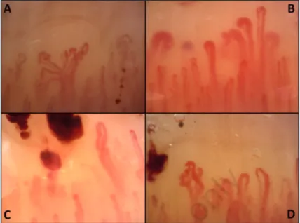
57-year-old woman presented with a five-month history of dry cough, dyspnoea, weakness, Raynaud’s phenom-enon and arthralgias. On physical examina-tion, we noted hyperkeratosis, scaling and fissuring of the fingers, digital clubbing, swollen joints and pulmonary crackles. Hematologic laboratory tests showed in-crease of CPK (873 U/L) and LDH (541 U/L). Anti-nuclear antibodies were posi-tive (1:160 speckled pattern), with anti-Jo1 positivity.She underwent pulmonary high-resolution computed tomography, showing interstitial lung disease (ILD). A diagnosis of antisyn-thetase syndrome (ASSD) was made. A nailfold videocapillaroscopy (NVC) showed a scleroderma pattern (Figure 1), characterized by disorganization of the vascular array (panels A, D), reduction of capillary density (panels A, B, D), enlarged and giant capillaries (panels B),
microhe-morrhages (panels C, D) tortuosities and ramified capillaries (panels A, D).
Alterations in capillary morphology have been reported in adults with inflamma-tory myositis (IM), such as polymyositis (PM) and dermatomyositis (DM)(1-3), but only recently have the differences in NVC findings between these two diseases been established(1). NVC alterations are sig-nificantly more frequent and severe in DM patients, in which a scleroderma-like pat-tern was described.
ASSD is currently classified as a subset of IM, characterized by the presence of serum autoantibodies directed to aminoacyl trans-fer RNA synthetases (anti-tRNA) along with features of ILD and arthritis, other than myositis(4). For this reason, only few studies in literature evaluate its specific hallmarks, showing nonspecific features of NVC in patients with PM and DM and an-tisynthetase antibodies. Corresponding author: Giulia Cassone Rheumatology Unit, Dpt. of Internal Medicine, University of Modena
and Reggio Emilia, Policlinico di Modena Via del Pozzo, 71 - 41125 Modena, Italy E-mail: [email protected]
Nailfold videocapillaroscopy
in antisynthetase syndrome
G. Cassone1,2,3, M. Sebastiani1, L. Cavagna4, K. Triantafyllias5,
V. Codullo4, C. Salvarani1,3, A. Manfredi1
1Rheumatology Unit, University of Modena and Reggio Emilia, Modena, Italy; 2Clinical and Experimental Medicine PhD Program, University of Modena and Reggio Emilia, Modena, Italy;
3Rheumatology Unit, IRCCS Arcispedale Santa Maria Nuova, Azienda Unità Sanitaria Locale-IRCCS
di Reggio Emilia, Reggio Emilia, Italy; 4Division of Rheumatology, University and IRCCS Policlinico
S. Matteo Foudation, Pavia, Italy; 5ACURA Rheumatology Clinics, Bad Kreuznach, Germany Reumatismo, 2018; 70 (4): 257-258
Non-commercial
CASE REPORT
258 Reumatismo 4/2018
G. Cassone, M. Sebastiani, L. Cavagna, et al. CASE
REPORT
n CONCLUSIONS
To our knowledge, this is the first descrip-tion of a scleroderma pattern at NVC in a patient with a diagnosis of ASSD.
Further studies are needed to define clearly frequency, typical features, and possible correlation with clinical and serological data of NVC changes in ASSD, differences between microangiopathy in ASSD and systemic sclerosis or DM.
Conflict of interest: the authors declare no
conflict of interest.
n REFERENCES
1. Manfredi A, Sebastiani M, Cassone G, et al. Nailfold capillaroscopic changes in dermato-myositis and polydermato-myositis. Clin Rheumatol. 2015; 34: 279-84.
2. Selva-O’Callaghan A, Fonollosa-Pla V, Tralle-ro-Araguás E, et al. Nailfold capillary micros-copy in adults with inflammatory myopathy. Semin Arthritis Rheum. 2010; 39: 398-404. 3. Mercer LK, Moore TL, Chinoy H, et al.
Quan-titative nailfold video capillaroscopy in pa-tients with idiopathic inflammatory myopathy. Rheumatology 2010; 49: 1699-705.
4. Monti S, Montecucco C, Cavagna L. Clinical spectrum of anti-Jo-1-associated disease. Curr Opin Rheumatol. 2017; 29: 612-7.
Figure 1 - Nailfold videocapillaroscopy showing a scleroderma pattern, characterized by
dis-organization of the vascular array (A, D), reduction of capillary density (A, B, D), enlarged and giant capillaries (B), microhemorrhages (C, D) tortuosities and ramified capillaries (A, D).