ROMA
“TOR VERGATA”
FACOLT `A DI SCIENZE MATEMATICHE, FISICHE E NATURALI DOTTORATO DI RICERCA IN FISICA
XXII CICLO
X-ray Absorption Spectroscopy: a powerful tool for structural studies of molecules involved in the pathogenesis of
neurodegenerative diseases
Francesco Stellato
Docente Guida: Prof. Silvia Morante
Preface 1 1 The molecular basis of neurodegenerative diseases 3
1.1 What is a protein? . . . 4
1.2 Protein Misfolding and Neurodegenerative Diseases . . . 6
1.3 Alzheimer’s Disease and Aβ Peptides . . . 10
1.3.1 Physiology and Pathology of AD . . . 11
1.3.2 Diagnostic and Therapeutic Strategies in AD . . . 16
1.3.3 Sequence and Structure of Aβ Peptide . . . 19
1.3.4 Interaction of Aβ Peptide with Metal Ions . . . 21
1.4 Transmissible Spongiform Encephalopathies and the Prion Protein 26 1.4.1 Physiology and Pathology of the TSEs . . . 27
1.4.2 The Structure of PrP . . . 29
1.4.3 Interaction of PrP with Metal Ions . . . 31
1.5 Parkinson’s Disease and Neuromelanin . . . 34
1.5.1 Physiology and Pathology of Parkinson’s Disease . . . 34
1.5.2 Diagnostic and Therapeutic Strategies in PD . . . 37
1.5.3 The Structure of Neuromelanin . . . 38
2 X-ray Absorption Spectroscopy 43 2.1 Theory of XAS . . . 44
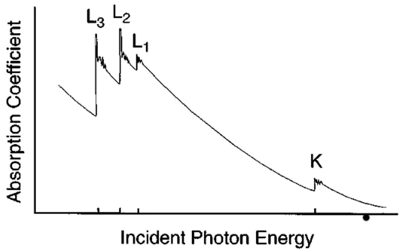
2.1.1 X-ray Absorption . . . 44
2.1.2 Single Scattering Approximation and EXAFS Equation . . 53
2.2 Experimental Overview . . . 57
2.2.1 Synchrotron Radiation . . . 57
2.2.2 X-ray Optics and Detectors . . . 59
2.2.3 Experimental Setup . . . 65
2.3 XAS data Analysis . . . 69
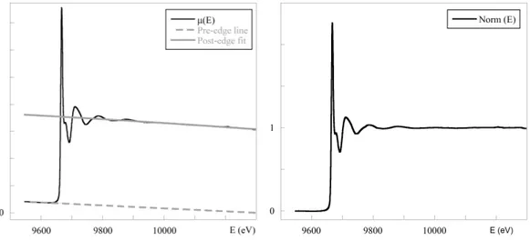
2.3.1 Signal Extraction and Data Reduction . . . 69
2.3.2 XANES Region . . . 71
2.3.3 EXAFS Region . . . 73
3 Experimental Results 77 3.1 Cu and Zn Aβ complexes . . . 78
3.1.1 Sample Description . . . 78
3.1.2 Optical Density Measurements . . . 79
3.1.3 FTIR Measurements . . . 82
3.1.4 Sample Preparation and Data Collection . . . 83
3.1.5 XAS Data Analysis . . . 84
3.1.6 Discussion and Conclusions . . . 92
3.2 Cu and Zn PrP complexes . . . 97
3.2.1 EPR Results . . . 97
3.2.2 XAS Data Analysis . . . 99
3.2.3 Discussion and Conclusions . . . 108
3.3 The S local environment in human Neuromelanin . . . 111
3.3.1 Samples Description . . . 111
3.3.2 XAS Data Collection . . . 114
3.3.3 XAS Data Reduction . . . 115
3.3.4 A semi-quantitative approach to XANES data analysis . . 117
3.3.6 Discussion and Conclusions . . . 123
4 The active site of human Prolidase 125 4.1 Structure and function of human Prolidase . . . 126
4.1.1 Prolidase and Prolidase Deficiency . . . 126
4.1.2 Experimental Details . . . 127
4.1.3 XAS Data Analysis . . . 130
5 Conclusions 139 5.1 Summary . . . 140
5.1.1 AD, TSEs and PD: Similarities and Inter-relationships . . 141
5.2 Future Perspectives . . . 144
5.2.1 Metal Ions-Aβ Peptides Complexes . . . 144
5.2.2 Metal Ions-PrP Complexes . . . 145
5.2.3 Neuromelanin . . . 146
Abbreviations 147
Just like XX century was dubbed the century of Physics, the XXI one is often claimed to be the century of Biology. It is in fact undeniable that the strong relationships already existing between the two disciplines are daily, and proficu-ously, increasing.
The work presented in this thesis is a good example of the fertile interaction between Physics and Biology. It is dealing with the application of X-ray Ab-sorption Spectroscopy (XAS) to the study of the structure of certain proteins and peptides, related to severe neurodegenerative diseases - Creutzfeldt-Jakob, Alzheimer and Parkinson - and of their interaction with metals.
The material of this thesis is organized as follows. Chapter 1 is devoted to the description of the main biophysical and medical issues related to the systems studied in this work. Chapter 2 is about the theoretical basis and the main experimental aspects of the XAS technique. A special attention is devoted to the data analysis methods. Chapter 3 is focused on the description of the exper-imental results on the prion protein, β-amyloid and neuromelanin and of their interpretation. Finally in Chapter 4 I report the application of XAS to prolidase (a protein related to the insurgence of a very rare, fatal disease) the structure of which is also influenced by its interaction with metals. Conclusions summariz-ing the main results of the thesis and describsummariz-ing possible future perspectives are given in Chapter 5.
All the work presented in the thesis has been done within the Biophysics group in the University of Rome ’Tor Vergata’ lead by Prof. Silvia Morante. Besides Prof. Morante, I would like to thank Prof. Giancarlo Rossi and Dr. Velia Minicozzi for
their scientific and practical support. Among the former group members I would like to thank Dr. Francesco Guerrieri and Dr.Stefania Alleva.
The molecular basis of
neurodegenerative diseases
Neurodegenerative diseases (a composite word from Greek ν²υρo, ”nerval” and Latin degenerare, ”to decline”) are a class of pathologies which affect the brain. Many of them are age related, therefore in a society in which population is getting older and older it is a very important issue to find effective therapies against them. The molecular basis of this class of pathologies is in many cases still unclear. However it is worth noticing how diseases with very different etiology show common molecular mechanisms, such as protein misfolding and aggregation processes.
This chapter starts with a short overview of neurodegenerative diseases, with particular attention to the role played in their pathogenesis by protein misfolding processes. Then it follows a more detailed description of the pathologies that are the main subject of the thesis, namely Alzheimer’s disease, the spongiform encephalopathies and Parkinson’s disease.
1.1
What is a protein?
Proteins are essential components of a living organism. They participate in al-most every process within cells and can have several functions. Many proteins are enzymes, i.e. molecules that catalyze biochemical reactions, but they can also have structural or mechanical functions, or play roles in cell signaling and immune responses [1].
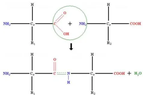
From a structural point of view, proteins are organic polymers made of a lin-ear chain of amino acidic monomers. The most common amino acids are 20. They share a common structure (the main chain) to which a variable (one for each amino acid) side chain is bound (see upper panel in Fig. 1.1). To help the further reading, in Table 1.1, the complete names of all aminoacids and their 3- and 1-letter codes are reported. The sequence of amino acids in a protein is determined by the sequence of nucleotides in the DNA (the gene), which is transferred to the mRNA (traduction process) and then from mRNA into the peptidic sequence (translation process).
Proteins are polymeric chains in which amino acids are joined together through a peptide bond between the carboxyl and amino groups belonging to two adjacent amino acid residues (see lower panel in Fig. 1.1). The sequence of amino acids along the chain is called ’primary structure’ of the protein.
After their synthesis, proteins can undergo post-translational modifications, which consists of attaching other biochemical functional groups such as acetate, phos-phate, various lipids and carbohydrates, or of the creation of structural changes, like the formation of disulfide bridges between Cysteine residues.
Due to the differences in the side chains, each amino acid has different physical and chemical properties. In particular, there are charged and neutral residues, polar and non-polar side chains. An important property strictly connected to this last feature is the hydropathicity, namely the preference for the side chain of being in a polar or non polar medium.
The local arrangement of the amino acidic chain is usually characterized by reg-ularly repeating structures (secondary structure), the most common of which are the α helix and the β sheet.
func-Amino Acid 3-Letter Code 1-Letter Code
Alanine Ala A
Arginine Arg R
Asparagine Asn N
Aspartic Acid Asp D
Cysteine Cys C
Glutamic Acid Glu E
Glutamine Gln Q Glycine Gly G Histidine His H Isoleucine Ile I Leucine Asn L Lysine Lys K Methionine Met M Phenylalanine Phe F Proline Pro P Serine Ser S Threonine Thr T Tryptophan Trp W Tyrosine Tyr Y Valine Val V
Table 1.1: The 20 Amino Acids: names, 3 and 1 letter abbreviations. tion, is the tertiary structure. Proteins composed by more subunits have also an higher, quaternary, level of structure, which depends on the relative positions of non-covalently bound peptidic subunits.
Figure 1.1: Amino Acid structure and peptide bond. Rn is the side chain that
is characteristic of a given amino acidic residue.
1.2
Protein Misfolding and Neurodegenerative
Diseases
It is well established that the function of a protein is strictly related to its struc-ture. In the 1950s Anfisen showed, in a series of now classical experiments, that protein folding depends only on the aminoacidic sequence of the molecule (the so-called primary structure)[1]. This is true for an isolated proteic molecule, but in a real case the situation is more complicated, because the correct folding can be influenced and guided by several factors, such as the pH, the presence of other biomolecules, membranes or metal ions [4].
Protein folding is indeed a very complicated process, that not always ends with the ’right’ three-dimensional configuration. Being protein function strictly re-lated to its folding, it is straightforwardly understood how errors in the folding can be at the basis of pathologies. The class of diseases in which alterations in protein folding are observed is composed of about 20 pathologies (see their list in Table 1.2). They are known as Protein Conformational Disorders (PCDs) [5]. Through the folding and unfolding processes specific types of cellular activity
Disease Fibril subunit Alzheimer’s Disease Amyloid β peptide Spongiform encephalopathies Full-length prion protein Hereditary cerebral haemorrhage Amyloid β peptide, cystatin C
Type II diabetes Amylin
Medullary carcinoma of the thyroid Procalcitonin Atrial amyloidoses Atrial natriuretic factor
Parkinson’s Disease α synuclein
Huntingtons Disease Glutamine
Amyotrophic lateral sclerosis Superoxide dismutase Primary systemic amyloidosis Immunoglobulin light chains Secondary systemic amyloidosis Serum amyloid A protein
Familial Mediterranean fever Serum amyloid A protein Familial amyloidotic polyneuropathy 1 Mutant transthoracic
Senile systemic amyloidosis Wild-type transthyretin Familial amyloidotic polyneuropathy II Apolipoprotein A-1
Haemodialysis-related amyloidosis β2-Microglobulin
Finnish hereditary amyloidosis Mutant gelsolin Lysozyme amyloidosis Mutant lysozyme
Insulin-related amyloid Insulin
Fibrinogen α-chain amyloidosis Fibrinogen α-chain Table 1.2: Most common PCDs.
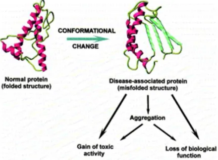
are generated or suppressed. Failure to correctly fold, or to remain correctly folded, will therefore give rise to the malfunctioning of living systems and hence to disease [6]. There are two main mechanism through which misfolding may affect protein function: the first one consists of directly impeding protein correct functioning, like in cystic fibrosis and some types of cancer; the second one is by giving to incorrectly folded proteins a high propensity to misfold and thus to form aggregates within cells or (more commonly) in extracellular space. Fig. 1.2 shows a schematic picture of the possible pathogenetic pathway.
extracel-Figure 1.2: Conformational changes related pathogenetic mechanisms.
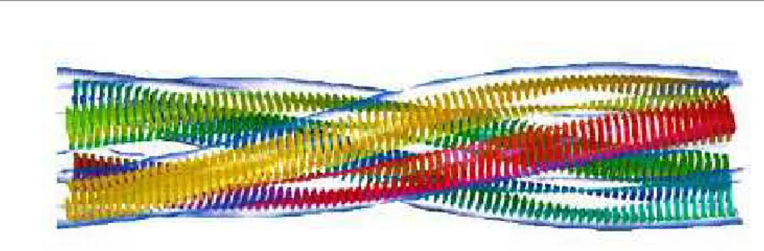
lular space of various tissues as insoluble deposits have been identified, and they are known as amyloidoses. It is worth noticing that, although the amino acidic sequence of the proteins that are the main component of the deposits are com-pletely different in each amyloidosis, the disorders have several key features in common. In particular, amyloid deposits have characteristic optical properties, such as birefringence, on binding certain dye molecules such as Congo red. The aggregates have peculiar morphologies, as shown by X-ray diffraction patterns which indicate the presence of fibrils of about 10 nm diameter. These fibrils are rich in β sheet conformation, and the peptide strands are arranged perpendicular to the long axis of the fibre (Fig. 1.3).
Of particular significance is the finding that globular proteins with diverse se-quences that are not currently associated with a protein-folding disease can un-dergo aggregation in vitro into fibrils indistinguishable from those found in the amyloidoses, thus suggesting that aggregation into β sheet rich fibrils is a generic property of polypeptide chains regardless of sequence.
Protein misfolding is thought to be connected to age-dependent neurodegenera-tion. During the past two decades, the pathogenesis of age related degenerations of the brain has become recognized as likely to be mediated by the progressive
Figure 1.3: Sketch of an amyloid fibril with peptide strands perpendicular to its long axis.
accumulation of β sheet rich protein aggregates. The most common neurodegen-erative disease, Alzheimer, is the main example of this mechanism.
The second most common, Parkinson disease, is increasingly considered to in-volve a protein-folding problem. For this reason, researches focused on under-standing the mechanisms at the basis of protein misfolding and aggregation have become of great scientific, biomedical, social and economical interest.
1.3
Alzheimer’s Disease and Aβ Peptides
In 1906 Alois Alzheimer gave a lecture at a congress in T¨ubingen, Germany, on the first case of the disease that Kraepelin some years later named Alzheimer’s disease (AD). In that single case, Alzheimer described typical clinical characteris-tics with memory disturbances and instrumental signs, and the neuropathological picture with miliary bodies (plaques) and dense bundles of fibrils (tangles), which today are considered the hallmarks of the disease [7].
Nowadays, AD is defined as an irreversible, progressive, neurodegenerative disor-der clinically characterized by memory loss and cognitive decline with impaired activities of daily living and a range of behavioral and psychological symptoms. It is the most common form of dementia, accounting for 50-60% of all cases. Dementia is indeed a generic term that describes chronic or progressive dys-function of cortical and subcortical dys-function that results in complex cognitive decline. These cognitive changes are commonly accompanied by disturbances of mood, behavior, and personality. A distinction is often made between primary degenerative dementias such as AD, dementia with Lewy bodies, frontotempo-ral dementia, and dementia secondary to another disease process, such as AIDS dementia.
AD shows an almost exponential increase with age, so that in people aged 85 years or older the prevalence is between 24% and 33% in the western world (see Fig. 1.4 for United States statistics). AD is thus very common and it is becoming
Figure 1.4: Percentage of U.S. population diagnosed with AD, versus age. one of the major public health problems.
Besides ageing, which is the main risk factor for the disease, epidemiological studies have suggested several further tentative associations. Some can be linked to a decreased reserve capacity of the brain, including reduced brain size, low educational and occupational attainment, low mental ability in early life, and reduced mental and physical activity during late life. The brain reserve capacity is determined by the number of neurons and their synaptic and dendritic ar-borization together with lifestyle-related cognitive strategies. Brain reserve can not only influence the incidence of the pathology, but also modulate the brain response to it.
Finally, it is worth recalling that the AD overall pathogenetic mechanism is still unknown, and that no effective therapies are up to now available.
1.3.1
Physiology and Pathology of AD
At a microscopic level, AD brains show two peculiar lesions, namely senile (or neuritic) plaques and neurofibrillary tangles, which are mainly found in the me-dial temporal lobe structures and cortical areas of the brain. These alterations can also be found in the brain of old people who do not show any symptom of the disease, but in AD patients they are definitely more relevant than in these people.
The two lesions may also occur independently one from the other. In particular, there are people who clearly show the symptoms of AD and have many neuritic plaques, but few or no neurofibrillary tangles. This last form of pathology devel-opment is characterized by a different kind of intra-neuronal inclusion, the Lewy body, and it is therefore known as Lewy variant of AD.
-Neuritic Plaques
Neuritic plaques contain extracellular deposits of amyloid-β peptide (Aβ) that occur principally in a filamentous form, i.e. as star-shaped masses of amyloid fibrils [8]. Electron microscopy studies (see Fig. 1.5) have shown that the fibrils in the plaques are 4-8 nm in diameter and are composed of 2-4 nm long filaments. X-ray fiber diffraction has recognized a β sheet structure with repeat distance in the fibers of 4.8 and 10.6A. The finding that exists a correlation between plaque◦
Figure 1.5: Plaques and tangles in the cerebral cortex of an AD patient. Plaques are extracellular deposits of Aβ surrounded by dystrophic neurites, reactive as-trocytes, and microglia, whereas tangles are intracellular aggregates composed of a hyperphosphorylated form of the microtubule-associated protein τ [7].
amount and dementia severity strongly suggested the involvement of plaques in the pathogenesis of the disease.
The Aβ peptide found in senile plaques was initially thought to be an abnor-mal protein, but in 1992 it was discovered [9] that it is produced during norabnor-mal cell metabolism. The Aβ is originated from the proteolytic cleavage of a long transmembrane protein, the so-called amyloid precursor protein (APP). APP is highly conserved in evolution and it is expressed in all mammals in the genome of which it is codified. In humans, APP is coded in chromosome 21.
The overall function of APP is unclear; however, it is believed to be important during the development of the Central Nervous System (CNS) and in response to stress or injury [10]. APP is a heterogeneous group of ubiquitously expressed
polypeptides mainly found in 3 isoforms of 695, 751, and 770 residues originated by alternative splicing. The APP splice forms containing 751 or 770 aminoacids are widely expressed in non-neuronal cells throughout the body, while the 695 form is mainly expressed in neurons.
The APP is cleaved by the proteolytic enzymes (or enzymatic complexes) α, β and γ secretases. As shown in Fig. 1.6, when the cut is operated by the α and γ secretases the non pathological peptide P3 is originated, while when it is oper-ated by β and γ secretases Aβ peptides of different length are originoper-ated. The protein BACE (β site APP cleaving enzyme) has been identified as β secretase, while the γ secretase is a multiprotein complex consisting of presenilin, nicastrin, Aph-1, and Pen-2, with presenilin containing the two catalytic aspartates that mediate peptide bond scission [14].
The major component of amyloid plaques associated with AD are the Aβ(1-40) and 42), although also other forms of the peptide (39) and Aβ(1-43)) have been sometimes observed. It has been shown that the Aβ(1-40) fibrils consist of dimeric units of Aβ stacked as in crossed β-sheets. Fibrils of Aβ(1-42) have an identical structure except that the two monomers that make up the dimeric propagating unit of the fibril are shifted relative to each other by two residues [13].
The hypothetic pathogenic mechanisms associated to Aβ peptides is known as amyloid cascade theory [15]. It proposes that Aβ is protein junk that spon-taneously self-aggregates into amyloid fibrils that are somehow neurotoxic and cause dementia. The self-aggregation hypothesis leaves unanswered important questions about the biology of AD, e.g. it does not explain why does Aβ, which is ubiquitous, precipitate only in the neocortex and why rats and mice, unlike other mammalian species, do not develop Aβ neuropathology with advancing age.
As pointed out in more details in paragraph 1.3.4, some answers to these ques-tions can be given by studying the metallochemical properties of Aβ, that is possibly connected with the neurobiology of metal ions metabolism in the brain. An important point that has still to be clarified is whether the amyloid plaques are one of the causes of AD or just its byproduct. It has been proposed indeed that Aβ oligomers are the proper pathogenetic agents, because they are known to
Figure 1.6: Metabolism of APP with Aβ generation [7].
inhibit hippocampal long-term potentiation and disrupt synaptic plasticity [7]. It has been also suggested that Aβ oligomers composed of no more than 12 Aβ peptides are related to memory disturbances in AD transgenic mice. This idea has been somehow confirmed by the work of Shankar et al.[11], who extracted soluble Aβ oligomers directly from the cerebral cortex of subjects with AD. They show that Aβ oligomers, dimers in particular, are significantly correlated with AD symptoms, while insoluble amyloid plaque cores from AD cortex do not have pathological effects unless they are first solubilized to release Aβ dimers, thus suggesting that plaque cores are inactive, but they sequester Aβ dimers that are synaptotoxic.
-Neurofibrillary Tangles
Neurofibrillary tangles are the second class of lesions characteristic of AD. They are composed of abnormally hyperphosphorylated τ protein. τ is a normal axonal protein that binds to microtubules through its microtubule-binding domains. τ phosphorylation is regulated by balancing multiple kinases and phosphates, and in AD patients the equilibrium of this process is found to be shifted toward an higher phosphorylation degree with respect to the healthy subjects. The process starts intracellularly and leads to sequestration of normal τ and other microtubule-associated proteins. Phosphorylated τ also becomes prone to aggre-gation into insoluble fibrils in tangles, further compromising neuronal function. Anyway, it is still unclear whether τ hyperphosphorylation and tangle formation are a cause or consequence of AD.
Genetics and Risk Factors
From a genetic point of view, AD is a heterogeneous disorder with both familial and sporadic forms. Familial AD is a rare autosomal dominant disorder with onset before 65 years. The first identified mutations causing the familial form of the disease are located in the APP gene on chromosome 21[12]. It has been observed that the overexpression of structurally normal APP due to the Down’s syndrome trisomy of chromosome 21 almost invariably leads to the premature, during middle adult years, occurrence of classical AD neuropathology. How-ever Down’s patients develop their first diffuse plaques at the end of the first or the beginning of the second decade of life and yet do not show full-blown AD histopathology until the end of the third or fourth decade. The study of AD de-velopment in these subjects could therefore help to obtain dynamic information about the disease cascade and understand the sequence of pathogenic events in the disorder [8].
However, APP mutations explain only a few familial cases, instead mutations in the presenilin genes (PSEN1 and PSEN2) account for most cases of familial disease. More than 160 different missense mutations have been identified within these two presenilin genes that cause an aggressive, early-onset form of AD, largely by producing longer and thus more aggregation prone species of Aβ.
The sporadic form of AD as well has been shown to have a significant genetic background. In particular, a study based on twins showed that heritability for the sporadic form is around 80%. In 1993 an association between the apolipopro-tein E ²4 allele and AD was reported. This allele increases the risk of the disease by 3 times in heterozygotes and by 15 times in homozygotes. The apolipopro-tein E is a cholesterol transporter in the brain, and the ²4 allele is less efficient than other variants in reuse of membrane lipids and neuronal repair. In addi-tion, apolipoprotein E is essential for the deposition of Aβ peptide, promoting its fibrilization and plaques formation.
Besides genetic causes, there are some risk factors which have been demonstrated to increase the risk of developing AD. Epidemiological studies have demonstrated that risk factors for AD include age, sex (females are at greater risk), head injury and cardiovascular and cerebrovascular diseases. The link between cerebrovas-cular pathology and AD is clearly demonstrated in the case of cerebral amyloid angiopathy (CAA)[16], where deposition of Aβ in the vascular media and adven-titia leads to loss of integrity of the vessel wall with resulting brain hemorrhages. CAA is also recognized as a cause of brain ischemia and cognitive impairment independent of stroke. Moreover, experimental data show that soluble Aβ can cause vascular reactivity in the absence of vascular deposition or vessel wall dysfunction and suggest that vascular dysfunction can be an early step in Aβ diseases and could even precede significant Aβ deposition.
1.3.2
Diagnostic and Therapeutic Strategies in AD
The first problem in AD therapy is the diagnosis, which is still mainly based on the clinical assessment of patients, as diagnostic markers remain elusive. Stan-dard criteria are used to determine the diagnosis of AD, which can be established at 3 different levels of certainty (definite AD, probable AD and possible AD). The clinical diagnostic accuracy for AD depends on the stage of disease and can overcome 90% in mid or late stages.
Laboratory techniques are also widely used to help in the AD diagnosis. In par-ticular, neuroradiology plays an important role in the investigation, because it allows to exclude alternative causes of dementia. Another useful technique is
magnetic resonance imaging (MRI), which has been used to examine atrophy of brain regions which are more affected by AD, such as temporo-mesial cortical regions, entorhinal cortex, perirhinal cortex, hippocampus and amygdala. How-ever, the low sensitivity of MRI makes it unsuitable to perform early diagnosis of degenerative disease.
Also Positron Emission Tomography (PET) has revealed a powerful tool in de-tecting early stages of the disease. In particular, PET images acquired using the so-called Pittsburgh Compound-B allowed to obtain an in vivo detection of the amyloid plaques formations [17] (see Fig. 1.7).
Figure 1.7: PET image showing amyloid plaques concentration in healthy and AD brain.
Once the AD diagnosis is made, the physiopathology of the disease suggests to try a few different therapeutic strategies. From a physiological point of view, AD is associated to a deficit in neurotransmission systems [18]. In particu-lar, in the ’70 it was observed that there is a strong decrease (60%-90%) in acetyl-cholintransferase in the cerebral cortex and in the hippocampus in AD patients. Acetyl-cholintransferase is an enzyme indispensable to the nervous sys-tem, because it catalyzes the synthesis of acetylcholine, which is an important neurotransmitter. The presence of cholinergic deficit in AD suggested the ad-ministration of acetylcholine precursors to improve the overall mental state of
patients, but the tests did not lead to significant results.
However still today the only somehow effective AD therapy is based on the acetyl-cholinesterase inhibitors that, decreasing the acetylcholine catabolism, increase its biodisposability. Due to its strong side effect, acetylcholine can not be directly administrated. In any case, this drugs class increases the cognitive behavior and slows down the AD evolution, but it is not able to completely remove the symp-toms nor to stop the pathology evolution. In addition, the effect of these drugs lasts for few months, and about one half of the treated patients does not have any improvement.
Other strategies have therefore to be developed. Possible targets can be the Aβ peptides and the τ protein, which are both involved in the development and progression of AD. However, pharmacological strategies directed at these targets have not yet proven to be able to modify the disease evolution in human stud-ies. In particular, in large-scale clinical trials no correlations have been found between a reduction in amyloid burden and improvement in cognitive functions. AD therapy is presently moving in various directions. Here below a short list of the main strategies nowadays followed is given [19]:
1: Administration of drugs able to inhibit the Aβ synthesis by decreasing, with-out blocking, β and γ secretases activities. This approach is found to be suc-cessful mainly in the first stage of the pathology, namely with patients with low cognitive disorders.
2: An alternative strategy under study consists of administrating drugs which bind Aβ monomers, thus preventing their aggregation and the formation of po-tentially toxic complexes. This approach requires a better understanding of the behavior of Aβ peptides alone and in association with the drugs. In particular, metastable oligomers formation, which are thought to be more dangerous than fibrils, has to be avoided. The great advantage of this strategy is that its tar-get is a purely pathologic event, therefore it does not interfere with the normal metabolic reactions catalyzed by β and γ secretases.
3: Administration of drugs which protect neurons from Aβ accumulation effects. The problem in this case is that the Aβ−neurons interaction mechanisms are still unclear, so it is difficult to act on them without causing strong collateral effects on neuronal cells.
4: Metal ions levels are significantly elevated in AD patients’ brain, and it has been demonstrated that APP is able to bind zinc and copper, that may influence Aβ aggregation level and associated toxicity. Therefore therapies based on the administration of metal chelators, i.e. of molecules that bind strongly to metal ions converting them into an inert form and decreasing the total pool of bioavail-able metals, is used. Tests made with Al, Cu, Fe and Zn chelators have shown that these molecules are indeed able to slow down the cognitive decline [20]. It has been also suggested that different metal ions can play different roles in the pathogenesis of AD. In particular, Crouch et al. [21] have shown that increased Cu bioavailability inhibits Aβ oligomers and τ phosphorylation.
5: τ aggregation inhibitors. Hyperphosphorylation of the microtubule-associated
τ −protein is likely to result from an imbalance in kinase and phosphatases
ac-tivities, and leads to destabilization of microtubules, loss of neuronal cytoskele-tal architecture and/or plasticity, impaired neuronal transport, dystrophy and ultimately neuronal cell death. Based on these findings, small molecules that in-terfere with the formation of τ -aggregates, selectively inhibit τ −kinases and/or activate τ −phosphatases are being pursued as therapeutic targets
1.3.3
Sequence and Structure of Aβ Peptide
As recalled in section 1.1, a peptidic sequence is characterized by three (or four) structural levels. In the following, details are given about primary and secondary structure of Aβ peptides.
-Primary Structure:
The primary structure of the Aβ peptide in the 42 aminoacids form is the fol-lowing
1—————— 11—————— 21—————— 31—————— 41 DAEFRHDSGY EVHHQKLVFF AEDVGSNKGA IIGLMVGGVV IA There are six negatively charged residues (D1, E3, D7, E11, E22, D23) and three positively charged ones (R5, K16, D23), yielding a net charge of -3. The long
hydrophobic tail (G29-V40) considerably reduces the solubility and enhances membrane adhesion.
An interesting analysis of the primary structure consists in looking at the hy-dropathicity profile of the peptide (Fig. 1.8), because it provides information on its aggregation tendency, being hydrophobic peptides more prone to aggregation in aqueous solution. In the case under analysis, there are two hydrophilic re-gions, namely (1-16) and (22-28), while the C-terminal region (29-42) is highly hydrophobic. The presence of this latter region is one of the causes of Aβ aggre-gation propensity in physiological conditions.
Figure 1.8: Aβ hydropathicity profile. Hydropathicity values reported here are calculated as the differences between the hydropathicity of the considered residue and that of glycine. Glycine is used as a standard because it has the smallest side chain (which consists of a single O atom) and its hydropathicity is therefore due essentially only to the main chain. Positive values indicate therefore a residue more hydrophobic than glycine, while negative ones a residue more hydrophilic than glycine.
-Secondary Structure:
The secondary structure is strictly related to the peptide behavior in solution, e.g. to its aggregation propensity and to the structure of possible aggregates. In particular, amyloid fibrils are composed by aggregates of Aβ peptide in β sheet secondary structure.
Different techniques, mainly Circular Dichroism (CD) and Fourier Transform Infrared spectroscopy (FTIR), have been applied to study the Aβ secondary
structure in different conditions, both in vivo and in vitro. In particular two re-gions, (10-20) and (29-42) have been identified where the structure is a β sheet. The study of Aβ fragments of different length is therefore useful in order to understand the role played by each region in the aggregation process. Many examples are provided in the following sections.
1.3.4
Interaction of Aβ Peptide with Metal Ions
A very important step in understanding the behavior of Aβ peptides in physiolog-ical conditions is the study of their interactions with metal ions. The transition metal ions Cu2+, Fe3+ and Zn2+ are present at total dry weight concentrations
of 70, 340, and 350 µM, respectively, in the neocortical parenchyma of healthy brain [22], but it has been observed in 1998 [23] that senile plaques associated with AD contain higher concentration of these metal ions, in particular 0.4 mM Cu2+, 1 mM Fe3+ and 1 mM Zn2+, thus suggesting a severe unbalance in their
homeostasis.
A recent work by Miller et al. [24] has used synchrotron FTIR to image the in situ secondary structure of the amyloid plaques in brain tissue of AD patients and synchrotron X-ray fluorescence (SXRF) microprobe to measure the metal ions concentration in the same sample. The authors observed that there is a strong spatial correlation between elevated β-sheet content in Aβ plaques and accumulated Cu and Zn ions, thus showing an association of metal ions with amyloid formation in AD.
It is interesting to note that non transgenic rats and mice do not develop AD, probably because of three differences in their Aβ sequence with respect to the human homologue. Significantly, all these mutations are in the 1-16 region of the peptide (Arg5 → Gly, Tyr10→ Phe, His13 → Arg), which, as discussed more in
details below, is thought to be the main metal binding region. Another observa-tion which supports this hypothesis is the fact that the non pathological peptide P3 corresponds to the Aβ(17-40) peptide, and therefore does not comprehend the 1-16 region.
It has also been demonstrated that Cu+2 and Zn+2 chelators can be used to
is accompanied by a modest increase in soluble Aβ and slows down the develop-ment of AD in these animals, thus supporting the connection among metal ion concentrations, amyloid plaques formation and AD development.
All together, the experimental evidences collected until now strongly increased the interest in elucidating the role of metal ions and many efforts have been made during the last years to study the structure and the behavior of Aβ pep-tides in complex with metal ions. It is important to recall that neither Aβ(1-40) nor Aβ(1-42) crystallize so there is no X-ray structure of the monomer of either form. However, several experimental techniques have been used in order to iden-tify the peptide structure, both in the absence and in the presence of metal ions: among them, Electron Paramagnetic Resonance (EPR) [26], Nuclear Magnetic Resonance (NMR) [29] [28] [27], CD [26] [29] and XAS [38] [33] are the most common.
Extensive reviews on metal-Aβ interaction are available [30] [31] [32], however in the following a short resume of the main experimental results, focused in par-ticular on Cu- and Zn-Aβ complexes which are of parpar-ticular interest for this thesis, will be given. It is important to note that the studies described below are performed on various fragments of the Aβ peptide and in different experimental conditions (nature and concentration of buffer, pH, incubation time). This can be one of the reasons why there is no uniform consensus on the metal ions coor-dination modes. Nevertheless, some major indications have emerged.
Cu studies:
Syme et al. [26] use a multi-technique (EPR, NMR, CD) approach to study the Cu binding mode of Aβ(1-28) peptide. They come to the conclusion that Cu coordinating ligands include the N-terminal amino group and the imidazole rings of His6, His13, and His14 in a square-planar geometry.
Ma et al.[35] obtain a slightly different result for Cu-Aβ(1-16) complex. A com-bination of CD and NMR measurements leads them to suggest that His6, His13, His14 and Tyr10 are involved in the square-planar coordination of Cu.
Another important work has been published by Strelstov and coworkers [33]. Their model, based on a combined XAS and Density Functional Theory (DFT) analysis, suggests a distorted six-coordinated (3N3O) geometry involving the
three histidines, glutamic or aspartic acid and axial water molecules.
The work of Karr et al. [34] is mainly focused on elucidating the role of the N-terminal region of the peptide in Cu binding. In their model, based on EPR experiments performed on Aβ fragments of different length, the Cu is bound to 2 histidine residues, a nitrogen atom belonging to a residue in the N-terminal region of the peptide and an oxygen atom (not belonging to Tyrosine10). A sim-ilar model had been previously proposed by Kowalik-Jankowska et al. [35], who, analyzing UV-Vis, CD and EPR data, reported for both Aβ(1-16) and Aβ(1-28) the binding of His13, His 14, N-terminus and an oxygen atom.
It is interesting to note how there is, with the remarkable exception of the two papers above [34][35], a consensus on the binding of the 3 His residues to the Cu, while the identity of the other residues involved in Cu binding is still debated. In conclusion, it appears that the nitrogen rich coordination sphere around the metal may include the three histidine residues, a fourth ligand which is most likely an oxygen donor atom [36] and possibly oxygen atoms belonging to water molecules.
Zn studies:
The situation in the presence of Zn ions is significatively less clear. The exper-imental findings can roughly be divided into two groups: many works suggest models where Zn coordination modes are definitely different from Cu coordi-nation mode, other studies indicate for Zn a binding mode which is essentially identical to the one found for Cu.
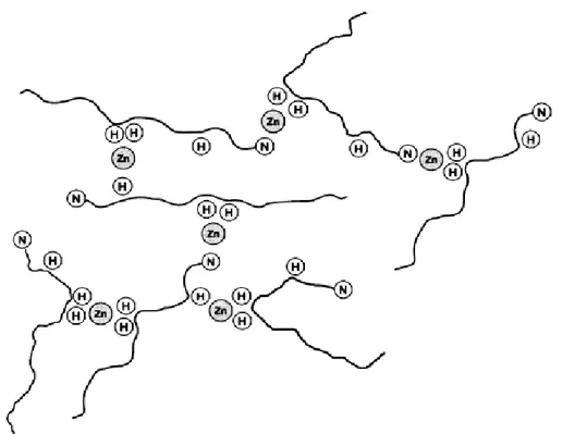
To the former group belong an interesting work by Syme et al.[28], who studied by NMR, UV and CD the binding mode of Zn in complex with Aβ(1-28). Their NMR data indicate that His6, His13 and His14 residues are implicated in Zn-Aβ binding. They show that high molecular weight polymeric species are formed, thus suggesting that Zn coordination is dominated by inter-molecular coordina-tion with consequent formacoordina-tion of polymeric species. In addicoordina-tion, they note that the UV spectra acquired either in the presence of Cu or in the presence of Zn ions are quite different, thus indicating that the coordination modes of these two metal ions are significantly different. A schematic model of the Zn coordination modes (taken from [28]) is represented in Fig. 1.9.
Figure 1.9: Intermolecular Zn2+ binding by Aβ peptide. Conjectural model
showing how various histidine (H) residues and the N-terminus (N) of the Aβ peptide may be binding Zn.
The model described above is consistent with the fluorescence and VIS-UV ex-periments of T˜ougu et al. [37]. They show that Aβ(1-40) forms a stable complex with Cu ions, while Zn promotes the partial aggregation of the peptide and the formation of polymeric species.
The possible role of Zn in promoting Aβ aggregation and fibrils formation has also been investigated. In particular, in a combined NMR, FTIR and XAS study Dong et al. [38] show that the Aβ(13-21) fragment reveals a β-sheet structure where parallel peptides are arranged in fibrils. They suggest a model where His13 residues on two different peptides chelate the metal, thus promoting fibrils formation.
To the latter group belong, among the others, the work of [27] and [39]. Daniels-son et al. suggest for Zn-Aβ(1-40) peptide a model similar to that of [26] for Cu, where Zn binding involves the three histidines and the N-terminus. Zirah
et al. study by NMR the Zn-Aβ(1-16) complex and identify the residues His6,
Zn cation. Both these works suggest therefore a monomeric state of the Zn-Aβ complex.
Even though the role played by Aβ oligomers and/or fibrils in the AD pathogen-esis is not completely understood, it is clear how a detailed knowledge about Cu and Zn binding sites, and in particular the behavior of these ions with respect to Aβ polymerization is very important. The XAS study presented in section 3.1 is indeed aimed at determining the metal coordination modes.
1.4
Transmissible Spongiform Encephalopathies
and the Prion Protein
In 18th century England, a disease affecting the behavior of sheep was identi-fied. This disease, which was recognized to be highly contagious, was termed scrapie because it caused the sheep to rub themselves against fences, trees and other hard objects scraping off their own wool. It also caused the sheep to walk poorly, display tremors, go blind, fall down and, at its last stage, die. When the brains of the sheep were dissected, the presence of many tiny holes, which made the tissues look like a sponge, was noticed. In addition to this characteristic sponge-like feature, the brains also showed shrunken nerve cells and proliferation of glial cells, known as astrogliosis.
In 1920, the German neurologists Hans Creutzfeldt and Alfons Jakob described a human disease with symptoms that included dementia, change in gait, pos-ture and movement, personality change, problems with speech and balance, and memory loss. This progressively degenerative disease always led to death. Upon autopsy, the brains of the affected individuals showed astrogliosis, the same char-acteristic as in scrapie brains. This human disease was called Creutzfeldt-Jakob disease (CJD), but no knowledge of its link to scrapie or its underlying cause was known.
In 1957, the American neurologist Gajdusek made a trip to Papua New Guinea and got in contact with a peculiar, apparently infectious disease who affected a local population, the Fore people, called kuru. It primarily infected women and children and its symptoms included unsteadiness of gait, hands, eyes and voice, tremor and shivering. The apparent source of transmission to the Fore people was through the practice of cannibalism. The Fore people, especially the women and children, did not have many sources of protein and had begun to cannibalize their dead as a way to conserve protein. The women and children, as lesser mem-bers of the Fore society, often received less desirable body parts. This included the brain and spinal cord. The theory of kuru transmission was that women and children ate the bodies, including the brains, of the kuru-afflicted people and the disease was passed on. A similarity between the kuru-infected brains and the description of the brains of patients with Creutzfeldt-Jakob disease was noticed:
they both showed astrogliosis and spongiform degeneration [40].
Nowadays all these pathologies are known to belong to the same class, the so-called Transmissible Spongiform Encephalopathies (TSEs), which comprise a group of fatal infectious neurodegenerative diseases that affect several animal species including mammals. They are all PCDs pathologies characterized by a fatal degeneration of the CNS.
In animals, the most common TSE is the scrapie observed in sheep. In the last decade of the XX century the bovine form of the disease (Bovine Spongiform En-cephalopathy, BSE) has gained much attention because of the possible relevance for human safety. In particular, because of the epidemic of BSE, clinical surveil-lance of the most common occurring TSE in humans, the CJD, was established in several countries.
1.4.1
Physiology and Pathology of the TSEs
All TSEs are characterized by spongiform degeneration (formation of fluid-filled holes in the brain tissue) and astrocytic gliosis (proliferation and branching of glial, structural non-neuronal, cells in the brain) of the CNS (see Fig. 1.10), that invariably lead to death. Interestingly this fatal process is accompanied by no febrile or immune system response, nor by the other common reactions shown by an organism which comes into contact with a pathogen. This observation suggests the presence of a peculiar infectious mechanism [41] the details of which will be given in the following paragraph 1.4.2.
There are at least three forms of TSE disease which affects humans [42]:
-Sporadic form, which is the most common form (about 85% of cases) of the disease;
-Heritable form, which is associated with mutations within the prion protein (PrP) gene (about 10% of cases). More than 20 mutations of the PrP gene are now known to cause in humans the inherited form of the diseases;
-Acquired form, which is a result of ingestion or inoculation of TSE-contaminated materials.
TSEs are unique among protein misfolding diseases because they have also a transmissible form. In fact, these diseases have all of the characteristics of more
Figure 1.10: Neuropathologic changes in Swiss mice brain following inoculation with scrapie prions. Spongiform degeneration is clearly visible in the left and upper parts of this section of the hippocampus [41]. Py is the pyramidal cell layer, SR is the stratum radiatum.
conventional viral or bacterial agents in the sense that they can replicate, have different strains of agent which are associated with different prion disease phe-notypes in vivo, and demonstrate very strong species specificities.
The annual incidence of the disease is uniform and accounts for about one case per million. The mean onset age of the disease is 65 years [43]. Main symptoms are: rapidly progressive dementia with myoclonus (rapid muscular contractions); cortical blindness (i.e. blindness due to cerebral damage); pyramidal and ex-trapyramidal signs (paralysis, anomalous muscular tone and rigidity).
From the physiological point of view, the disease hallmarks are characteristic pseudoperiodic sharp complexes of peaks in the electroencephalogram, associ-ated with cerebellar ataxia (non-coordination of movements). The accumulation of plaques in the brain (typical of the ’kuru’ form of the disease ) is observed only in a small minority of cases (5% of total cases). The death usually occurs within 3-4 months from the first manifestation.
1.4.2
The Structure of PrP
The discovery of the source of CJD and of all the other TSEs has been complex and still presents some uncertainty. However, TSEs are nowadays one of the best understood neurodegenerative disorders [43]. At the beginning of the XX century only the familial forms of the TSEs were recognized, while the infectious form of the pathology was unknown[44]. The discovery that apes inoculated with brain extracts from CJD patients would develop the disease was crucial in clarifying that some sort of pathogen was the cause of the disease, but the nature of this pathogen was unclear, since the responses typical of a organism in the presence of a pathogen was missing.
The main advance in understanding the nature of the pathogen agent was ob-tained by Stanley B.Prusiner’s, who was able to purify and quantify the amount of infective agent in a given sample, thus determining that scrapie infectivity could be reduced by procedures which hydrolyze or modify proteins. These ob-servations were interpreted by postulating that a proteic macromolecule was the pathogen agent for scrapie and CJD. Following Prusiner’s proposal, this pathogen was named prion, which defines a proteinaceous infectious particle that lacks nu-cleic acid.
Nowadays there are many experimental data that allow to conclude that prions are entirely made by a single protein, the PrP, although the possible involvement of other types of molecules or small proteins cannot be completely ruled out. The pathogenetic form of the protein is called PrPSc (Scrapie Prion Protein). It
has been shown that the infectivity titre is proportional to PrPSc concentration
[45]. Infectivity is retained also in the presence of highly purified preparations of PrPSc. Once the discovery that infective diseases can be mediated by a proteic
pathogen is accepted, it is necessary to understand the origin of the involved proteic material. It turns out that the PrPSc gene is contained in the genome of
all the species that can develop TSE. PrP is coded in a single gene, and the phys-iological protein, called Cellular Prion Protein (PrPC), is commonly expressed
[46] in the CNS, the brain, the heart, the lungs and the lymphoid system. It is also expressed at lower level in other tissues such as muscles, and very small traces can be found in blood.
N-terminal region of PrP, up to residue 120, is unstructured and highly flexible in solution. A characteristic hallmark of this region is the so-called octarepeat do-main composed of a number of repeats of the fundamental eight-residue sequence PHGGGWGQ. The C-terminal domain contains three α-helical segments, two short α-strands, two Cysteine residues linked by a disulfide bond, and a gly-cosylphosphatidylinositol anchor that tethers PrP to the membrane surface. A diagram with the main features of the primary structure of the PrP is given in Fig. 1.11.
Figure 1.11: A schematic diagram of PrP [51].
The octarepeat region is particularly interesting, because it is one of the more conserved regions of the PrP protein, thus proving that there is a selective pres-sure on it and suggesting that PrP should have a physiological role. The octare-peat region has been proved to be a binding site for metals[51]. The number of octarepeats depends on the organism in which the protein is expressed. For what concerns the more studied organisms, in cows there are five or six octarepeats [48], while in humans there are five octarepeats. In humans, mutations in the PrP that lead to the synthesis of mutant PrP with 6-14 octarepeats is one of the causes of the genetic form of CJD [49].
The two forms, PrPC and PrPSc, have identical primary structures, but exhibit
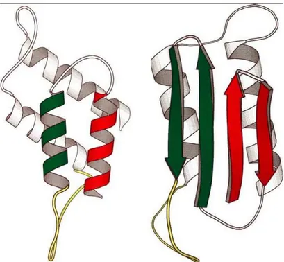
significant differences in their secondary structure [47]. PrPC contains ∼40% α-helix and has a minimal β-sheet content, whereas a 43% β-sheet and a
reduc-tion of α-helix contents are found in PrPSc. The PrPC-PrPSc conversion may
occur without any chemical modification, but the two isomers have considerably different physicochemical properties [50]. Fig. 1.12 shows a schematic picture of
both forms of the protein.
Figure 1.12: Secondary structure of PrPC (left panel) and PrPSc (right panel).
The finding that the brain of CJD patients contains deposits of PrP protein in its scrapie form lead to the theory that the disease is caused, at least in part, by the aggregation of PrPSc. Although the presence of deposits of PrP in the
brain is an indication of prion disease, it is now widely accepted that the real toxic species are the PrP oligomers. It has been shown that low molecular weight oligomers of the PrP are neurotoxic in vitro on cultures of neurons and in vivo after subcortical stereotaxic injection. On the contrary, monomeric PrP is not toxic, and large aggregates of PrP exhibits no toxicity in vitro and are less toxic than their oligomeric counterparts in vivo [52].
1.4.3
Interaction of PrP with Metal Ions
Observations from in vivo and in vitro studies using full-length recombinant PrP or its fragments led to important findings regarding PrP-metal interaction and its significance to prion disease pathogenesis [53]. It has been shown that re-combinant PrP binds several divalent cations, including Cu, Fe, Zn, Mn and Ni.
The octarepeat region of PrP is the principal metal binding site, and it has the highest affinity for Cu, followed by Ni, Zn and Mn [54].
The interaction of PrP with metals has been widely studied looking for the phys-iological and pathological implications of their association. For example, PrP is believed to mediate the uptake of Cu and Fe, suggesting a role in their cellular metabolism.
The association of PrP with Cu is better characterized than its interaction with other metals. Four Cu binding sites have been identified within the octapeptide repeat region of PrP with additional sites on the two histidine residues that do not belong to the octarepeat region [56] [102]. A drawing of PrP with its Cu binding sites is given in Fig. 1.13.
Figure 1.13: A model of PrP demonstrating the known copper binding sites in the octarepeat region and histidine residues 96 and 111 [53].
The interaction of PrP with Cu has also been studied using XAS [102]. Both the Cu-bovine PrP complexes and smaller Cu complexes of peptides containing one, two, and four copies of the octarepeat have been studied. Peptides containing one and two-octarepeat copies in sub-stoichiometric Cu complexes showed the direct binding to a single His compatible with can intra-repeat geometry. Alter-natively, the Cu-bovine PrP complex and Cu in complex with a four-octarepeat peptide at half-site-occupancy showed Cu directly bound to two His residues,
consistent with an inter-repeat binding mode.
In addition to Cu, other metals have been shown to bind PrP. Among them, zinc is the one with the highest affinity [54]. Zinc is also the only metal other than copper that induces PrP endocytosis. Zinc, inhibits fibril formation and promotes intermolecular interactions. It has been suggested that in vivo PrP may actually bind zinc rather than copper given the abundance of available zinc in the brain, with peak levels up to 300 µM in the synaptic cleft of glutaminer-gic neurons. However, even large excesses of zinc are unable to displace copper from either the octarepeat region or the full-length protein, but EPR reveals that physiologically relevant levels of zinc significantly alter the distribution of copper among the available binding modes [55]. These observations suggest that the interaction of PrP with Zn may be significant given the relative abundance of this metal in the brain.
From the physio-pathological point of view, observations from neuroblastoma cells suggest that PrP binds extracellular copper ions and delivers them to endo-cytic compartments, thus possibly functioning as a copper uptake protein [57]. The octarepeat region of PrP is essential for this process since its deletion in-hibits Cu uptake. Although in transgenic mice lacking PrP expression there is just a minimal difference in the Cu content of brains from wild type, it has been suggested that PrP may be a major Cu delivery protein [58].
1.5
Parkinson’s Disease and Neuromelanin
Parkinson’s disease (PD), which was first described by James Parkinson in 1817 in the book entitled ’An Essay of the Shaking Palsy’, is the second most preva-lent neurodegenerative disorder in the Western world. It is clinically defined as a neurological pathology characterized by 4 cardinal signs: resting tremor, bradykinesia, rigidity, and postural instability [59]. From a physiological point of view, it is characterized by depigmentation of the substantia nigra (SN), which is caused by the selective and progressive loss of dopaminergic neurons, and by the presence of intraneuronal proteinaceous inclusions known as Lewy bodies within the surviving neurons of the SN and other brain regions. These inclusions are enriched in filamentous α-synuclein and other proteins that are often highly ubiquitinated. In addition to the loss of '50% of neurons in the SN, there is widespread neurodegeneration in the CNS with the pars compacta and the locus
coeruleus (LC) being involved in midstages of the disease.
The incidence of the disease rises steeply with age, the median age of onset is 60 years and the mean duration of the disease from diagnosis to death is 15 years. The precise mode of death is difficult to identify in most cases, but pneumonia is the most common cause. PD is not related to race or religion, and literary and historical precedents before the publication of Parkinson’s monograph make it unlikely to be a postindustrial condition. The percentage of affected individuals within a population rises from 1% at 65 years to 5% at 85 years, making age the main risk factor for PD. The majority of cases are thought to be idiopathic, however, in 5-10% of cases there is a clear genetic component with both recessive and dominant modes of inheritance [60].
The molecular pathways leading to PD are still unclear, but the discovery of genes linked to rare familial forms of the pathology during the last decade have provided vital clues in understanding molecular pathogenesis of the more com-mon sporadic illness [61].
1.5.1
Physiology and Pathology of Parkinson’s Disease
PD commonly presents itself with impairment of dexterity or with a slight drag-ging the feet. The onset is gradual and the earliest symptoms might be unnoticed
or misinterpreted for a long time. Fatigue and stiffness are common but non-specific complaints. The early physical signs are often erroneously ascribed to old age, misery, introspection, or rheumatism, and a lag of 2-3 years from the first symptoms to diagnosis is not unusual. In the late stages of the disease, the face of patients is masked and expressionless, the speech is monotonous and slightly slurred, and posture is flexed with a severe tremor of the hands. Risk of dementia exists, particularly in those patients who show prominent gait and speech disorders or depression. The greatest risk factor for dementia, however, seems to be the age of the patient and not the duration of the disease.
Although PD is considered a sporadic disorder, remarkably few environmental causes or triggers have so far been identified. Similar to other neurodegenerative diseases, ageing is the major risk factor, although 10% of people with the disease are younger than 45 years of age. Never smokers are twice as likely to develop PD, and men and postmenopausal women who are not taking hormone replace-ment, who take no or very low quantities of daily caffeine, seem to be at increased (about 25% more) risk. These findings are probably related to dopamine’s role in reward pathways rather than to any neuroprotective effect of tobacco smoke, nicotine, or caffeine. Weak associations between PD and head injury, rural liv-ing, middle-age obesity, lack of exercise, well-water ingestion, and herbicide and insecticide exposure have also been reported.
From a genetic point of view, PD hereditary forms with autosomal dominant or autosomal recessive inheritance have been identified. They include auto-somal dominant point mutations or gene duplications or triplications in the
α−synuclein gene, and point mutations in the ubiquitin-C-terminal hydrolase-L1
and the leucine-rich repeat kinase genes, and also autosomal recessively inherited mutations in Parkin, PINK1, DJ-1 and the ATPase13A2 genes. By taking the biochemical function of these genes and mutations into account, it is possible to have an idea of the possible pathogenetic pathways which are at the basis of PD also in its more common sporadic form [62]. Essentially 3 mechanisms have been proposed:
1-Altered protein quality control
system leads to a disturbance in protein quality control. In particular, point mu-tations or gene duplications lead to the aggregation of α−synuclein and to the formation of α−synuclein fibrils. The misfolded protein can be either refolded by chaperone proteins, thereby regaining their normal function or have to be de-graded. They are marked by ubiquitin and are then directed to the proteasome. Dysfunction of one of these proteasome proteins therefore leads to a dysfunc-tion of the ubiquitin-proteasome system and to an accumuladysfunc-tion of aggregated
α−synuclein in the cytosol of dopaminergic cells (see Fig. 1.14). α−synuclein
Figure 1.14: Light microscopy of a surviving neuron in the substantia nigra of a PD patient. The neuron is full of α-synuclein Lewy bodies.
aggregation mainly occurs in the SN neurons, but it has been found also in the motor part of the nucleus vagus, in the olfactory bulb and in the LC.
α−synuclein is a small (14 kDa) natively unfolded presynaptic protein believed
to play a role in synaptic vesicle recycling, storage and compartmentalization of neurotransmitters and associates with vesicular and membranous structures. Structurally, it consists of an N-terminal amphipathic region, a hydrophobic middle region (containing the non-amyloid-β component domain) and an acidic C-terminal region. Three missense mutations in α-synuclein gene are associated with autosomal dominant PD [63].
α−synuclein has an increased propensity to aggregate due to its hydrophobic
major structural component of Lewy bodies in PD suggests a role of aggregated
α-synuclein in disease pathogenesis. As in the case of AD, however, most evidence
indicates that oligomers but not the fibrils of α−synuclein that are deposited in the Lewy bodies, are the real toxic species [64]. This would also imply that the rapid conversion of α−synuclein from an oligomeric to an aggregated state, de-posited in Lewy bodies, may help to detoxify the oligomeric form of α−synuclein. That the formation of Lewy bodies might be a protective response following the disturbance of protein quality control is also substantiated by the finding that at the end stage of the disease almost all of the few remaining and surviving dopaminergic neurons contain Lewy bodies. Interestingly, dopamine tends to stabilize the oligomeric form and prevents the aggregation of α−synuclein. This may be one reason for the selective vulnerability of dopaminergic neurons. 2-Mitochondrial dysfunction
Biochemical findings at autopsy as well as animal models studies suggest an al-tered mitochondrial function in PD, in particular a deficiency of complex I of the mitochondrial electron transport chain. Autosomal recessively inherited forms of PD with mutations in the PINK1 and the DJ-1 genes, which are related to mitochondrial activity, strongly support this hypothesis.
3-Altered kinase activity
Several aberrant kinase (enzymes that transfer phosphate groups from high-energy donor molecules) activities have been identified as contributing to the PD pathogenesis. Kinase related mutations represent indeed the most common monogenetic form of PD, affecting up to 16% of all patients of European origin and up to 40% in specific ethnic groups.
1.5.2
Diagnostic and Therapeutic Strategies in PD
The main symptom used for PD diagnosis is the unequivocal bradykinesia. Other indications are the facial expression, which can be immobile and rigid, the re-duced ability to express emotions, the speech which becomes slow, quiet, and lacking in rhythm and melody. PD is also characterized by a coarse, slow, pill
rolling tremor of the hands (4-6 cycles/s) and muscular rigidity.
Diagnostic techniques may also be used, in particular MRI shows extensive sub-cortical white-matter ischaemic changes. However, the ultimate diagnosis is the one done with post mortem analysis by detecting the presence of Lewy bodies and the loss of dopaminergic neurons in the substantia nigra pars compacta. From a therapeutic point of view, PD is still an incurable progressive disease, but treatments substantially improves quality of life and functional capacity. Several options exist for the symptomatic treatment of the pathology: oral treatment with L-Dopa, dopamine agonists, monoamine oxidase-B inhibitors and catechol-o-methyl-transferase inhibitors, continuous delivery of L-Dopa and dopamine ag-onists via transcutaneous or subcutaneous infusion, and deep brain stimulation. However, none of these therapies modifies the natural course of the disease[62]. Therapies aimed only at the execution of cell death, although possibly able to slow down the pathology development, are indeed unable to restore the function of neurons. Effective therapies may be found by interfering with the main patho-genetic mechanisms. A therapy with chaperones, small molecules that inhibit protein aggregation, and are able to enforce the ubiquitin- proteasome degrada-tion pathway, could stop the α−synuclein aggregadegrada-tion process and the formadegrada-tion of oligomers. Furthermore, mitochondrial dysfunction, oxidative stress and in-flammation can be addressed. Also some kinases may prove to be a promising target.
1.5.3
The Structure of Neuromelanin
The origin of the name melanin, from the Greek word µ²λας, black, is usually attributed to the Swedish chemist Berzelius [65]. Melanin in the brain has a similar appearance and structure to cutaneous melanins, and has thus been des-ignated neuromelanin (NM).
Based on their precursor molecules, melanins are classified into four groups [66]: 1. Eumelanin is formed from L-3,4-dihydroxyphenylalanine (L-dopa).
2. Pheomelanin is formed by oxidative polymerization of 5-S-cysteinyl-dopa or 2-S-cysteinyl-dopa.
or noradrenaline, with the possible involvement of cysteinyl-derivatives.
4. Allomelanin is formed by the oxidation of polyphenols, such as catechols and 1,8-dihydroxynaphtalene.
Although much is known about melanins outside the CNS, to which NM is thought to be related, many basic questions remain to be answered about NMs. It is unclear why some human dopamine neurons produce an insoluble pigment within their cytoplasm and others do not. Neurons rich in NM are indeed found especially in the SN and LC. (see Fig. 1.15). The structure and role of NM have
Figure 1.15: SN neurons containing NM.
been only partially characterized and are still a matter of investigation because of the possible involvement in brain aging and PD [67]. In fact, this pigment first appears in humans at 2-3 years of age and accumulates with aging.
The NM of SN shares some characteristics of melanins such as redox activity and chelating ability for metals. In NM isolated from SN and LC, a significant amount of iron was found. Due to its strong chelating ability for iron and other metals (cadmium, mercury and manganese), NM could play an important pro-tective role in neurons.
The process of neuromelanin formation is obscure, although some steps have been elucidated [69]. It has long been debated whether the synthesis of NM is enzy-matically mediated or whether it is a pure auto-oxidation process of dopamine derivatives. For the enzymatic synthesis, the enzyme tyrosinase catalyzes the conversion of tyrosine to L-dopa and then to dopa-quinone. Some authors pro-posed that tyrosinase could also be involved in neuromelanin biosynthesis, but
also other enzymatic mechanisms have been suggested, including tyrosine hy-droxylase mediated oxidation of dopamine. Alternatively, NM could derive from non-enzymatic oxidation. A dopamine-melanin can indeed be synthesized in
vitro by the auto-oxidation of dopamine, although there are several structural
differences between synthetic melanins and the natural one isolated from the
substantia nigra.
The proper melanic component of NM contains two classes of molecules in rather well-determined proportions. One is a benzothiazine-based molecule character-istic of pheomelanin that is formed through the incorporation of cysteine with dopamine; it constitutes 20-25% of the total melanic component of human NM. The other one is an indole-based molecule characteristic of eumelanin that is formed through the oxidation of dopamine [70]. The identification of dopamine and cysteinyl-dopamine as building blocks of natural NM lends support to the idea that its synthesis plays a detoxifying role, preventing an otherwise toxic intraneuronal accumulation of dopamine compounds.
NM has also been show to have high affinity for biomolecules, in particular lipids [68] and proteins. The lipidic component is mainly constituted by high molecu-lar weight lipids, while the peptide component of NM amounts to about 15% of its weight and is composed by several aminoacids found in fairly well preserved proportions. NMR studies [71] have shown that different α−synuclein immunore-active components are present in patients with Lewy bodies, thus suggesting a possible covalent bond between α−synuclein and NM.
From a structural point of view, X-ray diffraction studies [72] have shown that NM has a multilayer (graphite-like) three-dimensional structure made of pla-nar overlapping sheets of molecules containing indolebenzothiazine rings. Scan-ning probe and photoelectron emission microscopies have demonstrated that NM can be also found in spherical structures with a diameter of about 30 nm with pheomelanin at the core and eumelanin at the surface [73].
Several hypotheses have been proposed for the role of NM in the protection or degeneration of dopaminergic neurons in the SN. NM may play a cytoprotective role by sequestering redox active metals (Fe, Cu, Mn and Cr), toxic metals (Cd, Hg and Pb) and/or organic toxic compounds such as pesticides, which have been reported to be environmental risk factors for PD [70]. In contrast, it has been
proposed that NM increases the vulnerability of SN neurons and that it can be a source of free radicals by reaction with hydrogen peroxide.
A possible approach to this problem is to consider NM in terms of what is known about the better-characterized and more prominent peripheral melanins. Clearly, an increased understanding of the normal development of NM in the human sub-stantia nigra and changes that occur in PD will advance our understanding of this disorder and may provide targets for the development of novel interventions or treatments for the disease.
![Figure 1.13: A model of PrP demonstrating the known copper binding sites in the octarepeat region and histidine residues 96 and 111 [53].](https://thumb-eu.123doks.com/thumbv2/123dokorg/7605479.114798/38.892.219.617.497.802/figure-demonstrating-copper-binding-octarepeat-region-histidine-residues.webp)