PhD Course of Immunology
2000-2004
Coordinated by Professor Gino Doria
Title
Bacillus Calmette-Guerin (BCG) and environmental
mycobacteria divert Dendritic Cell functions interfering
with the differentiation of precursors into competent APCs
Implications for tuberculosis protection
Dr. Angelo Martino
Tutor: Professor Vittorio Colizzi
National Institute for Infectious Diseases Lazzaro Spallanzani- IRCCS 2004
“Lo scienziato non è l’uomo che fornisce le vere risposte:
è quello che pone le vere domande”
Alla memoria
di mia madre Giovanna
e a mio padre Giuseppe
Index
1. Abstract page 6
2. Introduction page 7
Immunology of Mycobacterium tuberculosis infection 7 Th-2 immune response in human tuberculosis: implication for vaccine design 16
BCG and tuberculosis: controversies about an old vaccine 18 Influence of environmental mycobacteria on BCG vaccination in tropical areas 21 Role of Dendritic Cells in tuberculosis infection and BCG vaccination 23
Aims of the study 28
3. Experimental procedures page. 29
Mycobacteria 30
Monocytes purification, infection and DCs generation 30
Fluorochrome-activated Cell Sorter (FACS) analysis 31 Confocal Laser-Scanning Microscopy (CLSM) analyses 31
Transwell experiments 32
Cytokines assay 32
T lymphocyte proliferation and functional polarization 33
Statistical analysis 33
4. Results page. 34
Phenotypic characterization of DCs derived from BCG-infected precursors 34 Distribution of CD1a molecules in BCG-DCs 40 Environmental Mycobacterium smegmatis affects DCs differentiation 42 The acquisition of mature phenotype by DC is independent on TNF−α 43 The induction of early mature phenotype requires mycobacteria-monocytes
interaction 45
T cell stimulation by BCG-DCs 47
T cell stimulation by Msm-DCs 48
Pro-inflammatory cytokines production by BCG-DCs 49 Immunoregulatory cytokine production by BCG-DCs and Msm-DCs 50
BCG-DCs induce Th2-like immune response in CBMCs impair the immune
response in naïve CD4 T cells 52 Msm-DCs direct naive T lymphocytes towards a mixed Th1/Th2
immune response 55
5. Discussion page 57
6. Acknowledgments page 64
7. References page 65
Abstract
Human monocytes can differentiate into dendritic cells (DCs) according to the nature of environmental signals. We tested here whether the infection with the live tuberculosis vaccine Bacillus Calmette-Guerin (BCG), that is known to be limited in preventing pulmonary tuberculosis, modulate monocytes and DCs differentiation. We found that monocytes infected with BCG differentiate into CD1a- DCs (BCG-DCs) in the presence of GM-CSF and IL-4 and acquired a mature phenotype in the absence of maturation stimuli. In addition, BCG-DCs produced pro-inflammatory cytokines (
TNF-αααα, IL-1ββββ, and IL-6) and IL-10 but not IL-12. BCG-DCs were able to stimulate allogeneic T lymphocytes to a similar degree as DCs generated in the absence of infection. However, BCG-DCs induced IL-4 production when co-cultured with human cord-blood mononuclear cells.
Since environmental mycobacteria can interfere with BCG efficacy in preventing tuberculosis in some tropical areas, we verify whether this capacity to interfere with DCs functions was a feature common to environmental mycobacteria. To this aim, we infected DCs precursors with Mycobacterium smegmatis (Msm) to generate DCs. We found that Msm-infected monocytes differentiate into DCs with the same phenotype of BCG-DCs but some differences in the capacity to produce immunoregulatory cytokines and polarize naïve CD4+ T cells.
The induction of IL-4 production by DCs generated from BCG-infected monocytes could explain one of the mechanisms for the failure of the BCG vaccine to prevent pulmonary tuberculosis.
1. Introduction
Immunology of Mycobacterium tuberculosis infection
Tuberculosis is a global emergency. One third of the world’s population is infected, and although only about 5-10 % develops active disease during the first few years following exposure 1, this still results in a massive case load, with eight million new cases each year, and three million deaths. Moreover, the percentage that progresses to disease is increasing. Tuberculosis is one of the first secondary infections to be activated in human immunodeficiency virus (HIV)-positive individuals 2. Moreover, the stresses of poverty, malnutrition and war, increase the rate of reactivation for reasons discussed later. Even in the developed countries, the disease distribution parallels the distribution of poverty 3. Meanwhile, the breakdown of healthcare systems is leading to incomplete case and contact tracing, incomplete treatment, and the increase in drug resistance. In some parts of the world, many of the available drugs are fake or out of date 4. In many areas, existing treatment is probably doing more harm than good, as incomplete treatment regimen select for drug resistance. Multidrug resistant tuberculosis is spreading at an alarming rate, and invading Western Europe from Eastern block countries such as Estonia. There were more cases of tuberculosis in 1999 than ever before in the history of humanity.
An important reason for the current failure to control tuberculosis is the fact that even the best available treatment must be continued for at least 6 months. This treatment regimen is not a realistic proposition in most developing countries, or even in the inner cities of rich ones, because the patients feel well after a few weeks and stop taking the drugs. The world health organization (WHO) now admits that directly observed therapy
short-course (DOTS), in which the patient is supervised while taking every dose of therapy helps but does not solve the problem 5.
There are two correlated reasons for the requirement for 6-month regimen. The first is obvious and often discussed. The chemotherapy kills the vast majority of the bacteria within a few days, but there are subpopulations of persisters 6. It is not clear whether these organisms are in true stationary phase 7 or merely replicating extremely slow. Nor is it clear where they are located. Most authors assume that they are in old lesions or sites of fibrosis or calcification, where oxygen availability may be low. However, in a forgotten paper published in 1927, Opie and Aronson reported that 80% of tuberculosis lesions were already sterile 5 years after the primary infection, whereas live bacilli could be found in macroscopically normal lung tissue 8. The fact that metronidazole, a drug that should be active in a model of latent infection in mice, implies that live organisms also persist in well-oxygenated sites in this species 9. Not only do persister organisms cause problems for the treatment, but they also constitute an important source of infection. They can persist for the rest of the life of the individual, and, at least in countries with low or moderate tuberculosis endemicity, many cases of tuberculosis result from reactivation of latent infection 10.
The other reason for the need for prolonged treatment is usually overlooked. Most tuberculosis patients have a necrotizing pattern of response to Mycobacterium
tuberculosis, analogous to the phenomenon first noted by Koch 11 in guinea pigs. There is overwhelming evidence that the Koch phenomenon does not correlate with optimal protective immunity to tuberculosis. Indeed pre-immunization of animals so that they have a Koch phenomenon before they are challenged with virulent Mycobacterium
tuberculosis results in a clear and reproducible increase in susceptibility to the disease,
pattern of response may not correct itself rapidly during treatment. Therefore if chemotherapy is stopped at 3 months, relapse rates are high even when chemotherapy was an optimal rifampicin-containing one that achieved sputum negativity well before 3 months, and in spite of the fact that there are very few live organism in the patients’ tissues at this time 13. We therefore need to understand the differences between protective immunity and the Koch phenomenon, and the factors that determine which response pattern is present. The ultimate objective is to learn to replace the pathological response with the protective one very large treatment.
Mycobacterium tuberculosis (MTB) is an intracellular pathogen with the capacity to persist in the early phagosomal compartment 14 in professional phagocytes. It arrests phagosome maturation at an early stage and strongly inhibits phagolysosome fusion 15. Based on Lurie’s studies in rabbits 16, four stages of pulmonary tuberculosis have been distinguished 17. Initially, there is the inhalation of tubercle bacilli (first stage). Alveolar macrophages ingest bacilli and often destroy them. At this stage, the destruction of mycobacteria depends on the intrinsic microbicidal capacity of host phagocytes and virulence factors of the ingested bacteria. Mycobacteria, which escape the initial intracellular destruction, will multiply, and this will lead to disruption of the macrophages. When this happens, blood monocytes and other inflammatory cells are attracted to the lung (second stage). These monocytes will differentiate into macrophages or dendritic cells, which again readily ingest but do not destroy the mycobacteria. In this symbiotic stage, mycobacteria grow logarithmically, and blood-derived macrophages accumulate, but little tissue damage occurs. Two to three weeks after infection, T cell immunity develops, with antigen-specific T lymphocytes that arrive, proliferate within the early lesions or tubercles, and then activate macrophages to
kill the intracellular mycobacteria. Subsequent to this phase, the early logarithmic bacillary growth stops (third stage). Central solid necrosis in these primary lesions inhibits extracellular growth of mycobacteria. As a result, infection may become stationary or dormant. Disease may progress, and haematogenous dissemination may take place after primary infection (fourth stage), as well as months or years after wards
(post primary tuberculosis), under conditions of failing immune surveillance.
Liquefied caseous foci provide excellent conditions for extracellular growth of MTB. Cavity formation may lead to rupture of nearby bronchi, allowing the bacilli to spread through the airways to other parts of the lung and outside environment. In summary, after entry in the human lung, MTB has a series of encounters with different host mechanisms. The outcome of infection with MTB depends on the balance between (i) outgrowth and killing of MTB and (ii) the extent tissue necrosis, fibrosis and regeneration.
The ability to manipulate the immune system of mice with neutralizing antibodies or gene knockout (KO) has provided strong evidence that in this species, immunity to tuberculosis correlates with a Type 1 response. In vivo T-helper (Th) 1 or Th2 cells act in concert with CD8+ T cells, and with numerous other cell types including macrophages, B cells and some stromal cells. Collectively these give rise to two patterns of cytokine release known as Type 1 (dominated by interleukin-2 (2), IL-12, and Interferon-γ γ γ γ (IFN-γγγγ)) and Type 2 (dominated by IL-4, IL-5 and IL-13) 18, 19. The term “Type 1” is used preferentially to Th1 referring to the overall pattern of cytokine release by all cell types in the infected site. Disruption of the Major Hystocompatibility Complex (MHC) class II genes or of the gene for the b chain of the
even to the avirulent Bacillus Calmette-Guerin (BCG). Disruption of the gene for the IFN-γ makes mice very susceptible to MTB (death within 3 weeks), and such mice may even die after many weeks from the challenge with BCG 21, 22, 23. IL-18 KO mice are also more susceptible, perhaps because IL-18 contributes to the induction of IFN-γ expression 24. A major inducer of Th1 pathway is IL-12. The exact role of IL-12 depends on the mouse strain 25, but IL-12 KO mice are not resistant to tuberculosis 26.
These data emphasize the crucial role of the Th1 immune response in tuberculosis immune response. In agreement with this, other data indicate that the Th2 immune response is not only unable to protect mice, but can seriously undermine the efficacy of the Th1 immunity. If a weak Th2 response to the shared mycobacterial antigens is deliberately induced before challenge, mice are found to be strikingly more susceptible to tuberculosis than non-immunized control animals 27. Similarly, in the Balb/c mouse model of pulmonary tuberculosis, the appearance of IL-4 in the lung lesions coincides temporally and spatially with the appearance of areas of pneumonia and necrosis, leading to rapid clinical deterioration and death 28. These observations are not contradicted by the claim that, in IL-4 gene disrupted mice; there is no evidence of increased resistance to the infection 29. First, such mice are not devoid of Th2 cytokine activity because IL-13 can substitute for many functions of the KO mice. Secondly, the detrimental role of the Th2 response is most apparent in the late progressive phase of the disease, particularly after 60 days 27, 28.
Some confirmation of the need for Th1 responses in man as in mice emerged from comparisons of patients and healthy contacts. For instance, patients produce relatively more antibody, whereas normal contacts produce relatively stronger T cell response to the 30 kDa antigens of MTB. Moreover, the cells from patients release less IFN-γ and more IL-10 in the presence of the antigen 30. Similarly, T cells from BCG-immunized
subjects respond more to the 16 kDa alpha-crystallin protein of MTB than do T cells from tuberculosis patients, who in contrast, have higher levels of antibody to it 31. Findings of this type suggest that Th1 responses are protective in men, as in mice. The study of serum concentrations of cytokines is uninformative in tuberculosis 32.
In humans, the elimination of MTB infection manly depends on the success of the interaction between infected antigen presenting cells (APCs) and T lymphocytes. Primary as well as acquired immunodeficiencies, especially human immunodeficiency virus (HIV) infection, have dramatically shown the importance of cellular immunity in tuberculosis. CD4+ T cells exert a protective effect by the production of cytokines primarily IFN-γ, after stimulation with mycobacterial antigens. The T cell response is mostly antigen specific, and attention has focused on the identification of immunodominant antigens, which may be used for the development of effective vaccine
33
. The acquired T cell response develops in the context of MHC complex and the polymorphism of MHC may contribute to differences in disease susceptibility of outcome 34, 35, 36.
Functional diversity of T lymphocytes may also be relevant. In addition to conventional CD4+ T cells, several other T cell types are also involved in the response to mycobacteria and represented in the figure below. Experiments, involving adoptive transfer, in vitro cell depletion and gene KO, have illustrated the importance of CD8+ T lymphocytes in the control of tuberculosis 37, 38. Protection of mice vaccinated with mycobacterial heat shock proteins 65 deoxyribonucleic acid (DNA) appears to be mediated mainly by CD8+ T cells 39. In an in vitro system, this ability to activate CD8+ T cells seemed to involve causing leakiness of the phagosome so that antigens reach the cytoplasm and hence join the conventional pathway for presentation on MHC class I 40,
41
, though another novel pathway may also be involved 42. A haemolysin-like molecule is in fact expressed by both MTB and BCG 43, and a BCG strain expressing the haemolysin from Listeria monocytogenes has been developed in the belief that this will increase the CD8+ T cell response 44. These CD8+ T cells have been shown to be cytotoxic, though the mechanism of this cell killing has been controversial. It has been thought that most cytotoxic T cells act to lyse infected cells and allow the released mycobacteria to be taken up by activated, uninfected macrophages that may kill them. However, it now appears that some CTLs directly kill MTB via granule-associated protein, granulysin acting with perforin 45. On the other hand, lysis by CD4+ cytotoxic T cells does not reduce the viability of the contained bacteria 46. Progression of tuberculosis in mice deficient in perforin is not different from progression in the wild type 47, 48. The major role of murine CD8+ T cells at this stage may be the secretion of IFN-γ 49. Recently, tuberculosis-specific CD8+ T cells have been identified in humans
50
, but their role in this species is equally uncertain. There are CD8+ T cells that will recognize tuberculosis-infected cells and secrete IFN-γ in blood from individuals with the disease 51, but these did not appear to contribute to control of intracellular proliferation of MTB in an in vitro system using human cells 52.
Most tuberculosis-specific CD8+ T cells recognize their antigens in association with MHC class I, however some are now known restricted by other molecules such as CD1 system 53. The relatively non-polymorphic CD1 family of molecules are MHC class I like, and possess cleft that binds lipid and glycolipid molecules 54 and allows their presentation to a variety of CD1-restricted cells, including αβ T lymphocytes negative for both CD4 and CD8 molecules (double-negative T cells), γδ T cells and certain CD4+, CD8+ and NK lymphocytes 55, 56. The exact role of CD1 and CD1 restricted cells, in either protection or pathology of tuberculosis, have proven difficult to evaluate, because mice possess a homologue of CD1d but no homologues of human CD1a, b or c. Indeed, mice deficient in CD1d have not been found to differ in the control of tuberculosis 57, though there is one claim that neutralization of CD1 resulted in exacerbation of the infection in mice at very early time points. The relevance of these findings to human disease is doubtful. Human CD1d has not been shown to present mycobacterial antigens, unlike classical CD1a, b and c that may present mycolic acid, lipoarabinomannan and other cell wall components 58. Double negative lymphocytes can recognize mycobacterial lipids in the context of CD1. Their predominant effector mechanism appears the secretion of IFN-γ and CD95/CD95L interactions 59, 60, and only rarely do they cause significant mycobacterial death. This has prompted suggestions that their role is the down regulation of local inflammatory responses by the removal of antigen-loaded antigen presenting cells. Many of the other types of human CD1-restricted cells also produce significant amounts of IFN-γ, but appear able to lyse infected cells and directly kill intracellular mycobacteria 61. It appears that MTB may be able to down-regulate CD1 expression on human antigen presenting cells, thereby potentially evading this component of the immune response 62.
Th-2 immune response in human tuberculosis: detrimental role and its implication for vaccine design
The susceptibility of children with defective receptors for IFN-γ or IL-12 provides definitive evidence of the importance of Th1 cytokines, suggesting a close parallel with the mouse models. Recently, the negative role of Th2 cytokines in human tuberculosis, again paralleling the murine models, has been established 63. Expression of IL-4, whether measured by flow-cytometry, or by sensitive quantitative RT-PCR on un-stimulated peripheral blood T cells, is increased and correlates with severity of diseases and with cavitation 64. The IL-4 mRNA copy number also correlates with total immunoglobuline E (IgE). Thus although it is true that actual cytokine levels and mRNA copy numbers are higher for Th1 than Th2 cytokines in tuberculosis, the major change in cytokine expression compared to healthy donors is not as previously stated, the small decrease in expression of Th1 cytokines, but rather a massive increase in expression of Th2 cytokines. This has resolved a long-running controversy, which deserves explanation. That there is a Th2 component in the response of human tuberculosis patients to MTB ought to have been accepted 10 years ago, because there is no other known explanation for the presence of specific IgE antibody 65656565. Interestingly, the other largely Type 2-cytokine dependent antibody, Immunoglobulin-G4 is also reported to be increased in patients 66. Similarly, immunochemistry reveals IL-4 expressing cells in the lymphoid organs of tuberculosis patients 67. Why then, has the matter been controversial? First, IL-4 is biological active at much lower concentrations than IFN-γ, and has a correspondingly lower mRNA copy number, so methods that reliably pick up IFN−γ or its mRNA fail to detect biologically significant levels of IL-4. Secondly, attempts to increase cytokine expression by stimulation of the cells in vitro do not yield an accurate reflection of the cytokine balance present in vivo, and rapid
early production of IFN-γ can suppress Th2 cytokine release. Finally, previous studies failed to take into account the presence of the IL-4 splice-variant. This variant of IL-4 may be an inhibitor of IL-4 activity, and is always co-expressed with IL-4, at about the same level 68. In lung cells, it may be expressed at higher levels than IL-4 itself. However, almost every study of IL-4 mRNA levels used primers that would amplify mRNA for both IL-4 and the splice variant. It is possible that some of the relative deficit in IFN-γ expression and lymphoproliferation in the peripheral blood of tuberculosis patients is due to the sequestration of the antigen presenting cells in the lymphonodes 69 or site of disease 70. However, this cannot fully explain the massive rise in expression of Th2 cytokines. What then, are the likely causes of this shift in cytokine profile? Increasing antigen load is likely to be one factor, since Th1/Th2 balance is strikingly linked to dose when immunizing with particulate antigens such as mycobacteria 71 or Leishmania 72. In some populations, excessive or inappropriate contact with environmental mycobacteria may have primed a Type 2 response to the crucial antigens. This mechanism is clearly demonstrable in mice, in which it can massively increase the susceptibility, and has been suggested, but not conclusively proven in man 73. During active tuberculosis, selective apoptosis of Th1 like T cells may be a factor, as may prostaglandin release and increased secretion of transforming growth factor-β (TGF-ββββ) and IL-10 74. However, the latter may be a consequence of the shift towards a Th2 immune response. Finally, there are now strong reasons for suggesting that endocrine interactions with the immune system are important.
BCG and tuberculosis: controversies about an old vaccine
Ever since its introduction in infant immunization, Bacillus Calmette-Guerin or BCG vaccine has attracted more controversies than any other vaccine. A vaccine in general is adjudged on two important parameters: a) ability to protect individual from the particular disease and b) control of the disease at community level and possibly eliminate and eradicate the same.
BCG is now being used routinely in over 100 countries in their National Immunization Programme. The main objectives are to prevent disseminated and other manifestations of MTB. However, BCG does not prevent infection of MTB. Because of genetic changes in the bacterial strains that have occurred over many years, various BCG vaccines used throughout the world differ 75. However, identification of complete BCG genome in 1998 has facilitated further research in development of more effective vaccine alternatives 76. From the earlier observation of to 80 % efficacy, recent meta analysis have shown greater efficacy of > 80% against meningeal and miliary tuberculosis in children and in one meta analysis >50% efficacy was recorded even against pulmonary tuberculosis 75. In short, endemicity of the disease, heavy bacillary load, constant contact with open tuberculosis case, concomitant HIV disease etc… are some of the factors which might affect the efficacy of BCG vaccine. In India, BCG vaccine is produced from Danish Strain 1331, based on the Copenhagen technology and standards. However, in the National Immunization Programme BCG vaccine produced from other strains are also supplied sometimes. Ever since the introduction of Universal Immunization programme in 1985, BCG vaccine, coverage has been uniformly stepped up to >85%. Sentinel centers under UIP have been constantly reporting low/nil incidence in meningeal tuberculosis, miliary tuberculosis and disseminated tuberculosis, which denote the field efficacy of BCG vaccine in controlling the haematogenous
spread of childhood tuberculosis. The identification of complete genome sequenced of MTB in 1998 has opened new vistas in developing newer anti-tubercular vaccines. Currently this research is mainly directed toward purification and synthesis of protein peptide and non-peptide antigens of MTB, creation of rationally attenuated mutant antigens from MTB, development of DNA vaccine based on published genome sequence, development of suitable vehicle for these potential subset vaccines using living vaccine carriers strain.
BCG is named after French scientists Calmette and Guerin who in 1908 isolated
Mycobacterium bovis from a cow with tuberculosis mastitis. They cultured this isolate
in a glycerinated, beef/bile/potato medium, subculturing every 3 weeks, until 13 years and 230 subcultures later, the bacillus had lost evidence of virulence 77. After this acceptance, as a vaccine for tuberculosis in humans, the BCG Pasteur Lille was widely distributed throughout the world to other institutions. As a result, many daughter substrains come into being, and recent molecular analysis has revealed numerous changes, gene deletions, as well as a suspicion that continued passage has attenuated BCG to almost complete avirulence 78.
BCG vaccination is remarkably safe, unfortunately, although BCG has been effective in certain controlled clinical trials, in other, it has been disastrous, with no effect at all. An appreciation of this depends on a “glass half empty” or “half full”. Certain a percentage of scientists falls into the half-empty group, believing that BCG is essentially worthless and needs to be completely replaced. Conversely, the half-full membership points to the fact that in some of the trials, BCG did work well indeed, whereas in other did not. So one question, still unanswered, is what is BCG doing when it does have positive effect?
In fact, the stark reality is that this question has never been adequately answered. If the working hypothesis is believed, that BCG induces memory immunity, but this immunity is gradually lost, then the literature shows that recent, serious studies of these parameters in models are counted on two hands. BCG can switch on a variety of mechanisms, as summarized in the figure below. Central to these is a potent, memory T cell response 79. In areas in which BCG is not effective, one possible explanation is that this T cell memory pool is consumed or gradually influenced leading to a loss of efficacy. This may the result of non-specific stimulation of memory T cell turnover and division, which may force a percentage of the pool into apoptosis, thus eventually reducing the pool size to a frequency whereby it is no longer effective. However, all available evidence suggests that the BCG is insufficient. The exact reasons for this unsuccessful are not completely understood. Theories proposed include natural cross presentation or suppression resulting from exposure to environmental mycobacteria in tropical and temperate climates that may mask or impair the protective effect of BCG, co-infection with other pathogens such as helminths, as well as the genetic variability of vaccines 80. Regardless of the explanation, it is generally agreed that a novel, more effective vaccine is required. Indeed, using mathematically modelling based on current global predictions; a new vaccine with only 50% efficacy could prevent 9 million deaths from tuberculosis by the year 2030. Of course, this level of protection would need to be achieved in many different populations, and not as a mere average of divergent groups. Therefore, it is critical that information concerning the host response to MTB and BCG is briefly reviewed when considering new potential vaccine candidates.
In particular, the primary habitats of mycobacteria are the macrophages and dendritic cells and all problems concerning the trigger of the effective immune response by these APCs will help to design a novel anti-mycobacterial vaccine.
Influence of environmental mycobacteria on BCG vaccination in tropical areas
The reason of BCG failure in some populations has been subject of debate since the 1950s, and many different hypotheses have been suggested to explain the observed variation. Some investigators have suggested that difference in the strain of BCG, the age of vaccination, or methodological differences are important factors for the variation reported. The most widely accepted hypothesis relates the efficacy of BCG to geographic location, with low to non-detectable levels of protection against tuberculosis seen in tropical regions such as Africa and India, where exposure to nontuberculous
mycobacteria is common 81. One exception from this general role is the consistent high efficacy when BCG is used to vaccinate newborn. Neonatal vaccination with BCG imparts protection against childhood manifestations of tuberculosis (in particular
meningitis) 82, but the efficacy wanes over a period of 10 to 15 years, and therefore it
does not prevent pulmonary tuberculosis in the adult population in the third world. There is convincing evidence that exposure of laboratory animals to environmental mycobacteria can provide some protection against infection of MTB 83. The prior sensitization with environmental mycobacteria can inhibit BCG multiplication and thereby prevent the induction of an efficient BCG-mediated immune response and protection against tuberculosis 80, 84. This can be explained through the particular sensitivity of BCG to the influence of pre-existing immune response to antigens shared between environmental mycobacteria and BCG. The multiplication of BCG is a precondition for the induction of immunity by BCG and killing of BCG by chemotherapy after administration has been demonstrated to abrogate subsequent immunity completely. This inhibitory effect provides an important argument in the ongoing discussion of live attenuated vaccines versus nonviable subunit vaccines against tuberculosis.
Role of Dendritic Cells in tuberculosis infection and BCG vaccination
It is well established that Dendritic cells (DCs) are APCs specialized for naïve T cell activation 85. Immature DCs are bone marrow-derived cells present in most non-lymphoid tissues, where they act as sentinel cells against incoming pathogens. DCs are characterized by a high capability for antigen capture and processing, but low T cell stimulatory activity. Inflammatory factors or microbial component, promote DCs maturation and migration out of the non-lymphoid tissues into the blood or afferent lymph. Maturing DCs then will rich lymphoid organs, where they home to T cell areas. At this stage, DCs have undergone a dramatic change in their properties, having lost the ability to capture antigens but having acquired an increased capacity to prime T cells 86,
87
. The summary of the dendritic cell-life is shown in the figure below.
This unique capacity of DCs to sample antigens at the site of infection, respond to microbial signals and activate naïve T cells suggests that they play a critical role in the initiation of antimicrobial immunity. Although DCs are efficient APC, it is also
apparent that they act as key modulators of immune response. The innate response of DCs to microbial antigens may preferentially drive CD4+ T cells to mature into Th1 or Th2 cell types, therefore controlling the generation of Th1 or Th2 immune responses to the pathogens 88. This immune activity has been clearly demonstrated in experiments involving the intravenous injection of mice with an extract of Toxoplasma gondii 89. After 3 hours, IL-12 production was detected in the spleen of animals. The IL-12 producing cells were identified as DCs localized in the T cell areas of the spleen. This IL-12 production was independent on T cell signals, thus demonstrating that DCs were the initial source of IL-12 in response to microbial stimulus. This IL-12 producing feature of DCs is of particular relevance in mycobacterial infections, since the production of IL-12 in response to MTB is a key event of protective immunity. The function of IL-12 is to activate naïve T cells and NK cells, as well as directing CD4+ T cells down the IFN-γ secreting pathway. In this way, IL-12 serves as a bridge between innate resistance and adaptive immune response against intracellular pathogens. The stimulation of IL-12 production by DCs in response to mycobacterial antigens may lead to accelerated Th1-oriented immunity and therefore to a better control of the disease. Research into the impact of mycobacterial infection on DCs has been facilitated by the development of culture conditions in which immature DCs of murine or human origin van be grown from precursors in the presence of growth factors 90, 91. DCs can internalize mycobacteria, either heat-killed or alive, with equal efficacy. Such phagosytosis may be enhanced by specific interactions between the bacilli and DCs surface molecules. Interaction of DCs with live MTB or BCG results in a direct cell maturation documented in DCs phenotype and in cytokine production and chemokines expression. Mycobacteria-infected DCs have a decreased endocytic activity, as shown by their low capacity to internalize latex beads or dextran after the infection 92.
Similarly to other bacterial products, such as LPS, mycobacterial stimuli up-regulate the DCs expression of co-stimulatory molecules such as CD80, CD86, the intercellular adhesion molecules (ICAM-1) and the signalling molecules CD40 93. MTB or BCG infection of DCs also up-regulate the expression of MHC molecules, suggesting that infected DCs have an increased capacity to stimulate T cells against mycobacterial antigens. Accordingly, BCG-infected DCs are more efficient than un-infected DCs at eliciting specific cellular responses in vivo 94.
In addition to the classical MHC system, DCs can present non-protein antigens to T cells by CD1 molecules. The impact of mycobacterial infection on CD1 expression is not clear, since MTB infection has no effect on CD1a, b, c expression on human PBL derived DCs 92. The presence of CD1+ DCs in human leprosy lesions correlates directly with effective immunity, suggest that CD1 presentation of antigens may play a role in augmenting host defence against mycobacterial infection 95. Indeed, the major mycobacterial cell wall component lipoarabinomannan (LAM) is presented by CD1b. similarly CD1a and CD1c have been found to present mycobacterial lipids to human T cells 56. Because CD1 expression is down regulated in MTB infected macrophages, it is tempting to postulate that MTB may have developed evasion mechanisms to escape mycobacterial antigens presentation through CD1 molecules 62. Mycobacterial infection of DCs results in the up-regulation of the regulatory cytokines such as IL-12 and
TNF-α. These molecules play a major role in protective immunity against MTB infection, because they contribute to the acquired specific resistance and to the development of delayed-type hypersensitivity (DTH) 96. The IL-12 production induced by mycobacterial infections potentiates the development of T cells that produce IFN-γ and TNF-α, both potent activators of bacterial killing by infected macrophages 97. The
generation of IL-1β, IL-6 and TNF-α contribute to the development of the inflammatory process, through the recruitment of leukocytes leading the granuloma formation and the containment of bacterial dissemination 98. Interestingly, these pro-inflammatory molecules are secreted in greater quantities from DCs cultured with live bacilli than with killed bacteria 92. This feature may explain the lack of efficacy of killed BCG compared to live BCG as a vaccine against MTB infection 33. DCs infection with MTB also induces the early production of IL-10 92. Because IL-10 is a potent suppressor of macrophages effector functions, this interaction between lung DCs and mycobacteria may result in local inhibition of the resident macrophages activation and alter the ensuing bacterial clearance. It has been demonstrated that IL-10 may inhibit the development of cellular responses to mycobacteria suppressing the release of IL-12 by DC 99. IL-12 production by BCG-infected DCs is significantly higher in IL-10 deficient cells and this effect is reinforced on CD40 ligation. This allows that IL-10 exerts an inhibitory effect on both mycobacteria and CD40 induced IL-12 production by DCs. These data are in accordance with previous studies showing that the addition of exogenous IL-10 to LPS-activated monocytes and LPS or CD40-activated DCs blocks their IL-12 synthesis. This blocking effect is likely to have a marked effect on the generation of protective immunity in vivo, because it would inhibit both innate and adaptive IL-12 responses of DCs to mycobacteria and may account for the increased antimycobacterial immunity of IL-10 deficient mice in response to BCG infection 100. The generation of proinflammatory cytokines is an essential component of resistance against tuberculosis infection. The production of chemokines at the site if infection is also an important element of the immune response to mycobacteria, because it controls the further recruitment of DCs precursors and the influx of cells participating to the inflammation process 101. These cells include monocytes that are the direct precursors of
DCs and activated lymphocytes, which contribute to granulomas formation. Summarizing these observations, mycobacterial infection of DCs induces cell maturation and up-regulation of the production of inflammatory cytokines and chemokines that are essential for the resolution of mycobacterial infections. Importantly, mycobacteria-infected DCs express high levels of IL-12 and the production is enhanced by CD40 stimulation, a fact that may contribute to the generation of the Th1 immune response. In this context, it is important also the contribution of IL-10 developed early in infection that can be a mechanism for mycobacteria to escape immunity down modulating the pre-existent Th1 immune response. In the figure below, the representative panel of the DCs function in mycobacterial infection is shown.
Aims of this study
Peripheral blood monocytes can differentiate into dendritic cells or macrophages depending on environmental factors encountered during their migration from blood to peripheral tissues 102, 103. Transendothelial trafficking and culture in the presence of serum from systemic lupus erythematous induce monocyte differentiation into immature DCs 104. Upon contact with IL-4 plus GM-CSF (cytokines that could be produced by
tissue mast cells), monocytes also differentiate into immature DCs. Addition of TGF-β
or exposition of monocytes to GM-CSF plus IL-15 lead to DCs features of Langherans cells. In contrast, M-CSF is a potent macrophage differentiation factor. IL-6 also shifts monocyte differentiation from DCs to macrophages. Tumor cells produce IL-6 and M-CSF that shift the differentiation of CD34+ progenitors from DCs to macrophages 105. In addition, cytokines such as IFNγ produced by NK cells or activated T cells can modify the pattern of differentiation of DCs.
At the sites of inflammation, cytokines and chemokines promote the activation of resident DCs and the recruitment of DCs precursors. Dendritic cell precursors may permeate peripheral tissues including the skin, where they receive stimuli from local infection or inflammation 106. Different signals of infection, inflammatory cytokines such as TNF-α or phagocytosis have been demonstrated to interfere with DCs differentiation 107, 108. Many pathogens have evolved mechanisms to exploit DC biology in their dissemination within the body, and/or interfere with DC functions to block or delay their elimination by the host. Several pathogens or their components can interact with either imDCs or their precursors influencing DC generation 109, 110. It has been demonstrated that MTB, is able to subvert the differentiation of infected monocytes into DCs, generating a subset of DCs population unable to induce an effective immune response and suggesting an escape mechanism which contribute to mycobacterial
intracellular persistence 111. Several properties of MTB are retained by BCG and other mycobacteria that can explain one of the mechanisms of the failure of vaccination and the influence of the habitat in the immune response to MTB. Indeed, the unsuccessful of tuberculosis vaccination may be at least partially explained by the induction of poor or inappropriate host responses induced by APCs.
Because of the role of DCs in the induction of immune response to M. tuberculosis and the contribution to the generation of protective cellular immunity against mycobacteria during the vaccination with BCG, we hypothesized that BCG can retain the capacity to influence DCs functions. In this work, we investigated the effects of BCG comparing to that of environmental mycobacteria on the differentiation of monocytes into imDCs and mDCs to obtain findings explaining some mechanisms underling the failure of the BCG vaccine to prevent pulmonary tuberculosis.
2. Experimental procedures
Mycobacteria
In all experiments, Bacillus Calmette Guerin or BCG vaccine from Aventis Pasteur (Paris, France) has been used. Lyophilized BCG was resuspended in physiologic solution at approximately 1 x 10⁶ Colony Forming Units (CFU) per 100 µl.
Mycobacterium smegmatis (Msm) MC2 155 strain has been used. Mycobacteria were grown in Middlebrook 7H9 medium for 3-4 days and frozen stocks were stored at -80 °C in medium and glycerol 15%. In some experiments, mycobacteria were inactivated by incubating them for 60 minutes at 80°C. The viability of bacteria was assessed by Colony Forming Units (CFU) analysis.
Monocytes purification, infection and DCs generation
Peripheral Blood Mononuclear Cells (PBMCs) were isolated from buffy-coats of healthy donors by density gradient centrifugation using Lympholyte-H (Cederlane, Canada). Monocytes were positively separated by anti-CD14 magnetic beads (MACS, Milteny Biothec, Germany) according to the manufacturer’s instructions. The cells were then resuspended in RPMI 1640 (Euroclone, UK) supplemented with 10% FCS (Biowittaker, Belgium), L-glutamine (2mM), Hepes buffer (10mM), sodium piruvate (0.1 M), non-essential amminoacids (0.1 M) (Euroclone, UK) and gentamycin (10µg/ml) (Sigma-Aldrich, Germany). Monocytes were infected for three hours with single cell suspensions of BCG at a multiplicity of infection (MOI) of 1, or stimulated with LPS (from E. coli, 1µg/ml) (Sigma-Aldrich, Germany). The infection was carried out in the absence of antibiotics. After the treatment, monocytes were washed and cultured with fresh complete medium for 5 days in the presence of
granulocytes-macrophages colony-stimulating factor (GM-CSF) (200 U/ml) and interleukin-4 (IL-4) (10ng/ml) (Euroclone, UK) to generate imDCs. To induce final maturation of imDCs, LPS (200ng/ml) was added at the fifth day of culture for further two days. Viability of infected cells was determined by trypan-blue exclusion.
Fluorochrome-Activated Cell Sorter (FACS) analysis
The following FITC- or PE-conjugated antibodies: anti-HLA class I, HLA DR, CD1a, CD11c, CD14, CD40, CD64, CD80, CD83, CD86, CCR7 (Becton Dickinson Bioscience Mountain View, CA, USA), were used for direct immunofluorescence staining to characterize the phenotype of DCs generated in different conditions. Briefly, the cells were washed twice in PBS, 1% BSA and 0.1% sodium azide and stained with the mAbs for 15 min at 4°C. The cells were then washed and acquired using a FACScalibur instrument running Cellquest Software (Becton Dickinson).
Confocal Laser-Scanning Microscopy (CLSM) analyses
For CLSM analyses, 5x10⁵ DCs were added per well in a U-bottom microtiter plate and washed once with PBS containing 0.2 mM NaN3 and 1% BSA. Cells were
incubated (30 min., +4°C) with appropriate dilution of anti-CD1a-FITC or anti-actin-PE mAbs. After extensive washes, the cells were fixed with 3% paraformaldehyde, washed once and mounted on the microscope slide with the Prolong antifade reagent (Molecular Probes). The control samples were treated in the same way. For intracellular staining, the cells were fixed and permeabilized with cold methanol (-20°C, 10 min.) and then incubated with the antibody for 30 min. at 37°C.
CLSM observations were done using a Leica TCS 4D apparatus, equipped with an Argon-Krypton laser, dichroic splitter (488 nm), 520-nm long-pass filter for
observations with FITC-conjugated antibody. Image acquisition and processing were conducted by using the Scanware (Leica Lasertechnik GmbH, Heidelberg, Germany) and Adobe Photoshop software programs.
Transwell experiments
In transwell experiments, the infected and uninfected monocytes were separated by a membrane (6.5 mm of diameter, 0.4 µm pore size) in 24-well plates (Corning-Costar, Cambridge, MA). The lower compartment of the wells contained untreated monocytes (1 x 10⁶ cells). The upper compartments contained either untreated cells, BCG-infected or LPS-treated monocytes (1 x 10⁶ cells). The cells were cultured in the presence of GM-CSF and IL-4 in 2 ml of culture medium. After five days, the cells were harvested from the lower and upper compartments, stained with the anti-CD1a mAb and analyzed for the CD1a expression by FACS.
Cytokine assay
Supernatants of DCs derived from treated or untreated precursors were collected at the fifth day of culture or after stimulation with LPS (200ng/ml) for 48 hours and stored at – 80°C. The levels of IL-10, IL-12, TNF-α, IL-1β and IL-6 were determined by ELISA kits (Pierce, Endogen, USA), according to the manufacturer’s instructions. Supernatants of CBMCs and purified CD4+CD45RA+ T cells co-cultured with DCs generated from untreated, BCG-infected or LPS-treated monocytes were tested for IL-4 and IFN-γ using commercially available ELISA kits (Pierce, Endogen, USA). Results are expressed as pg/ml. In some experiments, supernatanants of DCs co-cultured with naive T cells from cord-blood were tested for IL-2, IL-4, IFN-γ, TNF-α using
commercially available Cytokines Beads Assay (CBA) kit (Becton Dickinson Bioscience, Mountain View, CA, USA). Results are expressed as pg/ml.
T lymphocyte proliferation and functional polarization
DCs derived from untreated, BCG- or Msm-infected monocytes (0 to 1 x 10⁴ cells) were added to 5 x 10⁴ allogeneic PBMCs in 96 well U-bottom microplates (Corning-Costar, Cambridge, MA). The proliferative response was measured after 5 days of culture and 16 hours of incubation with {3H}thymidine (Amercharm, Little Chalfont, GB) (1µCi/well). Incorporated radioactivity was then assessed by a 1450 MicroBeta Trilux (Wallac, Turku, Finland). Results are expressed as means + SD of triplicate wells. T lymphocyte polarization was evaluated incubating DCs with Cord-Blood Mononuclear Cells (CBMCs) and purified CD4+CD45RA+ T cells at ratio of 1:5 for 8 days. CD4+CD45RA+ T cells were purified by incubating CBMCs with anti-CD45RA magnetic beads (MACS Milteny Biothec, Germany). According to the manufacturer’s instruction, after O/N incubation to allow beads degradation, CD4+ T cells were further purified. Supernatants were collected and analyzed for cytokine accumulation.
Statistical analysis
Statistical analysis was determined using a Student’s t test. Values of p<0.05 were considered statistically significant.
3. Results
Phenotypic characterization of DCs derived from BCG-infected precursors
In order to investigate the effect of BCG in DCs differentiation from peripheral monocyte precursors, we purified human monocytes from buffy-coats of healthy donors and infected with BCG at MOI of 1 for three hours. On attempt to evaluate the efficiency of BCG infection of human monocytes at MOI of 1 after 3 hours of incubation, we labelled BCG with FITC (1µµµµg/ml) by incubating the bacteria for 30 min at 4° C and then washed extensively. Labeled bacteria were used for the infection and after 3 hours of incubation, monocytes were washed 3 times with fresh medium and analyzed by FACS. More than 90 % of the cells were FITC positive, suggesting the percentage of infected cells. (Figure 1)
Figure 1. Purified monocytes (98%) were infected with BCG FITC labelled at MOI of 1 for three hours. After the infection, monocytes were washed 3 times in PBS and analyzed by FACS. Dot plots show the percentage of positive cells
We cultured infected monocytes in complete medium containing GM-CSF and IL-4 for five days. Human monocytes developed into imDCs characterized by the acquisition
of CD1a and the loss of CD14 and CD64 molecules. ImDCs can be induced to maturation by several stimuli, including LPS. The maturation process is associated with the expression of CD83 and CCR7 and a strong up-regulation of CD40, CD80, CD86 and MHC class I and II molecules. In contrast, imDCs generated from BCG-infected monocytes (BCG-DCs) did not express CD1a on their surfaces (Figure 2).
CD86 CD80 MHC class I MHC class II
Martino A. et al. Journal of Leukocyte Biology, 190:1167-1176, 2004
Figure 2. BCG-infected monocytes differentiate into mature DCs with a failure of CD1a expression. Histogram plots show the phenotype of DCs derived from BCG-infected (BCG-DCs) or untreated monocytes (imDCs) at the fifth day of culture (filled histogram) and after 48 hours of LPS stimulation (empty histogram). The cells were stained for the various markers and acquired by FACScalibur. Numbers indicate mean fluorescence intensity values at fifth day of culture (bold) and after LPS stimulation (normal). One representative result of ten independent experiments is shown.
However, BCG-DCs were CD14- and CD64- (data not shown), suggesting that they were not blocked at the monocyte/macrophage stage. Furthermore, BCG-DCs at the fifth day of culture, expressed higher levels of MHC class I, class II and CD80, CD86 co-stimulatory molecules than imDCs in the absence of maturation stimuli. Interestingly, BCG-DCs acquired the phenotype of mDCs at the fifth day of culture, as shown by the expression of the maturation markers such as CD83 and CCR7 (Figure 3). The presence of CCR7 suggests that the infection is sufficient to induce migratory receptor expression required by mDCs to reach lymphoid organs. Following stimulation with LPS (200ng/ml) for 48 hours, BCG-DCs did not further up-regulate their maturation markers. Moreover, imDCs after stimulation with LPS acquired a mature phenotype, showing the increase of MHC class I, class II, CD80 and CD86 and the induction of CD83 and CCR7.
Together with these data we wondered whether the acquisition of early mature phenotype by DCs was due to simply stimulation of monocytes with bacterial products. We used LPS to stimulate monocytes before culturing them for five days in the presence of GM-CSF and IL-4. After five days we compared the DCs generated by LPS treated monocytes (LPS-DCs) and DCs generated by BCG infection. In contrast to BCG-DCs, the phenotype of LPS-DCs was similar to that of imDCs except for the expression of MHC class II which was up-regulated and the CD1a expression that shows the slight reduction (Figure 3). After the stimulation for further 48 hours with LPS, LPS-DCs acquired the mature phenotype, showing the increase of MHC class I, class II, CD80 and CD86 and the new acquisition of CD83 and CCR7.
Figure 3. LPS-stimulated monocytes differentiate into DCs with a similar phenotype of imDCs derived from untreated monocytes, except for MHC class II.
All these effects were observed using monocytes isolated from different donors and each experiment was reproducible (Figure 4).
Martino A. et al. Journal of Leukocyte Biology, 190:1167-1176, 2004
Figure 4. Histograms show the mean of MFI (Mean Fluorescence Intensity) of ten independent experiments. Black bars indicate the MFI at fifth day of culture and white bars indicate the MFI after further 48 hours of LPS stimulation.
Furthermore, the incubation of monocytes that had phagocytosed heat killed BCG developed into DC (hkBCG-DCs) displaying a phenotype resembling that of BCG-DCs showing a strong up-regulation of CD86. However, CD1a expression was not completely negative and other markers such as MHC class I, class II and CD80 were less up-regulated indicating a low degree of maturation in comparison to BCG-DCs (Figure 5).
Figure 5. Histogram plots of a representative experiment of monocytes infected with heat killed BCG and cultured with GM-CSF and IL-4 for five days. Numbers indicate the MFI
Finally, the peculiar phenotype of BCG-DCs is dependent of mutiplicity of infection and the maximum effect was observed at MOI of 1. Higher multiplicity of infections resulted in a high mortality of monocytes and in no further variation of surviving cells
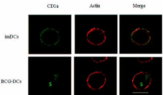
Distribution of CD1a molecules in BCG-DCs
It has been described that DCs with the CD1a- phenotype generated in different experimental conditions, have the capacity to produce IL-10 and to polarize T cells towards a Th2 response 112, 113. On the other hand, it has been reported that mycobacterial infections interfere with the surface expression of CD1 molecules and that mycobacterial antigens interact with different members of CD1 family 62. To investigate whether BCG-DCs are a subset derived from distinct precursors giving rise to CD1a- DCs population, or whether the down modulation of CD1a is induced by the infection, we analyzed the distribution of CD1a by confocal microscopy. The cells were analyzed by dual immunofluorescence staining using confocal laser-scanning microscopy analyses at the fifth day of culture. Immature DCs and BCG-DCs were fixed with cold methanol and labeled with anti-CD1a-FITC-conjugated and anti-actin-PE-conjugated mAbs for the intracellular staining. As demonstrated by merging the CD1a and Actin staining, BCG-DCs did not express the CD1a molecules on the surface, but CD1a molecules were present in intracellular compartment (Figure 6). This suggests that BCG induces a down modulation of CD1a, interfering with the differentiation of monocytes into DCs and not inducing the selection of a distinct DCs subset.
Martino A. et al. Journal of Leukocyte Biology, 190:1167-1176, 2004
Figure 6. Confocal laser-scanning microscopy (CLSM) analyses of localization of CD1a molecules in DCs derived from untreated (imDCs) or BCG-infected (BCG-DCs) monocytes. BCG-DCs and imDCs were fixed and permeabilized with cold methanol (-20°C, 10 min.) and then incubated with the appropriate dilution of the anti-CD1a-FITC (green) and anti-actin-PE (red) mAbs for 30 min at 37°C. image acquisition and processing were conducted by using the Scanware and Adobe Photoshop software programs.
Environmental Mycobacterium smegmatis affects DCs differentiation
In order to investigate whether the infection of monocytes with a different mycobacterium had the same influence on DCs differentiation, we infected monocytes with an environmental non pathogenic Mycobacterium smegmatis at the same MOI of infection for three hours and we evaluated the capacity to differentiate into competent DCs. DCs generated in the presence of live Msm (Msm-DCs) did not express CD1a on their surface (figure 7) and were CD14 and CD64 negative, suggesting that environmental mycobacteria have the same capacity of BCG to interfere with DCs differentiation phenotype. Moreover, their viability was not affected by the infection
(data not shown). On the other hand CD1a molecules on the Msm-DCs derived from
heat-killed Msm-infected monocytes (hkMsm-DCs) was not completely down-modulated. Msm-DCs derived from live or dead Msm-infected monocytes both up-regulate MHC class I, class II and CD80, CD86, compared to imDCs after five days of culture. They differentiated directly into mature DCs expressing CD83, CD25 and CCR7 acquiring the mature phenotype in the absence of maturation stimuli. Upon 48 hours stimulation with LPS, also Msm-DCs did not further up-regulate the expression of maturation molecules, suggesting that they acquired directly a fully mature phenotype.
Figure 7. Msm-infected monocytes differentiate directly into mature DCs. Histogram plots show the phenotype of DCs derived from untreated (imDCs), live (Msm-DCs) or heat killed (hkMsm-DCs) Msm-infected monocytes at fifth day of culture with GM-CSF and IL-4. Histogram plots of DCs derived from untreated monocytes and stimulated for further 48 hours with LPS are shown (mDCs). Cellular phenotype was analyzed by FACS and the numbers indicate mean fluorescence intensity values. One representative of seven independent experiments is shown.
The acquisition of mature phenotype is independent on TNF-αααα
It has been described that TNF-α treated monocytes differentiate into partially mature DCs 107. Since mycobacteria infected monocytes induce the production of
TNF-α and avirulent mycobacteria induce higher amounts of TNF−α than virulent mycobacteria 114, we tested whether the TNF-α produced following infection of monocytes was related to the acquisition of mature phenotype of DCs. The cells were infected with Msm at MOI of 1, washed and cultured for 5 hours in the presence of brefeldin A to block the exocytosis and the production of TNF-α was evaluated by
intracellular staining. We found that about 49% of infected monocytes produced high levels of TNF−α (Figure 8, A). To investigate whether the high amount of TNF-α produced by Msm-infected monocytes was involved in the induction of early mature phenotype observed in Msm-DCs, we performed experiments in the presence of neutralizing anti-TNF-α mAbs. We found that the phenotype of Msm-DCs differentiated in the presence of neutralizing anti-TNF−α mAbs was comparable to that of Msm-DCs (Figure 8, B), suggesting that additional factors than TNF-α are involved in the induction of early mature phenotype of DCs generated from Msm-infected monocytes.
Figure 8. The induction of mature phenotype of Msm-DCs is independent on TNF-α.α.α.α. A) Purified human monocytes were infected with Msm at MOI of 1 for three hours and then washed extensively. Then they were cultured for 5 hours in the presence of Brefeldin A to block the exocytosis and stained with anti-CD14 and anti-TNF-α . α . α . α . The percentage of TNF-αααα positive cells is shown. B) purified human monocytes were cultured in the presence or absence of blocking monoclonal anti TNF-αααα for 5 days and in the presence of GM-CSF and IL-4 to generate DCs. One representative of three independent experiments is shown.
The induction of early mature phenotype requires mycobacteria-monocytes interaction
To investigate whether the generation of BCG-DC or Msm-DCs was due to the interaction of mycobacteria with monocytes or to soluble factors produced upon mycobacterial infection, we performed a set of transwell experiments. The untreated monocytes were cultured in the lower chambers and either untreated, Msm- or BCG-infected monocytes were placed into the upper chambers in the presence of GM-CSF and IL-4 for five days. The expression CD1a, CD83 and CCR7 of the resulting DCs populations both in the lower and upper chambers was analyzed by FACS. DCs derived from monocytes cultured in the lower chamber were CD1a+ in all culture conditions, and showed the phenotype of imDCs, they were CD83 and CCR7 negative (Figure 9,
panel A) whereas BCG-DCs and Msm-DCs did not express CD1a and acquire CD83
and CCR7 as above described. In the panel B, the result regarding the CD1a expression (MFI) as mean + SD of 3 independent experiments is shown. This result suggests that the CD1a- phenotype of BCG-DCs and Msm-DCs requires the interaction of monocytes with mycobacteria and is not due to a soluble factor produced by infected cells.
Figure 9. monocyte interaction is necessary to generate Msm-DCs and BCG-DCs. Mycobacteria-infected and unMycobacteria-infected monocytes were separated by a membrane (6.5 mm of diameter, 0.4 ∝∝∝∝m pore size) in 24-well plates (Costar, Cambridge, MA). The lower compartments of the wells contained untreated monocytes (1 x 10⁶ cells). The upper chambers contained either untreated or Mycobacteria--infected monocytes (1 x 10⁶ cells). The cells were cultured in the presence of GM-CSF and IL-4 in 2 ml of culture medium. After five days, imDCs were harvested from the lower chamber and analyzed by FACS staining for the CD1a, CD83 and CCR7 expression. One representative of three independent experiments is shown (panel A.). The result regarding the CD1a expression (MFI) as mean + SD of 3 independent experiments is shown in the panel B.
T cell stimulation by BCG-DCs
To test whether the differentiation of infected precursors into BCG-DCs induces an alteration of their antigen presenting capacities, imDCs, DCs derived from BCG infected or LPS treated precursors were co-cultured with allogeneic PBMCs isolated from healthy donors at various DC: T cell ratios either before or after induction of maturation in the presence of LPS (Figure 10). Although BCG-DCs showed at the fifth day of culture the up-regulation of MHC class I, class II, CD80 and CD86 molecules, they were not more efficient to stimulate allogeneic PBMCs comparing to imDCs and LPS-DCs. However, upon further maturation stimuli, BCG-DCs increased their antigen presenting capacity as well as imDCs and LPS-DCs, suggesting that they are able to acquire the functions of fully mature DCs.
Martino A. et al. Journal of Leukocyte Biology, 190:1167-1176, 2004
Figure 10. Antigen presenting capacity of DCs derived from untreated (imDCs), BCG-infected (BCG-DCs) or LPS-treated (LPS-DCs) monocytes. The cells cultured in the presence of GM-CSF and IL-4 for five days (empty symbols) or induced to maturate in the presence of LPS (filled symbols), were co-cultured with allogeneic peripheral blood T lymphocytes. After 5 days, T cell proliferation was assessed by the addition of tritiated thymidine for 16 h. Results of 2 experiments are expressed as mean counts per minute (CPM) of triplicate cultures.
T cell stimulation by Msm-DCs
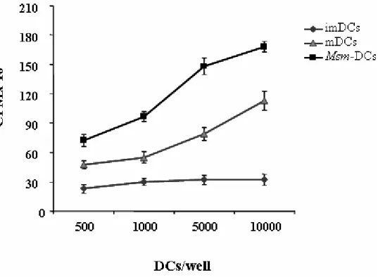
In the same manner, we tested whether the acquisition of mature phenotype by Msm-DCs resulted in an increased capacity of Msm-DC to stimulate T cell response, imMsm-DCs, mDCs (imDCs + LPS), and DCs derived from Msm-infected-precursors, were co-cultured with PBMCs isolated from healthy donors at various DC:T cell ratios and 3
H-thymidine incorporation was measured. Consisting with the early mature phenotype acquired from DCs generated from Msm-infected monocytes, we found that Msm-DCs showed higher capacities to stimulate allogeneic T cells compared to mDCs and even more compared to imDCs (Figure 11).
Figure 11. Msm-DCs induce a strong proliferation of allogeneic PBMC. ImDCs, Msm-DCs or DCs stimulated for 48 hours with LPS (mDCs) were used to stimulate allogeneic PBMC. After 6 days of culture T cell proliferation was assessed by the addition of tritiated thymidine for 16 h. Results are expressed as mean counts per minute (CPM) of triplicate cultures