Alma Mater Studiorum – Università di Bologna
in cotutela con Université Claude Bernard Lyon 1
DOTTORATO DI RICERCA IN
SCIENZE PSICOLOGICHE
Ciclo XXX
Settore Concorsuale: 11E/1
Settore Scientifico Disciplinare: M-PSI/02
THE PERIPERSONAL SPACE: A SPACE TO INTER-ACT
Action- and Social-related Modulations of the Space around Us
Presentata da:
dr. Ivan Patané
Coordinatore Dottorato
Supervisore
Prof.ssa Monica Rubini
Prof.ssa Francesca Frassinetti
Co-supervisore
Dr. Alessandro Farnè
THESE de DOCTORAT DE L’UNIVERSITE DE LYON
opérée au sein del’Université Claude Bernard Lyon 1 Ecole Doctorale N° ED 476
Neurosciences et Cognition (NSCo) Spécialité de doctorat :
Discipline : Neurosciences cognitives
Soutenue publiquement le 26/04/2018, par :
Ivan Patané
L'ESPACE PERIPERSONNEL: UN ESPACE POUR INTER-AGIR
Devant le jury composé de :
Prof Macaluso, Emiliano Professeur UCBL Lyon1 Président
Dr Ferri, Francesca Chercheure University of Essex Rapporteure
Prof Serino, Andrea Professeur University Hospital of Lausanne Rapporteur
Prof Ciaramelli, Elisa Professeure Università di Bologna Examinatrice
Dr Hadj-Bouziane, Fadila Chargée de Recherche CNRS CNRL Examinaterice
Dr Farnè, Alessandro Directeur de Recherche INSERM CNRL Directeur de thèse
UNIVERSITE CLAUDE BERNARD - LYON 1
Président de l’Université
Président du Conseil Académique
Vice-président du Conseil d’Administration
Vice-président du Conseil Formation et Vie Universitaire Vice-président de la Commission Recherche
Directrice Générale des Services
M. le Professeur Frédéric FLEURY M. le Professeur Hamda BEN HADID M. le Professeur Didier REVEL
M. le Professeur Philippe CHEVALIER M. Fabrice VALLÉE
Mme Dominique MARCHAND
COMPOSANTES SANTE
Faculté de Médecine Lyon Est – Claude BernardFaculté de Médecine et de Maïeutique Lyon Sud – Charles Mérieux
Faculté d’Odontologie
Institut des Sciences Pharmaceutiques et Biologiques Institut des Sciences et Techniques de la Réadaptation
Département de formation et Centre de Recherche en Biologie Humaine
Directeur : M. le Professeur G.RODE
Directeur : Mme la Professeure C. BURILLON Directeur : M. le Professeur D. BOURGEOIS Directeur : Mme la Professeure C. VINCIGUERRA Directeur : M. X. PERROT
Directeur : Mme la Professeure A-M. SCHOTT
COMPOSANTES ET DEPARTEMENTS DE SCIENCES ET TECHNOLOGIE
Faculté des Sciences et TechnologiesDépartement Biologie
Département Chimie Biochimie Département GEP
Département Informatique Département Mathématiques Département Mécanique Département Physique
UFR Sciences et Techniques des Activités Physiques et Sportives Observatoire des Sciences de l’Univers de Lyon
Polytech Lyon
Ecole Supérieure de Chimie Physique Electronique Institut Universitaire de Technologie de Lyon 1 Ecole Supérieure du Professorat et de l’Education Institut de Science Financière et d'Assurances
Directeur : M. F. DE MARCHI
Directeur : M. le Professeur F. THEVENARD Directeur : Mme C. FELIX
Directeur : M. Hassan HAMMOURI
Directeur : M. le Professeur S. AKKOUCHE Directeur : M. le Professeur G. TOMANOV Directeur : M. le Professeur H. BEN HADID Directeur : M. le Professeur J-C PLENET Directeur : M. Y.VANPOULLE
Directeur : M. B. GUIDERDONI Directeur : M. le Professeur E.PERRIN Directeur : M. G. PIGNAULT
Directeur : M. le Professeur C. VITON
Directeur : M. le Professeur A. MOUGNIOTTE Directeur : M. N. LEBOISNE
ACKNOWLEDGMENTS
My PhD has been nothing short of amazing… sometimes like a roller coaster, with its ups and downs, but I’ve always enjoyed the ride! However, no PhD thesis is possible without the support, help, guidance, commitment, and encouragement of numerous people and I take extreme pleasure in thanking here all of whom were inevitable in the completion of this thesis.
First and foremost, special mention goes to my Italian moka pot: your endless 24 x 7 support always gives me the strength to carry on. Kidding.
I thank my supervisors Francesca Frassinetti and Alessandro Farné with most happiness, gratitude and pleasure: it has been an honour to be your Ph.D. student - you’ve been the best supervisors a PhD student could ask for. Special thanks for taking a chance on this Sicilian guy showing up and asking to join your labs. Thank you for not only allowing me, but also encouraging me to explore new ideas and for your invaluable guidance in these endeavours. Also, thanks for your patience, time, motivation, passion, enthusiasm, and immense knowledge. Over the years, you made me realise that researchers like you are much more than their publications. You haven’t been just two wonderfully supportive supervisors. You’ve been my complementary guides and mentors.
No words will be enough to thank the help that was provided by Claudio Brozzoli. Around a cup of coffee or in front on a computer screen, it has been fun and stimulating to have the opportunity to work alongside you. Your enthusiasm for research has been contagious for me. The only thing that I regret is that I did not start this journey with you from the very beginning.
I extend my heartfelt thanks to my colleagues from Bologna: it was a pleasure to share with you knowledge and meals. A very special gratitude goes out to all down at Impact team, where I have been given unique opportunities... and taken advantage of them.
I owe big thanks to Alice, Alessandro and their sons: thank you for welcoming me with open arms, for your dinners, good wine and excellent company – always appreciated and enjoyed. I have very fond memories of my time with you.
For this dissertation I would like to thank my committee members and review ers for their time, interest, and helpful comments. I am also grateful to the master and Erasmus students who helped me collect data.
I deem it a pleasure to thank my “Bolognesi” and “Lyonnaises” friends who have supported me along the way. Thanks you all guys, especially for backing me up and putting up with me!
I also place on record, my sense of gratitude to one and all, who directly or indirectly, have lent a hand in this venture: thanks to the many people, in many countries, who so generously contributed to this work.
Finally, I must express my very profound gratitude to my family for providing me with unfailing support and continuous encouragement throughout my years of study - although it is likely that they have never grasped what it was all about! This accomplishment would not have been possible without them. Thank you.
The Peripersonal Space:
A space to inter-act
Action- and social-related modulations of the space around usContents
Abstract ... 11
Riassunto ... 13
Résumé ... 15
Chapter I: The space around the body: Peripersonal space ... 17
1.1 Multiple spaces... 17
1.2 Peripersonal space in the monkey brain ... 19
1.2.1 A multisensory space... 19
1.2.2 A dynamic multisensory-motor space ... 24
1.2.3 Mirroring the (peripersonal) space ... 27
1.2.4 Multisensory network of PPS coding ... 29
1.2.5 In a nutshell… ... 32
1.3 Peripersonal space in the human brain ... 34
1.3.1 Peripersonal space in the damaged human brain ... 34
1.3.2 Peripersonal space in healthy individuals ... 38
1.3.3 Neuroimaging PPS in the human brain ... 42
1.3.4 In a nutshell… ... 45
1.4 Peripersonal space in action... 47
1.4.1 A space for body-object interactions? Interim summary ... 54
1.5 Social PPS... 56
Chapter II: Action planning modulates peripersonal space ... 60
2.1 Methods ... 62
2.2 Statistics ... 65
2.3 Results ... 68
2.4 Discussion... 76
Chapter III The space between the body: Interpersonal space ... 80
3.1 The social space ... 80
3.2 Interpersonal space 2.0: a neuroscientific approach ... 87
3.3 In a nutshell... 93
3.4 How many spaces around the body?... 94
3.5 IPS=PPS?... 97
Chapter IV: Tool use differently shapes the space around us ... 98
4.1 Experiment 1 ... 98
4.1.1 Materials and method ... 99
4.1.2 Data analysis and results ... 101
4.1.3 Interim discussion ... 103
4.2.1 Materials and method ... 104
4.2.2 Data analysis and Results... 105
4.3 Discussion... 107
Chapter V: Cooperative Tool use reveals periperisonal and interpersonal space are dissociable ...109
5.1 Experiment 1: cooperative long tool use ... 110
5.2 Experiment 2: Cooperative short tool use ... 116
5.3 Relationships between participant-confederate distances, arm length and familiarity ... 121
5.4 Experiment 3: control tool use ... 123
5.5 Manipulation check questionnaires ... 125
5.6 Discussion... 127
Chapter VI: Mine, Thine, Ours ...131
6.1 Mine is special: the pervasive psychological advantage of self -owned objects... 132
6.2 Our-ness: our objects are special too?... 136
6.3 In a nutshell… ... 138
6.4 Tracking and controlling owned objects ... 139
6.5 Embodied object ownership? ... 141
6.6 In a nutshell… ... 144
Chapter VII: Our peri-personal property: Exploring the effect of object ownership on the space around us 145 7.1 Experiment 1 ... 147 7.1.1 Methods ... 147 7.1.2 Statistics ... 151 7.1.3 Results ... 152 7.1.4 Discussion ... 159 7.2 Experiment 2 ... 162 7.2.1 Methods ... 162 7.2.2. Statistics ... 164 7.2.3 Results ... 164 7.3 Discussion... 174
Chapter VIII: Conclusions and perspectives ...180
Abstract
The zone that surrounds our body is of vital importance: we carefully monitor the objects (both animate and inanimate) that enter the boundaries of the immediate space around the body to interact with them. In the neurocognitive field such a space is captured by the concept of peripersonal space (PPS), a highly plastic representation that integrates tactile and visual stimuli presented on, and close to, the body. This system seems to contribute to the efficient guidance of actions, yet, a clear demonstration of a prominent role of PPS in control of actions is critically lacking. Strong support for this would derive from evidence that PPS plastic changes occur before rather than after movement onset. The results from the first study (Chapter II) reveal that visual and tactile information strongly interact already during the planning phase of action and this visuo-tactile interaction is further enhanced during subsequent movement phases. Such a visuo-visuo-tactile remapping of PPS that temporally precedes and subsequently accompanies overt motor execution is ideally suited to planning and guiding actions.
Recently, it has been suggested a possible involvement of PPS in the guidance of motor interactions between individuals. In social psychology, the space around the body is termed interpersonal space (IPS), defined as the area individuals maintain around themselves into which others cannot intrude without arousing discomfort. Because of some similarities between the PPS and IPS constructs, some authors have raised the question of whether they share some functional features. The second aim of my thesis is to test this hypothesis by taking advantage of another PPS remapping, namely that one induces by tool-use. The results of the second study (Chapter IV) show that “standard” tool-use ‘extends’ PPS, as measured by reaching distance toward a peer, but does not affect IPS, as measured by the comfort distance toward the same peer. In the third investigation (Chapter V), we introduced a novel form of “social” tool-use setting to test for both sensorimotor and social plasticity of the two spaces. The findings that social tool–use ‘extends’ PPS and ‘reduces’ IPS, inducing opposite changes on each representation, clearly disconfirms the hypothesis that there might be functional overlap between these sectors of space. Such examples of functional dissociation may therefore be sufficient to warn scholars to refrain from risky conflations between the two concepts.
If the assumption of functional identity with IPS does not appear to be legitimate, it is true that PPS is sensitive to social features. The last study (Chapter VII) is thus aimed at probing this sensitivity of PPS to a so far unexplored but fundamental social dimension: ownership. The results from the forth study indicate that, whether considered to be as individual or shared property, ownership of an object is critical for the PPS dynamic properties to emerge. Visual stimuli affected
touch perception more strongly at the movement onset than before, but only when the object belonged to the acting participant. Interestingly, a similar remapping was found when simply observing the peer acting on her own belonging. In a follow-up experiment we investigated PPS plastic changes when property of the target object was shared between the two agents. In this case, PPS remapping emerged not only when acting in first person, but also when observing the peer acting upon the shared object.
Taken together, these findings critically inform current theoretical models about space around our body and about its function in our sensorimotor and social inter-actions.
Riassunto
La zona che circonda il nostro corpo è di vitale importanza: monitoriamo attentamente gli oggetti (sia animati che inanimati) che valicano tale confine allo scopo di interagire con essi. Nel campo delle neuroscienze cognitive tale spazio è catturato dal concetto di spazio peripersonale (PPS), una rappresentazione altamente plastica che integra stimoli tattili e visivi presentati, rispettivamante, sul e vicino al nostro corpo. Questo sistema sembra contribuire efficacemente alla guida delle azioni, tuttavia una chiara dimostrazione del coinvolgimento del PPS nel controllo delle azioni é ancora assente in letteratura. Sostegno a questa ipotesi deriverebbe dall’evidenza che la plasticità del PPS possa esser innescata prima, piuttosto che dopo, l'insorgenza del movimento. I risultati del primo studio (Capitolo II) mostrano che le informazioni visive e tattili interagiscono significativamente già durante la fase di pianificazione dell'azione e che questa interazione visuo-tattile aumenta ulteriomente durante le fasi successive del movimento. Un tale processo di ‘remapping’ visuo-tattile del PPS, poiché precede temporalmente e accompagna successivamente l'esecuzione dell’atto motorio, sembrerebbe ideale per pianificare e guidare le nostre azioni.
Recentemente è stato suggerito un possibile coinvolgimento del PPS nella guida delle interazioni motorie tra individui. In psicologia sociale, lo spazio intorno al corpo è definito spazio interpersonale (IPS): la distanza che gli individui mantengono attorno a sé. Tale distanza, quando é valicata dagli altri, desta un sentimento di disagio. Alla luce di alcune analogie tra PPS e IPS, alcuni autori si sono domandati se i due construtti possano condividere delle caratteristiche funzionali. Il secondo scopo di questa tesi è testare questa ipotesi sfruttando un altro processo di ‘remapping’ plastico del PPS, ovvero quello indotto all'uso di uno strumento (tool). Il secondo studio (Capitolo IV) mostra che l'uso di uno strumento ‘estende’ il PPS, misurato come distanza di raggiungimento verso un’altra persona, ma non influenza l'IPS, misurato come distanza di comfort verso la stessa persona. Nel terzo studio (Capitolo V), abbiamo introdotto una nuova variante più sociale dell’utilizzo di uno strumento per esaminare la plasticità sia sensorimotoria sia sociale dei due spazi. I risultati ottenuti rivelano che l'uso sociale di uno strumento ‘estende’ il PPS e al contempo ‘riduce’ l'IPS. La dimostrazione che si possano indurre cambiamenti direzionalmente opposti tra le due rappresentazioni, falsifica l'ipotesi secondo la quale possa esserci una sovrapposizione funzionale tra PPS e IPS. Questi esempi di dissociazione funzionale possono quindi essere sufficienti per mettere in guardia gli studiosi al fine di evitare rischiose sovrapposizioni tra i due concetti.
Se l'assunzione di identità funzionale con IPS non sembra essere legittima, è comunque vero che il PPS è sensibile a variabili sociali. L'ultimo studio (Capitolo VII) è perciò finalizzato a sondare tale sensibilità del PPS verso una dimensione sociale fondamentale, ma finora inesplorata: l’appartenenza. I risultati del quarto studio indicano che, sia che si consideri una proprietà individuale o condivisa, l’appartenenza di un oggetto è una variabile critica per far emergere le proprietà dinamiche del PPS. Gli stimoli visivi, difatti, influenzano la percezione tattile in maniera più marcata all'inizio del movimento, ma solo quando l'oggetto dell’azione é il proprio. Da notare che un effetto simile emerge anche quando si osserva l’altra persona agire sul suo oggetto. In un esperimento successivo abbiamo studiato le modifiche plastiche del PPS quando la proprietà dell'oggetto è condivisa. Qui il ‘remapping’ del PPS emerge non solo quando si agisce in prima persona, ma anche quando si osserva l’altra persona agire sull’oggetto condiviso.
Questi risultati complessivamente forniscono evidenze critiche rispetto agli attuali modelli teorici sulla funzione dello spazio attorno al nostro corpo nelle interazioni sensori -motorie e sociali.
Résumé
L’espace entourant notre corps est d'une importance vitale: nous surveillons attentivement les objets (animés et inanimés) qui entrent dans les limites de l'espace à immédiate proximité du corps pour interagir avec eux. Dans le domaine des neurosciences cognitives, cet espace est exemplifié par le concept d'espace péripersonnel (PPS), une représentation hautement plastique qui intègre des stimuli tactiles et visuels présentés sur et près du corps. Cette représentation semble contribuer au guidage efficace des actions, cependant dans la littérature on ne retrouve aucune preuve substantielle de l'implication du PPS dans le contrôle des actions. Un argument en faveurs de cette hypothèse dériverait de la preuve que la plasticité du PPS peut effectivement survenir avant le début du mouvement, plutôt que pendant le mouvement. Les résultats de la première étude (chapitre II) révèlent que les informations visuelles et tactiles interagissent de manière significative déjà au cours de la phase de planification de l'action et que cette interaction visuo-tactile augmente ultérieurement au cours des étapes successives du mouvement. Un tel processus de « remappage » visuo-tactile du PPS, qui précède temporellement et accompagne par la suite l'exécution de l’action motrice, semble donc idéalement adapté à pour aider au guidage de nos actions.
Récemment, il a été suggéré que le PPS pourrait jouer un rôle dans le guidage des interactions motrices entre individus. En psychologie sociale, l'espace autour du corps est appelé espace interpersonnel (IPS), défini comme l'espace que les individus maintiennent autour d'eux et dans lequel les autres ne peuvent pas pénétrer sans susciter d'inconfort. En raison de certaines similitudes entre les représentions du PPS et du IPS, certains auteurs ont soulevé la question d’un éventuel partage de certaines caractéristiques fonctionnelles entre ces deux représentations. Le deuxième objectif de ma thèse etait de tester cette hypothèse en exploitant un autre processus de «remappage» plastique du PPS, c'est-à-dire celui induit par l'utilisation d'un outil. Les résultats de la deuxième étude (chapitre IV) montrent que l'utilisation «standard» d’un outil «allonge» le PPS, mesuré par la distance d’atteignabilité d’une autre personne, mais n'influence pas l'IPS, mesuré par la distance de confort envers la même personne. Dans la troisième étude (chapitre V), nous avons introduit une nouvelle variante plus sociale de l'utilisation d'un outil pour examiner la plasticité sensorimotrice et sociale des deux espaces. Les résultats obtenus révèlent que l'utilisation sociale d'un outil «allonge» le PPS et en même temps «réduit» l'IPS. La démonstration que l'on peut induire des changements directionnellement opposés entre les deux représentations, falsifie l'hypothèse selon laquelle il peut y avoir un chevauchement fonctionnel entre PPS et IPS. Ces exemples de
dissociation fonctionnelle peuvent donc servir à éviter une association inappropriée entre les deux concepts.
Si l'hypothèse de l'identité fonctionnelle avec l’IPS ne semble pas légitime, il n'en demeure pas moins que le PPS est également sensible aux variables sociales. La dernière étude (Chapitre VII) vise donc à explorer cette sensibilité du PPS vers une dimension sociale fondamentale, encore inexplorée: la propriété privée. Les résultats de la quatrième étude indiquent que la propriété d'un objet, qu’elle soit considérée comme individuelle ou partagée, est essentielle pour l'émergence des propriétés dynamiques du PPS. Les stimuli visuels influencent effectivement la perception tactile d'une manière plus marquée au début du mouvement, mais seulement lorsque l'objet appartient au participant. Il convient de noter qu'un effet similaire apparaît également lorsque l'on observe une autre personne agir sur l’objet qui lui appartient. Dans une expérience ultérieure, nous avons étudié les modifications plastiques du PPS lorsque la propriété de l'objet était partagée entre les deux participants. Dans ce cas, le «remappage» du PPS émerge non seulement lorsque le participant agit, mais aussi lorsqu'il observe l'autre personne agissant sur l'objet partagé.
Dans l'ensemble, ces résultats apportent une preuve critique par rapport aux modèles théoriques actuels de la fonction de l'espace autour de notre corps dans les interactions sensori -motrices et sociales.
Chapter I: The space around the body: Peripersonal space
“Il n'y a de réalité que dans l'action’’ Jean-Paul Sartre
1.1 Multiple spaces
Our surrounding environment is often perceived as a unitary and seamless. Although it does not seem to be reflected in the way in which we subjectively experience the outer world, an ever-growing body of scientific evidence reveals that the brain constructs various functionally distinct representations of space. Over the years, many authors tried to establi sh valid segmentations of space. Notwithstanding all had different reasons to hit the mark and different visions about how the space is divided, all agreed to maintain, to various degrees, a sort of boundary between who/what is close to our bodies from who/what is further away (Previc, 1990, 1998; Grusser, 1983; Cutting and Vishton, 1995; see also Hediger 1955; Hall, 1969).
In cognitive neuroscience a key division is between peripersonal space (hereafter PPS), that is the region of near space immediately surrounding our bodies in which objects can be grasped and manipulated, and extrapersonal space representations, that is the farther space where exploratory eye movements occur and objects cannot be reached without moving toward them (Holmes and Spence, 2004; Ladavas and di Pellegrino, 2015). This division was initially suggested by Brain (1941), who distinguished a grasping distance within arm’s reach from a walking distance to account for the selective impairment in one or the other sector of space following right-brain lesions. From a computational point of view, it makes indeed sense that the brain may represent objects situated in PPS differently from those in extrapersonal space. The best use of the brain limited resources might well be to plan only grasping movements to those targets of interest within reach, and to plan only locomotive movements to those targets situated at a distance. Obviously, the two action modules can be combined when somebody walks towards a desk to grasp a glass of water from its surface.
Hence, the hypothesis that space may be represented by multiple sub-spatial maps partly delimited by the body was further supported by seminal neurophysiological studies in the non-human primate brain (Hyvärinen and Poranen, 1974; Mountcastle, 1976; Leinonen and Nyman, 1979a, 1979b; Rizzolatti et al., 1981a, 1981b). For example, lesion studies in macaque monkey (Rizzolatti et al., 1983) revealed that the unilateral surgical ablations of the postarcuate cortex resulted in a severe neglect limited to the PPS associated with a deficit in the use of the contralateral hand. Namely, the animal exhibited a failure to grasp food with the mouth when presented contralesionally and a reluctance to use the contralateral hand. By contrast, unilateral ablation of the pre-arcuate cortex (area 8), corresponding to the frontal eye-field or FEF, resulted in a decrease of eye movements contralateral to the lesion and a neglect of the contralateral hemispace, more prominent for ‘far’ space than for that near the animal (Rizzolatti et al., 1983; Schieber, 2000). Since these early works from 70-80’s, a plethora of studies in monkeys and humans (in both healthy and pathological states) have explored the functional features of the specific area close to the body, viz. PPS.
In this introductory session I will describe the single cells, areas, cortical functional networks in monkey’s brain proposed to underlie the PPS representation, as the definition of PPS itself originates from single-unit electrophysiological studies in non-human primates. Despite their heterogeneous nature, the majority of these papers will be framed within an action-based perspective of space pointing to dynamic and bidirectional links between PPS representation(s) and actions.
1.2 Peripersonal space in the monkey brain
Objects at a distance can typically be perceived through a limited number of senses such as vision, audition, and olfaction. Conversely, objects nearer to, or in contact with, the body surface can virtually impact upon all our sensory systems. The brain’s processing of stimuli in PPS is therefore quite complex ad involves more modalities of sensory information. Besides, it is not the case that any one single brain area is responsible for maintaining a representation of space, as once thought, but PPS is rather an emergent property of a network of interacting and well -connected cortical and subcortical areas (Rizzolatti et al., 1997; Graziano and Gross, 1998; Spence and Holmes, 2004; Clery et al., 2015a).
1.2.1 A multisensory space
Pivotal single-cell recording studies on the macaque monkey brain releveled a special set of neurons within the ventral premotor cortex strongly responding to tactile stimulation. Neurons in the histochemical area F4 (the caudal portion of the inferior part of the premotor cortex, see Figure1.1) containing representations of arm movements, also features relatively large tactile receptive fields. Such respective fields are mainly located on the animal’s hands, arm, face, and neck (Gentilucci et al., 1983, 1988; Rizzolatti et al. 1981a, 1981b), as if to form a broad map of the body. A large proportion (85%) of these tactile neurons also discharges in response to visual stimulation. Specifically, such cells in area F4 respond weakly to visual stimuli presented far from the body, whereas the most effective response, in terms of spike discharge rate, is evoked primarily by three-dimensional objects moving close to the tactile receptive field. According to the depth of the visual receptive fields protruding out of the skin surface, these bimodal visuo-tactile neurons were originally subdivided into pericutaneous (54%) and distant peripersonal neurons (46%). The former class of neurons responds best to visual stimuli presented in the very close vicinity (up to 10 cm away from the body), while the latter class responds also to visual stimuli presented at a further distance from the skin, but always within monkey’s reaching distance (Rizzolatti et al., 1981a,1981b). For the sake of simplicity, I will refer to both as “peripersonal” neurons, as one of distinctive features of either subpopulation is that their visual receptive fields are limited in depth from the somatosensory receptive fields of a specific body part and remain “anchored” to that body part. Indeed, the size of the visual receptive field not only matches and spatially overlaps the size of the tactile one, but also extends from the body for varying distances (typically from ~5 to ~50 cm).
That is to say, the visual and tactile receptive fields of these bimodal cells create a single responsive region mapping the skin and the space closely surrounding it. Since the visual responses are generally independent of gaze direction, the visual information in F4 region is spatially related to the body parts on which the tactile receptive fields are located (Fogassi et al., 1992; Gentilucci et al. 1983; see Figure 1.2). Here is to note that the visual receptive field seems to be “anchored” to the somatosensory receptive field on a body part because it moves congruently with it, keeping thus a rough spatial match between the locations of the visual RF and the body part with every displacement (Graziano et al., 1994). This is demonstrated by the fact that passive displacement of the arm causes a congruent shift of the location of the visual receptive field of neurons presenting somatosensory receptive field on the limb. In a similar vein, when the head is turned, but not when visual fixation changes, the visual response of neurons with tactile receptive fields on the face is updated following the spatial position of the face (Graziano et al., 1997a, 1997b). This result provides strong evidence for a body part-centred reference frame, since the neuronal responses were tested both when the monkey was fixating a particular position, and when fixation was not controlled. Regardless of the direction of gaze, neurons with somatosensory receptive fields on a given body part are specifically activated by visual stimuli presented near that given body. For some neurons the spontaneous or evoked firing rate is modulated by eye position, but the portion of space eliciting the maximal visual response does not change when the eyes moved.
Such a peculiar interaction between different receptive fields is not limited to visuo-tactile cells, since neural activity in F4 can be trigged by visual, as well as auditory signals. Indeed, some neurons responding to a somatosensory stimulus located on the back and the side of the head also discharge when a visual and/or auditory stimulus is administered next the body part being stimulated tactilely (Graziano et al., 1999). To anchor their multisensory receptive fields to a given body part and to maintain their spatial alignment, these neurons integrate proprioceptive information too (see also Figure 1.3). When exposed to conflicting visuo-proprioceptive conditions, the firing rates of the F4 neurons elicited by approaching stimuli is modulated by the position of the monkey’s real arm as well as by the position of a fake arm placed in a realistic posture (Graziano et al., 1999).
Figure 1.1 Selected cortical areas of the macaque monkey brain. A. Lateral view of the whole brain.
Thick black lines represent major cortical boundaries and sulci. Thin black lines represent cortical area boundaries. Area 4/F1 - primary motor cortex; Area 2/SI – primary somatosensory cortex; Area 5/PE – posterior parietal association cortex/superior parietal lobule; Area F5/PMv and Area F4/PMv – ventral premotor cortex; IPS – intraparietal sulcus; CS – central sulcus. B. The intraparietal sulcus has been opened up to reveal multiple and heterogeneous visual and somatosensory posterior parietal areas. Thick lines represent the superficial border of the sulcus; thin black lines mark the fundus of the sulcus. Other lines indicate the boundaries of cortical areas as follows: MIP – medial intraparietal sulcus; LIP – lateral intraparietal sulcus; AIP – anterior intraparietal sulcus; VIP –ventral intraparietal sulcus; PEip – intraparietal portion of area PE (from Holmes and Spence 2014; redrawn from Rizzolatti et al., 1998).
Figure 1.2 Multimodal neurons in the monkey brain that encode the space near the body. Each neuron
responds to touching a specific part of the body called the neuron's tactile receptive field. The same neuron responds to visual stimuli in the space near the tactile receptive field. Two examples are depicted. From Graziano and Gross (1998).
Another premotor area, i.e. the rostral subregion F5 of area 6, harbours similar visuo-tactile neurons (see Figure 1.1). However, as compared to those present in F4 region, bimodal neurons in F5 are less numerous and show smaller tactile receptive fields, which are frequently located on the face, the hand, or both. It is important to acknowledge another significant difference: albeit visual stimuli presented near the body result in stronger firing rate, what seems to be crucial in triggering the response of these neurons is instead the size of the stimuli (Rizzolatti et al., 1988; see also session 1.2.4).
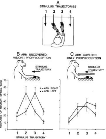
Figure 1.3 Visual responses of a typical premotor neuron with a tactile RF (hatched) on the forearm and hand, and a visual RF within 10 cm of the tactile RF. (A) On each trial, the arm contralateral to the
neuron was fixed in one of two positions and the visual stimulus was advanced along one of four trajectories. For this neuron, the two arm positions were chosen to align the visual RF near the hand and forearm with trajectories 2 and 3. For other neurons, the arm was moved to different extents depending on the location of the visual RF, to better capture the movement of the visual RF with the arm. (B) Responses of the neuron to the four stimulus trajectories when the arm was visible to the monkey. When the arm was fixed on the right, the response was maximum at position 3. When the arm was fixed on the left, the maximum response moved to the left, to position 2. (C) Responses of the neuron when the arm was covered. The movement of the visual RF with the arm was reduced but not eliminated, indicating that the neuron combined both proprioceptive and visual information about the position of the arm. Each point is a mean of 10 trials. Error bars are standard error. Adapted from Graziano et al., 1997.
Visuo-tactile neurons with similar properties have been also discovered in two regions of the posterior parietal cortex heavily connected with F4 (Matelli et al., 1984a, 1984b), namely area 7b and the ventral intraparietal (VIP) area. Electrophysiological studies in awake monkeys demonstrated that, similarly to neural cells in the ventral premotor cortex, visuo-tactile integration in these areas arises at the single unit level (Hyvärinen and Poranen 1974; Mountcastle et al. 1975; Hyvärinen 1981; Leinonen et al. 1979; Leinonen and Nyman 1979; Robinson et al. 1978; Robinson and Burton 1980a, 1980b; Hyvärinen 1981). In particular, area 7b presents a coarse somatotopic organization with a face representation on the upper inferior parietal convexity, followed by arm, hand, and foot representations located laterally, along the inferior parietal convexity (Hyvärinen and Shelepin 1979; Hyvärinen 1981, Robinson and Burton 1980a, 1980b). Although this region is prevalently a somatic area with most of its neurons being somato-motor or somatosensory, a part of the neurons studied in this area is, in fact, visuo-tactile (Gross and Graziano, 1995; Hyvärinen, 1981). In the regions representing the face and arm about one third of the cells are described as bimodal, with visual receptive fields being spatially aligned with the tactile receptive fields (Hyvärinen and Porane, 1974, Hyvärinen and Shelepin 1979; Lionene and Nyman 1979), whereas another portion of bimodal neurons has bilateral receptive fields located on the limbs, sometimes covering the whole body (Leinonen et al., 1979). Most of the cells in 7b respond preferentially to visual stimuli moving toward the skin, within about 10 cm of the tactile receptive fields, although stimuli presented further away, but still within a reachable distance, are also effective. Notably, the neural response in 7b can be both independent (Graziano and Gross 1995) and dependent of the position of the arm (Leinonen et al., 1979).
The ventral intraparietal area (area VIP), located in the fundus of the intraparietal sulcus and receiving projections from the middle temporal visual areas, as well somatosensory, auditory, and vestibular regions (Graziano and Cooke, 2006, see Figure 1.1), contains visual as well as visuo -tactile neurons (Colby and Duhamel 1991; Colby et al., 1993; Duhamel et al., 1998, Avillac et al., 2005). VIP bimodal neurons mainly respond to visual stimulation presented within a few centimetres of the tactile receptive field and show a strong sensitivity to speed and direction of motion of both visual and tactile stimuli (Duhamel et al., 1998). Unlike area 7b neurons, somatosensoty receptive fields in VIP are primarily located on the face and head and their visual receptive fields are anchored to the region of space around the face (Colby et al., 1993). Moreover, some VIP neurons are trimodal, responding to visual, auditory and tactile stimuli, with the three different receptive fields usually aligned (Schlack et al., 2003).
Some studies have revealed a similar pool of multisensory neurons devoted to represent the space near the body in the putamen (Graziano and Gross 1993, 1995), a subcortical structure of the primate brain that receives projections from inferior area 6 and area 7b (Cavada and Goldman-Rakic 1991; Matelli et al., 1986). The putamen has a complete somatotopic map of the body and, like the bimodal neurons described above, visual and tactile receptive fields of bimodal cells in this structure display a rough spatial correspondence, with the visual receptive field anchored to the tactile one. A large proportion of bimodal neurons has cutaneous receptive fields cantered on the face (Graziano and Gross, 1993) and responds best to visual stimulation administered within 10-20 cm from the face skin. Visuo-tactile neurons centred on the arm respond visually when the arm under the monkey’s view, but not when the arm is moved out view (Graziano and Gross 1995).
1.2.2 A dynamic multisensory-motor space
Peripersonal space has been so far depicted as a static functional representation, determined by fixed body constraints such as the within reach space around the body or around the head. Yet, there is abundant evidence that PPS should rather be considered as extremely dynamic and rapidly adjusting to both endogenous and exogenous factors, being readily modifiable and shaped by sensorimotor experience.
Again, such a dynamic signature of PPS was first captured by single-cell recording studies in monkeys. For example, it has been demonstrated that the depth of visual receptive fields of F4 neurons is not fixed, but can increase with increases in the velocity (20–80 cm/s) of a visual stimulus approaching the tactile receptive fields, such that fast-moving stimuli are signalled earlier than slow-moving ones (Fogassi et al., 1996). Visual receptive fields of bimodal neurons in the medial anterior intraparietal sulcus and in the post-central gyrus seem to have a somewhat similar plasticity. Iriki and colleagues (1996) trained monkeys to use a rake as a tool to reach for food pellets placed out of their reaching distance. After this training with the tool, the visual receptive field of some bimodal neurons were elongated along the axis of the rake. The elongated visual receptive fields seemed to have expanded toward the tool tip, such that the rake appeared to be included within the visual receptive fields. This was the case for both “distal” cells, whose tactile receptive field was on the surface of the hand, and “proximal” cells, whose tactile receptive field was on the skin of the shoulder. Remarkably, a few minutes after withholding the active use of the rake, the visual receptive fields apparently shrank back to their original size. Such a change to occur requires active tool-use: no modification was observed if the rake was just passively held by the animal, therefore suggesting the tool has to be actively employed to perform an action (see Figure
1.4). Moreover, a positron emission tomography (PET) study from the same group further extended this finding, describing the cortical activation of the presupplementary motor area and the premotor cortex at locations matching F4 and F5 areas (Obayashi et al., 2001). Parietal regions are also crucial, since the tool-dependent changes increased cortico-cortical afferents to the intraparietal sulcus (Hihara et al., 2006) and the expression of neuronal plasticity markers in this cortical region (Ishibashi et al., 2002a, 2002b) .
Figure 1.4. Changes in bimodal receptive field properties following tool-use. The somatosensory
receptive fields of cells in this region were identified by light touches, passive manipulation of joints or active hand-use. The visual receptive field was defined as the area in which cellular responses were evoked by visual probes (the most effective ones being those moving towards the somatosensory receptive field). (a) somatosensory receptive field (blue area) of the ‘distal type’ bimodal neurons and their visual receptive field (pink areas) (b) before tool-use, (c) immediately after tool-use and (d) when just passively grabbing the rake. (e) somatosensory receptive field (blue area) of ‘proximal type’ bimodal neurons, and their visual receptive field (pink areas) (f) before and (g) immediately after tool-use. From Maravita and Iriki 2004.
The dynamic changes of visual receptive fields observed in case of objects approaching the body (Fogassi et al. 1996) or after active tool-use (Iriki et al. 1996) point to another critical aspect: the link of PPS coding with motor responses. The neural response of visuo-tactile cells in inferior premotor area 6 (Gentilucci et al. 1988; Rizzolatti et al. 1981c, 1987, 1988, 1997), parietal area 7b
(Hyvärinen 1981; Hyvärinen and Poranen 1974; Hyvärinen and Shelepin 1979; Leinonen 1980; Leinonen et al. 1979; Leinonen and Nyman 1979; Robinson et al. 1978), and the putamen (Crutcher and DeLong 1984) is elicited by passive visual and tactile stimulation as well as during motor activity.
In the premotor cortex the visual responses of some neurons are enhanced when performing reaching and grasping movements toward an object (Godschalk et al., 1981, 1985; Kurata and Tanji 1986; Rizzolatti et al., 1981c; 1990). Interestingly, the active movements and the sensory receptive fields appear to share related functional roles. Neurons with visuo-tactile receptive fields around and on the face also responded during reaching movements of the arm toward the upper part of space that corresponds to their visual receptive fields. In other words, not only visual and tactile receptive fields, but also motor response fields are in spatial register: the motor activity of these neurons is maximal when the movement is directed to reach into the region of space coded by the bimodal neurons. As a result, the sensory and motor responses appear to be expressed in a common coordinate system (Caminiti et al., 1990). In addition, the posterior parietal cortex is accordingly related to approaching movements of a body-part toward external objects and show motor properties, similarly to the premotor region (Rizzolatti et al., 1997, Debowy et al., 2001; Fogassi and Luppino 2005; Ferraina et al., 2009a, 2009b). Note that the activation during grasping actions in monkey's posterior parietal areas starts before the beginning of the movement and persists during the whole action. The predominant activation moves then to SI only when the hand enters in contact with the target object (Gardner et al., 2002). Remarkably, this posterior parietal activation before and during the overt motor activity involves areas of the monkey's brain where bimodal neurons have been found, such as area 7b. Lastly, ablation and reversible inactivation of premotor and parietal region seem to produce very similar patterns of deficits, most of which impairs the execution of visually guided reaching actions (Moll and Kuypers 1977; Battaglini et al. 2002; Deuel and Regan 1985; Ettlinger and Kalsbeck 1962; Faugier-Grimaud et al., 1978; Gallese et al., 1994; Halsban and Passingham 1982). As mentioned earlier, removal of post-arcuate lesions to area 6, including areas F4 and F5, results in a severe impairment in grasping with mouth (Rizzol atti et al., 1983). The hand shaping that relies on the visual properties of the object to be grasped is disrupted following the inactivation of F5, and a similar impairment is observed following AIP inactivation (Fogassi et al., 2001; Gallese et al., 1994). To conclude this paragraph, these results therefore contribute collectively to raise the compelling possibility that the multisensory representation of PPS might serve some motor function.
1.2.3 Mirroring the (peripersonal) space
In accordance with the motor properties of PPS, the regions devoted to near space coding are immediately adjacent to, or coextensive with, brain areas containing another particular set of visuomotor neurons. In the ventral premotor cortex, visuomotor cells of area F5 are cla ssically subdivided into two categories: "canonical" and "mirror" neurons. "Canonical" neurons discharge to visual presentation of objects and to actions towards such objects, both in the dark and in the light. Because of this, such class of neural cells is proposed to underlie visuomotor transformation for grasping (Murata et al., 1997; 2002; Raos et al., 2006). On the other hand, “mirror” neurons selectively respond during action execution as well as during observation of someone else performing the same action. Such class of neural cells is instead thought to be involved in action understanding (di Pellegrino et al.; 1992, Gallese et al., 1996; Rizzolatti et al., 1996; Rizzolatti and Matelli, 2003). In the rhesus monkey’s premotor cortex, mirror neurons not only are activated by both the execution and the observation of motor acts, but they are also modulated differentially by the location in space of the observed motor acts relative to the monkey. A subset of mirror neurons show selectivity from actions performed within the observer’s PPS rather than in its extrapersonal space, while another subset show the opposite selectivity for actions performed in extrapersonal space. Even more interestingly, a portion of these spatially selective mirror neurons encodes space according to a metric representation, whereas other neurons encode space in operational terms, changing their properties according to the possibility that the monkey will interact with the object (Caggiano et al., 2009). When accessibility to PPS is limited, for instance by placing a transparent barrier in front of the monkey, the firing rate of several PPS mirror neurons are reduced during observation of actions performed in portion of the space now inaccessible. In other terms, when the border changed the previously reachable space of the monkey into non-reachable space, the PPS mirror neurons did not respond anymore. Accordingly, extrapersonal mirror neurons start to respond to observation of actions performed in the inaccessible PPS, as indicati ng that when PPS is inaccessible for action, it is represented as farther extrapersonal space. Such results therefore suggest that it was the currently reachable space that was important in evoking a response in these “extrapersonal” and “peripersonal” mirror neurons.
Along with this, a subpopulation of F5 neurons actually shares both canonical and mirror properties, termed therefore “canonical-mirror” neurons. The responses of canonical and “canonical-mirror” neurons to the presentation of graspable objects typically require the stimulus to be in PPS. In contrast, the responses of mirror and canonical-mirror neurons evoked by action
observation are present irrespectively of whether the action being observed by the monkey is performed in peripersonal or in extrapersonal space. Also in these hybrid visuo-motor cells, space-constrained coding of objects mostly relies on an operational (action possibility) rather than metric (absolute distance) reference frame. Moreover, such canonical-mirror neurons appear to code object as target for both one's own and other's action, suggesting that they could play a role in predictive representation of others' impending actions (Bonini et al., 2014). In addition, mirror neurons can also be found in parietal area 7b (Fogassi et al., 2005; Fogassi and Luppino, 2005). Like the “canonical” 7b neurons, these cells discharge differentially to simple movements and to complex goal-directed sequences of actions, and are proposed to play an important role in the organization of natural ecological actions (Bonini et al., 2010).
All together, these experiments reveal that a set of neurons encodes the observed motor acts not only for action understanding (Gallese et al., 1996; Rizzolatti et al., 1996; Rizzolatti and Fogassi, 2014) but also to analyse such acts in terms of features that are relevant to generating appropriate behaviours within and beyond the action space (Caggiano et al., 2009). If, on one hand, the space where the executed or observed action constrains the neural responses of the visuo-motor cells, on the other hand, coding of space could potentially benefit of similar mirroring processes.
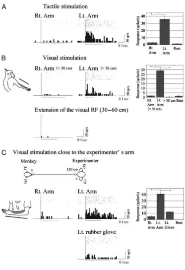
As proof of the existence of a mirror neuron-like mechanism encoding the PPS of other individuals, Ishida et al., (2010) described in area VIP “body-matching neurons” that respond to visual stimuli presented near a specific body part of the monkey being recorded from, as well as to visual stimuli presented near the corresponding body part of the human experimenter. Like the typical PPS neurons, the majority of the recorded neurons exhibit visual receptive fields around the tactile receptive fields anchored on a single body part (e.g., cheek, forearm, and trunk), selectively discharging when visual stimuli were delivered within monkey’s PPS (approximately 30 cm from the skin). However, the novel finding to emerge from this study was the some bimodal neurons exhibit both visuo-tactile receptive fields on the monkey’s body and visual receptive fields close to the experimenter’s body. Such neurons respond selectively to a visual stimulus delivered at a distance of 120 cm from the monkey’s body parts (i.e., beyond the monkey’s PPS) but close to the corresponding experimenter’s body parts (i.e., within the experimenter’s PPS). As an example, a neuron with a tactile receptive field on the arm responds to visual stimuli both presents around the monkey's own arm and delivered around another individual's arm, the experimenter in this case (see Figure 1.5). Importantly, the neuron failed to respond when the same stimulus was presented close to other body parts of the experimenter or in the absence of the experimenter, ruling out visual attention as an alternative interpretation of the findings.
Figure 1.5. An example of body-matching neuron. Location of tactile receptive field of this neuron is on
the monkey’s left forearm, with visual receptive field anchored close to the same part of the tactile RF (A and B). A tactile stimulus (hand touch by the experimenter) moved from proximal to distal across the tactile receptive field of the neuron. The neuron preferred nearby stimuli within 30 cm of the monkey’s left forearm when the experimenter was absent (<30 cm). The neuron was most active for visual motion of the experimenters’ hand in the same direction. (B, below) Responses to visual stimuli more than 30 cm from the monkey’s left forearm. In both modalities, responses to stimuli on/near the left arm were significantly stronger than those in other conditions. (C) Visual responses to experimenter’s forearms. This neuron was active when the monkey observed the experimenter stroking his own left forearm from proximal to distal. (C, below) Response to a left-hand rubber glove similar to the arm of the experimenter. Visual responses to the left forearm of the experimenter were significantly stronger than those under other conditions. From Ishida et al., 2010.
1.2.4 Multisensory network of PPS coding
The neurophysiological contributions outlined above revealed a pool of at least four characteristic regions in the macaque brain, namely premotor inferior area 6, parietal areas 7b and VIP, and the putamen, featuring similar visuo-tactile proprieties. These areas are heavily interconnected, forming a tight network (Matelli and Luppino 2001; Rizzolatti et al. 1997, 1998,
2002; Graziano and Gross, 1998; Rizzolatti and Sinigaglia, 2010). The inferior parietal area 7b and superior parietal area 5 send projections respectively to inferior and superior part of premotor area 6 (Pandya and Vignolo 1971; Strick and Kim 1978; Godschalk et al. 1984; Matelli et al. 1984a, 1984b). Reciprocal connections are also sent back from premotor area 6 to the respective parietal areas (Rizzolatti et al. 1997). Likewise, this parieto-frontal network projects also to the putamen (Kunzle 1978; Weber and Yin 1984; Cavada and Goldman-Rakic 1991).
As a whole, neurons in this network mainly share some common features:
- visual and tactile receptive fields are in spatial register, visual receptive fields matching the location of tactile receptive fields on body surface;
- visual receptive fields are limitedly extended in depth, being restricted to the space immediately surrounding the body part;
- visual stimuli moving close the primate’s skin modulate the neurons’ responses stronger than farther stimuli, the discharge decreasing as the distance between visual stimulus and somatosensory receptive fields increases;
- visual receptive fields operate in coordinate systems centred on body parts, remaining anchored to the tactile receptive fields of a given body part when this is moved;
- visual receptive fields feature dynamic properties in such a way that they can be modified as a function of the interaction with the environment;
- bimodal neurons are functionally and dynamically related to motor actions, so that the dynamic aspects of the visual receptive fields may depend on the execution of specific motor actions.
Notwithstanding such similarities, some key functional differences need to be highlighted. For illustrative purposes only, within the parieto-frontal network implicated in near space coding I will distinguish two functional, partially distinct, sub-circuits subserving PPS representation (see also de Vignemont and Iannetti 2015 for a review about the functional distinction of PPS in humans). The first PPS sub-circuit is formed by VIP and F4 (Rizzolatti and Luppino 2001, Rizzolatti and Sinigaglia, 2010; Clery at al., 2015a). In premotor area F4 visual information is primarily anchored to the arm/hand, while in parietal area VIP is primarily anchored to the head/face. Besides having strong anatomical connections, the two regions show some functional homologies. Specifically, the electrical microstimulation of VIP region produces eye blinking and defensive-like movements, including flinching as if avoiding or protecting the head from attack or collision (Cooke and Graziano 2003; Graziano and Cooke 2006). In F4 a similar motor repertoire is elicited by electrical microstimulations of visuo-tactile neurons on the head. At sites with visual and tactile receptive fields anchored to the arm or hand, fast withdrawal of the hand to a protective
posture behind the back is evoked (Cooke and Graziano, 2004; Graziano et al., 2002; Graziano and Cooke, 2006). Although distinct contributions of area VIP and area F4 to such a motor repertoire seem to co-exist, as highlighted by the comparative electrical microstimulation study performed by Graziano and colleagues, these observations document the involvement of this VIP-F4 circuit in defence and obstacle avoidance behaviour. Hence, one could speculate that this network could sub-serve the function of protecting the animal by maintaining a margin of safety around the body, with a specific emphasis on two vulnerable body parts, viz. the head and the arm/hand (Graziano and Cooke, 2006; Clery et al., 2015a).
The second circuit within the parieto-premotor network is formed by AIP and 7b in the parietal pole and area F5 in the frontal pole (Rizzolatti and Sinigalgia, 2010; Matelli and Luppino, 2001; Rizzolatti and Matelli, 2001). Notably, AIP is not described to contain bimodal visuo-tactile neurons, but it rather represents 3D object structure, possibly contributing to the definition of the motor affordances of objects. AIP is in fact functionally specialized in the visuomotor transformation involved in grasping actions and in the online adjustment of the hand and finger configuration for a secured interaction with the objects (Murata et al., 2000). Although AIP region does not appear to contribute to PPS coding per se, given the strong anatomical connections and functional homologies with F5 and 7b, it may provide the sensorimotor transformations aimed at guiding movements in PPS. The 7b-F5 circuit has visual and tactile as well as motor proprieties that might play a central role in unfolding grasping actions. As described earlier, some cells in 7b area also discharge during motor activity (Hyvärinen 1981, Hyvärinen and Shelepin, 1979; Leinonen, 1980; Leinonen et al., 1979; Leinonen and Nyman, 1979; Robinson et al., 1978). Their activity is evoked by the execution of different actions, from simple movements such as grasping a specific object, to more complex sequences of movements such as grasping to bring to the mouth (Fogassi et al., 2005; Fogassi and Luppino, 2005). Further, a small portion of area F5 neurons discharge when visually presenting 3D objects, similarly to AIP neurons, exhibiting selective responses for specific shape, size, and object orientation. Such a visual response is independent of whether an action toward the target is performed or not (Murata et al., 1997). In light of this multisensory hand-related features that are tightly associated with goad-direct action, one could think that this parieto-frontal circuit is more dedicated to planning and execution of actions to interact with objected located with the nearby environment.
It is worth mentioning here another network involving area 5 that, although it is not mainly devoted to the representation of PPS, is specialized for sensorimotor transformations necessary for reaching (Buneo et al., 2002). Some studies indicate that medial regions of area 5 combine tactile, proprioceptive, and visual signals with motor commands to generate a plan of action toward targets
in space, and subsequently monitor its execution (Kalaska et al., 1983; Andersen et al. 1997; Batista and Andersen 2001; Buneo and Andersen 2006; Fattori et al. 2004; Ferraina et al. 2001; Galletti et al., 1993, 2003). For example, in the macaque, neurons located in the caudal part of the medial intraparietal area (MIP, see Figure 1.1) are involved in the coordination of hand movements and visual targets. MIP projects to dorsal premotor cortex area F2 and responds to visual or visual/somatosensory stimuli, accordingly with the direction of hand movements toward a visual target.
The monkey V6 complex is another area of particular importance for the visuo-motor transformations needed to plan and execute reaching movements. The V6 complex consists of a purely visual V6 area that receives input from early visual areas and sends output to the V6A visuo-motor area and MIP. In particular, area V6A is located on the anterior bank of the parietooccipital sulcus and on the medial surface of the parietal lobe and projects predominantly to area F2 in the dorsal premotor cortex (Matelli et al., 1998). This region codes the direction of arm movement and the hand ⁄ arm position in space (Fattori et al., 2005). MIP and part of V6A are sometimes referred to as the parietal reach region (PRR, Andersen and Buneo 2002). However, reaching-related responses are present also outside PRR, in areas 7a, 7m, PEc (caudal part of superior parietal area PE), and 5 (Kalska et al, 1983; Sakata and Taira 1994; Ferraina et al., 2001; Breveglieri et al., 2006; Graziano 2001). In addition, PRR responds not only during arm movements but also during eye movements (Batista et al., 1999), suggesting a relative functional specialization of these areas (Andersen and Cui 2009). Therefore, it remains unclear how much these parieto-frontal circuits are functionally specific and segregated and how they interact each other to represent the body and its relation to external near space during reaching. Though these circuits are at least partially dissociable, together seem to play a major role in the guidance of goal-directed arm movements.
1.2.5 In a nutshell…
Summarizing what we have seen thus far, the special role of the space in terms of multisensory interactions involving the stimulation of the body has been inspired by the results of neurophysiological investigations documenting the existence of visuo-tactile neurons in macaque’s parieto-frontal areas. One of the peculiar characteristic common to all these cells is that they specifically respond to stimuli presented on and close to the body, but not to those stimuli that happen to be presented further from the body (e.g., Rizzolatti et al., 1981a; Graziano and Gross 1994; Graziano et al., 1999). As for the functions of such particular multisensory interactions in this
region of space, the integration of visual/auditory, proprioceptive, and somatosensory stimuli seems to contribute to the efficient guidance of actions and defensive behaviours in response to those events that are observed to happen in PPS (Rizzolatti et al., 1997; Graziano and Cooke 2006; Holmes and Spence 2004, Brozzoli et al., 2014; Clery et al., 2015a). Such a representation operates in body part-centred reference frames and demonstrated significant plasticity, appearing thus a functional system to assist the acting body in daily interactions with objects.
In humans, a functionally homologous coding of PPS is largely supported by behavioural experiments in brain-damaged [paragraph 1.3.1] and healthy individuals [paragraph 1.3.2] as well as by recent functional neuroimaging studies identifying multisensory integrative structures coding the space in the human brain [paragraph .1.3.3]. In the session that follows, I will describe evidence in humans corroborating the existence of an integrated representation of space.
1.3 Peripersonal space in the human brain
By close analogy with monkey data, a wealth of experiments demonstrates stronger multisensory interaction in near rather than far space in humans. The strong dependence of the multisensory interactions on the distance of visual (or acoustic) information from the body has been taken as proof of the existence of a PPS coding in the human brain similar to what I previously described in monkeys brain. Numerous reports on behavioural effects of brain lesions have played a critical role in understanding of such a system, often employing direct adaptations of animal paradigms in order to seek as well as to exploit homologies. In this respect, seminal scientific assessments of neurological condition such as extinction and neglect has provided considerable insight into the behavioural characteristics of multisensory spatial representations (di Pellegrino et al., 1997; Ladavas and Farnè 2004; Halligan and Marshall, 1991; Berti and Frassinetti, 2000). Convincing evidence for visuo-tactile interactions is also available in healthy people, in the form of distance-dependent crossmodal and multisensory behavioural effects that are seen under appropriate experimental conditions (Spence et al., 2004a, 2004b; Blanke et al., 2015). Furthermore, several functional neuroimaging studies in the human brain provide neural support the existence of multisensory integrative structures involved in PPS (Makin et al., 2007; Sereno and Huang 2006; Brozzoli et al., 2011; Ferri et al., 2015a).
1.3.1 Peripersonal space in the damaged human brain
Extinction is a clinical sign following brain damage, typically the right frontal and parietal cortex. Patients are able to detect a single stimulus presented ipsi - or contra-lesionally, but fail to report the contralesional stimulus when a concurrent stimulus is presented on the ipsilesional side. In other terms, they cannot detect contralesional stimuli under conditions of double simultaneous stimulation, thus revealing the competitive nature of this phenomenon (di Pellegrino and De Renzi 1995; de Hann et al., 2012). Several works have shown that extinction can emerge even when concurrent stimuli are presented in different sensory modalities. For instance, a visual stimulus presented near to the ipsilesional hand can extinguish a touch delivered on the contralesional hand (di Pellegrino et al., 1997). Critically, such a crossmodal visuo-tactile extinction appears to be stronger when visual stimuli are presented in near as compared to far space: less modulatory effects of vision on touch perception were indeed observed when visual stimulation was presented far from the space immediately around the patient’s hand. This phenomenon has been interpreted as a result of multisensory processes coding a PPS presentation centred on the hand (di Pellegrino et al., 1997).