PLEASE SCROLL DOWN FOR ARTICLE
This article was downloaded by: [Franzini, Maria] On: 6 November 2009
Access details: Access Details: [subscription number 916564472] Publisher Informa Healthcare
Informa Ltd Registered in England and Wales Registered Number: 1072954 Registered office: Mortimer House, 37-41 Mortimer Street, London W1T 3JH, UK
Biomarkers
Publication details, including instructions for authors and subscription information: http://www.informaworld.com/smpp/title~content=t713693137
Cultured human cells release soluble -glutamyltransferase complexes corresponding to the plasma b-GGT
Maria Franzini a; Alessandro Corti b; Irene Fornaciari a; Michela Balderi b; Francesca Torracca b; Evelina Lorenzini b; Angelo Baggiani b; Alfonso Pompella b; Michele Emdin c; Aldo Paolicchi b
a Scuola Superiore S. Anna, Pisa, Italy b Department of Experimental Pathology, University of Pisa, Italy c Cardiovascular Medicine Department, G. Monasterio Foundation, CNR-Regione Toscana, Pisa, Italy Online Publication Date: 01 November 2009
To cite this Article Franzini, Maria, Corti, Alessandro, Fornaciari, Irene, Balderi, Michela, Torracca, Francesca, Lorenzini, Evelina, Baggiani, Angelo, Pompella, Alfonso, Emdin, Michele and Paolicchi, Aldo(2009)'Cultured human cells release soluble
-glutamyltransferase complexes corresponding to the plasma b-GGT',Biomarkers,14:7,486 — 492 To link to this Article: DOI: 10.3109/13547500903093757
URL: http://dx.doi.org/10.3109/13547500903093757
Full terms and conditions of use: http://www.informaworld.com/terms-and-conditions-of-access.pdf This article may be used for research, teaching and private study purposes. Any substantial or systematic reproduction, re-distribution, re-selling, loan or sub-licensing, systematic supply or distribution in any form to anyone is expressly forbidden.
The publisher does not give any warranty express or implied or make any representation that the contents will be complete or accurate or up to date. The accuracy of any instructions, formulae and drug doses should be independently verified with primary sources. The publisher shall not be liable for any loss, actions, claims, proceedings, demand or costs or damages whatsoever or howsoever caused arising directly or indirectly in connection with or arising out of the use of this material.
O R I G I N A L A R T I C L E
Cultured human cells release soluble
G-glutamyltransferase complexes corresponding
to the plasma b-GGT
Maria Franzini
1, Alessandro Corti
2, Irene Fornaciari
1, Michela Balderi
2, Francesca Torracca
2,
Evelina Lorenzini
2, Angelo Baggiani
2, Alfonso Pompella
2, Michele Emdin
3, and Aldo Paolicchi
21Scuola Superiore S. Anna, Pisa, Italy, 2Department of Experimental Pathology, University of Pisa, Italy, and
3Cardiovascular Medicine Department, G. Monasterio Foundation, CNR-Regione Toscana, Pisa, Italy
Address for Correspondence: Maria Franzini, Scuola Superiore Sant’Anna, Pisa, Italy; c/o Istituto di Fisiologia Clinica – CNR, Via Moruzzi, 1, I-56124 Pisa, Italy. Tel.: (39)-050-3153309. Fax: (39)-050-3152166. E-mail: [email protected]
(Received 10 May 2009; accepted 03 June 2009)
Introduction
G-Glutamyltransferase (GGT) is a cell membrane enzyme that catalyses the hydrolysis of extracellular glutathione. Despite virtually all cell types displaying GGT activity and high GGT activity being attained in several tissues such as kidney, bile ducts, choroid plexus and testis, serum GGT is thought to derive exclusively from the liver (Whitfield 2001). While serum GGT is widely used as a biomarker of liver dysfunction and excessive alcohol use (Whitfield 2001), studies of the past decade have revealed that GGT values are positively associated with the risk of cardiovas-cular events, either in unselected populations (Ruttmann et al. 2005, Meisinger et al. 2006, Wannamethee et al. 1995), or in patients with ascertained cardiovascular dis-ease (Emdin et al. 2001). GGT values have also been found to predict hypertension, type II diabetes and metabolic
syndrome (Lee et al. 2003, 2004, 2007), and recent studies have shown a predictive value of GGT for diseases such as renal failure (Ryu et al. 2007) and cancer, even unrelated with hepatic involvement (Kazemi-Shirazi et al. 2007). This raises the suspicion that GGT derived from diseased tissues other than liver might contribute to serum GGT activity, thus explaining its broad predictive value.
GGT activity in blood has long been known to be associated with several complexes of different molecu-lar weights, thought to be lipoproteins (Huseby 1982, Wenham et al. 1984, Nemesanszky & Lott 1985), but these studies were performed, due to the modest sensitivity of the methods adopted, in patients with above-range values of serum GGT. Only recently a method has been devised that allows the sensitive and reproducible deter-mination of four GGT fractions in the blood of healthy individuals (Franzini et al. 2008): b-GGT ( > 2000 kDa),
ISSN 1354-750X print/ISSN 1366-5804 online © 2009 Informa UK Ltd DOI: 10.3109/13547500903093757
Abstract
Serum γ-glutamyltransferase (GGT) is thought to derive from the liver, but its values predict morbidity and mortality for several diseases, such as cardiac infarction, stroke, diabetes, renal failure and cancer. We assessed total GGT and its fractions in the culture supernatants of human cell lines (melanoma, prostate cancer, bronchial epithelium) by gel filtration chromatography. We also compared the GGT elution pro-file in plasma and the corresponding very-low-density lipoprotein (VLDL) fraction. All the cell lines tested released soluble GGT whose activity increased in parallel with the cell growth. Released GGT presented a molecular weight of 2000 kDa, identical to the b-GGT fraction of human plasma and corresponding to that of VLDL. But ultracentrifugation studies showed that b-GGT had a higher density than VLDL. The b-GGT present in human plasma can be produced by tissues other than the liver, thus explaining the increase of serum GGT observed in diseases of other organs.
Keywords: γ-Glutamyltransferase activity; γ-glutamyltransferase fractions; gel filtration chromatography;
γ-glutamyltransferase release
http://www.informahealthcare.com/bmk
Fractions of GGT 487
m-GGT (940 kDa), s-GGT (140 kDa), f-GGT (70 kDa). The knowledge of the precise nature, tissue origin and pathophysiological significance of these GGT fractions might allow the identification of extrahepatic tissue-specific forms of this enzyme, which could improve the clinical utilization that has been envisaged based on the existing methods (Huseby 1982, Wenham et al. 1984, Nemesanszky & Lott 1985, Sacchetti et al. 1988).
In the present study we show that cultured cancer and immortalized human cells of different origin are a source of soluble GGT, and that the enzyme released by these cells displays characteristics corresponding to those of the b-GGT fraction present in the plasma of healthy individuals.
Materials and methods
ChemicalsUnless otherwise indicated, all reagents were from Sigma Chemical Co. (St Louis, MO, IL, USA).
Cell lines and culture conditions
Three different human cell clones expressing differ-ent GGT activities were obtained by stable transfection of low-expressing GGT activity clones ( < 1 mU mg−1
cellular proteins) with a vector containing the full-length cDNA of human GGT, as previously described (Franzini et al. 2006, Corti et al. 2008): the c21/GGT clone (90.78 ± 3.40 mU mg−1 protein) was obtained from
Me665/2/21 human melanoma clone (c21); the DU145/ GGT clone (185.5 ± 6.6 mU mg−1 protein) was obtained
from DU145 human prostatic carcinoma clone; BEAS/ GGT (‘E1 clone’; 4.1 ± 0.2 U mg−1 cellular proteins) was
obtained from BEAS-2B human bronchial epithelial cells. Both the c21/GGT and DU145/GGT cell lines, and the corresponding lines transfected with the empty vector were routinely grown in RPMI 1640 medium, supplemented with 5% (v/v) heat-inactivated fetal calf serum, 2 mM L-glutamine (L-Gln) and 0.2 mg ml–1 G418 (Invitrogen/
Gibco, Milan, Italy), at 37°C in a 5%/95% CO2/air atmos-phere. BEAS/GGT and BEAS/basal cells were routinely grown in BEGM® medium (Cambrex, Walkersville, MD, USA), supplemented with 2 mM L-Gln and 0.2 Mg ml–1
G418, at 37°C in a 5%/95% CO2/air atmosphere. Preparation of very-low-density lipoproteins by ultracentrifugation
Blood was obtained from six subjects after an overnight fasting, collected into tubes containing EDTA and cen-trifuged (1500g, 10 min). Plasma was mixed to obtain two samples (4.5 ml) having total GGT activity of 19 and 60 U l−1, respectively. VLDL were separated from
plasma by ultracentrifugation (100 000g, 24 h, 15°C) in a Beckman ultracentrifuge with a fixed angle type 40 rotor. Top fractions containing VLDL were collected to analyse cholesterol and GGT activity elution profile by high-performance gel filtration chromatography. Determination of total GGT activity
Incubation media were centrifuged at 10 000g (10 min, 4°C) before GGT activity determination in order to remove floating whole cells or rough cell debris. Cell monolayers were harvested with hypotonic lysis buffer (10 mM Tris–HCl, pH 7.8).
Determination of GGT activity was performed accord-ing to Huseby and Strømme (1974) usaccord-ing
G-glutamyl-p-nitroanilide as a substrate and glycyl-glycine as a
transpeptidation acceptor.
Fractional GGT analysis by high-performance gel filtration chromatography
Determination of GGT fractions was performed as previously described (Franzini et al. 2008), by a HPLC System Gold apparatus (Beckman 126) equipped with a 100 Ml loop and a spectrofluorometric detector (821-FP; Jasco, Tokyo, Japan). Separation was performed by gel filtration chromatography using a Superose 6 HR 10/300 GL column (GE Healthcare, Amersham, UK) by iso-cratic elution with 0.1 mol l−1 sodium phosphate buffer
(pH 7.4) containing 0.2 mol l−1 NaCl, 0.1 mmol l−1 EDTA
and 5.4 mmol l−1 GlyGly to support GGT reaction. Flow
rate was set at 0.5 ml min−1. The column was calibrated
for molecular weight determination using a mixture of standard proteins of known molecular weight. Media samples were filtered with a 0.45-Mm PVDF filter (Millipore) before injection. Total run time was 50 min.
The flow eluting from the HPLC column was mixed post-column in a low-dead-volume mixing tee with the G-glutamyl-7-amido-4-methylcoumarin (gGluAMC) flu-orescent substrate delivered by a syringe pump (Braun) operating at a flow rate of 0.1 ml min−1. Enzymatic reaction
proceeded for 4.5 min in a reaction coil (PFA, 2.6 ml) kept at the constant temperature of 37°C in a water bath. The AMC signal was specifically detected by a fluorescence detector operating at excitation wavelength Lex = 380 nm and emission wavelength Lem = 440 nm. Intensity of the fluorescence signal was expressed in arbitrary fluores-cence units (f.u.).
Cholesterol analysis by high-performance gel filtration chromatography
Distribution profile of cholesterol in VLDL fractions was obtained by applying fractional GGT analysis method, but a commercially available reagent for total cholesterol
determination (Giesse Diagnostic, Rome, Italy) was used as post-injected reagent. Reaction product was detected by recording the absorbance at 510 nm.
Western blot analysis
Aliquots of cell homogenates (10 Mg of proteins) were sep-arated by 10% SDS-PAGE, and gels were blotted onto nitro-cellulose membranes. Blots were incubated overnight with a rabbit anti-GGT antiserum (1:10 000 in PBS, containing 0.01% Tween-20) directed against the C-terminal 20 amino acids of human GGT heavy chain. Visualization of protein bands was obtained using a peroxidase- conjugated antir-abbit IgG antibody and an enhanced chemiluminescence detection kit (Roche, Basle, Switzerland).
Other determinations
Protein content was assayed by the method of Bradford (Bio-Rad protein assay). Statistical analysis of data was performed by one-way ANOVA, followed by post test for linear trend.
Results
Comparison of the physical properties of VLDL and b-GGT
In both human plasma pools the expected four GGT fractions (b-, m-, s-, f-GGT) were found. VLDL obtained by plasma ultracentrifugation showed the same
Fluorescence (f.u ) Absorbance (mAU ) 100 80 60 40 20 0 10 15 20 25 0 2 4 6 2000 kDa 66 kDa Elution volume (mL) (a) Fluorescence (f.u ) Absorbance (mAU ) 100 80 60 40 20 0 10 15 20 25 0 2 4 6 b-GGT m-GGT s-GGT f-GGT Elution volume (mL) (b)
Figure 1. Online G-glutamyltransferase (GGT)-specific elution profile of two plasma samples (continuous line) with total GGT activity of 19 U l−1 (a) and 60 U l−1 (b). Corresponding elution profiles of cholesterol (dashed line) and GGT activity associated with very-low-density lipoprotein fraction (dotted line) are shown.
7.5 5.0 2.5 0.0 7.5 5.0 2.5 0.0 0 24 48 72 96 120 0 24 48 72 96 120 GGT activity (mU/ml) Time (hours) (a) (b)
Figure 2. G-Glutamyltransferase (GGT) release in culture media of melanoma c21/GGT cells (a) and prostate cancer DU145/GGT cells (b). Cells were seeded at a density of 2 × 10⁴ cells cm−2 (c21/GGT) and 1.5 × 10⁴ cells cm−2 (DU145/GGT), respectively. At the established times aliquots of culture media were collected and analysed for GGT activity. Results are means ± SD of three separate experiments. Data were analysed by one-way ANOVA with post test for linear trend; p < 0.0001 for both clones.
Fractions of GGT 489
molecular weight (MW) as b-GGT, but no GGT activity (Figure 1A, B), all of the plasma b-GGT activity being recovered in the VLDL-depleted plasma (not shown). GGT release from different cell lines
A time-dependent increase of GGT activity was observed in the supernatants of c21/GGT and DU145/GGT clones during culture (Figure 2A, B). The increase of enzyme activ-ity in the supernatant was found to parallel the increase of the protein content of the cultures, both in c21/GGT and in the DU145/GGT clone. The DU145/GGT clone, display-ing a higher GGT activity, released a proportionally higher GGT activity in the medium (Figure 3A, B).
The specific GGT activity of cell homogenates did not change in the c21/GGT clone during the 96 h incubation, while a statistically significant increase was found in DU/145 cells (Figure 4a, b). Stability tests of the GGT released in the culture medium did not reveal enzyme activity loss during the 96 h duration of the experiment (not shown).
GGT analysis of cell culture supernatants by gel filtra-tion chromatography
Gel filtration chromatography of the culture media of c21/GGT and DU145/GGT clones revealed in both the presence of one major peak of activity eluting at 11.2 ml (corresponding to a MW > 2000 kDa), and of a second
120 90 60 30 0 120 90 60 30 0 0 24 48 72 96 120 0 24 48 72 96 120 GGT activity
(mU/mg cell protein
)
Time (hours)
(a) (b)
Figure 3. Specific G-glutamyltransferase (GGT) activity in culture media of melanoma c21/GGT cells (a) and prostate cancer DU145/GGT cells (b). Cells were seeded at a density of 2 × 10⁴ cells cm−2 (c21/GGT) and 1.5 × 10⁴ cells cm−2 (DU145/GGT), respectively. Results are means ± SD of three separate experiments and were obtained by normalizing GGT activity in cell culture media on total cellular protein content. Data were analysed by two-way ANOVA; p < 0.0001 for c21/GGT vs DU145/GGT.
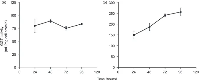
125 100 75 50 0 25 300 250 200 100 50 150 0 0 24 48 72 96 120 0 24 48 72 96 120 GGT activity
(mU/mg cell protein
)
Time (hours)
(a) (b)
Figure 4. Cellular G-glutamyltransferase (GGT) activity of c21/GGT melanoma cells (a) and DU145/GGT prostatic cancer cells (b). Cells were seeded at a density of 2 × 10⁴ cells cm−2 (c21/GGT) and 1.5 × 10⁴ cells cm−2 (DU145/GGT), respectively. At the established times cells were har-vested, homogenized and analysed for their GGT activity. Results are means ± SD of three separate experiments. Data were analysed by one-way ANOVA with Newman–Keuls test for multiple comparisons. Data were analysed by one-way ANOVA, post-test for linear trend gave the following results: not significant for 21/GGT cells, p < 0.0001 for DU145/GGT cells.
14 12 10 8 0 2 4 6 24h 2000 kDa 66 kDa 14 12 10 8 0 2 4 6 24h 2000 kDa 66 kDa 14 12 10 8 0 2 4 6 48h 14 12 10 8 0 2 4 6 48h 14 12 10 8 0 2 4 6 72h 14 12 10 8 0 2 4 6 72h 14 12 10 8 0 2 4 6 96h 14 12 10 8 0 2 4 6 96h Fluorescence (f.u.) 10 15 20 25 10 15 20 25 Elution volume (mL) (a) (b)
Figure 5. Online G-glutamyltransferase (GGT)-specific elution profile of representative medium samples from cultured DU145/GGT prostatic cancer cells (a) and c21/GGT melanoma cells (b).
Fractions of GGT 491
peak, less represented, eluting at 21.1 ml (correspond-ing to a MW of 66 kDa). The progressive increase of GGT in the medium with culture time was due to a selective increase of the first peak (Figure 5A, B).
To exclude the possibility that the high MW of the GGT released by c21/GGT and in DU145/GGT clones was due to the association of the released GGT with components present in the bovine serum contained in the culture medium, the human immortalized bronchial epithe-lial cell line BEAS/GGT, routinely grown in serum-free culture medium, was also tested for the release of GGT in the medium. Analysis of the BEAS/GGT supernatant showed the same two GGT peaks previously observed in cancer cell medium, with a prevalence and progressive increase of the one peak with a higher MW, correspond-ing to b-GGT (Figure 6).
Western blot analysis of cell culture supernatants Western blot analysis of the cell homogenates, con-ducted with an antibody directed against the heavy chain of GGT, showed in all the cell lines, the presence of the expected peptide in all the cell lines adopted in the present study. Only in BEAS/GGT homogenates there was a second band with a higher MW present, corresponding to the MW of the uncleaved GGT propeptide (Figure 7).
Discussion
The main finding of this study is that cultured melanoma, prostate cancer and bronchial epithelial cells release soluble GGT that corresponds to a specific GGT frac-tion (b-GGT) found in human plasma (Franzini et al. 2008); tissues other than liver might thus contribute to the levels of circulating GGT by increasing the plasma levels of a specific form of the enzyme. These findings might help explain why GGT is increased in several unrelated diseases not involving the liver, including cancer (Kazemi-Shirazi et al. 2007), and might add to the complex understanding of this sensitive biomarker (Whitfield 2001).
The enzyme fraction released in vitro is stable in the culture medium, is not degraded to other GGT fractions with lower MW, and the amount released is propor-tional to the specific GGT activity and to the number of cells present in the cultures, thus acting as a potential biomarker of the presence, amount and specific activity of GGT-positive cells.
Ultracentrifugation studies also showed that the corresponding fraction found in plasma, the b-GGT, despite showing the same MW as VLDL, displays a higher density, thus showing that b-GGT found in plasma is not simply due to the absorption of GGT over VLDL, but
corresponds to a specific particle whose properties are similar to the b-GGT obtained in vitro.
Only liver GGT is thought to circulate in blood, as peptidase-purified GGT obtained from other tissues is quickly removed in vivo due to its low content of sialic acid and consequently to its reduced surface charge (Mortensen & Huseby 1997). Anyway, as the GGT found in all the transfected cell lines showed the expected large subunit peptide (Visvikis et al. 1991), the 2000 kDa b-GGT necessarily includes component molecules still to be identified to meet the observed MW. These molecules are likely to determine the biological properties of the particles – including its half-life in blood – much more than GGT itself, thus
40 30 20 10 0 Fluorescence (f.u.) 10 15 20 25 Elution volume (mL) 2000 kDa 66 kDa
Figure 6. Online G-glutamyltransferase (GGT)-specific elution pro-file of a representative medium sample from cultured BEAS/GGT (continuous line) and BEAS-2B cells, the basal clone (dotted line) after 72 h in culture. 50 75 100 150 1 2 3 kDa
Figure 7. Western blot analysis of the cell homogenates, con-ducted with an antibody directed against the heavy chain of G-glutamyltransferase (GGT). Proteins were separated by 10% SDS-PAGE and gels were blotted onto nitrocellulose membranes. Anti-GGT antiserum was directed against the C-terminal 20 amino acids of human GGT heavy chain. Lane 1, c21/GGT; lane 2, DU145/GGT; lane 3, BEAS/GGT.
opening the possibility of a longer half-life of extrahe-patic b-GGT in vivo.
b-GGT is the only fraction of serum GGT found in atherosclerotic plaques (Franzini et al. 2009) co-localized with oxidized LDL lipoproteins and CD68+ foam cells
(Paolicchi et al. 2004), potentially involved in the pro-gression of the atherosclerotic plaque (Emdin et al. 2005); understanding in detail its properties might help to elucidate the precise role of GGT in the pathogenesis of atherosclerotic plaques, and the connection between organ damage, GGT levels and individual risk of disease. In conclusion, cells of different origin (melanoma, prostate cancer and bronchial epithelium) share the ability to release a GGT form that corresponds to the b-GGT fraction found in serum of healthy individuals; despite its MW, b-GGT does not correspond to GGT-laden VLDL. While supporting the hypothesis that liver is not the only source of serum GGT, the present findings point to the need of a better understanding of the nature and properties of the plasma GGT fractions, for a better clinical utilization of GGT as a biomarker of disease, and a better understanding of the pathogenesis of diseases associated with its increase in serum.
Acknowledgments
Decleration of interest: The authors report no conflicts of interest. The authors alone are responsible for the content and writing of the paper.
References
Corti A, Franzini M, Casini AF, Paolicchi A, Pompella A. (2008). Vitamin C supply to bronchial epithelial cells linked to glutathione avail-ability in ELF – a role for secreted gamma-glutamyltransferase? J Cyst Fibros 7: 174–8.
Emdin M, Passino C, Michelassi C, Titta F, L’Abbate A, Donato L, Pompella A, Paolicchi A. (2001). Prognostic value of serum gamma-glutamyl transferase activity after myocardial infarc-tion. Eur Heart J 22: 1802–7.
Emdin M, Pompella A, Paolicchi A. (2005). Gamma-glutamyltransferase, atherosclerosis, and cardiovascular dis-ease: triggering oxidative stress within the plaque. Circulation 112: 2078–80.
Franzini M, Corti A, Lorenzini E, Paolicchi A, Pompella A, De Cesare M, Perego P, Gatti L, Leone R, Apostoli P, Zunino F. (2006). Modulation of cell growth and cisplatin sensitivity by mem-brane gamma-glutamyltransferase in melanoma cells. Eur J Cancer 42: 2623–30.
Franzini M, Bramanti E, Ottaviano V, Ghiri E, Scatena F, Barsacchi R, Pompella A, Donato L, Emdin M, Paolicchi A. (2008). A high performance gel filtration chromatography method for gamma-glutamyltransferase fraction analysis. Anal Biochem 374: 1–6. Franzini M, Corti A, Martinelli B, Del Corso A, Emdin M, Parenti
GF, Glauber M, Pompella A, Paolicchi A. (2009).
Gamma-glutamyltransferase activity in human atherosclerotic plaques– biochemical similarities with the circulating enzyme. Atherosclerosis 202: 119–27.
Huseby NE, Strömme JH. (1974). Practical points regarding routine determination of gamma-glutamyl transferase (gamma-GT) in serum with a kinetic method at 37 degrees C. Scand J Clin Lab Invest 34: 357–63.
Huseby NE. (1982). Multiple forms of serum gamma-glutamyltrans-ferase. Association of the enzyme with lipoproteins. Clin Chim Acta 124: 103–12.
Kazemi-Shirazi L, Endler G, Winkler S, Schickbauer T, Wagner O, Marsik C. (2007). Gamma-glutamyltransferase and long-term survival: is it just the liver? Clin Chem 53: 940–6.
Lee DH, Jacobs DR Jr, Gross M, Kiefe CI, Roseman J, Lewis CE, Steffes M. (2003). Gamma-glutamyltransferase is a predictor of incident diabetes and hypertension: the Coronary Artery Risk Development in Young Adults (CARDIA) Study. Clin Chem 49: 1358–66.
Lee DH, Silventoinen K, Jacobs DR Jr, Jousilahti P, Tuomileto J. (2004). gamma-Glutamyltransferase, obesity, and the risk of type 2 dia-betes: observational cohort study among 20,158 middle-aged men and women. J Clin Endocrinol Metab 89: 5410–4.
Lee DS, Evans JC, Robins SJ, Wilson PW, Albano I, Fox CS, Wang TJ, Benjamin EJ, D’Agostino RB, Vasan RS. (2007). Gamma glutamyl transferase and metabolic syndrome, cardiovascular disease, and mortality risk: the Framingham Heart Study. Arterioscler Thromb Vasc Biol 27: 127–33.
Meisinger C, Doring A, Schneider A, Lowel H, KORA Study Group. (2006). Serum gamma-glutamyltransferase is a predictor of incident coronary events in apparently healthy men from the general population. Atherosclerosis 189: 297–302.
Mortensen B, Huseby NE. (1997). Clearance of circulating gamma-glutamyltransferase by the asialoglycoprotein receptor. Enzyme forms with different sialic acid content are eliminated at different clearance rates and without apparent desialylation. Clin Chim Acta 258: 47–58.
Nemesanszky E, Lott JA. (1985). Gamma-glutamyltransferase and its isoenzymes: progress and problems. Clin Chem 31: 797–803. Paolicchi A, Emdin M, Ghliozeni E, Ciancia E, Passino C, Popoff G,
Pompella A. (2004). Human atherosclerotic plaques contain gamma-glutamyl transpeptidase enzyme activity. Circulation 109: 1440.
Ruttmann E, Brant LJ, Concin H, Diem G, Rapp K, Ulmer H. (2005). gamma-Glutamyltransferase as a risk factor for cardiovascu-lar disease mortality. An investigation in a cohort of 163,944 Austrian adults. Circulation 112: 2130–7.
Ryu S, Chang Y, Kim DI, Kim WS, Suh BS. (2007). Gamma-glutamyltransferase as a predictor of chronic kidney disease in nonhypertensive and nondiabetic Korean men. Clin Chem 53: 71–7.
Sacchetti L, Castaldo G, Salvatore F. (1988). The gamma- glutamyltransferase isoenzyme pattern in serum as a signal discriminating between hepatobiliary diseases, including neoplasias. Clin Chem 34: 352–5.
Visvikis A, Thioudellet C, Oster T, Fournel-Gigleux S, Wellman M, Siest G. (1991). High-level expression of enzymatically active mature human gamma-glutamyltransferase in transgenic V79 Chinese hamster cells. Proc Natl Acad Sci USA 88: 7361–5. Wannamethee G, Ebrahim S, Shaper AG. (1995).
Gamma-glutamyltransferase: determinants and association with mortality from ischaemic heart disease and all causes. Am J Epidemiol 142: 699–708.
Wenham PR, Horn DB, Smith AF. (1984). Physical properties of G-glutamyltransferase in human serum. Clin Chim Acta 141: 205–18.
Whitfield JB. (2001). Gamma glutamyl transferase. Crit Rev Clin Lab Sci 38: 263–355.