FISIOPATOLOGIA CLINICA E SCIENZE DEL FARMACO Direttore: Prof. Paolo Miccoli
Programma: Fisiopatologia medica e farmacologia Direttore: Prof. Eleuterio Ferrannini
Tesi di Dottorato
Biomarcatori ecografici di funzione e struttura vascolare nell'uomo
Imaging biomarkers of vascular function and structure in humans
Dottoranda: Tutor:
Dott.ssa Rosa Maria Bruno Prof. Stefano Taddei
Table of contents
Chapter 1. Introduction Page 3
Chapter 2. Imaging biomarkers in cardiovascular disease: review of current literature
2.1 Carotid intima-media thickness 2.2 Carotid-femoral pulse wave velocity 2.3 Carotid distensibility 2.4 Flow-mediated dilation Page 7 Page 7 Page 12 Page 16 Page 21
Chapter 3. Rationale and objectives Page 27
Chapter 4. Methods
4.1 Carotid intima-media thickness and distensibility 4.2 Carotid-femoral pulse wave velocity
4.3 Flow-mediated dilation 4.4 Renal vasodilating capacity
Page 33
Page 33 Page 34 Page 34 Page 35
Chapter 5. Does vascular aging have a “vascular component”?
5.1 Experimental protocol 5.2 Results
5.3 Discussion Tables and figures
Page 37
Page 37 Page 38 Page 41 Page 47
Chapter 6. Is vascular aging similar in different districts? Is it related to cardio-renal damage?
5.1 Experimental protocol 5.2 Results
5.3 Discussion Tables and figures
Page 53 c Page 53 Page 55 Page 57 Page 61
Chapter 7. How to evaluate renal vascular damage?
7.1 Experimental protocol 7.2 Results
7.3 Discussion Tables and figures
Page 65
Page 65 Page 67 Page 69 Page 74
Chapter 8. Vascular consequences of environmental and therapeutic radiation
8.1 Rationale
8.2 Experimental protocol 8.3 Results
8.4 Discussion Tables and figures
Page 79 Page 79 Page 80 Page 82 Page 84 Page 88
Chapter 9. Vascular function in isolated obstructive sleep apnea syndrome
9.1 Rationale
9.2 Experimental protocol 9.3 Results
9.4 Discussion Tables and figures
Page 91 Page 91 Page 92 Page 96 Page 98 Page 103
Chapter 10. Vascular function in healthy Himalayan high-altitude dwellers
10.1 Rationale
10.2 Experimental protocol
Page 108
Page 108 Page 109
10.3 Results 10.4 Discussion Tables and figures
Page 112 Page 115 Page 120
Chapter 11. Conclusions Page 124
Chapter 1: Introduction
Cardiovascular (CV) disease is the first cause of mortality and morbidity in the world (Ezzati et al., 2002). Prevention of this condition, which is responsible of more than 2200 deaths per day only in United States, is a public health priority (Roger et al., 2012). Thus in the last decades great efforts have been made in the search of non-invasive biomarkers, able to identify the individual at risk for CV events in the asymptomatic, subclinical stage (Figure 1) (Duivenvoorden et al., 2009). Some biomarkers are currently recommended in order to improve stratification of CV risk, whereas other are considered useful only for research purposes (Mancia et al., 2007;Mancia et al.;Greenland et al., 2010). In particular, increased intima-media thickness of the common carotid artery (C-IMT), representing a marker of early atherosclerosis significantly correlated with coronary or cerebrovascular disease (Chambless et al., 1997;Witte et al., 2005), has been considered as an intermediate stage in the continuum of vascular disease and as a predictor of CV risk. Current guidelines also introduced other vascular parameters evaluating mechanical and functional arterial properties of peripheral and central arteries (Mancia et al., 2007;Mancia et al., 2009). Increased aortic stiffness has been shown to predict future CV events (Vlachopoulos et al., 2010) and it has been recognized as a marker of subclinical target organ damage in hypertensive patients (Mancia et al., 2007). Earlier vascular abnormalities, such as endothelial dysfunction in the peripheral arteries (Charakida et al., 2010), have been also mentioned for their possible use in future. However, several questions in this field are still open, limiting the wide use of these tools in the clinical practice. The open issues concern methodological as well as pathophysiological and prognostic aspects, and in this thesis we will discuss only a small part of these ones. First, C-IMT and arterial stiffness represent two sides of vascular aging, atherosclerosis and arteriosclerosis respectively, and are generally considered structural alterations. The identification of a “functional” component in these alterations would be of interest, since it will suggest a possibility of reversibility of vascular aging.
Second, vascular aging is a process involving the whole organism, while different techniques explore different districts. The quantification of the impact of different CV risk factors on different vascular districts might indicate the most adequate biomarker to use for future studies and suggest specific mechanisms of disease in different conditions. Third, vascular aging is often accompanied by cardiac and renal damage, but the relationship between organ damage in different districts is still largely unexplored. Fourth, while hypertension and diabetes have become the main cause of end-stage renal disease, and chronic kidney disease has been recognized as a main cause of CV events, to date early markers of renal vascular damage have not been developed.
For this PhD thesis I examined cross-sectionally a cohort of healthy subjects and patients with traditional CV risk factors in order to elucidate some of the abovementioned aspects. My original contribution to the knowledge in this field consists in:
-‐ the demonstration of a “functional” component in aortic stiffness, that is present only in diabetic patients and relies on endothelium-mediated mechanisms;
-‐ the demonstration of a differential impact of different CV risk factors on carotid and aortic stiffness;
-‐ the demonstration, in hypertensive patients, of an additive role of carotid and aortic stiffness in determining cardiac organ damage;
-‐ the identification of a new marker of renal vascular damage.
In the second part of the PhD thesis, I sought to demonstrate the usefulness of imaging biomarkers of vascular function and structure not only for risk stratification in patients with traditional CV risk factors, but also to explore CV consequences of non primarily CV diseases and conditions. We hypothesized that a comprehensive, multiparametric approach would be the best strategy to detect early vascular damage in one or more districts, possibly in a subclinical, reversible stage. This approach could allow identifying the “CV footprint” characterizing each condition, with a double aim: to elucidate the pathophysiology of CV complications in non-CV diseases and to propose the most useful test to be used in the clinical practice for screening and follow-up.
During my PhD thesis I applied this strategy to some primarily non-CV conditions that might qualify as emerging CV risk factors, such as exposure to environmental and iatrogenic radiation doses, obstructive sleep apnea syndrome (OSAS) in the absence of traditional CV risk factors, and chronic exposure to hypobaric hypoxia at high altitude.
My original contribution to the knowledge in this field consisted in:
-‐ the demonstration of a selective reduction of circulating endothelial progenitor cells, in the presence of preserved vascular function and structure, in young adults exposed during childhood to environmental radiation doses after the Chernobyl disaster and to therapeutic radioiodine treatment after thyroid cancer;
-‐ the demonstration that conduit artery endothelial dysfunction and impaired renal vasodilating capacity are part of the vascular phenotype of OSAS per se, since they are present even in the absence of traditional CV risk factors, while structural alterations such as arterial stiffness and increased C-IMT characterized only obese and/or hypertensive OSAS patients;
-‐ the demonstration that Himalayan high altitude dwellers, chronically living above 2500 meters of altitude, present a mainly microcirculatory endothelial dysfunction and a maladaptive carotid remodeling even in the absence of traditional CV risk factors.
Figure 1. Some biomarkers of vascular function and structure in humans proposed for CV risk stratification. From (Koenig, 2007)
Chapter 2. Imaging biomarkers in cardiovascular disease:
review of current literature
2.1 Carotid intima-media thickness
C-IMT, as measured by high resolution B-mode ultrasound of extra-cranial carotid arteries, is the most widely accepted non-invasive marker of subclinical atherosclerosis (Stein et al., 2008). C-IMT has been considered for a long time an intermediate phenotype of atherosclerosis suitable for use in large-scale population studies (Salonen and Salonen, 1993). Increased C-IMT has been associated with augmented CV risk (Oren et al., 2003;Paul et al., 2005) as well as with presence of advanced atherosclerosis at different vascular sites, including peripheral, cerebral and coronary districts (Geroulakos et al., 1994;Amato et al., 2007). Most importantly, large epidemiological studies, including the Atherosclerosis and Risk in Communities Study (ARIC), the Rotterdam Study and the Cardiovascular Health Study, have consistently reported the predictive value of C-IMT for myocardial infarction or stroke independent of traditional CV risk factors (Bots et al., 1997;Chambless et al., 1997;Hodis et al., 1998;Chambless et al., 2000;Rosvall et al., 2005;Lorenz et al., 2006). A recent meta-analysis of 8 population-based studies involving a total of 37197 subjects followed for a mean of 5.5 years confirmed the strong independent predictive value of cross-sectional C-IMT for future CV events (Lorenz et al., 2007). The predictive value of C-IMT for CV events has been confirmed also in asymptomatic type 2 diabetic patients, in whom, in combination with Framingham Risk Score (FRS), showed a greater predictive value than FRS alone (Yoshida et al., 2012).
Negative results on the independent predictive value of C-IMT for CV events have been also reported. The Thromso study, a prospective population-based study, in which total plaque area and C-IMT were measured in over 6000 healthy participants followed up for 6 years(Johnsen et al., 2007), and the Three-City Study, a large prospective study in which 5895 adults aged 65-85 years
with no history of coronary heart disease were followed up for 6 years (Plichart et al., 2011), showed that carotid plaques were a stronger predictor of myocardial infarction than was C-IMT. A recent meta-analysis of 11 population-based studies (54336 subjects) provided further evidence of a stronger predictive value for future CV events of carotid plaque than C-IMT (Inaba et al., 2012). It has been proposed that reduced progression of C-IMT corresponds to a reduction in CV events (Espeland et al., 2005). This hypothesis has been documented in clinical trials designed to study the efficacy of statins where C-IMT was used as surrogate end-point (Mercuri et al., 1996;Smilde et al., 2001;Taylor et al., 2002), even if conflicting results exist (Crouse et al., 2007). The European Lacidipine Study on Atherosclerosis (ELSA), a randomized trial in which 2334 hypertensive patients under effective antihypertensive treatment were followed-up for 3.75 years, showed that, despite baseline C-IMT strongly predicted CV events rate, differences in C-IMT compared with baseline did not (Zanchetti et al., 2009). Furthermore, a meta-analysis including 41 trials with 18307 participants showed that, despite significant reduction in CV events and all-cause death induced by active treatments, there was no significant relationship between C-IMT regression and events, suggesting that regression or slowed progression of C-IMT, induced by CV drug therapies, could not reflect reduction in CV events (Costanzo et al., 2010).
Taken together, the results of these studies question the accuracy of C-IMT as a marker of atherosclerosis. There are several explanations for these findings. First C-IMT, even if detected by high-resolution ultrasound, is not been able to distinguish between intima and media. The carotid artery is an elastic artery and C-IMT in healthy subjects consists almost entirely of media. While the carotid artery is unaffected by age or gender until 18 years of age, thereafter a progressive intimal thickening or medial hypertrophy occurs, determined by age, gender and hypertension, that do not necessarily reflect the atherosclerotic process (Finn et al., 2010). The observation that the classical risks factors for atherosclerotic disease poorly correlate with C-IMT further supports this hypothesis (Zanchetti et al., 2001;de Groot et al., 2004;Junyent et al., 2008). Istopathological studies indicate that C-IMT mainly represent hypertensive medial hypertrophy or thickening of smooth muscle
media, whereas atherosclerosis is largely an intimal process (Spence, 2006). In line with the hypothesis that C-IMT is biologically distinct from plaque, age-related thickening of intima-media layers of common carotid artery has been observed in the absence of overt atherosclerosis (Finn et al., 2010). Thus, it is not surprising that atherosclerosis plaque shows a better prognostic value than C-IMT for CV events. Furthermore, there have been great heterogeneity in methodological approaches to C-IMT measurements, which could have hampered its predictive value (O'Leary and Bots, 2010). C-IMT value can be influenced by: location of the measure, type of ultrasound data and features of the reading system. Inaba et al (Inaba et al., 2012) observed that 77% of the studies included in their meta-analysis did not explicit whether plaques were actually included in C-IMT analysis. In addition, 63% of the studies used maximal C-IMT, more likely reflecting focal thickening or plaque, instead of mean C-IMT. Furthermore, study design of C-IMT trials was heterogeneous since the definition of the landmarks of carotid segments (Common Carotid Artery, CCA, Carotid Bulb, CB or Internal Carotid Artery, ICA) selected to measure C-IMT may differ significantly (Lorenz et al., 2007). The far wall of CCA is the easiest of the three anatomical segments to be examined, being the most used measurement in clinical studies, but unfortunately plaques are rare at this site and studies on the relationship of C-IMT at this site are conflicting (O'Leary and Bots, 2010). No firm conclusion can be taken about which carotid district should be studied until large epidemiological studies will include measurement of C-IMT at different sites following standardized protocols. Ultrasound protocols that include common carotid CIMT measurements at multiple angles and multiple sites have been recently demonstrated to give the best results in terms of reproducibility, progression rate and treatment effect detection (Dogan et al., 2011), but it is still unknown whether this kind of approach can improve C-IMT predictive value. Noteworthy, the majority of the large prospective trials on predictive value of C-IMT used manual measurement by caliper, whereas automated systems are preferable. Regarding the latter, two main types are commercially available: B-mode image processing based devices and Radio-frequency (RF)-based echo-tracking systems (Figure 2) (Molinari et al., 2010). RF-based devices are
considered very accurate since RF signal has higher spatial resolution than B-mode (Brands et al., 1999;Laurent et al., 2006). However, when comparing the performance in terms of reproducibility of RF-based techniques with that of robust image-based systems, similar results are obtained (Bianchini et al., 2010;Molinari et al., 2010). However, it has to be pointed out that quality of the scans and system setting are crucial for the quality of the final result of B-mode based systems. In particular, dynamic range and gain settings, but also other parameters such as depth gain compensation or filtering, should be standardized as suggested by international guidelines (Touboul et al., 2007;Potter et al., 2008). Furthermore, Rossi et al. found that non-linear processing, such as compression and saturation, generally used in standard ultrasound equipment for a better image visualization, significantly influences carotid measurements (Rossi et al., 2009).
Future analysis providing the agreement between different kind of measurements and reference values for risk classification are needed in order to implement C-IMT assessment in clinical practice. In this sense, an important step forward has been recently made by Reference Values for Arterial Measurements Collaboration, which established reference values for C-IMT obtained by echotracking, based on subject-level data from different study centres worldwide (Engelen et al., 2012). This cross-sectional analysis, performed on 24871 subjects (4234 without any CV risk factor) enables now a comparison of C-IMT values for groups with different CV risk profiles, helping interpretation of such measures obtained both in research and clinical settings.
Figure 2. B-mode (on the left) and RF-mode (on the right) based echotracking systems for C-IMT measurement.
2.2 Carotid-femoral pulse wave velocity
Arterial stiffness represents the ability of the arterial system to cope with the systolic ejection volume through the distension of compliant large arteries during systole, and to relay cardiac contraction during diastolic runoff, through return to their initial dimensions, thus transforming the pulsatile ejection from the heart into continuous blood flow to the organs. Aorta is the vessel that gives the greatest contribution to the capacitive function of the arterial tree, and is a major determinant of the amplitude of the incident/forward pressure wave.
During ventricular contraction, part of the stroke volume is momentarily stored in the aorta and central arteries, stretching the arterial walls because of raising local blood pressure (BP). The arterial pressure wave generated in the aorta (forward or incident wave) is propagated to arteries throughout the body and is partly reflected back towards the aorta (reflected pressure wave). Arterial stiffness determines the propagation velocity of the pressure wave (Figure 3). Pulse wave velocity (PWV) works bidirectional, from the proximal aorta towards peripheral vessels and back. If aortic stiffness is low, the reservoir function during systole, as well as the recoiling during diastole, are maintained; furthermore the reflected pressure wave is delayed enough to impact on central arteries during end-systole and diastole, thus increasing the aortic pressure in early-diastole but not during systole. This situation is physiologically advantageous, since the higher diastolic pressure boosts coronary perfusion, without increasing the left ventricular pressure load. Increased arterial stiffening causes loss of the reservoir function and disrupts the above-described desirable timing of reflected wave. Thus, an increased PWV acts amplifying aortic and ventricular pressures during systole, and reducing aortic pressure during diastole. As a consequence, raising myocardial pressure load and oxygen consumption are increased, favoring the development of left ventricular hypertrophy and impaired ventricular relaxation (Ghiadoni et al., 2009;London and Pannier, 2010). On the other hand, diastolic BP and subendocardial blood flow are decreased in the presence of
stiffer large arteries: this phenomenon is responsible for decreased coronary perfusion pressure and myocardial ischemia. Increased aortic stiffness also decreases the stiffness gradient because peripheral arteries stiffness does not increase with aging, the consequence being microvascular pressure overload with concomitant alterations (capillary rarefaction, inward remodeling of small arteries) (Avolio et al., 1983;O'Rourke and Safar, 2005;Mitchell, 2008).
Aging is physiologically accompanied by arterial dilatation, increase in wall thickness and reduction of the elasticity and compliance, all features characterizing atherosclerosis (O'Rourke, 2007). The main structural change occurring with aging is the degeneration of the tunica media, which causes a gradual stiffening of large elastic arteries (Kaplan and Opie, 2006). This corresponds on the one hand to a reduced synthesis and an increased degradation of elastin, and on the other hand to an increased synthesis and reduced degradation of type-1 and -3 collagen (Lakatta and Levy, 2003). Arterial stiffness seems to be a common pathophysiological mechanism for many conditions associated with CV disease, including aging, menopausal status, lack of physical activity, genetic background or polymorphisms, and CV risk factors (smoking, hypertension, hypercholesterolemia, diabetes) or diseases (coronary heart disease, congestive heart failure, stroke) (Laurent et al., 2006). Arterial stiffness also has been demonstrated in primarily non-CV diseases, such as end-stage renal disease or moderate chronic kidney disease, rheumatoid arthritis, systemic vasculitis, and systemic lupus erythematosus (Laurent et al., 2006).
Arterial stiffness can serve in clinical practice as “intermediate” or “surrogate” end points for CV events. A major reason for its routine measurement comes from the recent demonstration that these parameters have an independent predictive value for CV morbidity and mortality. The largest amount of evidence has been given for carotid-femoral PWV (Figure 3), which showed independent predictive value for all-cause and CV mortality, fatal and nonfatal coronary events, and fatal strokes in patients with uncomplicated essential hypertension, type 2 diabetes, and end-stage renal disease, in the elderly and in the general population (Laurent et al., 2006;Vlachopoulos et al.,
2010). The independent predictive value of aortic stiffness has been demonstrated after adjustment to classic CV risk factors, including brachial PP, suggesting that aortic stiffness adds value to a combination of CV risk factors (Boutouyrie et al., 2002). This finding may be related to the fact that aortic stiffness integrates the damage to the aortic wall of CV risk factors over a long period, whereas BP, blood fasting glucose, and lipids can fluctuate over time and the values recorded at the time of risk assessment may not reflect the true damage to the arterial wall. Another explanation may be that the identification of aortic stiffness reveals the patients in which risk factors are translated into real risk (Ghiadoni et al., 2009).
An indirect argument to support the predictive value of arterial stiffness for CV mortality is the relationship with well-accepted intermediate end points such as left ventricular hypertrophy (Watabe et al., 2006), microalbuminuria and estimated decrease in glomerular filtration rate (GFR) (Hermans et al., 2007).
Although measures of stiffness are useful in predicting the occurrence of CV events, the value of reduction in arterial stiffness as a measurement of the reduction by treatment of the risk of such events has not yet been unequivocally proven. A meta-analysis of individual data in 294 patients demonstrated that long-term and short-term antihypertensive treatment is able to reduce PWV and that this reduction is not entirely explained by BP reduction (Ong et al., 2011). Indeed, PWV is highly BP-dependent on short term, but not on long term (Ait-Oufella et al., 2010;Ong et al., 2011). This seems to suggest that antihypertensive treatment can improve aortic stiffness beyond BP reduction in essential hypertensive patients. The only clinical evidence that reducing arterial stiffness is associated to a decreased risk of CV events was obtained in patients with end-stage renal disease (Guerin et al., 2001). In a mean follow-up of 50 months, the absence of PWV decrease in response to BP decrease was one of the predictors of all-cause and CV mortality, together with increased left ventricular mass, age, and preexisting CV disease. After adjustment for all confounding factors, the risk ratio for the absence of PWV decrease was 2.59 for all-cause mortality
and 2.35 for CV mortality(Guerin et al., 2001). However, the effect of aortic stiffness attenuation on CV morbidity and mortality remains to be established in other populations, particularly those at lower but still high CV risk, such as those with hypertension, dyslipidemia, diabetes, and moderate chronic kidney disease.
2.3 Carotid distensibility and stiffness
The measurement of aortic stiffness as PWV by arterial tonometry is generally accepted as the most simple, non-invasive, robust, and reproducible method to determine arterial stiffness (Laurent et al., 2006). However, it should be recognized that carotid-femoral PWV is not a direct measurement, since it is based on the acceptance of a propagative model of the arterial system. Furthermore, other arterial sites have potential interest: the measurement of local carotid stiffness (Figure 4) may also provide important prognostic information, since the carotid artery is a frequent site of wall-thickening and plaque formation, as above discussed (Laurent et al., 2006). Arterial stiffness can be estimated at the systemic, regional and local level. Arterial stiffness evaluation at the local level, generally obtained at the common carotid site (a large, easily accessible superficial artery) presents as advantages a great accuracy, since, unlike the systemic and regional evaluation, it is estimated directly by BP changes, which in turn determine the changes of volume of the vessel (Figure 4) (Laurent et al., 2006).
Aging, along with BP, is the main determinant of stiffness both of carotid and aortic stiffness (Paini et al., 2006). However, histological differences exist between large artery districts, responsible for different behaviors in the presence of CV risk factors. In particular, even if the carotid artery and the aorta are both classified as elastic vessels, the ultra-structure of the carotid artery is intermediate between muscular and elastic arteries, being more similar to the abdominal than to the ascending aorta. The radial, brachial and femoral arteries, which have a muscular structure, seem to be resistant to age-induced stiffening when compared to the carotid artery (Laurent et al., 1994). This implies that carotid and arterial stiffness are strictly correlated in the healthy population, while the correlation becomes weaker as soon as the number of CV risk factors increases (Paini et al., 2006). Therefore, although carotid-femoral PWV and carotid stiffness provide similar information on the aging impact on the stiffness of large arteries in healthy subjects, this is not the same for the
hypertension and / or diabetes. In these cases, the aorta seems to stiffen more than the carotid artery due to age and other CV risk factors (Paini et al., 2006).
Several studies investigated the physiopathology of carotid stiffness in essential hypertension. The increased arterial stiffness observed in patients with essential hypertension was generally attributed to arterial wall hypertrophy (Safar and Frohlich, 1995). However, further studies have shown that the increased carotid stiffness, observed in hypertensive patients, was due to an increase in distending pressure and not to hypertension-associated changes in structural properties, suggesting a functional adaptation of the wall material (Boutouyrie et al., 2000). Young's incremental elastic modulus of the common carotid artery, which estimates arterial stiffness controlling for C-IMT, has been shown to be greater in young never-treated hypertensive patients than in age- and gender-matched normotensive subjects, at a given circumferential wall stress, whereas it did not differ between the two groups in middle-aged and older individuals (Bussy et al., 2000). Thus, the mechanisms involved in the arterial stiffening in younger hypertensive patients likely differ from those advanced to explain the stiffening of large arteries with aging (O'Rourke, 2007).
The Atherosclerosis Risk in Communities (ARIC) Study, a population study recruiting a biracial sample of 4701 men and women 45 to 64 years of age, investigated the relationship between carotid stiffness and different CV risk factors. Carotid stiffness was associated with hypertension, diabetes, trait anger, physical activity, and ethnicity (Salomaa et al., 1995;Liao et al., 1999;Schmitz et al., 2001;Din-Dzietham et al., 2004;Williams et al., 2006). In particular, metabolic factors, such as elevated glucose, insulin, and triglycerides, had a synergistic effect on Young’s elastic modulus (Salomaa et al., 1995). Metabolic factors such as body mass index and triglycerides were independent correlates of Young’s elastic modulus also in the Bogalusa study, enrolling a younger multiracial population sample (516 asymptomatic subjects aged 25-38 years), beyond BP values, sex and age (Urbina et al., 2004). In the Baltimore Longitudinal Study on Aging, an independent association between suppressed anger and carotid stiffness was reported, as well as an increase in stiffness with metabolic syndrome and decreasing levels of testosterone (Scuteri et al., 2004). In a
aged population with high prevalence of CV risk factors and disease, enrolled in the Hoorn study, low-grade inflammation appeared to have an important role in determining increased carotid stiffness, mainly through arterial enlargement (van Bussel et al., 2012). In the same population, metabolic syndrome has been associated with stiffness of muscular arteries (brachial and femoral), but not of musculo-elastic arteries (carotid and aorta) (Henry et al., 2009), again confirming that impact of different risk factors varies depending on the district considered. In the Second Manifestations of ARTerial disease (SMART) Study, decreased carotid distensibility was a marker of increased CV risk but in patients who already had vascular disease (Simons et al., 1999). Carotid stiffness was also able to predict incident hypertension in the ARIC cohort (Liao et al., 1999). Arterial stiffness has been shown to be an independent predictor of CV morbidity and mortality (Vlachopoulos et al., 2010) and it is thought to play a crucial role in the development of CV disease. The predictive value of arterial stiffness has been shown mainly for aortic PWV (Vlachopoulos et al., 2010), while only few studies, with inconsistent results, were specifically aimed at investigating the predictive value of carotid stiffness for CV events. Blacher and colleagues first analyzed a cohort of 79 patients with end-stage renal disease undergoing hemodialysis, followed-up for 25 months, during which there were 10 fatal CV and 8 non-CV events. The study showed that an increased carotid stiffness is a powerful independent predictor of all-cause and CV mortality (Blacher et al., 1998). Carotid artery distensibility proved to be an independent predictor of CV disease after renal transplantation (Barenbrock et al., 2002). In the abovementioned SMART study, increased carotid stiffness was associated with an increased risk of CV events and mortality in not-corrected analysis, whereas the relationship disappeared after controlling for age (Dijk et al., 2005). However these study results are limited by the use of brachial instead of the local BP (BP) for the calculation of the carotid stiffness parameters, possibly leading to underestimation of the relationship between arterial stiffness and CV events. In contrast, in the Three-City study, enrolling 3337 elderly subjects (mean age 73 years) followed for a median of 44 months, patients who at baseline had a higher distension, showed an increased risk of coronary events compared to the ones
with lower distension (Leone et al., 2008). Finally, an independent association between increased carotid stiffness and a first-ever acute ischemic stroke has been reported (van Popele et al., 2001;Tsivgoulis et al., 2006), although other studies did not find the same relationship (Mattace-Raso et al., 2006). A tighter cause-effect relationship with cerebral rather than to coronary CV events is not surprising, given that the brain is the downstream district of the carotid artery. Accordingly, as a recent analysis conducted in 10407 subjects of the ARIC study followed-up for 13.8 years, showed that, after adjusting for CV risk factors, ultrasound measures of carotid arterial stiffness are associated with incident ischemic stroke but not incident coronary events, despite that the 2 outcomes sharing similar risk factors (Yang et al., 2012).
With an ever-increasing attention focused on the clinical implications of arterial stiffness analysis, it is extremely important to take into consideration methodological aspects. Indeed, inconsistent findings in trials exploring the clinical significance of carotid stiffness can be attributed, at least in part, to the large variance in technical measurements of carotid stiffness and local BP. The assessment of local stiffness can be obtained by measuring the diameter of the vessel and its variations during the cardiac cycle (stroke change in diameter or distension) by ultrasound signal, in conjunction with local pulse pressure (PP) estimation by tonometry (Laurent et al., 2006). As already discussed also for C-IMT, two main approaches are available for arterial diameter assessment by ultrasound data, with similar accuracy (Bianchini et al., 2012): B-mode image processing based devices (Faita et al., 2008;Bianchini et al., 2010) and Radio-frequency (RF)-based echo-tracking systems (Figure 2) (Brands et al., 1999). In particular, when adopting B-mode based systems, attention should be posed to the quality of the scans and the system settings (Potter et al., 2008;Rossi et al., 2009;Bianchini et al., 2010). In addition, both RF- and B-mode based devices can also provide the automatic measure of carotid C-IMT, and thus are able to provide both functional and structural parameters of the analyzed vessel, as suggested by the international expert consensus (Laurent et al., 2006).
2.4 Flow-mediated dilation
Endothelium plays a primary role in the control of vascular function by the production of nitric oxide (NO), which derives from the transformation of L-arginine into citrulline by the constitutive endothelial enzyme NO synthase. NO is produced under the stimulus of agonists (acetylcholine, bradykinin, and others), acting on specific endothelial receptors, and of mechanical forces, namely shear stress (Figure 5) (Furchgott and Zawadzki, 1980;Luscher and Barton, 1997). In pathological conditions, the same stimuli determine to the production of endothelium-derived contracting factors (e.g. thromboxane A2 and prostaglandin H2), which counteract the relaxing activity of NO, and reactive oxygen species, which impair endothelial function by causing NO breakdown (Luscher and Barton, 1997). In such conditions, reduced NO availability and endothelium-derived contracting factors not only exert an opposite effect on vascular tone, but also facilitate the pathogenesis of thrombosis and atherosclerotic plaque by promoting platelet aggregation, vascular smooth muscle cell proliferation and migration, and monocyte adhesion (Figure 5) (Ross, 1999).
This pivotal role of the endothelium in the atherosclerotic process led to the development of a different of methods to assess endothelial function, providing novel insights into pathophysiology and a clinical opportunity to detect early disease, quantify risk, judge response to interventions designed to prevent progression of early disease, and reduce later adverse events in patients (Deanfield et al., 2005;Deanfield et al., 2007).
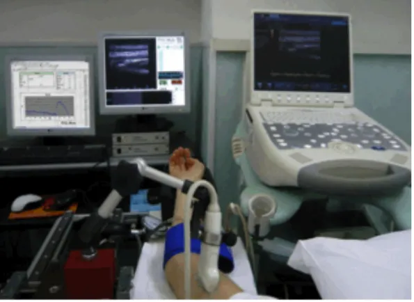
Endothelial function in clinical research is mainly tested by vascular reactivity studies (Deanfield et al., 2005). The most widely used technique is the so-called “flow-mediated dilation” (FMD) (Figure 6). This is a non-invasive, ultrasound-based method, introduced in 1992 (Celermajer et al., 1992). FMD occurs as a result of local endothelial release of NO and it is measured as brachial artery diameter changes in response to increased shear stress, induced by reactive hyperemia (Joannides et al., 1995;Ghiadoni et al., 2007). To this aim, a sphygmomanometer cuff placed on the forearm distal to the brachial artery is inflated to 300 mmHg and subsequently released 5 minutes
later. Endothelium-independent dilator response can be tested by low dose sublingual nitroglycerin (Ghiadoni et al., 2001). FMD has been studied widely in clinical research as it enables serial evaluation of young subjects, including children (Celermajer et al., 1992) It also permits testing of lifestyle and pharmacological interventions on endothelial biology at an early preclinical stage, when the disease process is most likely to be reversible (Deanfield et al., 2007).
Impaired FMD has been show in hypertensive patients and in the presence of the other CV risk factors (Ghiadoni et al., 2001;Ghiadoni et al., 2003;Brunner et al., 2005;Ghiadoni et al., 2008a;Bruno et al., 2012b). A report from the Framingham study showed a progressive inverse relation between by FMD, and the increasing FRS (Benjamin, 2004). A meta-analysis, performed in over 200 available studies, observed that the relationship between FMD and risk factors was more evident in patient with lower CV risk (Witte et al., 2005).
Several studies have shown that endothelial dysfunction is an early indicator of atherosclerotic damage and is associated with target organ damage, including increased C-IMT (Juonala et al., 2004;Rundek et al., 2006;Halcox et al., 2009), and left ventricular hypertrophy (Ercan et al., 2003) Importantly, impaired FMD has been associated with major CV events (Modena et al., 2002;Muiesan et al., 2008;Yeboah et al., 2008;Yeboah et al., 2009). A meta-analysis, evaluating longitudinal studies on the prognostic impact of endothelial dysfunction and including around 2500 patients with atherosclerotic coronary disease or characterized by high CV risk, showed that endothelial dysfunction, evaluated also as FMD, significantly predicted CV events, independently of traditional CV risk factors (Lerman and Zeiher, 2005). These studies suggested prognostic relevance of endothelial dysfunction in high-risk patients. This concept could be extended to lower risk populations according to the results of a more recent meta-analysis of 4 population-based cohort studies and 10 cohort studies, involving 5547 participants, showed a pooled relative risks of CV events per 1% increase in brachial FMD, adjusted for confounding risk factors of 0.87 (95% CI, 0.83- 0.91), consistent among all subgroups evaluated (Inaba et al., 2010). However, the authors highlighted that the presence of heterogeneity in study quality, the remaining confounding factors,
and publication bias in the available literature prevent a definitive evaluation of the additional predictive value of brachial FMD beyond traditional CV risk factors (Inaba et al., 2010).
Lack of restoration of endothelial function despite conventional treatment might identify a subset of “non-responders”, who might be suitable for more intensive or new therapeutic approaches. In a study conducted in 251 Japanese men with newly diagnosed stable coronary artery disease and concurrent endothelial dysfunction, FMD was repeated after 6 month of optimized individualized therapy. Those patients with persistently impaired FMD had significant higher event rates in the follow up period (26% over 31 months) (Kitta et al., 2009). Thus, correction of endothelial dysfunction might lead to an improvement of CV prognosis. Similarly, in a group of postmenopausal hypertensive women with impaired endothelial function, FMD was re-evaluated after 6 months of antihypertensive treatment (Modena et al., 2002). At 5-year follow-up, the incidence of CV events was significantly lower in the subgroup of women whose FMD was improved as compared to the subgroup without improvement, despite similar reduction in BP (Modena et al., 2002).
Furthermore, several studies have demonstrated that FMD can be improved with specific modifications of CV risk factors and with the use of drugs known to reduce CV risk (Ghiadoni et al., 2003;Plantinga et al., 2007;Charakida et al., 2009). Since the change in FMD occurring as a result of treatment can be obtained in a much shorter time (few months) than required for other vascular endpoints such as C-IMT (Halcox et al., 2009) or arterial stiffness (Laurent et al., 2006;Ghiadoni et al., 2009), FMD testing has a potential role for being included in clinical trials as a surrogate end-point (Charakida et al., 2010). Despite these considerable evidences, further large scale clinical trials are needed to demonstrate conclusively whether reversal of impaired FMD independently offers a better prognosis to patients with CV risk factor and disease.
Assessment of brachial FMD in clinical investigation has increased because of it is noninvasive and apparently easy to perform. However, several challenges need to be overcome representing major limitations to a widespread application of this method in clinical studies (Corretti et al.,
2002;Deanfield et al., 2005;Ghiadoni et al., 2008a;Charakida et al., 2010;Thijssen et al., 2011a). These challenges include the need for highly trained operators, an adequate equipment including probe-holder with stereotactic clamp and automated image analysis (Figure 6), and also the care required to minimize the effect of environmental or physiological influences (Ghiadoni et al., 2000). Furthermore, other caveats should be considered in designing a study where FMD is investigated for the biological and technical variability of its measurement, including appropriate study design and sample size and efforts to achieve a uniform technique and to minimize operator-dependency (Corretti et al., 2002;Ghiadoni et al., 2008b;Charakida et al., 2010;Thijssen et al., 2011a).
It is important to note that variations in technique, such as the position of the occluding cuff and duration of inflation, may produce results that are less representative of local NO activity, since FMD is also determined by the magnitude of post-ischemic forearm vasodilatation, which is a measure of microcirculatory function (Deanfield et al., 2007). Interestingly, in a meta-analysis of 14 studies, FMD derived using upper cuff occlusion was at least as predictive as that derived using distal cuff placement, despite the latter being more NO dependent, suggesting that that any direct measure of vascular function may provide independent prognostic information in humans (Green et al., 2011). Training and certification of sonographers to FMD procedure has been well described in guidelines (Corretti et al., 2002), and proven by results in recent multicenter trials by the low number of rejected examinations, because of poor quality and/or instability of the images (Ghiadoni et al., 2012;Luscher et al., 2012). To date, only few experienced research centers apply a rigorous methodology to achieve a high standard of accuracy and reduce FMD variability (Donald et al., 2008). The lack of uniform methodology represents one of the major limitations for the application of FMD assessment in large multicenter studies. The time-dependent variability of FMD measurements obtained was recently evaluated in more than 130 healthy volunteers by trained operators according to a standardized technique (Ghiadoni et al., 2012). This included centralized analysis by an automated edge detection system, composed of a special-purpose hardware/software
device for measuring changes of the brachial artery diameter (Gemignani et al., 2007;Gemignani et al., 2008). The study showed for the first time that the adherence to a rigorous protocol, with certified operator training as well as defined experimental settings, is feasible and provides an optimal time-dependent reproducibility of FMD (Ghiadoni et al., 2012).
Figure 5. Role of NO in endothelial function
TGF: transforming growth factor. AT: angiotensin. ATG: angiotensinogen. ET: endothelin. ACE: Angiotensin converting enzyme. TX: tromboxane. PG: prostaglandin. NADPH: nicotinamide dinucleotide phosphate. eNOS: endothelial NO synthase. L-Arg: L-Arginine. Ach: acetylcholine. ADP: adenosin diphosphate. cyclic guanosine monophosphate: cGMP. EDHF: endothelium-derived hyperpolarizing factors. 5-HT: serotonin. BK: bradikynin.
Chapter 3. Rationale and objectives
There is increasing evidence that imaging biomarkers of vascular function and structure: 1) are associated with classical CV risk factors; 2) predict CV events independently of classical risk scores; 3) can be used as surrogate endpoints for pharmacological and non-pharmacological clinical interventions, thus possibly having a crucial role in CV drug development; 4) might identify a subset of patients in which conventional treatments are not sufficient. After decades of research, these non-invasive techniques are finally reaching solid standardization and good reproducibility. However, several questions in this field are still open, strongly limiting the wide use of these tools in the clinical practice. Methodological as well as pathophysiological and prognostic aspects still need to be clarified before a widespread use of imaging biomarkers for risk stratification in patients with traditional CV risk factors, as well as in primarily non-CV conditions. In this PhD thesis I focused mainly on the following issues:
Does vascular aging has a “functional component”?
Arterial stiffness has been commonly considered a consequence of structural alterations of the vessel wall. However, mechanistic studies have suggested that a “functional” component may contribute to the compliance of large arteries. In particular, endothelium-derived factors such as NO (Wilkinson et al., 2004) and endothelin-1 (McEniery et al., 2003) have been proposed as physiological modulators of arterial stiffness in healthy individuals. An inverse correlation between endothelial dysfunction and arterial stiffness has been reported in cross-sectional studies performed in mixed cohorts including healthy subjects as well as patients with isolated systolic hypertension (Wallace et al., 2007), type 2 diabetes (Ravikumar et al., 2002;Jadhav and Kadam, 2005;Gunarathne et al., 2009) and coronary artery disease (Nigam et al., 2003;Kobayashi et al., 2004;Kopec et al., 2009), although conflicting results were obtained in healthy subjects (McEniery et al., 2006;Koivistoinen et al., 2011). These aspects are of clinical relevance, since arterial stiffness is a powerful predictor of CV events in the diabetic population (Cruickshank et al., 2002).
Thus we hypothesized that the “functional” component of arterial stiffness might differ in relation to the presence of absence of CV risk factors. The aim of the study was to investigate whether conduit artery endothelial function could be a determinant of arterial stiffness in patients with arterial hypertension, and whether the concomitant presence of metabolic alterations such as type 2 diabetes mellitus might influence this relationship.
Is vascular aging similar in different districts?
Arterial tree ageing, expressed as stiffening of large arteries, is a key feature of arteriosclerosis and CV disease and is accelerated by the presence of CV risk factors. An increased carotid-femoral PWV was demonstrated in the presence of hypertension, smoking, diabetes, hypercholesterolemia, obesity and metabolic syndrome (Laurent et al., 2006). However it was recently suggested that only arterial hypertension and age are independently associated with increased aortic stiffness (Cecelja and Chowienczyk, 2009) and the role of CV risk factors on top of hypertension and aging is largely unexplored. Furthermore, vascular aging is a global process involving the whole organism, while different techniques explore different districts. The study of the impact of different CV risk factors on different vascular districts might indicate the most adequate biomarker to use for future studies and suggest specific mechanisms of disease in different disease conditions. The aim of the study was to evaluate whether classical CV risk factors on top of hypertension can worsen large artery stiffness in hypertensive patients. For this purpose we studied arterial stiffness in two different districts: aorta, which is the most important district in which stiffness is related to CV prognosis, by means of carotid-femoral PWV, and carotid artery, a musculo-elastic vessel which is the preferential site for development and assessment of arteriosclerosis but for which the prognostic significance of stiffness is less established, by means of an ultrasound-based analysis system.
Is arterial stiffness related to cardio-renal damage?
The presence of arterial hypertension and organ damage, at the cardiac, vascular and renal level, identifies a condition of high risk of future CV events (Jager et al., 1999;Verdecchia et al., 2001;Lorenz et al., 2007) and, in these patients, prompt initiation of pharmacological treatment is considered (Mancia et al., 2007). Carotid-femoral PWV was included among measures indicating target organ damage in hypertensive patients (Mancia et al., 2007) and has been shown to reclassify a significantly higher percentage of patients to the high risk category in comparison to routine workup but also added to microalbuminuria and to cardiac ultrasound in the general population (Muiesan et al., 2010), even if the prognostic significance of this reclassification is still unknown.
Early vascular aging is often accompanied by cardiac and renal damage, but the relationship between organ damage in different districts is still largely unexplored. Aortic stiffness, measured as PWV, has been associated in cross sectional studies with well-accepted intermediate endpoints such as left ventricular hypertrophy (Watabe et al., 2006), albuminuria, and estimated GFR (Hermans et al., 2007) in the general population. Arterial stiffness and albuminuria were associated even in nonhypertensive, nondiabetic individuals, which suggests the possibility of a similar pathophysiological mechanism involved in these two indices of subclinical target organ damage (Kim et al., 2011). To our knowledge, no data are available at the moment about the relationship between carotid stiffness and hypertensive organ damage. This would be of importance, because arterial hypertension and associated risk factors are able to damage in parallel different districts, but vascular damage might amplify organ damage in parenchymal districts per se (O'Rourke and Safar, 2005). Furthermore, stiffness in different arterial districts might cluster in a different manner with organ damage. Thus this study is aimed at evaluating whether carotid and aortic stiffness are associated in a similar way to cardiac and renal hypertensive organ damage.
How to evaluate renal vascular damage?
Early diagnosis of target organ damage has a relevant role for patients at high CV risk, since both the presence and the regression of initial alterations, including left ventricle hypertrophy, C-IMT and microalbuminuria, are independent determinants of CV prognosis (Mancia et al., 2007). On regard of large arteries, endothelial dysfunction and arterial stiffness have been proved to be indexes of early vascular pathological changes, which might lead to end-organ dysfunction (Laurent et al., 2006;Virdis et al., 2008;Ghiadoni et al., 2009). Microalbuminuria is recognized as a clinical marker of first stages of nephropathy in type 1 and 2 diabetes (de Zeeuw, 2007;Bakker et al., 2009), whereas in essential hypertension it is mainly considered an indicator of CV risk (Mancia et al., 2007). However, microalbuminuria identifies an already established glomerular damage, which is likely to be preceded by earlier structural and functional alterations undetectable so far, unless by kidney biopsy (Futrakul et al., 2009;Ruilope and Segura, 2009;Weir, 2009). Moreover, a variable percentage of type 2 diabetic patients shows an impairment of GFR even in the presence of normal albumin excretion (MacIsaac et al., 2004). Thus, new reliable markers of early renal dysfunction, specifically linked to vascular renal impairment, are needed, in order to better identify the responsible mechanisms and the ideal timing for the treatment, aiming to delay the onset and/or slow the progression of renal damage.
Renal resistive index (RI) measured by duplex ultrasound is a useful parameter for quantifying the alterations in intraparenchimal renalcirculation that may occur during the course of chronic renal disease. RI is tightly related to renal arteriolosclerosis, as demonstrated by bioptic studies (Ikee et al., 2005), and represents an integrated index of arterial compliance, pulsatility and downstream microvascular impedance (Derchi et al., 2005;Krumme, 2006). High RI has a negative prognostic value in diabetic patients in terms of progression of renal disease (Hamano et al., 2008) and in hypertensive patients with renal artery stenosis for the success of percutaneous revascularization (Radermacher et al., 2001). However, some limitations must be considered concerning the use of RI in the early phases of nephropathy. First, reference values of RI in the general population have not
been unanimously defined and validated; second, RI is critically influenced by the aging process (Platt et al., 1991a); third, in the presence of normal renal function, the narrow range of RI values might not be able to highlight initial renal microvascular alterations. Therefore, the use of a pharmacological stimulus inducing vasodilation applied to RI measurements (Dynamic Resistive INdex, DRIN) might increase the discriminating power of duplex ultrasound, configuring a method potentially able to unmask renal abnormalities even in the early phases. A reduced renal vasodilation to nitroglycerine has been demonstrated in patients with overt diabetic nephropathy (Frauchiger et al., 2000); whether an impaired renal vasodilation is present even before the onset of microalbuminuria in patients with type 2 diabetes or with other CV risk factors, and its potential relation to markers of systemic vascular dysfunction, is still unknown.
The aim of the study was to evaluate RI in basal conditions and under pharmacological vasodilator stimulus in newly diagnosed, treatment-naive type 2 diabetic patients with normal urinary albumin excretion and glomerular filtration rate, comparing them to a group of essential hypertensive patients, in order to investigate whether a reduced DRIN could be a specific feature of diabetes. As a secondary aim, this study investigated the possible association of DRIN with established indexes of systemic vascular damage, such as endothelial function and arterial stiffness, and increased oxidative stress.
Multiparametric assessment of vascular function and structure in primarily non-cardiovascular conditions
Imaging biomarkers of vascular function and structure can be useful not only for risk stratification in patients with traditional CV risk factors, but also to explore CV consequences of primarily non-CV diseases and conditions (Laurent et al., 2006). We hypothesized that a comprehensive, multiparametric approach would be the best strategy to detect early vascular damage in one or more districts involved in each condition, possibly in a subclinical, reversible stage. This approach could
allow identifying the “CV footprint” characterizing each condition, with a double aim: to elucidate the pathophysiology of CV complications in primarily non-CV conditions and, in long term, to propose the most useful test to be used in the clinical practice for screening and follow-up.
During my PhD thesis I applied this strategy to some primarily non-CV conditions that might qualify as emerging CV risk factors, such as exposure to environmental and iatrogenic radiation doses, obstructive sleep apnea syndrome (OSAS) in the absence of traditional CV risk factors, and chronic exposure to hypobaric hypoxia at high altitude.
Chapter 4. Methods
In order to fulfill the objectives of this PhD thesis, several cohorts of volunteers with or without CV risk factors and other CV conditions were examined in a cross-sectional design. A set of non-invasive biomarkers of vascular function and structure were performed.
4.1 Carotid intima-media thickness and distensibility
Common carotid artery scans were obtained by high-resolution ultrasound with a 10 MHz linear array transducer (MyLab25; ESAOTE). Two 10’-clips were acquired from each common carotid artery (1 cm proximal to the carotid bulb in a region 1 cm wide and free of plaques) and then analyzed offline by means of Carotid Studio (Quipu srl, Pisa, Italy), an algorithm for the automatic evaluation of the instantaneous carotid diameter (Figure 2). The method is based on a well-validated contour tracking technique, allowing automatic evaluation of diameter stroke changes during the heart cycle, and was validated for accuracy against the gold standard measurement by radiofrequency (Bianchini et al., 2010). The following parameters were calculated:
- Carotid distension (ΔD), that is the stroke change in diameter, calculated as the difference between the systolic and diastolic diameter values;
- Cross-sectional distensibility coefficient (DC), calculated as DC = ΔA/(A*carotid PP), where A is the diastolic lumen area, and ΔA is the stroke changein lumen area;
- Carotid stiffness (CS), calculated using the Moens-Korteweg equation,(ρ*DC)–1/2, where ρ is the blood density.
Carotid lumen area was derived from the diameter values, assuming the cross-section of the artery to be circular. Carotid systolic BP and PP were obtained by applanation tonometry from carotid pressure waveforms, using central diastolic and mean BP for calibration (SphygmoCor, AtCor Medical). C-IMT was automatically measured on the same image sequences as the mean of the
relative values of 10 s. Parameters are indicated as the mean of the right and left common carotid values.
4.2 Carotid-femoral pulse wave velocity
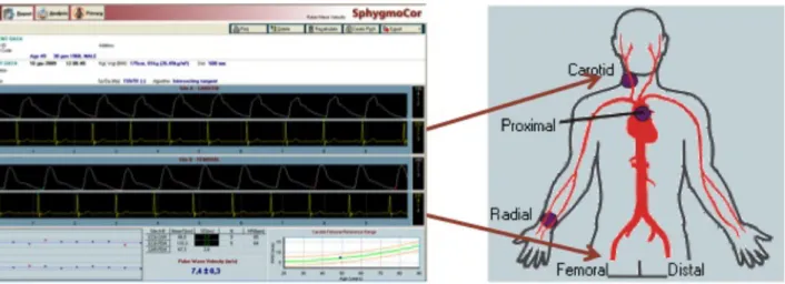
Arterial tonometry was performed according to international recommendations (Laurent et al., 2006). Carotid-femoral and carotid-radial PWV were assessed by SphygmoCor device (AtCor Medical, Sydney, Australia), recording waveforms at the two recording sites, sequentially. PWV was calculated as the ratio between the subtracted distance between the two recording sites and wave transit time (Figure 3 and 7). Applanation tonometry was also performed on the radial artery in order to obtain central BP values, by using a validated transfer function (SphygmoCor, AtCor Medical), and augmentation index normalized at 75 bpm (AIx). Two consecutive measurements were recorded and averaged. Carotid systolic BP and PP were then obtained from carotid pressure waveforms, using central diastolic and mean BP for calibration.
4.3 Flow-mediated dilation
Brachial artery scans were obtained by high-resolution ultrasound with a 10 MHz linear array transducer (MyLab25; ESAOTE, Florence, Italy). Endothelium-dependent response was assessed as dilation of the brachial artery in response to increased blood flow (flow-mediated dilation, FMD) (Bruno et al., 2011). Briefly, a pediatric cuff was positioned around the right forearm below the elbow and the right brachial artery was located and scanned longitudinally between 5 cm and 10 cm above the elbow using a 10 MHz linear array transducer (MyLab 25, ESAOTE Florence, Italy). The transducer has been held at the same point throughout the scan by a stereotactic clamp to ensure consistency of the image (Figure 6). After 1-min baseline recording, the cuff was inflated for five min at 300 mmHg and then deflated to induce reactive hyperemia (Figure 1). Endothelium-independent vasodilation was obtained by the sublingual administration of 25 µg GTN (Ghiadoni et
al., 2001). Brachial artery diameter measurements were performed by computerized edge detection system (Figure 6) (Gemignani et al., 2008). FMD and response to GTN were calculated as the maximal percent increase in diameter above baseline (mean of measures obtained during the first min).
Volume blood flow was calculated by multiplying duplex flow velocity (corrected for the angle) by heart rate and vessel cross-sectional area (πr2). Mean blood flow velocity in the brachial artery was determined by pulsed Doppler signal at 70°, with the range gate in the center of the artery, and continuously acquired throughout all the study. Resting and hyperemic shear rate (SR) were calculated according to the following equation: 8 *mean flow velocity / brachial artery diameter. Reactive hyperemia was calculated as the maximum percent increase in flow after cuff release as compared to baseline flow.
4.4 Renal vasodilating capacity
Renal scans were performed by a single trained operator using an ultrasound machine (MyLab 25, ESAOTE Florence, Italy) with a high-resolution multifrequency Convex probe (2.5 - 4.5 MHz). Three velocimetric measurements of the interlobar renal arteries adjacent to medullary pyramids were obtained by a translumbar or anterior approach (Figure 8). Renal resistive index (RI) was calculated in both kidneys according to the formula: (systolic peak velocity – end diastolic velocity)/systolic peak velocity. RI measurement was obtained at baseline and 5 min after pharmacological stimulus with a low-dose (25 µg) administration of sublingual GTN. Renal vasodilating capacity was calculated as absolute percent changes from baseline in response to GTN (Bruno et al., 2011).
Figure 7. Carotid PWV assessment
Chapter 5. Does vascular aging has a “functional” component?
The aim of this study was to investigate whether a “functional” component of arterial stiffness exists, based on endothelium-mediated mechanisms, in patients with arterial hypertension, and whether the concomitant presence of metabolic alterations such as type 2 diabetes mellitus might influence this relationship. The results of this research have been recently published (Bruno et al., 2012a).
5.1 Experimental protocol
Study population – A total of 341 individuals (259 patients and 82 age- and sex- matched healthy subjects) were enrolled at the Diabetes Outpatient Clinic of the Department of Endocrinology and Metabolism and the Hypertension Outpatient Clinic of the Department of Clinical and Experimental Medicine of the University Hospital of Pisa, Italy. For the patients’ group inclusion criteria were: diagnosis of essential hypertension and / or type 2 diabetes according to current guidelines (Mancia et al., 2007), or current treatment with antihypertensive or antidiabetic drugs. Exclusion criteria were: chronic kidney disease KDOQI stage 4 and 5, current insulin therapy, prior CV events, major co-morbidities (malignancies, chronic and acute inflammatory diseases, chronic heart failure and liver insufficiency, atrial fibrillation or frequent ectopic beats), non-CV medications interfering with vascular function (i.e. hormonal therapy, steroidal and non-steroidal anti-inflammatory drugs). Patients were divided in two groups according to the presence (n=84) or absence (n=175) of type 2 diabetes mellitus. All pharmacologically treated patients were on a stable therapeutic regimen for at least 3 months. The study conformed to the Declaration of Helsinki, was approved by the local Ethical Committee and all patients provided written informed consent prior to entering the study.
Experimental session – The volunteers were requested to refer to the local Diabetes Unit after an overnight fasting for collection of medical history, anthropometric parameters (body weight, height, and waist circumference) as well as blood and urine samples. On the following day, BP measurement and vascular assessment (FMD and carotid-femoral PWV) were determined at the Hypertension Unit. All measurements were performed in the morning after an overnight fasting, in a quiet air-conditioned room (22 to 24°C). For the duration of the study patients were kept on their usual pharmacologic treatment.
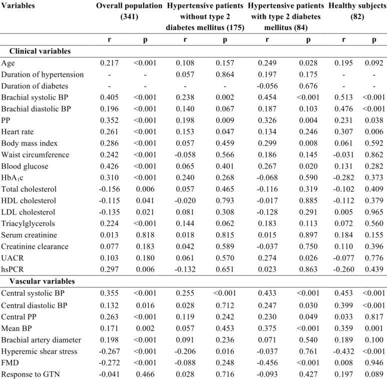
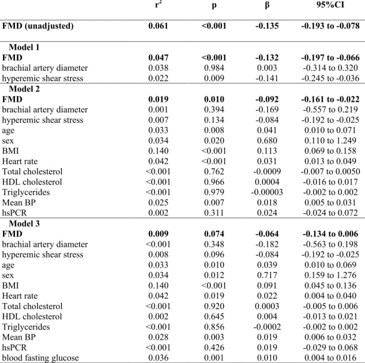
Statistical analysis - All statistical analyses were performed using NCSS 2004 (NCSS: Kaysville, Utah, USA). For normally distributed data, results are expressed as mean ± SD, whereas median value and 25%-75% interquartile range is used for non-normally distributed data. Differences in means among groups were analyzed using ANOVA and Bonferroni post-hoc analysis for normally distributed variables, or Kruskal-Wallis Z Test for not normally distributed variables; categorical variables were analyzed by χ2 test. Analysis of covariance was used to compare aortic PWV and FMD in different subgroups, as appropriate. Spearman’s rank was used to explore correlations among variables. Multiple linear regression analysis was performed including parameters with significant correlation with the dependent variable and building different models. The study was powered (90%) to detect a 1.3% difference in FMD, a 0.8 m/s difference in aortic PWV, and a difference of 0.20 in the slopes of regression lines, with a Type I error probability of 0.05.
5.2 Results
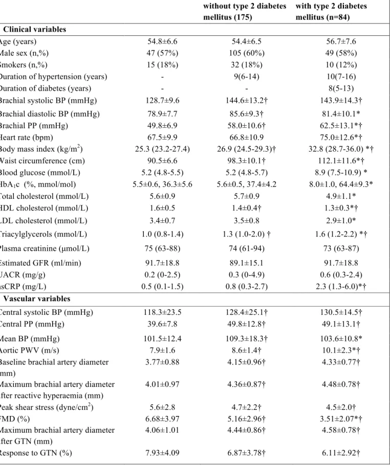
Clinical characteristics - Clinical characteristics of the study population are shown in Table 1. Diabetic and non-diabetic hypertensive patients were comparable for age, sex, duration of hypertension, percentage of smokers, renal function, and UACR. Diabetic hypertensive patients showed a higher prevalence of isolated systolic hypertension (78.8% vs. 63.1%, p=0.012) in comparison to non-diabetic hypertensive patients, in spite of similar systolic BP and lower diastolic
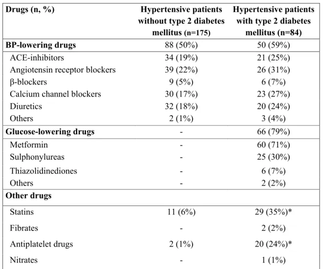
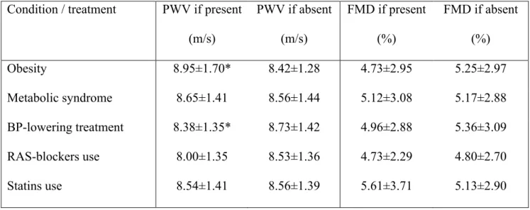
BP. Diabetic patients had also higher body mass index (BMI), waist circumference, blood glucose and HbA1c values. Due to the higher use of statins (35 vs. 6%, p<0.001), diabetic hypertensive patients had lower total- and LDL-cholesterol, and a higher prevalence of hypercholesterolemia (defined according ATP-III criteria or by current lipid-lowering therapy, 82% vs. 63%). HDL-cholesterol was lower and triglycerides higher in diabetic than in non-diabetic hypertensive patients. Diabetic hypertensive patients showed also higher levels of hsCRP. Obesity (defined as BMI ≥ 30 kg/m2) and metabolic syndrome, defined according to ATP-III criteria (Grundy et al., 2005), were significantly more prevalent among diabetic hypertensive patients than in normoglycaemic hypertensive patients (66 vs. 18%; 91 vs. 40%; p<0.001 for both). The majority of patients in both groups were on antihypertensive treatment (65% vs. 59%, p=ns). Details on CV drug therapy are shown in Table 2.
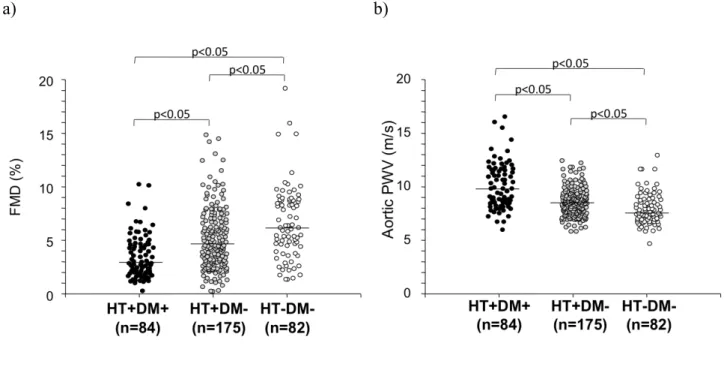
Vascular variables - Brachial artery diameter and hyperemic shear stress did not significantly differ between diabetic and non-diabetic hypertensive patients, although in both groups they were significantly higher than in healthy subjects (Table 1). FMD was lower in non-diabetic hypertensive patients than in healthy subjects, and further reduced in diabetic patients (Figure 1). When brachial artery diameter and hyperemic shear stress were considered as covariates, FMD was still significantly different among the three groups studied (p<0.001). On the contrary, GTN response was not different between diabetic and non-diabetic hypertensive patients, though similarly reduced as compared to healthy subjects (Table 1), even after adjustment for brachial artery diameter.
PWV was higher in non-diabetic hypertensive patients than in healthy subjects, and further increased in diabetic hypertensive patients (Figure 9). The significance was not affected after considering mean BP, age, and BMI as covariates (p<0.001). Central systolic BP and PP were similarly increased in diabetic and non-diabetic hypertensive patients, while mean BP was higher in the latter group (Table 1).
