UNIVERSITÀ
DELLA CALABRIA
Dipartimento di Farmacia e Scienze della Salute e della
Nutrizione
Dottorato di Ricerca in
Medicina Traslazionale
CICLO
XXIX
Stimulatory actions of IGF-I are mediated by
IGF-IR cross-talk with GPER and DDR1 in
mesothelioma and lung cancer cells
Settore Scientifico Disciplinare: MED/04
Coordinatore:
Ch.mo Prof. Sebastiano Andò
Supervisore/Tutor:
Ch.mo Prof. Marcello Maggiolini
Index
Abstract
...1Chapter I
Introduction
1.1 Lung cancer and Mesothelioma...21.1.1 Lung Cancer...2
1.1.2 Mesothelioma...5
1.2 G-protein coupled receptors (GPCRs)...7
1.2.1 G protein-coupled estrogen receptor (GPER)...10
1.3 Insulin-like growth factor-I ...12
1.3.1 The IGF-I/IGF-IR signaling pathway and its involvement in cancer...14
1.4 DDR1 expression and role in cancer...15
1.5 Aim of the study...18
Chapter II
Materials and Methods
2.1 Reagents...192.2 Cell cultures...19
2.3 Plasmids and luciferase assays...20
2.4 Gene silencing experiments ...20
2.5 Gene expression studies...21
2.6 Western blot analysis...21
2.7 Co-immunoprecipitation...22
2.9 Time-lapse microscopy...23 2.10 Statistical analysis...23
Chapter III
Results
3.1 IGF-I stimulates GPER expression through IGF-IR/ERK/p-38 transduction signaling...24 3.2 IGF-I triggers the expression of GPER target genes...28 3.3 IGF-IR and GPER are both involved in IGF-I regulation of DDR1 target genes...30 3.4 DDR1, IGF-IR and GPER contribute to the chemotaxis and migration of mesothelioma
and lung cancer cells...35
Chapter IV
Discussion...38
References
...43Abstract
Abstract
Insulin-like growth factor-I (IGF-I)/IGF-I receptor (IGF-IR) system has been largely involved in the pathogenesis and development of various tumors. We have previously demonstrated that IGF-IR cooperates with the G-protein estrogen receptor (GPER) and the collagen receptor discoidin domain 1 (DDR1) that are implicated in cancer progression. Here, we provide novel evidence regarding the molecular mechanisms through which IGF-I/IGF-IR signaling triggers a functional cross-talk with GPER and DDR1 in both mesothelioma and lung cancer cells. In particular, we show that IGF-I activates the transduction network mediated by IGF-IR leading to the up-regulation of GPER and its main target genes CTGF and EGR1 as well as the induction of DDR1 target genes like MATN-2, FBN-1, NOTCH 1 and HES-1. Of note, certain DDR1-mediated effects upon IGF-I stimulation required both IGF-IR and GPER as determined knocking-down the expression of these receptors. The aforementioned findings were nicely recapitulated in important biological outcomes like IGF-I promoted chemotaxis and migration of both mesothelioma and lung cancer cells. Overall, our data suggest that IGF-I/IGF-IR system triggers stimulatory actions through both GPER and DDR1 in aggressive tumors as mesothelioma and lung tumors. Hence, this novel signaling pathway may represent a further target in setting innovative anticancer strategies.
Chapter I
Chapter I
Introduction
1.1 Lung cancer and Mesothelioma
1.1.1 Lung cancer
Lung cancer is the leading cause of cancer-associated death worldwide and has one of the poorest prognoses among all cancer types (Ford et al. 2007). It is estimated that lung cancer is diagnosed in about 1.8 million patients and causes more than 1.5 million deaths each year (Fitzmaurice et al. 2015; Chen et al. 2008). Partly due to the increasing number of risk factors, as smoking and environmental pollution, the incidence of lung cancer was significantly increased in the past decades in many developing countries (Chen et al. 2008; Guo et al. 2012; Guo et al. 2015).
Studies of pulmonary carcinogenicity and respiratory inflammation have shown high risk for respiratory diseases and lung carcinogenesis in humans from exposures to various inhalable dusts, mineral fibers, airborne particulate matter (PM) and ozone. Ambient air pollution, containing PM smaller than aerodynamic diameter of 2.5 µm (PM2.5), has gained particular
attention in recent years as a main factor in the increased incidence of respiratory diseases, including lung cancer (Valavanidis et al. 2013; Aust et al. 2002; Nagai et al. 2010; Chuang et al. 2012; Strak et al. 2012). Tobacco smoke also plays an important role in augmenting the risk of epithelial inflammation and lung cancer due to its high carcinogenic potential and the synergistic effects with other respirable particulates leading to oxidative stress and increased production of mediators of pulmonary inflammation (Gardi et al. 2012; Sangani et al. 2011;
Chapter I
diverse tumors. In particular, the mechanisms by which inflammation can contribute to carcinogenesis include genomic instability, alterations in epigenetic events and gene transcription, enhanced cell proliferation and invasion, resistance to apoptosis, tumor neovascularization and metastasis (Azad et al. 2008).
Lung cancer is highly heterogeneous as it can arise in many different sites in the bronchial tree, therefore presenting variable symptoms and signs depending on its anatomic location. 70% of patients diagnosed with lung cancer present with advanced stage disease (Lemjabbar-Alaoui et al 2015). Lung cancer has been classified in different ways in order to minimize the number of unclassifiable lesions and to provide the basis for improved tumor diagnosis and therapy (Brambilla et al. 2001). The classification based on the type and the anatomic location of the tumor accounts (Table 1):
1) The Non-small-cell lung carcinoma (NSCLC) represents about 80% to 85% of lung cancers and its overall 5-year survival rate is 15%. Early stage tumors are treated primarily by complete surgical resection, yet 30 – 55% of patients will develop recurrence or die for the disease. NSCLC can be classified in:
• Squamous cell lung cancers (SQCLC), which represent about 25%–30% of all lung tumors and tend to arise in the main bronchi and advance to the carina.
• Adenocarcinomas (AdenoCA) that account for approximately 40% of all lung cancers and consist of tumors arising in peripheral bronchi. Adenocarcinoma is the most common histologic subtype of lung cancer in most countries, accounting for almost half of all lung tumors. It is the most frequent in younger males (<50 yrs old) and in women of all ages, in never smokers and in former smokers. Difficulties in adenocarcinoma sub-classification arise from its histologically heterogeneity. Mixed pattern adenocarcinomas are more common than tumors showing a single pattern (e.g. acinar, papillary, bronchioloalveolar, and solid adenocarcinoma with mucin
Chapter I
• Bronchioloalveolar cancers (BAC), now reclassified into adenocarcinoma in situ (AIS) and minimally invasive adenocarcinoma (MIA), arise in alveoli and spread through the interalveolar connections. AIS and MIA patients present a good disease-free survival after complete resection (5-year rate nears 100%) (Travis et al. 2013; Rubin et al 2012).
• Large cell anaplastic carcinomas (LCAC), also termed NSCLC not otherwise specified (NOS), are more proximal in location and locally tend to invade the mediastinum and its structures early. NSCLC-NOS comprises about 10% of all NSCLC and behaves similarly to small cell cancers with a rapid fatal spread.
2) Small cell lung cancers (SCLCs) derived from the hormonal cells of the lung, are the most differentiated tumors and tend to be central mediastinal malignancies. SCLCs comprise 10%–15% of all lung cancers, and are extremely aggressive disseminating rapidly into submucosal lymphatic vessels and regional lymph nodes, and almost always present without a bronchial invasion.
Chapter I
The international TNM classification of the lung is based on the staging system, which describes the anatomical extent of the disease (Fig. 1.1). The T category describes the size and extent of the primary tumor. The N category describes the extent of involvement of regional lymph nodes. The M category describes the presence or absence of distant metastatic spread. The addition of numbers to these categories describes the extent of the cancer. All possible combinations of the T, N, and M categories are then used to create TNM subset.
Figure 1.1
1.1.2 Mesothelioma
Smoking has been considered the main etiologic factor for lung cancer, however, several environmental contaminants like asbestos, arsenic, cadmium, nickel and silica, play an important role toward the development of lung tumor (Silvestri et al. 2013). Among the aforementioned environmental pollutants, asbestos has been particularly acknowledged as
Chapter I
from mesothelial cells lining lung, pleura or peritoneum (Rajer et al. 2014; Carbone et al. 2012). The pleural cavity is the most common site of origin of this tumor (Davidson 2015). The malignant pleural mesothelioma (MPM) is one of the most clinically aggressive malignancies, with the majority of patients succumbing to their disease within 2 years of diagnosis. The combination of surgery with chemotherapy has in recent years led to improvement in the survival and life quality of MPM patients, whereas targeted therapy has to date failed to have major clinical impact in this disease (Campbell et al. 2011). Asbestos refers to a family of silicate minerals divided into two major groups: the serpentine form of asbestos-chrysotile and the amphibole forms (crocidolite, anthophyllite, actinolite amosite and tremolite) (Carbone et al., 2002). Chronic inflammatory processes, which are caused by the deposition of asbestos fibers and the subsequent release of cytokines and growth factors by macrophages and mesothelial cells, have been shown to play an active role toward the development of pleural MM (Rascoe et al. 2012; Valavanidis et al. 2013). The duration, quality and intensity of the exposure are important variables in any mineral fiber related disease (Carbone et al. 2012). In particular, it has been recently demonstrated that asbestos induces necrotic cell death with resultant release of HMGB-1 in the extra-cellular space. HMGB-1 causes a chronic inflammatory response, macrophage accumulation and the secretion of TNF-alpha which, activating NF-kB, leads to HM survival (Yang et al., 2010) (Figure 1.2).
Chapter I
At the early stage of pleural mesothelioma, small nodules are found in the parietal pleura (not in the visceral pleura) that eventually extend along the pleural surface. Parietal and visceral pleurae show adhesion, and the tumor encloses the entire lung parenchyma. Few cases of peritoneal mesothelioma have been reported at the early stage and, consequently, little is known in terms of pathology and disease progression during the early stage (Inai 2008; Nonaka et al. 2005). Most of peritoneal mesothelioma is found as a diffuse tumor involving intestinal serosa or as a tumor located at the omentum or mesentery.
Histological classification of mesothelioma includes (Travis et al. 2013): • epithelioid type 60%
• sarcomatoid type 20% • biphasic type 20%
• desmoplastic (1–2%), and special variants only appear sporadically (several percentages).
Moreover, the last effect of the persistent inflammation caused by asbestos fibers accumulation is the pulmonary fibrosis, which in turn may create a favorable environment for the development of lung and pleura malignancies (Carbone et al. 2012; Mossman et al. 2011).
1.2 G-protein coupled receptors (GPCRs)
G-protein coupled receptors (GPCRs) are the largest family of cell-surface molecules involved in signal transmission and constitute the most prominent pharmacological targets in biomedicine (Dorsam and Gutkind 2007; Lappano and Maggiolini 2011; Pierce et al 2002). These proteins are characterized by a seven-transmembrane domain structure with an extracellular amino-terminus and an intracellular carboxyl-terminus (O’Hayre et al 2013). Over the last years, a number of studies has provided evidence on the crucial role elicited by
Chapter I
these receptors in various physiological processes including cardiac function, immune responses, neurotransmission and sensory functions (such as vision, taste and olfaction). However, their aberrant activity or expression also contributes to some of the most prevalent human diseases (O’Hayre et al 2013; Pierce et al 2002; Lappano and Maggiolini 2011). GPCRs owe their name to their interaction with heterotrimeric G proteins (composed of an α, β and γ subunit), which bind to the guanine nucleotide GDP in its basal state (Lappano and Maggiolini 2012; Pierce et al. 2002). A wide variety of ligands, including peptide and non-peptide neurotransmitters, hormones, amino acids, ions, lipids, odorant molecules and light, activate various GPCRs that regulate multiple biological responses including growth, migration, invasion and angiogenesis in normal and cancer cells (Lappano and Maggiolini 2012; Marinissen and Gutkind 2001). Upon activation by ligand binding, GDP is released and replaced by GTP, which leads to subunit dissociation into a βγ dimer and the GTP-bound α monomer (Lappano and Maggiolini 2012; Neves et al. 2002) (Figure 1.3). Consequently, the Gα and Gβγ complexes stimulate diverse effector molecules, which include adenylyl and guanylyl cyclases, phosphodiesterases, phospholipase A2 (PLA2), phospholipase C (PLC) and phosphoinositide 3-kinases (PI3Ks), thereby activating or inhibiting the production of a variety of second messengers such as cAMP, cGMP, diacylglycerol, inositol (1,4,5)-trisphosphate [Ins(1,4,5)P3], phosphatidyl inositol (3,4,5)- (1,4,5)-trisphosphate [PtdIns(3,4,5)P3], arachidonic acid and phosphatidic acid, in addition to promoting increases in the intracellular concentration of Ca2+ and the opening or closing of a variety of ion channels (Gutkind 2001).
Chapter I
Figure 1.3
Given the number, diversity and complexity of GPCRs, it is not surprising that their effectors extensively cooperate with other transduction pathways playing a critical role in signal transmission and integration (Lappano and Maggiolini 2011). For instance, activated GPCRs interact with cell-surface molecules including growth factor receptors which belong to the receptor tyrosine kinases (RTKs) family (Lappano and Maggiolini 2012; Daub 1996). In particular, multilayered cross-talk between GPCRs and growth factor receptors has an instrumental role in orchestrating downstream signaling molecules that are implicated in cancer development, angiogenesis and metastasis (Lappano and Maggiolini 2011). For instance, agonist-stimulated GPCRs have been shown to transactivate the epidermal growth factor receptor (EGFR) through the matrix metalloproteinases (MMP)-mediated release of EGF-like ligands and the subsequent generation of transduction signals that contribute to cancer progression (Blobel et al. 2005; Daub et al 1997; Filardo et al. 2000; Pierce et al. 2001). In a variety of tumors, like colon, lung, breast, head and neck, prostate and ovarian
Chapter I
development and metastasis (Filardo et al. 2000; Pierce et al. 2001; Hart et al. 2005). Likewise, a connection between the insulin-like growth factor-I receptor (IGF-IR) transduction pathway and GPCR-mediated signaling has been involved in many physiological functions and a variety of malignancies (Kisfalvi et al. 2009; Rozengurt 2010; Young et al. 2010). In addition, a strict dependence of the IGF-I signaling on GPCRs was reported in many physiological functions as well as in the development of diverse tumors (Young et al. 2010; Kisfalvi et al. 2009). On the basis of these findings, various GPCRs and their targets have been considered as promising therapeutic targets in drug discovery toward innovative anti-cancer strategies.
1.2.1 G-protein estrogen receptor (GPER)
In the last years, numerous studies have suggested that the G-protein estrogen receptor (GPER, formerly called GPR30) mediates estrogen signals in a wide number of normal and cancer cells (Maggiolini and Picard 2010). GPER was first identified as an orphan member of the 7-transmembrane receptor family by multiple groups in the late 1990s (Carmeci et al. 1997; Takada et al. 1997). GPER belongs to the rhodopsin-like receptor superfamily and its gene is mapped to chromosome 7p22.3 (Carmeci et al. 1997). Several studies have reported the presence of GPER at the plasma membrane, in the endoplasmic reticulum, in the Golgi apparatus as well as in the nuclear compartment (Filardo et al. 2007; Madeo and Maggiolini 2010; Pupo et al. 2014). GPER mediated signaling triggers the transactivation of EGFR, the rapid phosphorylation of the mitogen-activated protein kinases (MAPKs) ERK1/2 (Filardo et al. 2000), the activation of the PI3-kinase (PI3K) transduction pathway (Revankar et al. 2005), the increase in cAMP concentrations (Filardo et al. 2002; Thomas et al. 2005) and the mobilization of intracellular calcium (Revankar et al. 2005). Through these rapid actions, GPER induces a specific gene signature that in turn induces relevant biological responses as
Chapter I
cell proliferation, migration and angiogenesis (Maggiolini et al. 2004; Albanito et al. 2007; Pandey et al. 2009; Vivacqua et al. 2012; De Francesco et al 2014) (Fig.1.4).
Figure 1.4
GPER was also demonstrated to mediate the stimulatory action of estrogens in cancer-associated fibroblasts (CAFs), indicating its potential to contribute to cancer progression also through these important players of the tumor microenvironment (Madeo and Maggiolini 2010; De Francesco et al 2013).
GPER is expressed in diverse malignancies, including breast, endometrial, ovarian, thyroid, lung and prostate cancer cells, pancreatic epithelial neoplasms, choriocarcinoma and testicular germ cells (Maggiolini and Picard 2010; Bouskine 2009; Siegfried 2009; Liu et al 2015; Chan
Chapter I
2010; Glass 2011; Chevalier 2012). Moreover, our previous studies have shown that ligand-activated growth factor receptors up-regulate GPER expression in diverse types of cancer cells (Albanito et al. 2008; Vivacqua et al. 2009; De Marco et al. 2012). In particular, EGF and IGF-I were shown to induce GPER expression at both mRNA and protein levels (Albanito et al. 2008; Vivacqua et al. 2009; De Marco et al. 2012), hence highlighting the functional cross-talk which may occur between GPER, EGFR and IGF-IR signaling in estrogen-sensitive tumors (Lappano et al. 2013) (Fig.1.5).
Figure 1.5
1.3 Insulin-like growth factor-I
The IGF system includes in mammals at least three ligands, insulin and the insulin-like growth factors I and II (IGF-I and IGF-II), six high affinity binding proteins (IGFBP-1 to 6)
Chapter I
and four cell surface receptors (i.e. the IGF-I receptor (IGF-IR), the insulin receptor (IR), the insulin receptor-related receptor (IRR) and the Mannose-6-phosphate/IGF-II receptor (M6P/ IGF-IIR) (Bartella et al. 2012; Morgan, J.C.et al.1987) (Figure 1.6). The insulin and IGF-I are phylogenetically related peptides that play a major role as regulators of energy metabolism as well as development, growth and reproduction in response to nutrient availability (Bartke 2005).
Figure 1.6
IGFs are small, single-chain polypeptide ligands (7–8 kD) with an intact C-domain derived from prepropeptides in a manner similar to insulin (Beauchamp et al. 2010). The mature IGF-I and IGF-IGF-IGF-IIGF-I peptides consist of α and β domains that are homologous to β and α chains of insulin. Furthermore, in the cellular microenvironment, six IGF-binding proteins (IGFBP1–6) are present, which are not only crucial in regulating the bioavailability of IGFs by competing with IGFR and IGFBP proteases but also modulate the balance between IGFs and IGFBPs (Singh et al. 2014). IGFBPs and IGFs comprise a major superfamily of protein hormones that regulate mitogenesis, differentiation, survival, and other IGF-stimulated events in both normal and cancerous cells (Renehan et al. 2004). IGF-s are largely secreted from the liver, but also by extrahepatic tissues. IGF-I is mainly under the control of the pituitary growth hormone
Chapter I
(GH), while the regulation of IGF-II is less well understood (Casa et al. 2007; Reiss et al. 2001). The expression of IGFs is also influenced by several hormones including estrogens, thyrotropin, adrenocorticotropic hormone, as well as other growth factors such as EGF (Epidermal Growth Factor), FGF (Fibroblast Growth Factor), PDGF (Platelet-Derived Growth Factor). In physiological conditions, insulin and IGF-I are regulated by the endocrine system and act as hormones, and specifically bind to their cognate receptor (IR and IGF-IR respectively). However, in cancer tissues both IGF-I and IGF-II are often locally produced in a deregulated manner and act through autocrine and paracrine mechanisms (Samani et al. 2007).
1.3.1 IGF-I/IGF-IR signaling in cancer
IGF-IR is a protein tyrosine kinase (RTK) that belongs to the IGF system and regulates many crucial aspects of cellular physiology (Belfiore and Malaguarnera 2011). IGF-IR activation is initiated when ligands bind to the extracellular α-subunit, which undergoes to a conformational change that activates the tyrosine kinase activity and trans-phosphorylation of the β-subunits. Intra-chain phosphorylation allows the recruitment and activation of numerous docking proteins, including the IRS family members (IRS-1, IRS-2) and adaptors molecules as Shc and Grb2 (Pessin and Saltiel 2001). These substrates, in turn, are involved in the activation of transduction pathways that transmit the receptor signals to intracellular signaling cascades like MAPK, PI3K and the Janus kinase/signal transducer and activator of transcription pathway (JAK/STAT), which mediate important biological responses as glucose metabolism regulation, cell proliferation, inhibition of apoptosis, cell size, cell survival (Samani et al 2007; Belfiore 2007). In this regard, IGF-IR, which is often overexpressed in diverse cancer cell types, affects tumor development, progression and resistance to therapies (Carboni et al. 2005; Franks et al. 2016; Scagliotti et al. 2008; Sachdev et al. 2007). In
Chapter I
particular, the IGF system and related receptors as well as IGF-binding proteins have been established as important regulators of tumor initiation and progression in several malignancies, including pleural MM and lung cancer (Belfiore et al. 2009; Kai et al. 2009; Matà et al. 2016; Scagliotti et al. 2012). Moreover, a dysregulated IGF system has been shown to be implicated in various chronic diseases, such as pulmonary fibrosis (Lee et al. 2014; Hung et al. 2013).
Recently, GPER was shown to interact with IGF-IR in estrogen receptor (ER)-negative breast cancer cells (Pisano et al. 2016) as well as it has been demonstrated the capability of IGF-I to regulate GPER expression and function in ER-positive breast and endometrial cancer cells (De Marco et al. 2013). In particular, the increase of GPER levels induced by IGF-I occurred through the IGF-IR/PKCδ/ERK/ transduction pathway and involved also ER (De Marco et al. 2013).
In addition, an increasing body of data has demonstrated that important biological responses in cancer involve functional interactions of IGF-IR with the collagen receptor discoidin domain receptor 1 (DDR1), an other members of RTK family, that has been found overexpressed in diverse malignancies, including lung carcinomas, and implicated in cancer progression (Valiathan et al. 2012; Lappano and Maggiolini 2011; Liu et al. 2014; Song et al. 2004). Interestingly, this cross-talk occurs also independently of the collagen binding actions of DDR1 and, in human breast cancer cells, amplifies the stimulatory biological effects of IGF-I toward proliferation, migration and colony formation. Moreover, through a signaling pathway involving Akt/miR-199a-5p, IGF-I is able to upregulate DDR1 (Matà et al. 2016; Malaguarnera et al. 2015).
1.4 DDR1 and cancer
Chapter I
cell that directly or indirectly regulates almost all cellular behavior and the major developmental processes (Wiseman et al. 2003; Stickens et al. 2004; Rebustini et al. 2009; Lu et al. 2011). The ECM not only confers structural properties to tissues but it is also able to regulate cell proliferation, survival, migration and invasion (Lu et al. 2012). A diverse array of specialized cell surface receptors binds extrinsic factors such as mitogens, differentiation factors, cell membrane-bound molecules or extracellular matrix (ECM) proteins, and then transmit signals through the plasma membrane (Vogel et al. 2006). The Type I collagen, one of the most abundant ECM constituents, exhibits high density and altered architecture in cancer and is causally linked to tumor formation and metastasis (Provenzano et al. 2006, 2008). Many of the cell surface receptors belong to the family of RTKs characterized by an extracellular ligand binding domain, a single transmembrane domain and a catalytic tyrosine kinase domain. One subfamily of RTKs are the Discoidin Domain Receptors (DDRs) defined by the discoidin homology region in their extracellular domain and represented by two members: the discoidin domain receptor 1 and 2 (DDR1 and DDR2) (Fig.1.7).
Chapter I
DDR1 was first identified in the Dictyostelium discoideum and was shown to mediate cell aggregation (Rammal et al. 2016; Vogel et al., 1997). DDR2 shares highly conserved sequences with DDR1 (Carafoli et al., 2009). Both receptors are activated upon binding to collagen. In particular, DDR1 is activated by various types of collagen including type I, IV, V, VI, and VIII, whereas DDR2 is only activated by fibrillar collagens as type I, III, and type X (Shrivastava et al., 1997; Vogel et al., 1997; Leitinger and Kwan, 2006). In contrast with other RTKs as EGFR and fibroblast growth factor receptor (FGFR), which display a rapid and transient activation (Dengjel et al., 2007), DDR1 and DDR2 are unique in that they exhibit a delayed and sustained phosphorylation upon binding to collagen (Vogel et al., 1997). Furthermore, many RTKs undergo negative regulation such as receptor/ligand internalization and subsequent degradation or dephosphorylation by phosphatases (Avraham and Yarden, 2011). In the case of DDRs, phosphorylation levels may persist up to 18 h (Vogel et al., 1997).
Notably, numerous studies have linked the expression, activation and dysregulation of DDRs to the progression of diverse tumors (Rammal et al. 2016; Valiathan et al., 2012; Ford et al., 2007). In this context, recently it has been discovered that, in breast cancer cells and in transfected fibroblasts, the collagen receptor DDR1 associates with the IGF-IR in an IGF-I dependent manner, suggesting that the growth factor contributes to a functional cross-talk between IGF-IR and DDR1 in cancer (Malaguarnera et al. 2015). Of note, this interaction amplifies the stimulatory actions of IGF-I toward breast cancer cell proliferation, migration and colony formation (Matà et al. 2016; Malaguarnera et al. 2015).
Chapter I
1.5 Aim of the study
The aim of the present study was to provide novel insights into the molecular mechanisms by which IGF-I/IGF-IR system may trigger stimulatory responses, as gene expression changes, chemotactic motility and migration, in IST-MES1 mesothelioma and A549 lung cancer cells.
Chapter II
Chapter II
Materials and Methods
2.1 Reagents
IGF-I, SB202190 (SB) and collagen I from rat tail were obtained from Sigma-Aldrich Inc. (Milan, Italy). PD98059 (PD) and 3-bromo-5-t-butyl-4-hydroxybenzylidenemalonitrile (AG1024) were purchased from Calbiochem (DBA, Milan, Italy). All compounds were solubilized in dimethylsulfoxide, except PD and IGF-I, which were dissolved in ethanol and in water, respectively. DDR1-IN-1 dihydrochloride (DDR-1 in) was purchased from Tocris Bioscience (Space, Milan, Italy).
2.2 Cell cultures
IST-MES1 malignant mesothelioma cells were kindly provided by Dr. Orengo (Istituto Nazionale per la Ricerca sul Cancro, Genova, Italy). Cells were previously characterized (Orengo et al. 1999) and were grown in Nutrient Mixture F-10 Ham (Ham's F-10) medium supplemented with 10% fetal bovine serum (FBS) and 100 µg/ml penicillin/streptomycin. A549 lung cancer cells were obtained by ATCC, used <6 months after resuscitation and maintained in DMEM/F12 (Dulbecco’s modified Eagle’s medium) supplemented with phenol red 10% FBS and 100 µg/ml penicillin/streptomycin. All cell lines were cultured at 37°C in 5% CO2 and switched to medium without serum the day before immunoblots and reverse
Chapter II
2.3 Plasmids and luciferase assays
The GPER luciferase expression vector (promGPER) was previously described (Recchia et al. 2011). The CTGF luciferase reporter plasmid (promCTGF) (-1999/+ 36)-luc was a gift from Dr. Chaqour. EGR1-luc plasmid, containing the -600 to +12 5’-flanking sequence from the human EGR1 gene, was kindly provided by Dr. Safe (Texas A&M University). The plasmid DN/cfos, which encodes a c-fos mutant that heterodimerizes with c-fos dimerization partners but does not allow DNA biding (Gerdes et al. 2006), was a kind gift from Dr C Vinson (NIH, Bethesda, MD, USA). The Renilla luciferase expression vector pRL-TK (Promega, Milan, Italy) was used as internal transfection control. Cells (1x10⁵) were plated into 24-well dishes with 500 µl/well culture medium containing 10% FBS. Transfection were performed using X-treme GENE 9 DNA transfection reagent as recommended by the manufacturer (Roche Diagnostics, Milan, Italy), with a mixture containing 0.5 µg of reporter plasmid and 10 ng of pRL-TK. After 24 h, treatments were added and cells were incubated for 18 h. Luciferase activity was measured using the Dual Luciferase Kit (Promega, Milan, Italy) according to the manufacturer’s recommendations. Firefly luciferase activity was normalized to the internal transfection control provided by the Renilla luciferase activity. Normalized relative light unit values obtained from cells treated with vehicle were set as 1-fold induction upon which the activity induced by treatments was calculated.
2.4 Gene silencing experiments
Cells were plated onto 10-cm dishes and transfected by X-treme GENE 9 DNA Transfection Reagent for 24 h before treatments with a control vector, a specific shRNA sequence for each target gene. The shIGF-IR and the respective control plasmids (shRNA) were purchased from SA Bioscience Corp. (Frederick, MD, USA) and used according to the manufacturer’s recommendations. The short hairpin (sh)RNA constructs to knock down the expression of
Chapter II
GPER and the unrelated shRNA control construct have been described previously (Albanito et al. 2008).
2.5 Gene expression studies
Total RNA was extracted and cDNA was synthesized by reverse transcription as previously described (Rigiracciolo et al 2015, 2016). The expression of selected genes was quantified by real-time PCR using Step One sequence detection system (Applied Biosystems, Milan, Italy). Gene-specific primers were designed using Primer Express version 2.0 software (Applied Biosystems Inc. Milan, Italy) and are as follows: GPER Fwd 5′- ACACACCTGGGTGGACACAA-3′ and Rev 5′-GGAGCCAGAAGCCACATCTG-3’; HES-1 Fwd 5′-TCAACACGACACCGGATAAA-3′ and Rev
5′-CCGCGAGCTATCTTTCTTCA-3′; NOTCH 1 Fwd 5′-AATGGCGGGAAGTGTGAAGC-3′ and Rev
5′-GCATAGTCTGCCACGCCTCT-3′; MTN-2 Fwd 5′-CTCCGAGTGGGCCAGTAAAG-3′
and Rev 5′- CTGGCTCAGATTCTGTTGGCT-3′; FBN-1 Fwd
5′-GCCGCATATCTCCTGACCTC-3′ and Rev 5′-GTCGATACACGCGGAGATGT-3′; 18S Fwd 5′- GGCGTCCCCCAACTTCTTA-3′ and Rev 5′-GGGCATCACAGACCTGTTATT-3′. Assays were performed in triplicate and the results were normalized for 18S expression and then calculated as fold induction of RNA expression.
2.6 Western blot analysis
Cells were processed according to a previously described protocol (Santolla et al. 2015) to obtain protein lysate that was electrophoresed through a reducing SDS/10% (w/v) polyacrylamide gel, electroblotted onto a nitrocellulose membrane and probed with primary antibodies against antiphosphotyrosine antibody (4G10) (Merck Millipore, Milan, Italy), IGF-IR (7G11), GPER (N-15), CTGF (L-20), phosphorylated ERK1/2 (E-4), ERK2 (C-14), NOTCH 1 (C-20), EGR1 (588), phosphorylated p-38 (D-8), p-38 (A-12), β-actin (C2), (Santa
Chapter II
Cruz Biotechnology, DBA, Milan, Italy). Proteins were detected by horseradish peroxidase-linked secondary antibodies (DBA, Milan, Italy) and revealed using the ECL System (GE Healthcare).
2.7 Co-immunoprecipitation
Cells were lysed using 200 µl RIPA buffer with a mixture of protease inhibitors containing 1.7mg/ml aprotinin, 1mg/ml leupeptin, 200mmol/L phenylmethylsulfonyl fluoride, 200mmol/L sodium orthovanadate, and 100mmol/L sodium fluoride. A total of 100 µg proteins were incubated for 2 h with 2 µg of the appropriate antibody (GPER, N-15; IGF-1R, 7G11) and 20 µl of protein A/G agarose immunopreciptation reagent (Santa Cruz Biotechnology, DBA, Milan, Italy). Samples were centrifuged at 13,000 rpm for 5 min at 4°C to pellet beads. After four washes in PBS, samples were resuspended in RIPA buffer with protease inhibitors and SDS sample buffer. Western Blot analysis was performed as described above.
2.8 Migration assay
Migration assays were performed using Boyden chambers (Costar Transwell, 8 mm polycarbonate membrane, Sigma Aldrich, Milan, Italy). Cells were transfected in regular growth medium. After 8 h, cells were trypsinized and seeded in the upper chambers. Treatments were added to the medium without serum in the bottom wells where applicable, cells on the bottom side of the membrane were fixed and counted 8 hours after seeding.
Chapter II
2.9 Time-lapse microscopy
Cells (1 × 10⁵) were seeded in 6-well plates and maintained in regular growth medium for 24 h. For knockdown experiments, cells were transfected for 24 h with shRNA constructs directed against IGF-IR or GPER and with an unrelated shRNA construct. Thereafter, cells were treated and transferred into a time-lapse microscopy platform, equipped with a heated stage chamber (Cytation™3 Cell Imaging Multi-Mode Reader, Biotek, Winooski, VT). Cells were maintained at routine incubation settings (37 °C, 5% CO2) using temperature and gas
controllers. To evaluate chemotaxis the images were recorded using Cytation 3 Cell Imaging Multimode Reader and the software Gen5 (BioTek, Winooski, VT) in 10 min intervals for 8 hours. Then, the images were processed as a movie using the software Adobe Creative Cloud Premier Pro CC. Frames collected every 10 minutes are displayed at a rate of 10 frames s-1.
2.10 Statistical analysis
Statistical analysis was performed using ANOVA followed by Newman-Keuls’ testing to determine differences in means. P < 0.05 was considered as statistically significant.
Chapter III
Chapter III
Results
3.1 IGF-I stimulates GPER expression through IGF-IR/ERK/p-38
transduction signaling
On the basis of previous studies showing that IGF-I triggers stimulatory effects in malignant mesothelioma as well as in lung cancer cells (Kim et al. 2014; Liu et al. 2004), we began our study evaluating the transduction signaling activated by IGF-I in IST-MES1 mesothelioma and A549 lung cancer cells, which were used as model system.
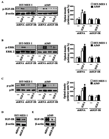
Figure 3.1 IGF-IR (A), ERK (B) and p-38 (C) phosphorylation in cells transfected for 24 h with shRNA or
shIGF-IR treated or not with vehicle (-) or 100 ng/ml IGF-I for 15 min. (D-E) Efficacy of IGF-IR silencing. Data shown are the mean ± SD of three independent experiments. (■) p<0.05 for cells receiving vehicle (-) versus
Chapter III
First, we determined that in both cell types IGF-I induces the phosphorylation of IGF-IR and both ERK and p-38. As expected, these responses were no longer observed after IGF-IR silencing (Fig. 3.1).
The activation of ERK triggered by IGF-I was abolished in the presence of the IGF-IR inhibitor AG and the MEK inhibitor PD, but it still persisted using the p-38 inhibitor SB. The phosphorylation of p-38 was prevented by AG and SB, but not in the presence of PD (Fig. 3.2).
Figure 3.2 ERK (A) and p-38 (B) activation in cells treated for 15 min with vehicle (-) or 100 ng/ml IGF-I alone
and in combination with either 1 µM IGF-IR inhibitor tyrphostin AG1024 (AG), or 1 µM MEK inhibitor PD98059 (PD) or 1 µM p38 inhibitor SB202190 (SB). Side panels show densitometric analysis of the blots normalized to β-actin, ERK2 and p38 that served as loading controls respectively for pIGF-IR, pERK and p-p38. Data shown are the mean ± SD of three independent experiments. (■) p<0.05 for cells receiving vehicle (-) versus treatments.
On the basis of our previous data showing that IGF-I signaling cooperates with several GPCR family members, including GPER, toward cancer progression (Lappano and Maggiolini 2011; De Marco et al. 2013), we evaluated whether IGF-I regulates GPER expression in IST-MES1 and A549 cells. In this regard, time-course experiments demonstrated that IGF-I up-regulates
Chapter III
that these responses to IGF-I occurred through IGF-IR, as the induction of GPER mRNA (data not shown) and protein levels (Fig. 3.3C-E) was abolished by knocking-down IGF-IR expression. Recapitulating the aforementioned findings, the transactivation of the GPER promoter by IGF-I was prevented by IGF-IR silencing (Fig. 3.3F), and the IGF-I induced GPER protein up-regulation was abrogated in the presence of AG, PD and SB (Fig. 3.3G). Taken together, these results indicate that the IGF-I/IGF-IR transduction pathway stimulates GPER expression through ERK and p-38 signaling. In order to further investigate this functional cross-talk between IGF-IR and GPER, we performed co-immunoprecipitation studies determining that IGF-I triggers also a direct interaction between these receptors in both IST-MES1 and A549 cells upon either 1 h (data not shown) or 8 h treatment with IGF-I (Fig. 3.3H-I), thus suggesting that the interaction between IGF-IR and GPER may occur without a newly protein expression of GPER.
Chapter III
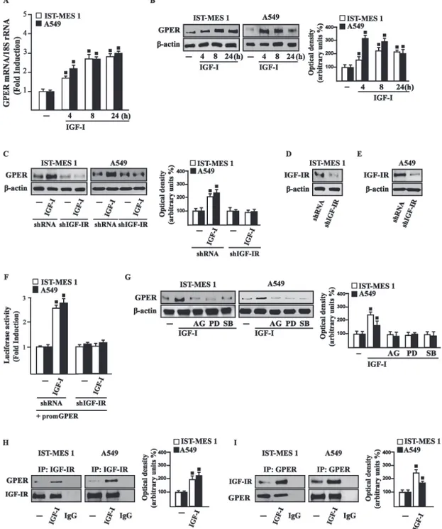
Figure 3.3 (A) mRNA expression of GPER in cells treated with either vehicle (-) or 100 ng/ml IGF-I, as
evaluated by real-time PCR. Results obtained from experiments performed in triplicate were normalized for 18S expression and shown as fold change of RNA expression compared to cells treated with vehicle. (B) GPER protein levels were evaluated by immunoblotting in cells treated with either vehicle (-) or 100 ng/ml IGF-I, as indicated. (C) GPER protein expression in cells transfected for 24 h with either shRNA or shIGF-IR and then treated for 8 h with vehicle (-) or 100 ng/ml IGF-I. (D-E) Efficacy of IGF-IR silencing. (F) Cells were transfected for 24 h with shRNA or shIGF-IR together with the GPER promoter construct. Then, cells were treated for 18 h with vehicle (-) or 100 ng/ml IGF-I. The luciferase activities were normalized to the internal transfection control, and values of cells receiving vehicle (-) were set as one fold induction upon which the activity induced by treatments was calculated. (G) GPER protein levels in cells treated for 8 h with vehicle (-) or 100 ng/ml IGF-I alone or in combination with 1 µM IGF-IR inhibitor tyrphostin AG1024 (AG), 1 µM MEK inhibitor PD98059 (PD) and 1 µM p38 inhibitor SB202190 (SB). Side panels show densitometric analysis of the blots normalized to β-actin. (H-I) Co-immunoprecipitation studies performed in cells treated for 8 h with vehicle (-) or 100 ng/ml IGF-I, as indicated. In control samples, non-specific IgG was used instead of the primary antibody. (H) Side panel show densitometric analysis of the blot normalized to IGF-IR. (I) Side panel show densitometric analysis of the blot normalized to GPER. Data shown are the mean ± SD of three independent
Chapter III
3.2 IGF-I triggers the expression of GPER target genes
Considering that in our previous study (Pandey et al. 2009) we demonstrated that GPER mediates a specific gene signature, here we evaluated whether, in IST-MES1 and A549 cells, IGF-I was able to affect the expression of certain GPER target genes like CTGF and EGR1, which have been involved in fibrotic responses in mesothelioma and lung cancer cells (Fujii et al. 2012; Wang et al. 2014; Shan et al. 2015). Indeed, in time-course experiments we found that IGF-I increases the mRNA and protein levels of both CTGF and EGR1 (Fig. 3.4).
Figure 3.4 (A-B) mRNA expression of CTGF and EGR1 in cells treated with either vehicle (-) or 100 ng/ml
IGF-I, as evaluated by real-time PCR. Results obtained from experiments performed in triplicate were normalized for 18S expression and shown as fold change of RNA expression compared to cells treated with vehicle. CTGF (C) and EGR1 (D) protein levels were evaluated by immunoblotting in cells treated with vehicle (-) or 100 ng/ml IGF-I, as indicated. Side panels show densitometric analysis of the blots normalized to β-actin and each data point represents the mean ± SD of three independent experiments. (■) p<0.05 for cells receiving vehicle (-) versus treatments.
Chapter III
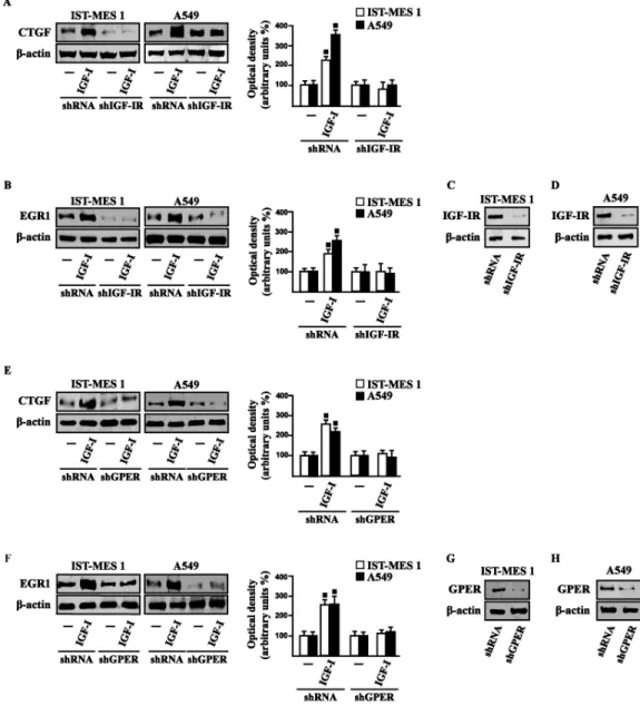
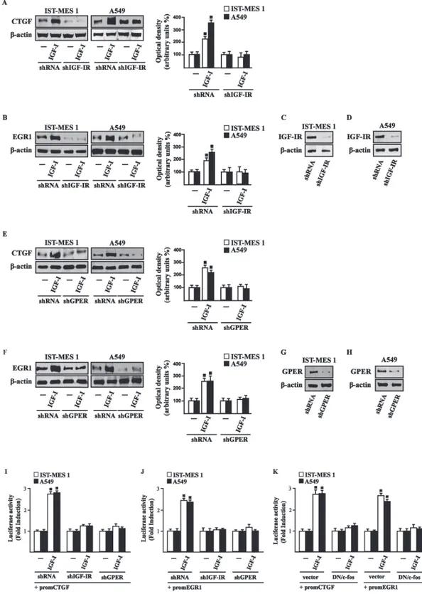
Figure 3.5 (A-F) CTGF and EGR1 protein levels in cells transfected for 24 h with shRNA, shIGF-IR or
shGPER and then treated for 8 h with either vehicle (-) or 100 ng/ml IGF-I. Efficacy of IGF-IR (C-D) and GPER (G-H) silencing. Side panels show densitometric analysis of the blots normalized to β-actin. Data shown are the mean ± SD of three independent experiments. (■) p<0.05 for cells receiving vehicle (-) versus treatments.
Next, we determined that this action of IGF-I involves not only the IGF-IR but also GPER, as the silencing of each of these receptors prevented gene changes (Fig. 3.5).
In accordance with these observations, the IGF-I transactivation of CTGF (Fig. 3.6A) and EGR1 (Fig. 3.6B) promoters required both IGF-IR and GPER, as demonstrated by knocking down the expression of these receptors.
Chapter III
As c-fos plays a main role in the up-regulation of GPER target genes (Pandey et al. 2009; Maggiolini and Picard 2010), we next determined that the promoter transactivation of both CTGF and EGR1 is abrogated by co-transfecting a dominant-negative form of c-fos (DN/c-fos) in IST-MES1 and A549 cells (Fig. 3.6C). Collectively, these findings provide novel mechanisms through which IGF-I/IGF-IR transduction signaling regulates GPER target genes like CTGF and EGR1 in mesothelioma and lung cancer cells.
Figure 3.6 (A-B) Cells were transfected for 24 h with shRNA, shIGF-IR or shGPER together with the CTGF or
EGR1 promoter construct. Then, cells were treated for 18 h with vehicle (-) or 100 ng/ml IGF-I. (C) Cells were transfected for 24 h with a dominant negative form of c-fos (DN/c-fos) together with the CTGF or EGR1 promoter construct. Then, cells were treated for 18 h with vehicle (-) or 100 ng/ml IGF-I. The luciferase activities were normalized to the internal transfection control, and values of cells receiving vehicle (-) were set as one fold induction upon which the activity induced by treatments was calculated. Data shown are the mean ± SD of three independent experiments. (■) p<0.05 for cells receiving vehicle (-) versus treatments.
3.3 IGF-IR and GPER are both involved in IGF-I regulation of
DDR1 target genes
Considering that in diverse model systems IGF-I stimulates the synthesis of collagen (Blackstock et al. 2014; Sukhanov et al. 2011; Sukhanov et al. 2007), we next established that
Chapter III
IGF-I regulates in both IST-MES1 and A549 cells the mRNA expression of COL1A1 (Fig. 3.7A) that encodes the major component of type I collagen (Inamori et al. 2007). We previously reported that IGF-IR functionally interacts with DDR1, which is activated by various collagen types including type I collagen. Therefore, we first ascertained that, in both IST-MES1 and A549 cells, several DDR1 target genes such as matrilin-2 (MATN-2), fibrillin-1 (FBN-1), NOTCH 1 and HES-1, are induced by the DDR1 agonist COL1 (Fig. 3.7B-C) and abrogated by the DDR1 inhibitor (DDR1 IN) (Fig. 3.7D-E).
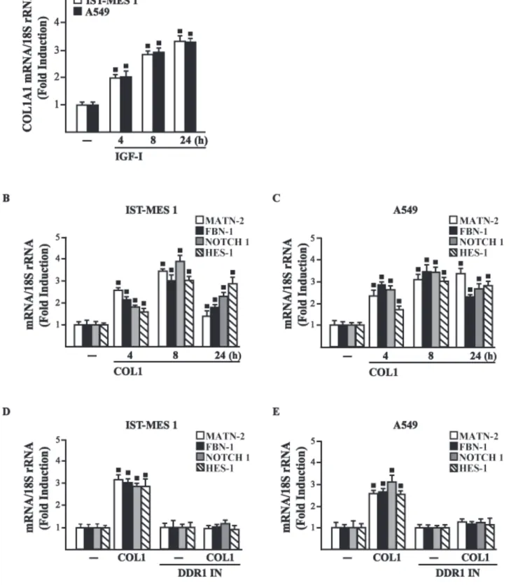
Figure 3.7 (A) mRNA expression of COL1A1 in IST-MES 1 and A549 cells treated with vehicle (-) or 100
ng/ml IGF-I, as evaluated by real-time PCR. mRNA expression of MATN-2, FBN-1, NOTCH 1 and HES-1 in IST-MES 1 (B, D) and A549 (C, E) cells treated with vehicle (-) or 10 µg/ml COL1 alone or in combination with 1 µM DDR1 inhibitor (DDR1 IN), as indicated. Results obtained from experiments performed in triplicate were normalized for 18S expression and shown as fold change of RNA expression compared to cells treated with vehicle. (■) p<0.05 for cells receiving vehicle (-) versus treatments.
Chapter III
Then, we assessed that these DDR1 target genes are also stimulated by IGF-I (Fig. 3.8A-B) and that this response was inhibited by DDR1 IN (Fig. 3.8C-D) as well as by silencing IGF-IR (Fig. 3.8E-F) or GPER (Fig. 3.8G-H).
Figure 3.8 (A-D) mRNA expression of MATN-2, FBN-1, NOTCH 1 and HES-1 in cells treated with vehicle (-)
or 100 ng/ml IGF-I alone or in combination with 1 µM DDR1 inhibitor (DDR1 IN), as indicated. (E-H) mRNA expression of MATN-2, FBN-1, NOTCH 1 and HES-1 in cells transfected for 24 h with shRNA, shIGF-IR or shGPER and then treated for 8 h with vehicle (-) or 100 ng/ml IGF-I. Results obtained from experiments performed in triplicate were normalized for 18S expression and shown as fold change of RNA expression compared to cells treated with vehicle. (■) p<0.05 for cells receiving vehicle (-) versus treatments.
Chapter III
In accordance with these findings, we determined that the NOTCH 1 protein induction by COL1 and IGF-I is prevented in the presence of the DDR1 IN in IST-MES1 and A549 cells (Fig. 3.9).
Figure 3.9 (A) NOTCH 1 protein levels in cells treated with vehicle (-) or 10 µg/ml COL1, as indicated. (B)
NOTCH 1 protein levels in cells treated for 8 h with vehicle (-) or 10 µg/ml COL1 alone and in combination with 1 µM DDR1 inhibitor (DDR1 IN). (C) NOTCH 1 protein levels in cells treated with vehicle (-) or 100 ng/ml I, as indicated. (D) NOTCH 1 protein levels in cells treated for 8 h with vehicle (-) or 100 ng/ml IGF-I alone and in combination with 1 µM DDR1 inhibitor (DDR1 IGF-IN). Side panels show densitometric analysis of the blots normalized to β-actin and each data point represents the mean ± SD of three independent experiments. (■) p<0.05 for cells receiving vehicle (-) versus treatments.
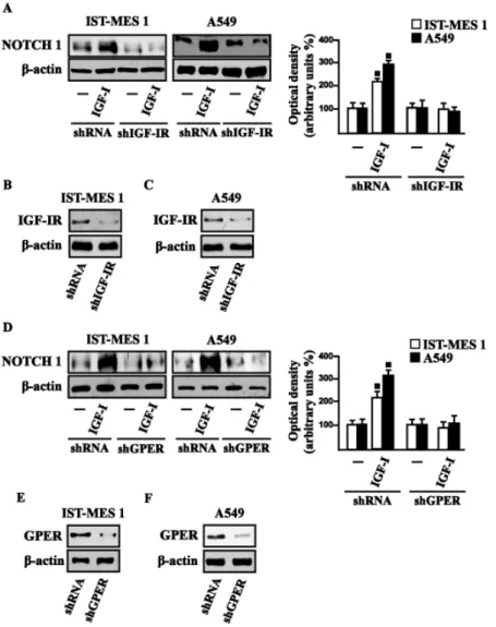
Accordingly, IGF-I was not able to trigger NOTCH 1 protein expression when IGF-IR (Fig. 3.10A-C) or GPER (Fig. 3.10D-F) was silenced. Altogether, these results indicate that, in
Chapter III
both mesothelioma and lung cancer cells, IGF-I may up-regulate DDR1 target genes, and that this action involves not only IGF-IR but also a cross-talk with GPER.
Figure 3.10 NOTCH 1 protein levels in cells transfected for 24 h with shIGF-IR (A) or shGPER (D) and then
treated for 8 h with vehicle (-) or 100 ng/ml IGF-I. Efficacy of IGF-IR (B-C) and GPER (E-F) silencing. Side panels show densitometric analysis of the blots normalized to β-actin. (■) p<0.05 for cells receiving vehicle (-) versus treatments.
Chapter III
3.4 DDR1, IGF-IR and GPER contribute to the chemotaxis and
migration of mesothelioma and lung cancer cells
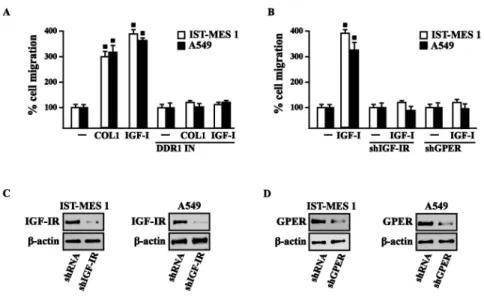
Previous studies have reported that IGF-I stimulates chemotactic and chemokinetic motility in mesothelioma cells (Liu and Klominek 2004). Moreover, DDR1 also plays an important role in promoting cell-cell interactions and cell migration in various cell contexts (Wang et al. 2009; Yeh et al. 2011; Eswaramoorthy et al. 2010; Hidalgo-Carcedo et al. 2011). Further extending these data, in IST-MES1 cells, we found that IGF-I induces chemotactic motility through DDR1, as this response was abolished by DDR1 IN (Fig. 3.11A). Moreover, we ascertained that the chemotactic motility induced by IGF-I requires also IGF-IR and GPER as the aforementioned effect was prevented silencing the expression of these receptors (Fig. 3.11B).
Chapter III
Figure 3.11. (A) Chemotactic motility in cells treated for 8 h with vehicle (-) or 100 ng/ml IGF-I, alone or in
presence of 1 µM DDR1 inhibitor (DDR1 IN). (B) Chemotactic motility in cells transfected for 24 h with shGPER or shIGF-IR, as indicated, and then treated for 8 h with vehicle (-) or 100 ng/ml IGF-I. Images shown were captured from time lapse microscopy experiments and are representative of three random fields from three independent experiments.
Similar findings occurred in A549 cells (data not shown). Likewise, we determined that IST-MES1 and A549 cell migration induced by both IGF-I and COL1 is abolished using DDR1 IN (Fig. 3.12A), whereas the silencing of IGF-IR or GPER abolished cell migration triggered by IGF-I, as determined by Boyden chamber assay (Fig. 3.12B). Collectively, our data indicate novel cross-talk and biological functions exerted by IGF-I toward tumor progression.
Chapter III
Figure 3.12. COL1 and IGF-I stimulate IST-MES 1 and A549 cell migration through DDR1, IGF-IR and GPER.
(A) The migration of IST-MES 1 and A549 cells upon 8 h treatment with vehicle (-), 10 µg/ml COL1 or 100 ng/ml IGF-I alone and in combination with 1 µM DDR1 inhibitor (DDR1 IN), as evaluated by Boyden Chamber assay. (B) The migration of IST-MES 1 and A549 cells induced by 8 h treatment with 100 ng/ml IGF-I was prevented knocking down IGF-IR and GPER expression, as evaluated by Boyden Chamber assay. Efficacy of IGF-IR (C-D) and GPER (E-F) silencing. Values represent the mean ± SD of three independent experiments. (●) indicates p < 0.05 for cells treated with vehicle (–) versus treatments.
Chapter IV
Chapter IV
Discussion
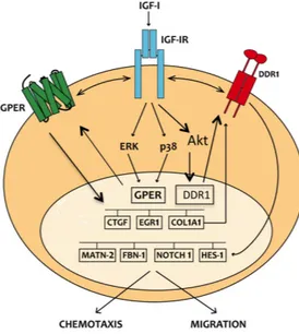
In the present study we provide novel evidence regarding the molecular mechanisms by which IGF-I triggers biological responses in mesothelioma and lung cancer cells. In particular, we show a complex functional cooperation involving IGF-IR, GPER and DDR1 through which IGF-I up-regulates first the expression of COL1A1 and certain DDR1 target genes, thereafter stimulating cancer cell motility and chemotactic response (Fig. 3.13).
Figure 3.13. Schematic representation of the signaling network between IGF-IR, GPER and DDR1 activated by
IGF-I. IGF-I stimulates the expression of GPER and its target genes, then IGF-IR and GPER trigger the IGF-I regulation of DDR1 target genes. The functional cross-talk of IGF-IR, GPER and DDR1 contributes to the chemotaxis and migration observed in cancer cells.
Lung cancer is a high heterogeneous tumor that can arise in different sites of the bronchial tree and one of the most common types of human malignancies (Travis et al. 2011; Guo et al. 2015). The incidence of lung cancer depends on toxic effects of inhaled substances such as tobacco, asbestos, arsenic, cadmium, nickel and silica (Ahuja et al. 2015). The environmental pollutant asbestos is also considered the main cause of the insurgence of malignant
Chapter IV
mesothelioma (MM), which is a rare and aggressive tumor that springs from mesothelial cells lining lung, pleura or peritoneum (Rajer et al. 2014; Carbone et al. 2012; Rascoe et al. 2012; Lenters et al. 2011; Straif et al. 2009). The deposition of asbestos fibers has been also related to chronic inflammatory processes as well as to pulmonary fibrosis, which in turn may create a favorable environment for the development of lung and pleura malignancies (Rascoe et al. 2012; Mossman et al. 2011). As it concerns the multifaceted mechanisms and factors involved in pulmonary fibrosis and neoplasia, an increased expression and activation of DDR1 have been reported (Avivi-Green et al. 2006; Lemeer et al. 2012; Matsuyama et al. 2005; Heinzelmann-Schwarz et al. 2004). To date, DDR1 has been shown to play an important role in cancer progression by regulating the interactions of tumor cells with the surrounding collagen matrix, therefore leading to pro-migratory and pro-invasive responses (Valiathan et al. 2012). Furthermore, collagen activated DDR1 triggers diverse pro-survival pathways toward anti-apoptotic, proliferative and aggressive features in cancer cells (Valiathan et al. 2012). In this regard, it should be noted that several types of collagen are able to bind to and activate DDR1, which then regulates cell and tissue homeostasis acting as a collagen sensor (Valiathan et al. 2012; Vogel et al. 2006). Of note, an abnormal expression and deposition of collagen has been associated with cancer development (Tavazoie et al. 2008; Ramaswamy et al. 2003). As it concerns the synthesis and extracellular accumulation of diverse types of collagen, cytokines and growth factors like IGF-I, the epidermal growth factor (EGF) and the transforming growth factor-βl have been reported to promote these effects (Blackstock et al. 2014; Sukhanov et al. 2011; Sukhanov et al. 2007; Grande et al. 1997). Notably, we previously showed that, in breast cancer cells, IGF-I may upregulate DDR1 expression through a signaling pathway involving the DDR1 regulatory miR-199a-5p (Matà et al. 2016). Moreover, the activation of one of the main I transduction signaling, the IGF-IR/PI3K/Akt cascade, inhibits miR-199a-5p expression, thus relieving its inhibition upon
Chapter IV
through post-transcriptional mechanisms and amplifies IGF-I downstream signaling and biological effects, such as proliferation, migration and colony formation (Matà et al. 2016). Indeed, previous studies showed that DDR1 directly interacts with IGF-IR, and that this interaction is enhanced by IGF-I stimulation, which promotes rapid DDR1 tyrosine-phosphorylation and co-internalization of the DDR1 - IGF-IR complex (Malaguarnera et al. 2015). This interaction was shown to occur in a panel of human breast cancer cells as well as in mouse fibroblasts (R- cells) co-transfected with the human IGF-IR and DDR1, indicating that it is not cell-specific. Notably, the formation of this DDR1 – IGF-IR complex did not require the presence of collagen, the canonical DDR1 ligand. In addition, the critical role of IGF-IR in DDR1 activation and biological actions is supported by the finding that collagen-dependent DDR1 phosphorylation was impaired in the absence of IGF-IR (Malaguarnera et al. 2015). Extending these previous studies, in the present study we show that IGF-I through the cognate receptor IGF-IR is able to induce COL1A1 expression (Vogel et al. 2006). Moreover, a panel of DDR1 target genes could be also induced by IGF-I through the previously described functional cross-talk involving IGF-IR and DDR1. Taken together, these findings show that DDR1, besides enhancing the activation of typical IGF-IR downstream cascades, the PI3K/Akt and the ERK1/2 cascades, following cell exposure to IGF-I, modifies significantly these IGF-I effects by allowing the induction of typical DDR1 target genes. These effects confirm the relevance of DDR1 in the amplification and diversification of IGF-I signaling pathways in cancer. We have previously demonstrated that IGF-IR may also functionally interact with the non-canonical estrogen receptor GPER. Indeed, through the IGF-IR/PKCδ/ERK/c-fos/AP1 transduction pathway, IGF-I up-regulates GPER, which plays an important role in sustaining proliferation and migration in response to IGF-I in breast and endometrial human cancer cells (De Marco et al. 2013). In close accordance with these findings, we now show that the functional cooperation between IGF-IR and DDR1 also
Chapter IV
stimulated by IGF-I in mesothelioma and lung cancer cells.Notably, we now show that GPER and IGF-IR co-immunoprecipitate in lung and mesothelioma cells (Fig. 3.3), indicating that GPER and IGF-IR also interact. Taken together all these data strongly suggest the possible formation of a ternary functional complex involving IGF-IR – DDR1 – GPER. However, further studies are needed to fully elucidate this aspect. These data may be of a particular interest as GPER expression has been associated with negative clinical features and poor survival rates in diverse types of malignancies (Filardo et al. 2006; Smith et al. 2007; Smith et al. 2009; Marjon et al. 2014). In the last years, extensive studies were therefore performed in order to better characterize the role of GPER in cancer development, including the mechanisms and factors involved in its expression. For instance, we determined that EGF and IGF-I, insulin and further tumorigenic factors like hypoxia and endothelin-1 up-regulate GPER expression in diverse cancer cell contexts (De Marco et al. 2013; De Marco et al. 2014; Albanito et al. 2008; Recchia et al. 2011; De Francesco et al. 2013; De Francesco et al. 2014; Bartella et al. 2016). Our present findings provide significant new insights on the well-established role played by the IGF axis in cancer (Belfiore et al. 2009; Belfiore et al. 2011; Kai et al. 2009; Carboni et al. 2005; Franks et al. 2016; Hoang et al. 2004; Liu et al. 2014; Rozengurt et al. 2010; Baserga et al. 2003; Yakar et al. 2005; Novosyadlyy et al. 2010) that involves also the interaction of IGF-IR with other RTKs and GPCRs in diverse tumor histotypes (Lappano and Maggiolini 2011 Rozengurt et al. 2010; Kisfalvi et al. 2009; Akekawatchai et al. 2005). In particular, our findings might be relevant in devising new therapeutical strategies in cancers with a dysregulated IGF system. In the last decade, much effort has been made in targeting the IGF-IR in these malignancies (Gombos et al. 2010). In particular, both small-molecule IGF-IR tyrosine kinase inhibitors, and humanized monoclonal antibodies with blocking activity to the IGF-IR, have been investigated in Phase III trials of advanced non-small cell lung cancers (Scagliotti and Novello 2012). Unfortunately, in spite of
Chapter IV
minority of malignancies do respond to target therapies when IGF-IR is the sole target (Fidler et al. 2012), because the frequent occurrence of resistance mechanisms arising by the complex signaling network involving the IGF-IR (Scotlandi and Belfiore 2012).
Overall, on the basis of our data the multifaceted signaling network between IGF-IR, GPER and DDR1 could be taken into account in setting innovative combined strategies targeting these pathways in mesothelioma and lung cancers.
References
References
Ahuja J., Kanne J.P., Meyer C.A. Occupational lung disease. Semin Roentgenol. 2015; 50: 40-51
Akekawatchai C., Holland J.D., Kochetkova M., Wallace J.C., McColl S.R. Transactivation of CXCR4 by the insulin-like growth factor-1 receptor (IGF-1R) in human MDA-MB-231 breast cancer epithelial cells. J Biol Chem. 2005; 280: 39701-8.
Albanito L., Madeo A., Lappano R., Vivacqua A., Rago V., Carpino A., Oprea T.I., Prossnitz E.R., Musti A.M., Andò S., Maggiolini M. G protein-coupled receptor 30 (GPR30) mediates gene expression changes and growth response to 17β- estradiol and selective GPR30 ligand G1 in ovarian cancer cells. Cancer Res. 2007; 67(4):1859-66.
Albanito L., Sisci D., Aquila S., Brunelli E., Vivacqua A., Madeo A., Lappano R., Pandey D.P., Picard D., Mauro L., Andò S., Maggiolini M. Epidermal growth factor induces G protein-coupled receptor 30 expression in estrogen receptor- negative breast cancer cells. Endocrinology, 2008; 149(8):3799–3808.
Arpino G., Weiss H., Lee A.V., Schiff R., De Placido S., Osborne C.K., Elledge R.M. Estrogen receptor-positive, progesterone receptor-negative breast cancer: association with growth factor receptor expression and tamoxifen resistance. J Natl Cancer Inst. 2005; 97(17):1254-61.
Aust A.E., Balla J.C., Hu A.A. Lighty, J.S.; Smith, K.R.; Straccia, A.M.; Veranth, J.M.; Young, W.C. Particle characteristics responsible for effects on human lung epithelial cells. Res. Rep. Health Effects Inst. 2002; 110:1-65;
Avivi-Green C., Singal M., Vogel W.F. Discoidin domain receptor 1-deficient mice are resistant to bleomycin-induced lung fibrosis. Am J Respir Crit Care Med. 2006; 174: 420-7. Avraham R., Yarden Y. Feedback regulation of EGFR signalling: decision making by early and delayed loops. Nat. Rev. Mol. Cell Biol. 2011; 12(2):104–117.
Azad N., Rojanasakul Y., Vallyathan V. Inflammation and lung cancer: Roles of reactive oxygen/nitrogen species. J. Toxicol. Environ. Health B Crit. Rev. 2008; 11:1–15.
Bartella V., De Francesco E.M., Perri M.G., Curcio R., Dolce V., Maggiolini M., Vivacqua A. The G protein estrogen receptor (GPER) is regulated by endothelin-1 mediated signaling in cancer cells. Cell Signal. 2016; 28: 61-71.
Bartella V., De Marco P., Malaguarnera R., Belfiore A., Maggiolini M. New advances on the functional cross-talk between insulin-like growth factor-I and estrogen signaling in cancer. Cellular Signalling, 2012; 24(8):1515–1521.
Bartke A. Minireview: role of the growth hormone/insulin-like growth factor system in mammalian aging. Endocrinology. 2005; 146(9):3718-23.
References
Barton M. Position paper: the membrane estrogen receptor GPER – clues and questions. Steroids, 2012; 77:935–942.
Baserga R., Peruzzi F., Reiss K. The IGF-1 receptor in cancer biology. Int J Cancer. 2003; 107: 873-7.
Beauchamp M-C., Yasmeen A., Knafo A., Gotlieb W.H. Targeting insulin and insulin-like growth factor pathways in epithelial ovarian cancer. J Oncol. 2010, 2010:257058.
Belfiore A. The role of insulin receptor isoforms and hybrid insulin/IGF-I receptors in human cancer. Curr Pharm Des. 2007; 13(7):671-86.
Belfiore A., Frasca F., Pandini G., Sciacca L., Vigneri R. Insulin receptor isoforms and insulin receptor/insulin-like growth factor receptor hybrids in physiology and disease. Endocr Rev. 2009; 30: 586-623.
Belfiore A., Malaguarnera R. Insulin receptor and cancer. Endocr Relat Cancer, 2011; 18(4):R125-47.
Blackstock C.D., Higashi Y., Sukhanov S., Shai S.Y., Stefanovic B., Tabony A.M., Yoshida T., Delafontaine P. Insulin-like growth factor-1 increases synthesis of collagen type I via induction of the mRNA-binding protein LARP6 expression and binding to the 5' stem-loop of COL1a1 and COL1a2 mRNA. J Biol Chem 2014; 289: 7264-74
Blobel C.P. ADAMs: key components in EGFR signalling and development. Nature Reviews Molecular Cell Biology, 2005; 6(1):32-43.
Bouskine A., Nebout M., Brücker-Davis M., Benahmed M., Fenichel P. Low doses of bisphenol A promote human seminoma cell proliferation by activating PKA and PKG via a membrane G-protein-coupled estrogen receptor. Environmental Health Perspectives, 2009; 17(7):1053-8.
Brambilla E., Travis W.D., Colby T.V., Corrinz B., Shimosato Y. The new World Health Organization classification of lung tumours, 2001; 18: 1059-68.
Campbell N.P., Kindler H.L. Update on malignant pleural mesothelioma. Semin Respir Crit Care Med. 2011; 32:102-10.
Carafoli F., Bihan D., Stathopoulos S., Konitsiotis A.D., Kvansakul M., Farndale R.W., Leitinger B., Hohenester E. Crystallographic insight into collagen recognition by discoidin domain receptor 2. Structure. 2009; 17(12):1573-81.
Carbone M, Kratzke RA, Testa JR. The pathogenesis of mesothelioma. Semin Oncol. 2002; 29: 2– 17.
Carbone M., Ly B.H., Dodson R.F., Pagano I., Morris P.T., Dogan U.A., Gazdar A.F., Pass H.I., Yang H. Malignant mesothelioma: facts, myths, and hypotheses. J Cell Physiol. 2012; 227: 44-58.
References
Carbone M., Ly B.H., Dodson R.F., Pagano I., Morris P.T., Dogan U.A., Gazdar A.F., Pass H.I., Yang H. Malignant mesothelioma: facts, myths, and hypotheses. J Cell Physiol. 2012; 227: 44-58.
Carboni J.M., Lee A.V., Hadsell D.L., Rowley B.R., Lee F.Y., Bol D.K., Camuso A.E., Gottardis M., Greer A.F., Ho C.P., Hurlburt W., Li A., Saulnier M., Velaparthi U., Wang C., Wen M.L., Westhouse R.A., Wittman M., Zimmermann K., Rupnow B.A., Wong T.W. Tumor development by transgenic expression of a constitutively active insulin-like growth factor I receptor. Cancer Res. 2005; 65(9):3781-7.
Carmeci C., Thompson D.A., Ring H.Z., Francke U., Weigel R.J. Identification of a gene (GPR30) with homology to the G-protein-coupled receptor superfamily associated with estrogen receptor expression in breast cancer. Genomics, 1997; 45(3):607-17.
Casa A.J. Dearth R.K., Litzenburger B.C., Lee A.V., Cui X., Frontiers in Bioscience 13, 2008; 3273–3287.
Casa A.J., Dearth R.K., Litzenburger B.C., Lee A.V., Cui X. The type I insulin-like growth factor receptor pathway: a key player in cancer therapeutic resistance. Front Biosci. 2008; 13:3273-87.
Chan Q.K., Lam H.M., Ng C.F., Lee A.Y., Chan E.S., Ng H.K., Ho S.M., Lau K.M. Activation of GPR30 inhibits the growth of prostate cancer cells through sustained activation of Erk1/2, c-jun/c-fos-dependent upregulation of p21, and induction of G(2) cell-cycle arrest. Cell Death and Differentiation, 2010; 17(9):1511-23.
Chen W., Zheng R., Zeng H., Zhang S. Epidemiology of lung cancer in China. Thorac Cancer. 2015; 6(2):209-15
Chevalier N., Vega A., Bouskine A., Siddeek B., Michiels J.F., Chevallier D., Fénichel P. GPR30, the non-classical membrane G protein related estrogen receptor, is overexpressed in human seminoma and promotes seminoma cell proliferation. PLoS ONE, 2012; 7(4):e34672 Chiang S.H., Baumann C.A., Kanzaki M., Thurmond D.C., Watson R.T., Neudauer C.L., Macara I.G., Pessin J.E., Saltiel A.R. Insulin-stimulated GLUT4 translocation requires the CAP-dependent activation of TC10. Nature, 2001; 410(6831):944-8.
Chuang H.C., Fan C.W., Chen K.Y., Chang-Chien G.P., Chan C.C., Vasoactive alteration and inflammation induced by polycyclic aromatic hydrocarbons and trace metals of vehicle exhaust particles. Toxicol. Lett. 2012; 214(2):131–136.
Curado M.P., Edwards H.R, Shin H., Storm J., Ferlay M.H., Boyle P. Cancer Incidence in Five Continents, Vol. IX. Lyon: IARC Scientific Publications, 2007.
Daub H., Wallasch C., Lankenau A., Herrlich A., Ullrich A. Signal characteristics of G protein-transactivated EGF receptor. EMBO Journal, 1997; 16(23):7032-44.
Daub H., Weiss F.U., Wallasch C., Ullrich A. Role of transactivation of the EGF receptor in signalling by G-protein-coupled receptors, Nature, 1996; 379(6565):557-60.