U
NIVERSITÀ
D
EGLI
S
TUDI
D
I
M
ESSINA
T
ESI DID
OTTORATO DIR
ICERCA INB
IOLOGIAA
PPLICATAE
M
EDICINAS
PERIMENTALEC
URRICULUM IN BIOLOGIA APPLICATAXXIX CICLO
SSD BIO/19
Phage Display as a Tool for
Theranostic and Nanomedicine Application
Candidato:
D
OTT.
D
OMENICOF
RANCORelatore:
Ch.mo Prof.
S
ALVATOREP
IETROP
AOLOG
UGLIELMINOCoordinatore:
Ch.mo Prof.
SALVATORE CUZZOCREA
ABSTRACT
Theranostic nanomedicine allows co-development of different nanotechnology-based diagnostic and therapy strategies in order to combine detection, diagnosis, and treatment of disease into a single unit. Usually, constructs of multifunctional systems for theranostics include a selective probe both as diagnostic tool and as selective agent for targeted therapy.
In the present work, several proofs of concept of diagnostic and teranostic systems, based on the phage display technology, are presented. In particular, the phage display has been used for the selection of peptides specific to bacterial and tumor cellular targets.
First, phage display technology has been employed to select phage clones, expressing peptides able to bind molecular targets of pathogenic bacteria and cancer cells. Then, the whole phage structure as "biomaterial" has been evaluated with fluorchromes, beads, noble metal nanoparticles and PHA microspheres for the development of multifunctional systems for theranostics. The proofs of concept have been developed using both conventional detection methodologies (agglutination, fluorescence microscopy, PCR, and ELISA) and new optical methods, such as Raman spectrometry and SERS-Raman, which have been carefully considered in the development of some of the described methodologies.
The technical combinations and the obtained results here presented demonstrated the importance of intersections among different fields of theoretical and applied science and the need of a common language among different professionals and research teams.
i
INDEX
INTRODUCTION
Bibliography
CHAPTER 1: Probes selection by Phage Display
Introduction
Selection of phage peptides targeting bacterial membrane receptors
Materials and Methods
Results and Discussions
Selection of phage peptides targeting cancer membrane receptors
Materials and Methods
Results and Discussions
Phage-display as tool for biomarker discovery in neurodegenerative
diseases
Materials and Methods
Results and Discussions
Acknowledgements
Bibliography
CHAPTER 2: Raman spectroscopy applied to cellular systems
Introduction
Identification of pathogenic microbes by Raman spectroscopy
Materials and Methods
Results and Discussions
Application of Raman spectroscopy to discriminate cancer cells
Materials and Methods
X
1
4
5
5
10
10
12
16
16
18
20
20
21
23
23
26
26
28
28
29
33
33
ii
Results and Discussions
Acknowledgements
Bibliography
CHAPTER 3: Selective phage clones for fluorescence imaging
Introduction
Bacterial fluorescence imaging
Materials and Methods
Results and Discussions
Leukaemia fluorescence imaging
Materials and Methods
Results and Discussions
Acknowledgements
Bibliography
CHAPTER 4: Phage-based capture and concentrating system for single step
detection of pathogens
Introduction
Materials and Methods
Results and Discussions
Acknowledgements
Bibliography
CHAPTER 5: Selective phage clones assembled to metal nanoparticle
Introduction
Materials and Methods
Results and Discussions
35
44
44
47
47
50
50
51
54
54
54
56
56
59
59
60
63
73
73
75
75
76
79
iii
Acknowledgements
Bibliography
CHAPTER 6: Selective phage clones assembled to nanocarriers
Introduction
Poly(3-hydroxy)alkanoates
Materials and Methods
Results and Discussions
Acknowledgements
Bibliography
SUMMARY AND PERSPECTIVES
89
89
94
94
95
97
99
104
104
106
INTRODUCTION
The problem of public health expenditure and the proper allocation of economic resources is one of the main Europe's problems. The need to ensure economic and financial sustainability of public health systems is due to the increase in life expectancy that requires treatment for prolonged and continuous periods. According to Farma-factoring Foundation estimations, between 2008 and 2016, health expenditure in Italy has substantially declined in recent years (Atella and Cincotti)
In this context, research is moving to the development of diagnostic and therapy tools, at low cost and highly efficient.
Basic approach of conventional therapies is to remove diseased cells faster than healthy cells. Moreover, traditional diagnosisprocedures are usually used to examine only the current disease status (discriminating between normal and disease state) and rarely give information on disease progression and therapeutic response. In this scenario, the therapeutic effects can be brought out only after an extended therapeutic treatment, with a significant delay in the implementation of a truly effective therapy and unnecessary patient exposure to serious side effects.
The rationale arose from the fact that diseases are heterogeneous, and all existing treatments are effective for only limited patient subpopulations and at selective stages of disease development (Xie et al, 2010).
Nanomedicine takes care of nanotechnology applications to medicine, through a conventional biochemical approach. In contrast to conventional therapies, nanomedicine attempts to use sophisticated approaches to either kill specific cells or repair them one cell by using a biosensor to detect, for example, when a drug should be released (Riehemann et al, 2009).
This approach has opened new opportunities in therapeutic and diagnostic fields, and new concepts as personalised medicine, pharmacogenetics and point-of-care testing have been introduced.
In recent years, there is a growing interest towards the development of personalized
medicine, which is defined as: “the concept which marks the expected reform in
medicine that is projected to arrive at the clinic in coming decades, harnessing genomics and proteomics technologies for tailoring the most suitable pharmacotherapy for each patient; based on individual profiling, it is also projected to allow improved treatment efficacies for many diseases” (Gurwitz and Livshits, 2006). Pharmacogenetics deals
with the study of variability in drug response due to heredity (Nebert, 1999). Variation within the human genome is seen about every 500±1000 bases (Roses, 2010). Although there are a number of different types of polymorphic markers, most attention recently has focused on single nucleotide polymorphisms (SNPs), and the potential for using these to determine the individual drug response profile. Since SNPs occur at a frequency of 1% or greater in the population, theoretically, this could be used to create individual SNP profiles that correlate with individual drug response (Gray et al, 2000). It may be possible to tailor drug prescription and drug dosage to the individual, thereby maximizing efficacy and minimizing toxicity (Roses, 2000; Wolf et al, 2000; Meyer, 2000). The promise of personalized medicines is also of obvious interest and importance
to the pharmaceutical industry since it may allow streamlining of the drug development, drug testing and drug registration process, reducing the time from chemical synthesis to introduction into clinical practice, and therefore the cost of the drug development process (Roses, 2000).
On a technical level, broad implementation of many aspects of personalized medicine will require the introduction of a new class of inexpensive diagnostic instruments that do not require time-consuming sample preparation. Ideally, such instruments could be operated in a doctor’s office by unskilled personnel.
Point-of-care testing (POCT) is an immediate, convenient, and easy-to-use medical
diagnostic testing at or near the time and place of patient care (Nichols, 2003). Rapid diagnostic information permits immediate clinical management decisions to be made that will improve patient safety and clinical outcomes. Major benefits are obtained when the output of a POCT device is made available immediately within an electronic medical record. Results can be shared instantaneously with all members of the medical team through the software interface enhancing communication by decreasing turn around time (TAT). A reduction in morbidity and mortality has been associated with goal-directed therapy (GDT) techniques when used in conjunction with POCT and the electronic medical record (Rossi and Khan, 2004). POCT has become established worldwide and finds vital roles in public health (Tran and Kost, 2006).
New diagnostic systems, such as Lab-on-a-chip (LOC), can integrate multiple functions in a single miniaturized chip and allow to carry out medical diagnostic testing near the point of patient care (Nichols, 2003). Excluding economic benefits by system miniaturization, rapid diagnostic information allows immediate clinical management decisions to be made that will improve patient safety and clinical outcomes.
Nanotechnology allows combinations and co-development of different diagnostic and therapy strategies in order to obtain into single agent detection, diagnosis, and treatment of disease, principle on which is based the concept of theranostic nanomedicine.
Possible constructs of multifunctional systems for theranostics include a selective
probe both as diagnostic tool and as target agent for therapy.
In the present thesis, several proofs of concept of diagnostic and teranostic systems, based on the phage display technology, are presented. In particular, the phage display has been used for the selection of peptides specific to bacterial and tumor cellular targets, using the whole phage structure as "biomaterial" for the correct exposure of these peptides.
In such a way, biosensing and diagnostic systems, with low cost of production, more efficient and much more stable than conventional systems based on antibodies or purified peptides, have been obtained.
Simultaneously, the use of the whole phage structure allowed building metal nanoparticles-phages stable networks for use in both diagnostic and therapeutic fields. Finally some preliminary data on a potential drug targeting system, aimed to convey and concentrate the encapsulated drug at the target site, are presented. The system is based on a complex between a biopolymer as carrier for controlled drug delivery and a phage expressing specific and selective peptide for the target of interest.
The proofs of concept have been developed using both conventional detection methodologies (agglutination, fluorescence microscopy, PCR, and ELISA) and new optical methods, such as Raman spectrometry and SERS-Raman, which have been carefully considered in the development of some of the described methodologies.
Bibliography
Atella V, Cincotti F. I conti della sanità in Italia: consuntivi e prospettive future. 2015. Quaderni della Fondazione Farmafactoring, n. 3/15.
Gray IC, Campbell DA, Spurr NK. Single nucleotide polymorphisms as tools in human genetics. 2000. Hum Mol Genet. 9, 2403–08
Gurwitz D, Livshits G. Personalized medicine Europe: health, genes and society: Tel-Aviv University, Tel-Tel-Aviv, Israel, June 19-21, 2005. 2006. Eur J Hum Genet. 14, 376-80.
Meyer UA. Pharmacogenetics and adverse drug reactions. 2000. Lancet. 356, 1667–71. Nebert DW. Pharmacogenetics and pharmacogenomics: why is this relevant to the
clinical geneticist? 1999. Clin. Genet. 56 247-58.
Nichols J. Point of Care Testing – Performance Improvement and Evidence Based Outcomes. 2003. CRC Press.
Riehemann K, Schneider SW, Luger TA, GodinB, Ferrari M, Fuchs H. Nanomedicine-Challenge and Perspectives. 2009. Angew. Chem. Int. Ed., 48, 872 – 97.
Roses AD. Pharmacogenetics and the practice of medicine. 2000. Nature 405, 857-65. Wolf CR, Smith G, Smith RL. Science, medicine, and the future: Pharmacogenetics.
2000. Br Med J. 320, 987–90.
Rossi AF, Khan D . Point of care testing: improving pediatric outcomes. 2004. Clin. Biochem. 37, 456–61.
Tran NK, Kost GJ. Worldwide point-of-care testing: compendiums of POCT for mobile, emergency, critical, and primary care and of infectious diseases tests. 2006. Point of Care: the Journal of Near-Patient Testing & Technology. 5, 84–92.
Xie J, Lee S, Chen X. Nanoparticle-based theranostic agents. 2010. Adv Drug Deliv Rev. 62(11), 1064–79.
CHAPTER 1
Probes selection by Phage Display
Introduction
A molecular repertoire is a collection of several molecular variants of peptide or protein structures, from which it can select specific ligands against molecular targets of interest. One of the methods most commonly used in the construction of molecular repertoire is based on the use of filamentous phages that infect Escherichia coli, able to display amino acid sequences in a "tolerant" region of its capsid proteins. This experimental technology, called "phage display", offers the advantage to associate in the same phage particle the phenotype, represented by the protein / peptide exposed on the viral surface, to a specific genotype, that is included in the phage genome. So this technology allow to exploring, efficiently selecting and rapidly structure characterizing of very large repertoires of biological molecules by the construction of "encoded" pools (Smith, 1985; Parmley and Smith, 1988).In the mid-1980s, Dr. George Smith of the University of Missouri highlighted the link between capsid proteins expression and nucleic acid encapsulated in M13 filamentous bacteriophage. Particularly, foreign DNA, inserted into upstream of the gene for one of viral coat proteins (pIII), produced phage particles that “displayed” the protein encoded by the foreign DNA at the amino terminal portion of that coat protein (Smith, 1985). The ability to use bacteriophage as tools for the display of ligands became clearer over the next several years, after developing of methodology for fusing larger molecules, such as antibody fragments, to pIII (McCafferty et al, 1990; Barbas et al, 1991). Nowadays Phage Display has established itself as a powerful biological tool for rapid and inexpensive identification of novel ligands (e.g. receptor agonists) or mapping the interactions between two known proteins (e.g. antibody epitope mapping). In fact antibodies, enzymes, signal transduction proteins, cell surface receptors and structures, and also non-protein targets, such as carbohydrates, polymers or other surface materials, have all been successfully utilized as targets in screening experiments, and have allowed the isolation of specific molecules (Smith and Petrenko,1997; Petrenko and Sorokulova, 2004).
Phage display technology is based on the insertion of foreign nucleotide sequences into genes encoding for various bacteriophage coat proteins, resulting in heterogeneous mixtures of phages, each carrying a distinct peptide sequence (encoded by the corresponding nucleotide insert) as fusion to the coat protein. The physical link between
a displayed fusion protein and the DNA encoding for it, represented by the bacteriophage particle, makes specific phage clones selectable and
The strength of the phage display approach results from the powerful combination of affinity selection and biological amplification. Libraries can be affinity selected with a ligate in small reaction volumes (i.e. at high concentratio
foreign peptides, displayed on the surface of the bacteriophage particles, can be surveyed at once for their ability to interact with a given target.
The phage particle is a thin and cylindrical shape, of size about 900 nm x 9 containing a single-stranded DNA genome (6,407 base pairs) which encodes 11 genes, five of which are coat proteins. The major coat protein is the gene 8 protein (g8p), present in 2,700 copies and responsible of the phage DNA encapsulation
The distal end of the phage particle is capped by five copies each of gene 7 protein (g7p) and gene 9 protein (g9p). At the proximal end, four to five copies each of g6p and g3p are present. M13 bacteriophage infects male bacteria, i.e. which bear the F
encoding for the F-pilus. Infection is mediated by the interaction between viral g3p and the F-pilus, and does not cause lysis of host cell. In fact, respect to the lytic phages T4 and T7, filamentous phages are able to replicate and are released f
membrane, allowing
greatly simplifies the intermediate phage purification steps between rounds of panning, as a simple PEG precipitation step is sufficient to separate the phage f
contaminating cellular proteins.
Figure 1.1 M13 phage structure Phage genome consists in single
Products of genes 1, 4 and 11 are used for genome assembling in phage capsid encode for a DNA endonuclease for double
stranded circle; gene 5 encodes for a single and 9 encode for capsid proteins.
displayed fusion protein and the DNA encoding for it, represented by the bacteriophage particle, makes specific phage clones selectable and
The strength of the phage display approach results from the powerful combination of affinity selection and biological amplification. Libraries can be affinity selected with a ligate in small reaction volumes (i.e. at high concentration), so that a vast number of foreign peptides, displayed on the surface of the bacteriophage particles, can be surveyed at once for their ability to interact with a given target.
particle is a thin and cylindrical shape, of size about 900 nm x 9 stranded DNA genome (6,407 base pairs) which encodes 11 genes, five of which are coat proteins. The major coat protein is the gene 8 protein (g8p), present in 2,700 copies and responsible of the phage DNA encapsulation
The distal end of the phage particle is capped by five copies each of gene 7 protein (g7p) and gene 9 protein (g9p). At the proximal end, four to five copies each of g6p and g3p are present. M13 bacteriophage infects male bacteria, i.e. which bear the F
pilus. Infection is mediated by the interaction between viral g3p and pilus, and does not cause lysis of host cell. In fact, respect to the lytic phages T4 and T7, filamentous phages are able to replicate and are released f
membrane, allowing to the host cell continues to continue to grow and divide. This greatly simplifies the intermediate phage purification steps between rounds of panning, as a simple PEG precipitation step is sufficient to separate the phage f
contaminating cellular proteins.
M13 phage structure.
Phage genome consists in single-stranded DNA of 6,407 base pairs which encodes 11 genes. Products of genes 1, 4 and 11 are used for genome assembling in phage capsid
encode for a DNA endonuclease for double-stranded replicative form and DNA replication single stranded circle; gene 5 encodes for a single-stranded DNA-binding protein; finally genes 3, 6, 7, 8 and 9 encode for capsid proteins.
displayed fusion protein and the DNA encoding for it, represented by the bacteriophage particle, makes specific phage clones selectable and easily identifiable. The strength of the phage display approach results from the powerful combination of affinity selection and biological amplification. Libraries can be affinity selected with a n), so that a vast number of foreign peptides, displayed on the surface of the bacteriophage particles, can be
particle is a thin and cylindrical shape, of size about 900 nm x 9 nm, stranded DNA genome (6,407 base pairs) which encodes 11 genes, five of which are coat proteins. The major coat protein is the gene 8 protein (g8p), present in 2,700 copies and responsible of the phage DNA encapsulation (Figure 1.1). The distal end of the phage particle is capped by five copies each of gene 7 protein (g7p) and gene 9 protein (g9p). At the proximal end, four to five copies each of g6p and g3p are present. M13 bacteriophage infects male bacteria, i.e. which bear the F-plasmid, pilus. Infection is mediated by the interaction between viral g3p and pilus, and does not cause lysis of host cell. In fact, respect to the lytic phages T4 and T7, filamentous phages are able to replicate and are released from the cell to the host cell continues to continue to grow and divide. This greatly simplifies the intermediate phage purification steps between rounds of panning, as a simple PEG precipitation step is sufficient to separate the phage from almost all
stranded DNA of 6,407 base pairs which encodes 11 genes. Products of genes 1, 4 and 11 are used for genome assembling in phage capsid; genes 2 and 10 stranded replicative form and DNA replication
Filamentous phage display systems are generally based on N
coat proteins pIII or pVIII. pIII is present at 5 copies per virion, of which all 5 can be fused to short peptides without interfering with phage infectivity. The major coat protein pVIII is present at ~2700 copies per virion, of which ~10% can be reliably fused to peptides or proteins. As a result, peptides expressed as pIII fusions are present at low valence (1-5 copies per virion), while pVIII fusions are present at high
copies per virion). In this last case, highly multivalent display libraries can be produced by generating fusions to pVIII, usually achieved via two
phage particles displaying a mixture of wild
1991). The increased avidity effect of high valency pVIII display permits selection of very low affinity ligands
display limits selection to higher affinity ligands.
The success of phage derived peptides essentially depends on the quality of the library screened.
Phage-display systems can be distinguished according to the arrang protein genes (Figure
In a “type 3” vector, there is a single phage chromosome, with a single gene III which accepts foreign DNA inserts and encodes for a single type of pIII peptide. Therefore, the foreign peptide is theoretically displayed
Similarly, type 8 vectors display foreign peptides on every copy of pVIII.
Figure 1.2 Types of phage display systems. The line inside each virion represents the single proteins pVIII and pIII
boxes represent foreign coding sequences spliced into a coat on pIII (green circles) are either appended to the
N-terminal domain (type 3+3 and most type 33 systems). In type 8 systems, the foreign peptide (red circles) is displayed on all copies of the major coat protein pVIII (2700 copies in wild
whereas in type 88 and 8+8 sy
Filamentous phage display systems are generally based on N-terminal fusions to the coat proteins pIII or pVIII. pIII is present at 5 copies per virion, of which all 5 can be tides without interfering with phage infectivity. The major coat protein pVIII is present at ~2700 copies per virion, of which ~10% can be reliably fused to peptides or proteins. As a result, peptides expressed as pIII fusions are present at low
5 copies per virion), while pVIII fusions are present at high
In this last case, highly multivalent display libraries can be produced by generating fusions to pVIII, usually achieved via two-gene systems that result in
age particles displaying a mixture of wild-type and recombinant proteins
The increased avidity effect of high valency pVIII display permits selection of very low affinity ligands (such as mimic and bioactive peptides), while low
display limits selection to higher affinity ligands.
The success of phage derived peptides essentially depends on the quality of the library
display systems can be distinguished according to the arrang
Figure 1.2; Smith, 1993; McConnell 1994).
In a “type 3” vector, there is a single phage chromosome, with a single gene III which accepts foreign DNA inserts and encodes for a single type of pIII peptide. Therefore, the foreign peptide is theoretically displayed on all five pIII molecules on a virion. Similarly, type 8 vectors display foreign peptides on every copy of pVIII.
Types of phage display systems.
The line inside each virion represents the single-stranded viral DNA, the segments encoding coat being designated by black and grey boxes, respectively, while red and green boxes represent foreign coding sequences spliced into a coat-protein gene. Foreign
on pIII (green circles) are either appended to the N-terminal domain (type 3
domain (type 3+3 and most type 33 systems). In type 8 systems, the foreign peptide (red circles) is displayed on all copies of the major coat protein pVIII (2700 copies in wild
whereas in type 88 and 8+8 systems, only a minority of the pVIII copies display the foreign peptide.
terminal fusions to the coat proteins pIII or pVIII. pIII is present at 5 copies per virion, of which all 5 can be tides without interfering with phage infectivity. The major coat protein pVIII is present at ~2700 copies per virion, of which ~10% can be reliably fused to peptides or proteins. As a result, peptides expressed as pIII fusions are present at low 5 copies per virion), while pVIII fusions are present at high valence (~200 In this last case, highly multivalent display libraries can be produced gene systems that result in type and recombinant proteins (Felici et al, The increased avidity effect of high valency pVIII display permits selection of , while low valency pIII
The success of phage derived peptides essentially depends on the quality of the library
display systems can be distinguished according to the arrangement of the coat
In a “type 3” vector, there is a single phage chromosome, with a single gene III which accepts foreign DNA inserts and encodes for a single type of pIII peptide. Therefore, the on all five pIII molecules on a virion. Similarly, type 8 vectors display foreign peptides on every copy of pVIII.
stranded viral DNA, the segments encoding coat being designated by black and grey boxes, respectively, while red and green protein gene. Foreign peptides displayed terminal domain (type 3 systems) or replace the domain (type 3+3 and most type 33 systems). In type 8 systems, the foreign peptide (red circles) is displayed on all copies of the major coat protein pVIII (2700 copies in wild-type virions),
Although only short foreign peptides can be displayed on every copy of pVIII, they comprise a substantial fraction of the virion’s mass and can dramatically alter its physical and biological properties (Kishchenko et al, 1991; 1994)
Differently by type 8 vector, in a type 88 vector the phage genome bears two genes VIII, encoding two different types of pVIII molecule: one is ordinarily recombinant (i.e., bears a foreign DNA insert) and the other wild-type. The resulting virion is a mosaic of both wild-type and recombinant pVIII molecules. These vectors allow hybrid pVIII proteins with quite large foreign peptides to be displayed, since phage assembly is made possible by wild-type pVIII molecules that is the former predominating. The same property applies to a type 33 vector.
Finally, a type 8+8 system consists the two VIII genes are on several genomes, in wild-type phage, called “helper” phage, and in a special kind of plasmid, called “phagemid” (Mead et al, 1998; Cesareni et al, 1988). Phagemid carries a plasmid replication origin that allows it to replicate normally in an E. coli host, an antibiotic resistance gene for its selection and that allows a filamentous phage replication origin, which is inactive until the cell is infected with the helper phage.
By phagemid vectors, it is possible to achieve a large number of transformants (until 1010 clones), creating highly diverse libraries with increased size and improved performance (Ponsel et al, 2011). Libraries of short peptides of random sequence have been displayed on phage, and screened by antibodies or other molecules, leading to the identification of new ligands (Zwick et al, 1998; Rodi and Makowski, 1999; Deroo and Muller, 2001).
Ligands selected from phage-displayed random peptide libraries can be directed toward biologically relevant sites on the surface of the target protein. Consequently, they can modulate the target protein’s activity, acting as delivery and targeting systems in drug design.
Selection consists of culling an initial population of phage-borne peptides to give a subpopulation with increased “fitness” according to some user-defined criterion. Generally, the input to the first round of selection consists in a very large library (109 clones), while a selected subpopulation, against a molecular target, is represented by a little fraction of the initial population. A subpopulation consists in a mixture of several phage clones, each with a specific affinity against the target that can be “amplified” by infecting fresh bacterial host cells, so that each individual phage clone in the subpopulation is represented by millions of copies in the amplified stock. The amplified
population can then be subjected to further rounds of selection (perhaps accompanied by mutagenesis) to obtain a phage clones population with high stringency and yield, the most important parameter to enhance the efficacy of selection.
Stringency is the degree to which peptides with higher fitness are favored over peptides with lower fitness; yield is the fraction of particles with a given fitness that survive selection. The ultimate goal of selection is usually to isolate peptides with high fitness, but this does not mean that stringency should be increased without bound, since increasing stringency usually entails decreased yield. High yield of the fittest clones is of paramount importance in the very first round of selection, whose input consists of all clones in a very large initial library.
In this section, phage display technology is employed as a tool for the selection of peptides able to bind molecular targets of pathogenic bacteria and cancer cells. Moreover, it is presented a new approach for biomarkers discovery in neurodegenerative diseases by Phage Display technology. Regardless of the molecular target of interest, selection of ligands involves the sequential enrichment of specific binding phage from a large excess of nonbinding clones. This result is accomplished by multiple rounds of phage library “panning”, comprising incubation with molecular target, washing to remove nonspecific phage, elution to retrieve specific binding phage, and reinfection and propagation in Escherichia coli. Generally three to four rounds of panning are required to enrich for specific binders.
A more detailed discussion about selections made is presented in materials and methods following subsections.
Selection of phage peptides targeting bacterial membrane receptors
Rapid, selective and sensitive detection technologies for pathogenic bacteria are critical in clinical diagnosis and disease control.Actually, immunoselection methods based on the use of antibodies or synthetic peptides are commonly used for detection of viruses and different bacterial species in various research fields including clinical diagnosis. However, in both cases, there are some disadvantages like their sensivity to physico-chemical variations of environment such as pH, temperature, mechanical stresses and organic solvent and their extremely high production and purification costs. M13 bacteriophage, engineered to express specific and selective peptides on its capsid proteins, can be used as a good substitute for antibodies in recognition of specific targets. The viral mass consists mainly of about 2700 copies of its major coat protein pVIII, about 50% of which are engineered to display selective and specific target-binding peptides. They are easy to produce in large quantities at a relatively low cost, as their propagation on host bacterial cells is cheaper compared to both antibodies and peptides production. Furthermore, phages are structurally robust, and have a strong resistance to pH, and denaturing compounds such as urea, alcohols and other organic solvents, and events stretching (elongation) and bending without losing bacterial infectivity and/or binding capability (Petrenko and Smith, 2000; Petrenko and Vodyanoy, 2003). Bacteriophage can represent suitable surrogates of antibodies or synthethic peptides, possessing distinct advantages while achieving equivalent specificity and sensitivity (Petrenko and Vodyanoy, 2003). This section describes the selection techniques for phage clones displaying peptides capable of specific and strong binding to the cell surface of microorganisms. Particularly, the following describes the procedures for phage clones selection against Staphylococcus
epidermidis and Staphylococcus aureus.
Materials and Methods
Bacteria and growth media. S. epidermidis ATCC 12228 and S. aureus ATCC 29213
obtained from the American Type Culture Collection (ATCC, LGC Promochem, Milan, Italy) were propagated in Tryptone Soya Broth (TSB) and Tryptone Soya Agar (Oxoid).
Escherichia coli TG1 was used for propagation of phage clones. Stock organisms were
Phage display libraries. The selections were performed through affinity-selection
procedures described by Carnazza et al. (Carnazza et al., 2007; 2008). For the affinity selection procedures, two phage display libraries (pVIII-9aa and pVIII-12aa) were used (kind gift of Prof. F. Felici). These libraries were previously constructed (Felici et al., 1991) and consist of filamentous phages displaying random 9- and 12-mer peptides, respectively, fused to the major coat protein (pVIII). The nonapeptide and dodecapeptide libraries were constructed in the vector pC89 (Felici et al., 1991), by cloning a random DNA insert between the third and fifth codon of the mature pVIII-encoding segments of the pVIII gene (Luzzago and Felici, 1998). The relative binding of the phage clones from each round of affinity selection was estimated by ELISA assay. Then the most reactive individual phage clones were amplified and their DNA inserts were sequenced to determine the amino acid sequences of the displayed peptides.
Phage peptide selection. Library screening was performed using four rounds of affinity
selection. Selection was performed by incubating 1012 phages with S. epidermidis and S.
aureus cells (OD660 = 0.5) respectively, in Phosphate-Buffered Saline (PBS, 137mM
NaCl, 2.7mM KCl, 10mM phosphate buffer, pH 7.4) for 60 min at Room Temperature (RT) with gentle shaking. Bacteria and phages were precipitated by spinning for 5 min at 16000 xg, and separated from unbound phages in solution by a series of 10 washing and centrifugation steps (16000 xg, 5 min) with 1ml TBS/Tween buffer (50mM Tris– HCl (pH 7.5), 150mM NaCl, 0.05% (v/v) Tween 20) each time. Bound phages were pelleted with cells and finally eluted with 200 µl of 0.2M glycine–HCl (pH 2.2) with gentle shaking at RT for 20 min, followed by neutralization with 150 µl of 1M Tris– HCl (pH 9.1). Phages eluted from the final round of affinity selection were used to infect E. coli TG1 cells. These cells were plated on ampicillin-containing LB agar plates to select for phage-infected bacteria. Bacterial colonies, each containing phages from a single library clone, were randomly selected and propagated for subsequent analyses.
ELISA test. Representative clones demonstrating the highest ELISA signals were
investigated for their ability to preferentially interact with the select target in comparison to other potential targets.
A first ELISA test was carried out in order to verify positive reactivity of selected phage clones against bacterial target of interest. Wells of a 96-well ELISA plates were coated overnight at 4 °C with 100 µl suspensions of each bacterial strain (OD660 = 1) in
washing buffer (0.05% w/v Tween 20 in PBS) by automatic plate washer; blocked with Blocking buffer (6% non-fat dry milk, 0.05% w/v Tween 20 in PBS) for 2 h at 37 °C; washed again; reacted with 100 µl of specific phage clone (1012 PFU/ml) in blocking buffer for 2 h at 37 °C with shaking; washed again; reacted with 100µl of anti-M13 peroxidase conjugate antibody (Amersham Biosciences, Buckinghamshire, UK) at a dilution of 1:2500 in Diluition Buffer (1% non-fat dry milk, 0.05% w/v Tween 20 in PBS) for 1 h at 37 °C; washed again; reacted with 100 µl of TMB (3,3,5,5-tetramethylbenzidine) liquid substrate system for ELISA for 45 min at RT and stopped with 100 µl of 1M H2SO4. Optical absorbance was recorded at 450 nm Multiscan FC
(Thermo Scientific Type: 357). The positive clones were assayed again using a modified ELISA assay, in which Urea 4M is added to washing buffer for binding avidity and, finally, a cross-reactivity ELISA test, against other bacteria, for binding specificity.
Peptide sequence analysis. The insert DNA of phage clones was amplified by
Polymerase chain reaction (PCR) and sequenced. The PCR products were purified using QIAquick PCR purification kit (Qiagen) and sequenced by the DNA sequencing service of CRIBI (University of Padova, Italy) using the M13 primer-40 (5'-GTTTTCCCAGTCACGAC-3').
Results and Discussions
9-mer and 12-mer random M13 phage display libraries were used to affinity select for peptides able to bind S. epidermidis and S. aureus cell surface epitopes. Selected phage clones were separated by centrifugation, separately amplified in E. coli strain TG1 and used for subsequent rounds of selection. The relative binding of the phage clones was estimated by ELISA test, using as negative control the insert-less vector pC89. In
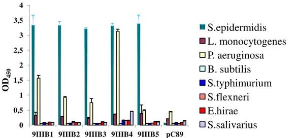
Figure 1.3 ELISA test values of selected phage clones against S. epidermidi by 9-mer
Figure 1.3 Reactivity ELISA test for clones selected against S. epidermidis. Clones named 9IIIB derive by the third round of selection of the 9-mer random M13 phage display libraries. Clones named 12IVB derive by the fourth round of selection of the 12-mer random M13 phage display libraries. A pC89 vector was used as a negative control. More details about the procedure are shown in the materials and methods section.
0 0,5 1 1,5 2 2,5 3 3,5 4
9IIIB1 9IIIB2 9IIIB3 9IIIB4 9IIIB5 12IVB112IVB2 12IVB312IVB412IVB5 pC89
O
D
4
5
0
Data underline that all selected phage clones from both libraries had a similar reactivity. In order to determine clone binding specificity and avidity, selected clones from both libraries were investigated by means of further ELISA tests.
Below are presented selectivity results of all clones previously selected (Figure 1.4,
1.5).
Figure 1.4 Cross-reactivity ELISA test for clones by 9-mer random M13 phage display libraries.
In these phage-capture ELISA assays, each organism was incubated with an equal amount of phage clone, and bound virions were detected by using anti-M13 peroxidase conjugate antibody and then TMB substrate colour development. More details about the procedure are shown in the materials and methods section. 0 0,5 1 1,5 2 2,5 3 3,5 4
9IIIB1 9IIIB2 9IIIB3 9IIIB4 9IIIB5 pC89
O D4 5 0
S.epidermidis
L. monocytogenes
P. aeruginosa
B. subtilis
S.typhimurium
S.flexneri
E.hirae
S.salivarius
9IIIB5 and 12IVB5 clones showed the highest selectivity against S. epidermidis, compared to other bacteria.
Below are presented the results of ELISA test about the avidity of both clones (Figure
1.6).
A similar procedure was carried out for the selected clones against the other bacterium of interest, namely S. aureus. In this case, the most reactive phage clone appeared to be arising from pVIII-9aa library, labeled as StauIVS5 (Figure 1.7)
Figure 1.6 Avidity ELISA test for 9IIIB5 and 12IVB5 clones.
In these phage-capture ELISA assays, Urea 4M was added to washing buffer to verify binding avidity of selected clones. A pC89 vector was used as a negative control. More details about the procedure are shown in the materials and methods section.
0 0,5 1 1,5 2 2,5 3 3,5 4 9IIIB5 12IVB5 pC89 O D4 5 0 Control 4M UREA Figure 1.5 Cross-reactivity ELISA test for clones by 12-mer random M13 phage display libraries.
In these phage-capture ELISA assays, each organism was incubated with an equal amount of phage clone, and bound virions were detected by using anti-M13 peroxidase conjugate antibody and then TMB substrate colour development. More details about the procedure are shown in the materials and methods section. 0 0,5 1 1,5 2 2,5 3 3,5 4
12IVB1 12IVB2 12IVB3 12IVB4 12IVB5 pC89
O D4 5 0
S.epidermidis
L. monocytogenes
P. aeruginosa
B. subtilis
S.typhimurium
S.flexneri
E.hirae
S.salivarius
Moreover, ELISA test avidity, using Urea 4M, confirmed that StauIVS5 bind with high-avidity its bacterial target (Figure 1.8).
Figure 1.8 Avidity ELISA test for StauIVS5 clone.
In these phage-capture ELISA assays, urea 4M was added to washing buffer to verify binding avidity of selected clones. A pC89 vector was used as a negative control. More details about the procedure are shown in the materials and methods section.
0 0,5 1 1,5 2 2,5 StauIVS5 pC89 O D4 5 0 Control 4M UREA Figure 1.7 Cross-reactivity ELISA test for clone StauIVS5 by 9-mer random M13 phage display libraries.
In these phage-capture ELISA assays, each organism was incubated with an equal amount of phage clone, and bound virions were detected by using anti-M13 peroxidase conjugate antibody and then TMB substrate colour development. More details about the procedure are shown in the materials and methods section. 0 0,5 1 1,5 2 2,5 O D4 5 0
Selection of phage peptides targeting cancer membrane receptors
The discovery of novel approaches for the rapid and accurate detection of cell types, such as neoplastic cells, is one of the principal objectives in early-stage cancer diagnostics and monitoring of Minimal Residual Disease. Leukemia is one of the most blood malignancy affecting hematopoietic stem cells, bone marrow and lymphatic system. In this malignancy, the clonal population of neoplastic cells exhibits marked heterogeneity with respect to proliferation and differentiation (Bonnet and Dick, 1997; Reya et al., 2001). The diagnostic gold standard of leukemia involves several methods, for individual treatment strategies and for the evaluation of treatment response, since there are different types of leukemia. For this reason, those strategies are often ineffective and time consuming, as well as expensive and often ineffective.As alternative, M13 phage clones by a 9-mer pVIII display library can be used for detection and neoplastic cell imaging in vitro, including same advantages described in previous section. In this section a 9-mer pVIII M13 phage display library is screened against histiocytic lymphoma cell (U937) to identify peptides that selectively recognize these cells. This section describes the selection techniques for phage clones displaying peptides capable of specific and strong binding to the cell surface of U937, used as an in
vitro model of cancer cells. Materials and Methods
Cell line, bacteria and growth media. U937 cell line was purchased from American
Type Culture Collection (ATCC® CRL-1593.2™) and maintained in RPMI 1640 with L-Glutamine (Lonza, BE12-702F) supplemented with 10% fetal bovine serum (FBS, Lonza DE14-801F), penicillin (100 units/ml) and streptomycin (100 µg/ml) at 37 °C in humidified 5% CO2 incubator. TG1 Escherichia coli was used for propagation of phage
clones. Stock organisms were maintained in LB broth containing 20% (v/v) glycerol at −80 °C.
Phage display libraries. A 9-mer random phage display library (kind gift of Prof. F.
Felici) was constructed in the vector pC89, by cloning a random DNA insert between the third and fifth codon of the mature pVIII protein encoding segments of the pVIII gene (Felici et al., 1991). This library was selected towards whole U937 cells in suspension.
Phage peptide selection. U937 cells were harvested by centrifugation at 800 rpm for 5
min, washed twice in Hank’s buffered salt solution (HBSS Sigma-Aldrich pH 7.6) and resuspended at a concentration of 1·106 cells/ml. Selection procedure was according to Cao et al. with some modifications (Cao et al., 2003). Three rounds of selection were performed and recombinant phages were identified by plating infected E. coli TG1 cells on LB agar added with X-Gal (5-bromo-4-chloro-3-indolyl-beta-D-galactopyranoside), IPTG (isopropyl thiogalactoside) and ampicillin. The presence of the insert was detected by X-gal which produces a characteristic blue dye when cleaved by β-galactosidase. Blue bacterial colonies, from third round of selection, were randomly selected. Each colony contained a single phage clone.
Elisa Test. The binding capacity of the thirteen phage clones to the target cells was
tested in ELISA using as a negative control vector pC89.
2.5·104 U937 cells in Phosphate-Buffered Saline (PBS) were dispensed in each well of a 96-well plate (Orange scientific) and incubated over night at 4 °C. They were fixed with methanol for 10 min at RT, washed three times with Washing Buffer (PBS + 0.05% Tween 20) and blocked with 100 µl of PBS + 5% Lactalbumin + 0.05% w/v Tween20 for 2h at 37 °C at 30 rpm. 1·1010 phage clones, diluted in PBS + 1% Lactalbumin + 0.1% w/v Tween20, were added in each well and the plate was incubated for 1 h at 37 °C at 30 rpm. After three washing steps in Washing Buffer, cells were incubated with monoclonal anti-M13 peroxidase conjugate antibody (Amersham Biosciences, Buckinghamshire, UK) at a dilution of 1/5000 in PBS + 1% Lactalbumin + 0.1% w/v Tween20 (100 μl/well) for 1 h at 37 °C. After further three washing steps, antibody binding was detected by adding a 3,3′,5,5′-Tetramethylbenzidine (TMB) liquid substrate system for ELISA (Sigma-Aldrich), incubated for 45 min at RT and stopped with 25 μl of 1M H2SO4. Optical absorbance was recorded at 450 nm (MultisKan FC
ThermoScientific).
Peptide sequence analysis. The insert DNA of phage clones was amplified by
Polymerase chain reaction (PCR) and sequenced. The PCR products were purified using QIAquick PCR purification kit (Qiagen) and sequenced by the DNA sequencing service of CRIBI (University of Padova, Italy) using the M13 primer-40 (5'-GTTTTCCCAGTCACGAC-3'). The amino acid sequences were aligned according to their similarity by using the Clustal X 2.1 sequence alignment program using the IDENTITY series matrix (available at [http://clustalx.software.informer.com/2.1/]).
GeneDoc (available at [http://iubio.bio.indiana.edu/soft/molbio/ibmpc/genedoc-readme.html]) was used as a tool for visualizing, editing and analyzing multiple sequence alignments of the peptides (Thompson et al., 1994; Aiyar, 2000). Statistical analysis was performed in order to calculate amino acid frequency and diversity within the pool of peptides in the selected phage clones.
Results and Discussions
A 9-mer pVIII M13 phage display peptide library was screened against U937 cells in suspension. The most reactive phage clones showing a specific binding to U937 cells were amplified and their DNA was sequenced to determine the amino acid sequences of the displayed peptides. The deduced amino acid sequences were aligned by using CLUSTALX 2.1 software based on IDENTITY matrix, and the consensus sequence was obtained by using GENEDOC software. All sequences contained two positively charged amino acid residues (lysine and arginine) in the amino terminal region and non-polar amino acid residues in the central positions. These regions may be directly involved in the cell-target interaction. Among all peptides, EIII1 phage sequence showed the most significant similarity with consensus when all the clone sequences were re-aligned with consensus when all the clone sequences were re-aligned with this (Figure. 1.9).
Figure 1.9 Alignment of phage-displayed peptides.
The deduced amino acid sequences were aligned by using CLUSTALX 2.1 with the consensus sequence obtained by GENEDOC. Dashes indicate gaps used to maximize the alignment. This alignment shows conserved positively charged amino acid residues in the N-terminal region (K and R), flanked by nonpolar amino acids such as Valine (V), Isoleucine (I), Leucine (L) and Alanine (A). Data have been published in Biosensors and Bioelectronics (Lentini et al, 2015)
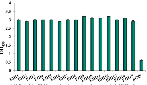
The relative binding of the phage clones was estimated by ELISA test, using as negative control the insertless vector pC89. The results showed that all selected phage clones had a similar reactivity and specificity for U937 cells (Figure 1.10)
Figure 1.10 Reactivity ELISA test for clones selected against whole U937 cells
Clones named EIII derive by the third round of selection of the 9-mer random M13 phage display libraries. More details about the procedure are shown in the materials and methods section.
Data have been published in Biosensors and Bioelectronics (Lentini et al, 2015) 0 0,5 1 1,5 2 2,5 3 3,5 4 O D4 5 0
Phage-display as tool for biomarker discovery in neurodegenerative
diseases
Alzheimer’s disease (AD) is a chronic neurodegenerative disorder, with a slow progression to dementia. In the past few years, the development and validation of specific tests, based on AD biomarkers detectable from serum, such as antibodies against β-amyloid (Aβ), has been the major goal of many research groups. Auto-antibodies are naturally present in human blood, in free form or complexed with Aß, both in AD patients and in healthy subjects. Although antibody levels change to disease progression, data reported about this topic are not consistent.
As proposed by some authors, it is reasonable to assume that it is unlikely that the unmodified antigens are the primary autoantigen to trigger the autoimmune response. Since the deposition of amyloids at diverse size, sequence, native structure, biological function can misfold and self-assemble, it is more plausible to assume that this could generate new epitopes (e.g.discontinuous) derived from amyloid-based aggregation. At this purpose, it is proposed a new approach to identify homologous mimotopes to Aβ42 as capture agents for diagnostically useful autoantibodies for AD (IgG-AD).
By bioinformatics tools, it has been verified a conformational homology between several Aβ42 and other proteins domains, particularly a chaperon protein (Caf1-M) of
Yersinia pestis.
In this section a 12-mer random M13 phage display (12-mer and 12-mer cys) is used for a double binding screening against YPF19 a monoclonal antibody against the Caf1, and IgG-AD, obtained from several pools of AD sera. The selected clones were used as a probe to identify sera of AD subjects.
Materials and Methods
This section describes the selection techniques for phage clones displaying peptides able to specific and strong binding to IgG-AD.
Bioinformatic analysis . Several peptide sequences spanning the C-terminal regions of
Aβ42 protein were predicted according to hydrophilicity, exponential surface
accessibility, flexibility, antigenicity and amphipathicity scales using the Protean program of DNAstar software package. Afterword 3D structures of misfolding of Aβ42,
peptides and fibril forms were compared with all proteins present Bank), in order to identify the possible conformational mimotopes.
“Double Binding” screening.
(mean age= 77.4 years, mean MMSE v tied on paramagnetic
phage display libraries was performed by alternating DYN mAbYPF19 (sequential double binding).
Affinity analysis. ELISA and im
peptides with conformational homology to reactivity Elisa test.
Results and Discussions
Yersinia pestis Chaperon Protein Caf1
conformational structure ( panel), respectively.
The residues that occupy an equivale 161 and 20-29 of Chaperon Protein Caf1 (dark colors).
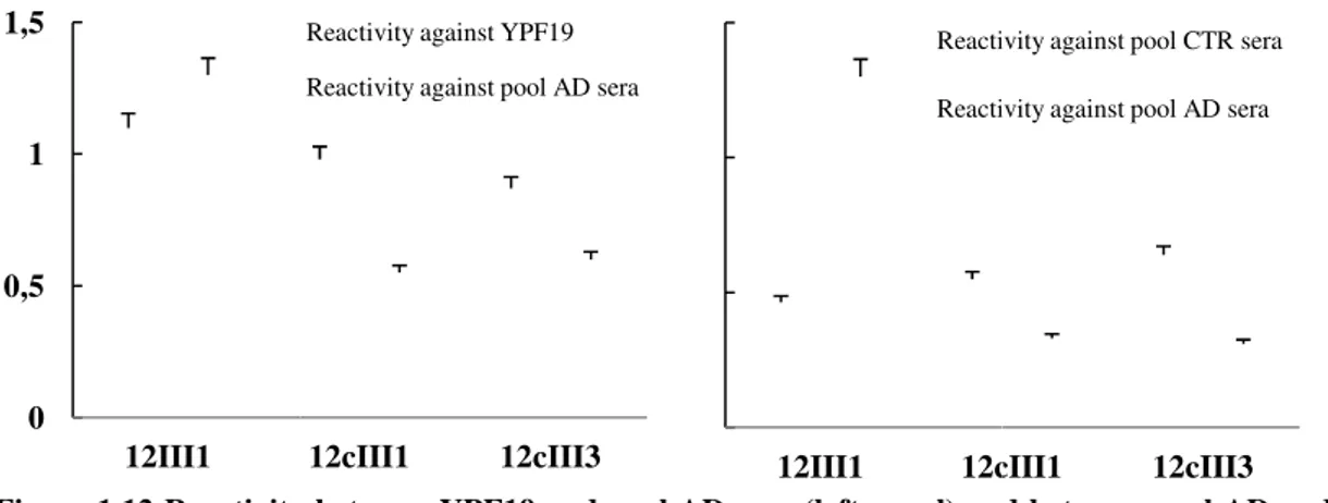
Figure 1.12 shows the most reactive clones against both pool of AD sera and mAb
YPF19 (left panel) and
healthy individuals (CTR) sera
Figure 1.11 Bioinformatic analysis by PDBeFOLD tool Peptides (PDB ID= 1z0q) and
in PDB (Protein Data Bank), in order to identify the possible conformational mimotopes.
peptides and fibril forms were compared with all proteins present Bank), in order to identify the possible conformational mimotopes.
“Double Binding” screening. IgGs from a pool of five sera, obtained from AD cases
4 years, mean MMSE value = 15.2), and mAb-YPF19, were covalently tied on paramagnetic-beads, Dynabeads®-proteinG (DYN). Biopanning of pVIII M13 phage display libraries was performed by alternating DYN
mAbYPF19 (sequential double binding).
ELISA and immunoblot tests were used to select clones
peptides with conformational homology to Aβ42. The same were then evaluated
and Discussions
Chaperon Protein Caf1-M (PDB ID=1p5u) showed similarity of conformational structure (Figure 1.11) to Aβ42, peptide (left panel) and fibril (right
The residues that occupy an equivalent geometric shape in space were of Chaperon Protein Caf1-M (light colors) and 20
the most reactive clones against both pool of AD sera and mAb and the reactivity of the same clones against both pool of AD and healthy individuals (CTR) sera (right panel).
Bioinformatic analysis by PDBeFOLD tool .
eptides (PDB ID= 1z0q) and fibril (PDB ID= 2NAO) forms were compared with all in PDB (Protein Data Bank), in order to identify the possible conformational mimotopes.
peptides and fibril forms were compared with all proteins present in PDB (Protein Data Bank), in order to identify the possible conformational mimotopes.
pool of five sera, obtained from AD cases YPF19, were covalently proteinG (DYN). Biopanning of pVIII M13 phage display libraries was performed by alternating IgG-AD and
DYN-munoblot tests were used to select clones displaying 42. The same were then evaluated by
M (PDB ID=1p5u) showed similarity of , peptide (left panel) and fibril (right
shape in space were: amino acid 149-M (light colors) and 20-36 and 1-9 of Aβ42
the most reactive clones against both pool of AD sera and mAb-the reactivity of mAb-the same clones against both pool of AD and
fibril (PDB ID= 2NAO) forms were compared with allproteins present
Results by test Elisa reactivity suggest that 12III1 clone significantly discriminates levels of IgGs between AD and CTR sera,
0.03). Clone was tested in ELISA (Figure 1.13).
Data show levels of IgGs in AD patients significantly higher than (mean AD=0.93, CTR=0
bind with high-avidity the 12III1 phage clone, as confirmed by avidity test using Urea 4M (mean AT= 91.43).
Figure 1.13 Reactivity of 12III1 against AD and healthy sera Figure 1.12 Reactivity
healthy individuals (CTR) sera (right panel). 0
0,5 1 1,5
12III1
Results by test Elisa reactivity suggest that 12III1 clone significantly discriminates levels of IgGs between AD and CTR sera, with a 95 % confidence interval
was tested in ELISA against 38 serum samples (20
Data show levels of IgGs in AD patients significantly higher than 93, CTR=0.55) (p-value = 0.0044). Intriguingly, IgGs
avidity the 12III1 phage clone, as confirmed by avidity test using Urea 43).
Reactivity of 12III1 against AD and healthy sera.
Reactivity between YPF19 and pool AD sera (left panel) and between pool healthy individuals (CTR) sera (right panel).
12cIII1 12cIII3
Reactivity against YPF19 Reactivity against pool AD sera
0 0,5 1 1,5
12III1 12cIII1
Reactivity against pool CTR sera Reactivity against pool AD sera
Results by test Elisa reactivity suggest that 12III1 clone significantly discriminates confidence interval (p-value = against 38 serum samples (20-AD and 18-CTR)
Data show levels of IgGs in AD patients significantly higher than healthy individuals 0044). Intriguingly, IgGs present in AD sera avidity the 12III1 phage clone, as confirmed by avidity test using Urea
between YPF19 and pool AD sera (left panel) and between pool AD and
12cIII1 12cIII3
Reactivity against pool CTR sera Reactivity against pool AD sera
Acknowledgements
This activity was partially funded by Italian Ministry of Education, University and Research (MIUR) by means of the national Program PON R&C 2007–2013, project project HIPPOCRATES – Development of Micro and Nano-Technologies and Advanced Systems for Human Health (PON02_00355_29641931).
Phage-display libraries were a kind gift of Prof. Franco Felici.
The activity “Phage-display as tool for biomarker discovery in neurodegenerative
diseases” was carried out in collaboration with:
Professor A. Nicoletti and Professor M. Zappia, Section of Neurosciences, Department G.F. Ingrassia, University of Catania, Italy
Doctor S. Conoci and S. Petralia, STmicroelectonic Catania, Italy
Bibliography
Aiyar A. The use of CLUSTAL W and CLUSTAL X for multiple sequence alignment. 2000. Methods Mol. Biol. 132, 221-41.
Barbas CF, Kang AS, Lerner RA, Benkovic SJ. Assembly of combinatorial antibody libraries on phage surfaces: The gene III site. 1991. Pro Nat Acad Sci U S A. 88, 7978-82.
Bonnet D, Dick JE. Human acute myeloid leukemia is organized as a hierarchy that originates from a primitive hematopoietic cell. 1997. Nat. Med. 3, 730–37.
Cao J, Zhao P, Miao XH, Zhao LJ, Xue LJ, Qi ZT. Phage display selection on whole cells yields a small peptide specific for HCV receptor human CD81. 2003. Cell. Res. 13, 473-79.
Cesareni G. Phage-plasmid hybrid vectors. 1988. Biotechnology. 10, 103-11.
Carnazza S, Gioffrè G, Felici F, Guglielmino SPP. Recombinant phage probes for Listeria monocytogenes. 2007. J. Phys. Condens. Matter 19,13.
Carnazza S, Foti C, Gioffrè G, Felici F, Guglielmino SPP. Specific and selective probes for Pseudomonas aeruginosa from phage-displayed random peptide libraries. 2008. Biosens. Bioelectron. 23, 1137–44.
Deroo S, Muller CP. Antigenic and immunogenic phage displayed mimotopes as substitute antigens: Applications and limitations. 2001. Comb. Chem. High Through Screen 4, 75-110.
Felici F, Castagnoli L, Musacchio A, Jappelli R, Cesareni G. Selection of antibody ligands from a large library of oligopeptides expressed on a multivalent exposition vector. 1991. J. Mol. Biol. 222, 301-10.
Kishchenko GP, Minenkova OO, Ilyichev A A, Gruzdev AD, Petrenko VA. Study of structure of phage-M13 virions containing chimeric B-protein molecules. 1991. Mol. Biol.-Engl. Transl. 25, 1171-76.
Kishchenko G, Batliwala H, Makowski L. Structure of a foreign peptide displayed on the surface of bacteriophage M13. 1994. J. Mol. Biol. 241, 208-13.
Lentini G, Fazio E, Calabrese F, De Plano LM, Puliafico M, Franco D, Nicolò MS, Carnazza S, Trusso S, Allegra A, Neri F, Musolino C, Guglielmino SPP. 2015. Phage-AgNPs complex as SERS probe for U937 cell identification. Biosens. Bioelectron. 74, 398–405.
Luzzago A, Felici F. Construction of Disulfide-Constrained Random Peptide Libraries Displayed on Phage Coat Protein VIII. 1998. Methods Mol. Biol. 87, 155–64.
McCafferty J, Griffiths AD, Winter G, Chiswell DJ. Phage antibodies: Filamentous phage displaying antibody variable domains. 1990. Nature 348, 552-4.
McConnell SJ, Kendall ML, Reilly TM, Hoess RH. Constrained peptide libraries as a tool for finding mimotopes. 1994. Gene, 151, 115-8.
Mead, D. A.; Kemper, B. Chimeric single-stranded DNA phage-plasmid cloning vectors. 1988. Biotechnology. 10, 85-102.
Parmley SF, Smith GP. Antibody-selectable filamentous fd phage vectors: affinity purification of target genes. 1988 Gene 73, 305-18.
Reya T, Morrison SJ, Clarke M F, Weissman IL. Stem cells, cancer, and cancer stem cells. 2001. Nature. 415, 105–11.
Smith GP. Filamentous fusion phage: novel expression vectors that display cloned antigens on the virion surface. 1985. Science 228, 1315-17.
Smith GP. Gene 1993, 128, 1-2.
Smith G P, Petrenko VA. Phage display. 1997. Chem. Rev. 97, 391-410.
Petrenko VA, Smith GP. Phage from landscape libraries as substitute antibodies. 2000. Protein Eng. 13, 101–04.
Petrenko VA, Vodyanoy VJ. Phage display for detection of biological threat agents. 2003. J. Microbiol. Methods 53, 253–62.
Petrenko VA, Sorokulova IB. Detection of biological threats. A challenge for directed molecular evolution. 2004. J. Microbiol. Methods, 58, 147-68.
Ponsel D, Neugebauer J, Ladetzki-Baehs K, Tissot K. High affinity, developability and functional size: the holy grail of combinatorial antibody library generation. 2011. Molecules 16, 3675-700.
Rodi DJ, Makowski L. Phage-display technology - finding a needle in a vast molecular haystack. 1999. Curr. Opin. Biotechnol. 10, 87-93.
Thompson JD, Higgins DG., Gibson TJ. CLUSTAL W: improving the sensitivity of progressive multiple sequence alignment through sequence weighting, position-specific gap penalties and weight matrix choice. 1994. Nucleic Acids Res. 22, 4673-80.
Zwick MB, Shen J, Scott, JK. Phage-displayed peptide libraries. 1998. Curr. Opin. Biotechnol. 9, 427-39.
CHAPTER 2
Raman spectroscopy applied to cellular systems
Introduction
The label-free imaging of cell systems allows clustering of cells based on their inherent chemical makeup. Imaging based on label, such as fluorescent labels, distinguish one cell type from another by the binding between label and a cell surface marker. Differently the use of Raman analysis includes contributions not only from surface markers, but also influences from the signaling pathways and intracellular molecules which relate to the current state of the cell as well as cell type and function.
Spectroscopy can be defined as an analysis technique to study the interaction of electromagnetic radiation with atoms and molecules. The scattering of a light after interaction with matter grades into its component energies (colors). The resultant dissection of energies can be used to analysis that matter’s physical properties.
When the energy of an incident photon is unaltered after collision with a molecule, the scattered photon keeps same frequency (elastic or Rayleigh scattering). Differently, if energy is transferred from the molecule to the photon or vice versa, the scattered photon has less or more than the energy of the incident photon (inelastic or Raman scattering). Scattered photons with more energy will be shifted to a lower frequency (red-shifting or Stokes shift) (Diem et al, 2008; Wachsmann-Hogiu et al, 2009), while photons with less energy will be shifted to a higher frequency (blue-shifting or anti-Stokes shift) (Diem et
al, 2008; Wachsmann-Hogiu et al, 2009), to balance in both cases the total energy of the
system. Raman scattering is an example of inelastic scattering because of the energy transfer between the photons and the molecules during their interaction. The difference in energy between the incident and scattered photons corresponds to the energy of the molecular vibration (Wachsmann-Hogiu et al, 2009).
The loss (or gain) in the photon energies corresponds to the difference in the final and initial vibrational energy levels of the molecules participating in the interaction. The resultant spectra are characterized by shifts in wave numbers (inverse of wavelength in cm−1) from the incident frequency. The frequency difference between incident- and Raman-scattered light is termed the Raman shift, which is unique for individual molecules and represented as 1/cm. Raman peaks are spectrally narrow, and in many cases can be associated with the vibration of a particular chemical bond (or a single
functional group) in the molecule (Choo-Smith et al, 2002). The vibrations are molecular bond specific allowing a biochemical fingerprint to be constructed of the material (Harris et al, 2009).
It is important to note that, depending on whether the bond length or angle is changing, vibrations are subdivided into two classes: stretching (symmetric and asymmetric) and bending (scissoring, rocking, wagging and twisting).
There are six different vibrational modes. In symmetric and asymmetric stretching types, the lengths of the bonds become unstable. In another four types, the length remains stable and the angulations of the bonds changes (Figure 2.1).
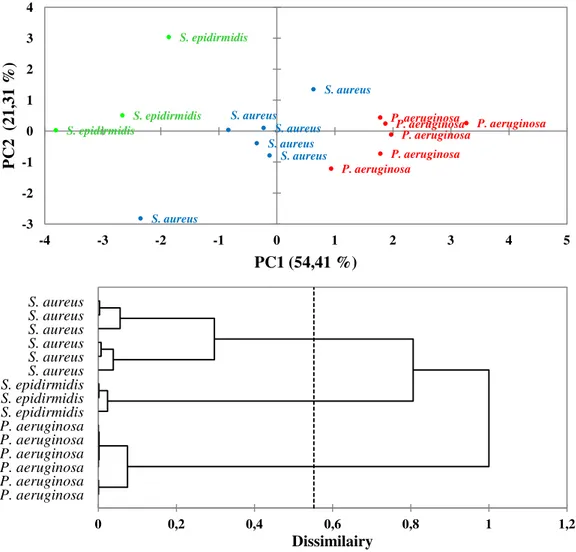
In this section, it's verified the ability of Raman measurement to discriminate cellular systems, such as bacterial pathogens or neoplastic cells. The large amount of information present in the vibrational spectra were evaluated by multivariate technique, including principal component analysis (PCA) and Hierarchical Cluster Analysis (HCA), that allowed these many subtle spectral differences to be clarified and used to classify cell types. Particularly, Raman spectroscopy allows identifying pathogenic bacteria, namely Staphylococcus epidermidis ATCC 12228, Staphylococcus aureus ATCC 29213 and Pseudomonas aeruginosa ATCC 27853, without the use of any label type. Moreover, Raman spectroscopy represents a good tool for discrimination of cell type, belonging to different cell lines. At this purpose, cells by four different cell lines, namely histiocytic lymphoma (U937), laryngeal carcinoma (HEp-2) and myelome multiple (MM1 and U266B1), were spectrophotometrically evaluated. Subsequently the same cell lines are evaluated individually on the basis of their different metabolic state and chemo-resistance to some drugs.