International PhD Program in Neuroscience
XXIX Cycle
Coordinator: Prof. Salvatore Salomone
miRNA EXPRESSION PROFILES IN RETINAL
NEURODEGENERATIVE DISEASES
PhD thesis
Giovanni Luca Romano
Tutor: Prof. Claudio Bucolo
BIOMETEC
Department of Biomedical and Biotechnological Sciences Section of Pharmacology.
Medical School - University of Catania
2016
3
TABLE OF CONTENTS
ACKNOWLEDGEMENTS ... 4
LIST OF ABBREVIATIONS ... 5
ABSTRACT ... 7
RETINAL NEURODEGENERATIVE DISEASES ... 9
Glaucoma ... 15
Age-Related Macular Degeneration (AMD) ... 18
MiRNAs ... 24
miRNA expression in the eye ... 27
CHAPTER I ... 29
CHAPTER II ... 66
DISCUSSION AND CONCLUSIONS ... 92
REFERENCES ... 95
4
A
CKNOWLEDGEMENTSI would like to thank prof. Claudio Bucolo, who has fully and patiently supported me during the years of my PhD studies. With his great experience and knowledge, he taught me how to approach the research in the field of ocular pharmacology.
My biggest thanks go to prof. Filippo Drago, his leadership and his precious suggestions have been crucial in my professional and human training.
Prof. Drago, along with prof. Bucolo and prof. Salomone, has welcomed me in his research group, which is the best I’ve ever worked for.
An important acknowledgment goes to Dr. Maria Pia Aiello, her friendship and her kindness have been precious for my inclusion in this wonderful environment.
I want to thank also all the former and actual lab. mates, researchers and friends of the Section of Pharmacology of BIOMETEC, in particular Dr. Chiara Platania who shared with me all the good and bad of my PhD path.
At least, my thanks go to my beloved parents and my whole family, for their efforts in supporting me during university studies, till the highest degree.
5
L
IST OF ABBREVIATIONSAβ amyloid β
AD Alzheimer’s disease ALS amyotrophic lateral sclerosis AMD Age-Related Macular Degeneration APP amyloid precursor protein
ARNT aryl hydrocarbon receptor nuclear translocators ATPase Adenosine Triphosphate Phosphatase ATP Adenosine Triphosphate
ADP Adenosine Diphosphate BACE1 β-secretase
BBB Blood Brain Barrier
BDNF Brain-derived neurotrophic factor BM Bruch’s membrane
BRB Blood Retinal Barrier
CC chemokine
CDS coding sequences CK/CK-R cytokine/cytokine-receptor CNS central nervous system CNV choroidal neovascularization DAMPs damage-associated molecular patterns FC Fold-changes
GCL ganglion cell layer
HIF hypoxia-inducible transcription factor HMDD Human miRNA Disease Database INL inner nuclear layer
IPL inner plexiform layer IVT intravitreal injection IOP intraocular pression JNK Jun N-terminal kinase
KO knockout
miRNAs microRNAs mRNAs messenger RNAs
NF-kB Nuclear factor kappa-light-chain-enhancer of activated B cells NFL nerve fiber layer
NFTs neurofibrillary tangles; NGF Nerve growth factor NT Neurotrophins
OCT Optical coherence tomography ONH optic nerve head
6
OPL outer plexiform layerPD Parkinson disease
PEDF pigmented-epithelium-derived-factor PIGF placental growth factor
p-tau phosphorilated tau RGCs retinal ganglion cells
RISC RNA-induced silencing complex RPE retinal pigment epithelium TFs transcription factors
TFBSs transcription factors binding sites TLRs Toll-like receptors
Trk tyrosine kinase
VEGF vascular endothelial growth factor VEGFRs VEGF receptors
7
A
BSTRACTMicroRNAs (miRNAs) are non-coding small RNAs, which have been found to regulate gene expression at the post-transcriptional and translational levels. A lot of studies demonstrated that miRNAs regulate various cellular processes, including differentiation, development, aging, apoptosis, oncogenesis and metabolism. Moreover, dysregulation of specific miRNAs is associated with a variety of diseases, including neurodegenerative disorders. Identification of differenzial pattern expression of miRNAs could be of value for development of novel biomarkers and discovery of new pharmacological targets for human diseases.
The aim of our research was to investigate miRNAs regulation in neurodegenerative diseases. Glaucoma is a progressive optic neuropathy and it is one of the leading cause of blindness in the industrialized countries. Age related macular degeneration (AMD) is the leading cause of blindness among people aged 50 and over. Signs of irreversible neurodegeneration in glaucoma, AMD and Alzheimer’s disease (AD), are usually evident at least a decade after onset of disease; thus early diagnosis is an urgent need in order to start effective therapy against neurodegenerative process. Identification of deregulated miRNA and associated pathways common to glaucoma, AMD and AD might help in the challenging search of biomarkers and novel therapeutic strategies.
We found, from literature search, 8 deregulated miRNAs in glaucoma, 9 and 23 in AMD and AD, respectively. One miRNA was found to be commonly deregulated in glaucoma and AMD (miR-23a), two miRNA (miR-29a, miR-29b) in glaucoma and AD, and four miRNAs in AMD and AD (miR-9, miR-31, miR-21, miR-34a, miR-146a). Predicted miRNAs common to the three neurodegenerative diseases were 9 (miR-107, miR-137, miR-146a, miR-181c, miR-197, miR-21, miR-22, miR-590, miR-9), which demonstrate to be involved in the regulation of inflammation pathways. Based on prediction of miRNA and associated biochemical pathways, inflammation could represent a therapeutic target common to glaucoma, AMD and AD.
Then we evaluated the differential expression profile of miRNAs in a rat model of AMD and in patients with AMD. Analysis of rat retina revealed that miR-27a, miR-146a and miR-155 are up-regulated in comparison to control rats. Seven miRNA (miR-9, miR-23a,
8
miR-27a, miR-34a, miR-126, miR-146a and miR-155) have been found to be dysregulated in serum of AMD patients in comparison to control group. Dysregulated miRNAs, both in the AMD animal model and in AMD patients, can target genes regulating pathways linked to neurodegeneration and inflammation.
Our findings support the assessment of specific miRNAs as potential biomarkers and therapeutic targets in retinal neurodegenerative diseases by means of preclinical and clinical studies.
9
RETINAL NEURODEGENERATIVE DISEASES
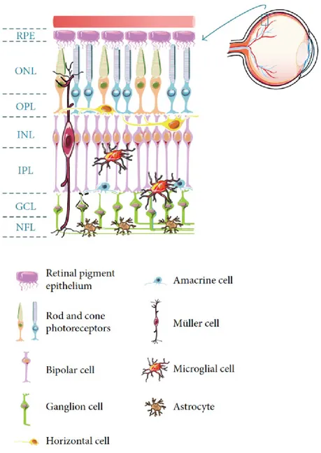
The retina is part of the central nervous system (CNS) due to its neuroectodermal origin and derivation from the anterior neural tube. Figure 1 shows a schematic representation of the major retinal cell types and their organization in the retina. The outer layer is the retinal pigment epithelium (RPE), a monolayer of cuboid, pigmented cells between the photoreceptors and the choroid. The retina is then constituted by three laminar layers: the outer nuclear layer (ONL) that contains cell bodies of photoreceptors; the inner nuclear layer (INL) formed by cell bodies of the bipolar, horizontal, and amacrine cells; the ganglion cell layer (GCL) composed by nuclei of retinal ganglion cells (RGCs) and of displaced amacrine cells. Axons of RGCs form the nerve fiber layer (NFL), beneath the GCL. These cells are interconnected through synapses that occur in the outer and inner plexiform layers (1).
Neurodegeneration describes the slow and progressive dysfunction and loss of neurons or their axons in the CNS. Retinal degenerative diseases, such as glaucoma and age-related macular degeneration (AMD) are chronic neurodegenerative conditions that affect various retinal neurons and lead to progressive and irreversible loss of the vision.
The pathological features of these retinal disorders have been commonly compared to that of the neurodegenerative condition Alzheimer’s disease (AD) (Table 1). Although these diseases have very different symptoms,they may result from similar mechanisms.
Age is a risk factor common to glaucoma/AMD of the eye and AD of the brain. (2).
AD progressively impairs brain structures and consequently cognitive ability and is characterized by enhanced amyloid β (Aβ) levels and its deposition in the central nervous system (CNS) (3).
Several human and animal studies reported ocular changes in AD and pathological accumulation of Aβ in the retina (4, 5). Aβ (Figure 2), a peptide of 39-43 amino acids, is the main constituent of senile amyloid plaques in the brains of AD patients and is also deposited in the drusen of eyes with AMD.Interestingly, Aβ and phosphorilated tau (p-tau), have been detected also in glaucoma patients and have a role in retinal ganglion cells (RGCs) death and progression of visual loss.
Therefore, retinal Aβ accumulation can be regarded as a common feature of these three distinct disorders.
Secreted Aβ exists mainly in two isoforms, Aβ 1-40 and Aβ 1-42 forms. Monomers of Aβ1-40 are prevalent in the brain than the Aβ1-42 form.Small amounts of Aβ is deposited
10
in the normal brain (6) and in the normal retina, and the levels of these deposits increase with increasing age.
Aβ spontaneously aggregates into multiple coexisting physical forms which may have differential neurotoxicity. One form consists of 2-6 peptide oligomers, which combine into intermediate aggregates. Soluble oligomers and intermediate amyloids are considered the most neurotoxic forms of Aβ (7). Moreover, Aβ fibrils can arrange themselves into β-pleated sheets to form insoluble fibres of advanced amyloid plaques.
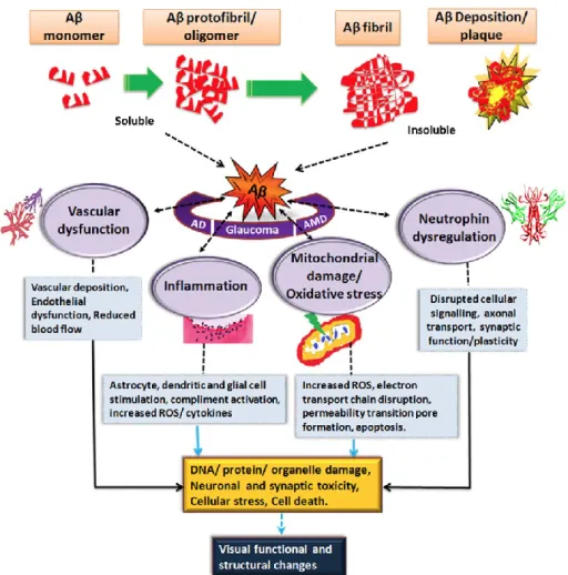
Aβ deposition induces different effect in cell types involved, resulting in vascular dysfunction, inflammation, oxidative stress and neurotrophin impairment (Figure 3). The major cell types affected in the brain are neural cells and microglia: Aβ activates microglial cells and causes neuronal cell death.
The major retinal cell type affected by Aβ is retinal pigment epithelium (RPE) cells. Kurji et al. (8) examined changes in gene expression of RPE cells stimulated with Aβ1-40 and found up-regulation of genes of inflammatory and immune response categories, in particular, IL-1β and IL-8, known as angiogenesis molecules. These findings suggest that Aβ triggers inflammatory and angiogenic responses in the RPE/choroidal layers, resulting in the development of choroidal neovascularization (CNV) in wet type of AMD.
Abnormal APP metabolism and increased Aβ expression in RGCs has been associated with the development of RGC apoptosis in experimental models of glaucoma (9).
A greater understanding of the link between these disorders will help in the identification of overlapping molecular mechanisms as well as development of novel and effective treatment strategies.
11
Table 1: Similarities and Differences between AD, Glaucoma, and AMD.
NFL: nerve fiber layer; NFTs: neurofibrillary tangles; ONH: optic nerve head; pTau: hyperphosphorylated tau protein; RGC: retinal ganglion cell; RPE: retinal pigmented epithelium.
12
Figure 1: Schematic representation of the major retinal cell types and their organization in the retina.
RPE: retinal pigment epithelium; ONL: outer nuclear layer; INL: inner nuclear layer; GCL: ganglion cell layer; NFL nerve fiber layer; OPL: outer plexiform layer; IPL: inner plexiform layer.
13
Figure 2: The amyloid cascade and major therapeutic approaches targeting amyloid β (Aβ) (K.
Ohno-Matsui. Progress in Retinal and Eye Research. 2011). The transmembrane amyloid precursor protein (APP) is sequentially cleaved by two proteases, β-secretase (BACE1) and γ-secretase, to release various isoforms of the Aβ peptide. The fragment Aβ1-42 aggregates to form toxic oligomers and is deposited in amyloid plaques. A major therapeutic effort is aimed at reducing Aβ1-42 production with BACE1-inhibitors and with γ-secretase inhibitors and modulators. Another therapeutic approach aims to enhance the clearance of Aβ, with antibodies or vaccines directed at soluble monomeric Aβ and/or oligomers and/or plaques.
14
Figure 3: impact of Aβ on the visual structure and function in AD, AMD and glaucoma (V. Gupta et
15
GLAUCOMA
Glaucoma is a progressive optic neuropathy that involve optic disc degeneration and visual field aberration. It is one of the leading cause of blindness in the industrialized countries. It has been described as an “optic vasobaropathy” indicating that both vascular and mechanical pressure processes contribute to the optic nerve damage (27). Besides high intraocular pressure (IOP), aging is one of the mainly recognized risk factor of glaucoma, and it has been proposed to have a neurodegenerative imprint.
The relative increase in IOP is related to an imbalance between aqueous humor (AH) production (by the ciliary body) and drainage (through the trabecular meshwork and Schlemm’s canal).
Glaucoma is principally divided into open-angle and angle-closure subtypes. Angle closure is characterized by an obstruction of the trabecular meshwork and elevated IOP.
The most common form of glaucoma is the primary open angle glaucoma (POAG) associated to increased intraocular pression (IOP). However, there are about 20-52% of glaucomatous patients with normal IOP and those patients develop damage at the optic nerve head similar to POAG patients. Ocular hypertension (> 21 mmHg) can arise asymptomatically and patients develop symptomatic visual field loss when damage is irreversible.
Both open-angle glaucoma and angle-closure glaucoma can be further subdivided into primary or secondary diseases, where primary refers to forms not correlated to previous and concomitant ocular disease (28). Secondary gluacoma refers to an elevated IOP with an identifiable pathological cause, such as inflammation, trauma, neovascularisation or iatrogenic causes.
Age-dependent accumulation of Aβ in RGCs has been suggested to contribute to glaucoma pathology. Human post-mortem and animal studies have found aggregation of Aβ and other proteins in the retina exposed to increased IOP which then promotes RGC apoptosis.
Diagnostic evaluation
The diagnosis of glaucoma is achieved using functional and structural assessments. The standard automated perimetry (SAP) is considered the gold standard for visual field assessment and monitoring of visual field loss. Structural diagnostic tools are principally based on the quantification of retinal nerve fibre layer (RNFL) thickness changes by optical
16
coherence tomography (OCT) and disc tomography (confocal laser-scanning tomography, cSLO) to assess changes of the optic nerve head (ONH).
IOP is assessed using tonometry devices, and the gold standard remain Goldman applanation tonometry (GAT).
Pharmacological treatments of glaucoma
Up to know the only therapeutic approach in glaucoma therapy is aimed at decreasing the IOP, also in normotensive glaucomatous patients. Reducing the baseline IOP by at least 20% generally will prevent progression of visual field loss, even in patients with IOP within the normal range (29-37).
AH inflow and outflow facilities are the pharmacological targets of ocular hypotensive drugs.
The canonical treatment remains the topical use of β receptor antagonists or prostaglandin analogues. Common alternate treatment modalities include carbonic anhydrase inhibitors or α2 adrenergic agonists.
Beta-blockers antagonizes β-adrenergic receptors of iris-ciliary body system inhibiting AH production. Several beta-blockers have been approved for treatment of glaucoma: timolol, betaxolol, levobunol and carteolol. Because of the possible side effects, they should be avoided in patients with asthma or decompensated heart failure.
Alpha-adrenergic agonists also decrease the production of AH, activating presynaptic adrenergic receptors in the ciliary body and inhibiting the release of noradrenaline. Sympathomimetics have shown not only to block the inflow of AH, but also to increase the uveo-scleral outflow.
Prostaglandin (PG) analogs are first-line, along with beta-blockers, drugs for treatment of ocular hypertension. PG analogs work by increasing the outflow of AH, especially through the uveoscleral pathway Topical treatment with PG analogs is not associated to systemic side effects. PG analogs local side effects are mostly not severe: changes in eye color, increase eyelash length, eye redness, itching, burning, impaired visual function.
Additional IOP-lowering drugs include cholinomimetics, such as pilocarpine, and carbonic anhydrase inhibitors, including acetazolamide. Cholinomimetics act by contracting the ciliary muscle, which opens the TM, increasing the outflow of AH.
Carbonic anhydrase inhibitors decrease production of AH, inhibiting the release of HCO3
17
Research is focused on development of neuroprotective therapies, which could stop the disease and even restore visual function through the regeneration of cells lost.
18
AGE-RELATED MACULAR DEGENERATION (AMD)
During the natural process of aging, the human eye undergoes physiological changes that include changes in the distribution of photoreceptors (30% loss of rod photoreceptors [24]), thickening of Bruch’s membrane (BM) (10) and accumulation of retinal pigment epithelium (RPE) debris, including drusen (11-15) composed by esterified cholesterol, phospholipids, lipofuscin, inflammatory components and other intra- and extraocular degenerative materials (16, 17, 18).
Age-related macular degeneration (AMD) is a progressive degenerative disease of the central part of the retina (the macula), a small, pigmented area responsible for visual acuity. Damage to the macula results in central vision loss. It is most common among people 55 years of age and older and is the leading cause of blindness among the elderly in developed countries. Risk factors implicated for AMD include older age, smoking, genetic predisposition and cardiovascular disease.
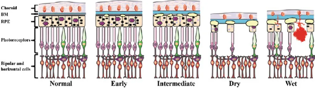
Initial modifications of AMD are seen as focal hyperpigmentation of the RPE and accumulation of sub-RPE deposits, including drusen, both between BM and the RPE and within the RPE itself (Figue 4).
The histopathology and clinical progression contribute to the definition of four major AMD subtypes (19-21):
1. Early AMD; 2. Intermediate AMD;
3. Advanced non-neovascular (“dry” or geographic atrophy) AMD; 4. Advanced neovascular (“wet” or exudative) AMD.
Early AMD is characterized by few small or medium-sized drusen and pigmentary abnormalities in the RPE, resulting in either mild visual impairment or can be asymptomatic. The transition from early to intermediate AMD occurs with the appearance of at least one large druse. At this point, the disease can progress to one of the two advanced forms of AMD: dry AMD (non-neovascular), characterized by presence of drusen and atrophy of the RPE and choroid; or wet AMD (neovascular), with newly formed vessels and RPE detachment.
The result of both of these processes (gradually in the dry form and suddenly in the wet form) is photoreceptor cell death and vision loss.
19
Diagnostic evaluation
Fluorescein angiography has been the gold standard for detecting and confirming the presence of neovascularization. However, it is an invasive imaging test in which serial retinal photographs are taken after fluorescein dye is injected into a vein. Optical coherence tomography (OCT) is a non-invasive method of imaging for posterior structures of the eye, frequently used to detect and monitor morphological changes associated with choroidal neovascularization.
Treatment opstions
Experimental and clinical evidence has shown that vascular endothelial growth factor (VEGF) is a key component in promoting neovascularization. Drugs that block vascular endothelial growth factor-A (VEGF-A) have shown to be effective in neovascular AMD. These drugs, administered through intravitreal injection, act by decreasing accumulated fluid in the back of the eye and by causing regression of the new fragile vessels.
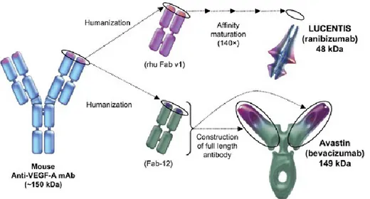
Available medications in this class include ranibizumab (Lucentis, Genentech Inc.), bevacizumab (Avastin, Genentech Inc.), and aflibercept (Eylea, Regeneron Pharmaceuticals, Inc.) (Table 2) (22).
Ranibuzimab is a recombinant, humanized, monoclonal, antibody fragment that inhibits all VEGF-A isoforms. Bevacizumab is a monoclonal, humanized, VEGF-specific antibody, developed for various cancers and used off-label for treatment of neovascular AMD (Figure 5). Treatment with ranibizumab or bevacizumab usually requires monthly injections into the eye or injections given as needed on the basis of monthly assessments to monitor disease activity.
Frequent eye injections may result in rare but serious adverse events. The effectiveness (23) and safety (24) of ranibizumab and bevacizumab for the treatment of patients with neovascular AMD have been analyzed in published Cochrane reviews and documented to be similar.
Aflibercept (VEGF-Trap), a relatively new medication for neovascular AMD, is a fusion protein made of key domains of VEGF receptors 1 (VEGFR1) and 2 (VEGFR2) fused with a portion of human antibody (25). Unlike ranibizumab and bevacizumab, aflibercept binds VEGF-A, VEGF-B, and another protein - placental growth factor (PIGF) - that is believed to play a role in progression of neovascular AMD. The binding affinity of aflibercept for VEGF is stronger than that of bevacizumab or ranibizumab (26), leading to potentially
20
longer duration of action in the eye that allows longer intervals between treatments. Less frequent dosing should lead to reduction in the risk of harm associated with intraocular injections.
21
Figure 4: Diagram of the outer layers of the human central retina in normal and in AMD (Tan et al.
Human Genomics. 2016). As the disease progresses, Bruch’s membrane (BM) increases in thickness. Early AMD is associated with small drusen and retinal pigmented epithelium (RPE) pigment abnormalities. As the disease progresses to the intermediate form, additional drusen are observed. In the two late forms of AMD (dry and wet), there is extensive drusen and photoreceptor cell death, with atrophy of the RPE and choroid in the dry form and choroidal neovascularization (CNV), hemorrhaging, and RPE detachment in the Wet form.
22
23
Table 2: Comparative anti-VEGF medications with the approved protocols for wet AMD (Mansour
AM, et al. Br J Ophthalmol 2015). *Affinity expressed as equilibrium dissociation constant (KD) in the picomolar (Pm) range. AMD, age-related macular degeneration; PlGF, placental growth factor.
24
Mi
RNA
SGene expression is largely regulated, in particular by transcription factors (TFs) and their binding sites (TFBSs). Recently, another group of molecules, namely microRNAs (miRNAs), have been found to regulate gene expression at the post-transcriptional and translational levels through interaction with target messenger RNAs (mRNAs) (38).
MiRNAs belong to a class of non-coding small RNAs. The first miRNA was found by Victor Ambros and his colleagues (39, 40). Its mature sequence contains only 21~24 nucleotides. The miRNA precursors are converted to mature miRNA duplexes through sequential processing by the RNaseIII family of endonucleases Drosha and Dicer (41, 42). (Figure 6). They bind to the 3’-untranslated regions (3’-UTRs) of target mRNAs through base pairing to complementary sequences, resulting in the cleavage of target mRNAs or repression of their translation (43-45). Translational inhibition, which seems to be the major mode of action, is performed by a riboprotein complex called RNA-induced silencing complex (RISC) consisting of the miRNA and proteins of the argonaute family (46-48). One miRNA can have multiple target sites in the mRNA transcript, while one mRNA can be targeted by multiple miRNAs.
The human genome has been estimated to encode up to 1000 miRNAs that regulate approximately one third of human genes, and each miRNA could target more than 200 genes (49, 50).
Emerging in vivo and in vitro experiments are showing that miRNAs regulate various cellular processes, including differentiation, development, aging, apoptosis, oncogenesis and metabolism (51-56). Expression studies have shown that miRNAs have expression patterns tissue-specific or developmental-stage-specific (57, 58).
Moreover, the dysregulation of specific miRNAs has been associated with a variety of diseases. miRNAs are shown to play important roles in tumorigenesis, with some miRNAs acting as oncogenes and some as tumor suppressor genes. miRNA profiles differ between normal tissues and tumor tissues and among tumor types (59-62) and can be used to accurately identify tumor tissue origin and prognosis.
Several studies suggest that some miRNAs are differentially expressed in human brain and regulate the expression of genes associated with specific neurodegenerative disorders,
25
whereas just few studies are available about miRNA deregulation in ocular degenerative diseases.
MiRNAs have been detected in body fluids such as blood, saliva or urine (63). Circulating miRNAs, found in plasma or serum, could be involved in cell-cell signaling [48]. Since they can be easily evaluated through a blood draw, they could represent potential minimally invasive biomarkers used for screening and/or monitoring diseases (64).
Moreover, novel classes of chemically engineered oligonucleotides, termed ‘‘antagomirs’’ or ‘‘antimiRs’’, have been developed and proved to be efficient in the modulation of miRNAs levels, representing potentially future treatment options.
Therefore, identification of different pattern of miRNAs expression could be a potential approach in order to develop novel biomarkers and to discover pharmacological targets in human diseases, such as age-related neurodegenerative diseases.
Finally, once their role in the pathogenesis of human diseases will be definitely clarified, miRNAs could represent novel targets for drug development.
26
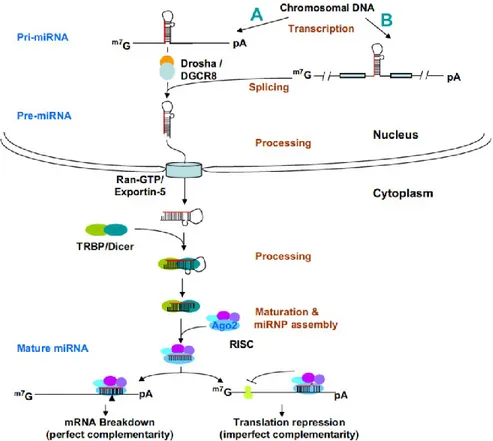
Figure 6. miRNA biogenesis and functions (S. Xu et al. 2009). In the nucleus, the primary
transcript of miRNA, pri-miRNA, is transcribed by RNA polymerase II (A). Many miRNAs are derived from introns of protein-coding genes (B). The pri-miRNAs fold into hairpin structures, which are cleaved by an RNase III endonuclease, Drosha in the Drosha-DGCR8 complex, to form 60–70-nt intermediates, known as pre-miRNAs. Pre-miRNAs are transported to the cytoplasm by Ran-GTP and an Exportin 5, and are cleaved by another RNase III endonuclease, Dicer, to form and 22-bp miRNA duplexes. TRBP recruits Agonaute protein, Ago2, to initiate the assembly of the RNA-induced silencing complex (RISC). When the miRNA and the target sites have precise or nearly precise complementarity, miRNAs specify cleavage of the target mRNAs by Ago2; when the base pairing is imperfect, miRNA with the RISC can destabilize the mRNA by deadenylation and subsequent decapping, and repress translation of the targeted mRNA by blocking translation initiation and/or inhibiting elongation.
27
MiRNA expression in the eye
The role of miRNA in the development and function of the eye is not well understood but is currently being explored and several reports on miRNA expression in retina and other ocular tissues, e.g. lens and cornea, helped to define it better.
A lot of miRNAs highly expressed in the mouse retina have been identified (65, 66).
In a group of inherited retinal degenerations, characterized by progressive photoreceptor loss and visual impairment and known as retinitis pigmentosa (RP), an altered pan-retinal expression of specific miRNAs has been identified, suggesting a potential involvement of miRNAs in this retinal disease (67).
Moreover, miRNAs have been identified in human blood samples and in many ocular tissues and showed to play a role in lens and retina development, ocular physiology, and several ocular diseases.
Increasing research efforts on small RNAs suggests that dysregulated miRNAs may regulate key aspects of AMD pathology. Data on circulating miRNA in AMD are still limited: four studies published between 2014 and 2016 analyzed the circulating miRNA expression profiles of AMD patients but showed little overlap in the findings.
Findings on dysregulated ocular miRNA showed more promise. Three studies profiled miRNA from retina samples and RPE cells, and one study reported miRNA measurements from vitreous humor (VH) of AMD patients. MiR-146a was found to be upregulated in both forms of AMD, and in different tissue. It is a well characterized miRNA and has been linked to progressive, age-related, inflammatory neurodegenerative disorders (68). It is supposed to modulate innate immune responses, inflammation, and the microglial activation state. It is under transcriptional control by nuclear factor-kappaB (NF-jB), and has been found to be upregulated by reactive oxygen species, pro-inflammatory cytokines, and amyloid peptides. As mir-146a, miR-155 was found to be dysregulated in AMD retina and it is a regulator of inflammation and microglial activation state in response to stress.
Recent studies revealed that a number of miRNAs are involved in the process of angiogenesis (69, 70, 71): miR-31, miR-150 and miR-184 have shown to be downregulated in oxygen-induced retinopathy mice models; angiogenesis and response to varying oxygen levels resulted to be modulated by miR125b, and miR-17, which may also regulate apoptosis.
28
These key candidate miRNAs could be useful as novel therapeutic approaches in AMD (64).
Finally, miRNA-based novel treatment of retinal and ocular diseases may be developed once the roles of miRNAs in the pathogenesis of these diseases will be identified.
29
CHAPTER I
30
Prog Brain Res. 2015;220:217-40. doi: 10.1016/bs.pbr.2015.04.013. Epub 2015 Jun 30
microRNA target prediction in glaucoma
Giovanni Luca Romano1*, Chiara Bianca Maria Platania1*, Stefano Forte2, Salvatore
Salomone1, Filippo Drago1, Claudio Bucolo1
1Department of Biomedical and Biotechnological Sciences, Section of Pharmacology,
University of Catania, Catania, Italy;
2IOM Ricerca srl, Viagrande-Catania, Italy.
Abstract
Glaucoma is a progressive optic neuropathy and it is one of the leading cause of blindness in the industrialized countries. The aim of the present study is to investigate miRNA regulation in glaucoma and other neurodegenerative diseases, that share similar pathways, by means of in-silico approaches such as bibliographic search and access to bioinformatic resources. First of all, data mining was carried out on Human miRNA Disease Database (HMDD) and mir2Disease databases. Then, predictions of deregulated miRNAs were carried out accessing to microrna.org database. Finally, the potential combinatorial effect of miRNAs, on regulation of biochemical pathways, was studied by an enrichment analysis performed by DIANA-miRPath v.2.0. We found, from literature search, 8 deregulated miRNAs in glaucoma, 9 and 23 in age-related macular degeneration (AMD) and Alzheimer’s disease (AD), respectively. One miRNA is commonly deregulated in glaucoma and AMD (miR-23a). Two miRNA (miR-29a, miR-29b) are common to glaucoma and AD, and four miRNAs where identified to be commonly deregulated in AMD and AD (miR-9, miR-31, miR-21, miR-34a, miR-146a). The match of the miRNA common to glaucoma and the other two neurodegenerative diseases (AMD and AD) did not generate any output. Enrichment of information has been reached through miRNAs prediction: 88 predicted miRNA are common to glaucoma and AMD; 19 are common to glaucoma and AD, 9 are common to AMD and AD. Indeed predicted miRNA common to the three neurodegenerative diseases are 9 (107, 137, 146a, 181c, miR-197, miR-21, miR-22, miR-590, miR-9). DIANA-miRPath predicted that those nine miRNAs might regulate pathways involved in inflammation. The findings hereby obtained provide a valuable hint to assess deregulation of specific miRNA, as potential biomarkers and therapeutic targets, in glaucoma and other neurodegenerative diseases by means of preclinical and clinical studies.
32
Introduction
Glaucoma is a progressive optic neuropathy and it is one of the leading cause of blindness in the industrialized countries. Aging is one of the mainly recognized risk factor of glaucoma, and it has been proposed to have a neurodegenerative imprint. Incidentally, aging is also a risk factor to develop Alzheimer disease (AD), a well-known neurodegenerative pathology, characterized by accumulation of amyloid plaques. An important characteristic common to both diseases is amyloid β (Aβ) deposition in the brain and in the retina of AD and glaucoma patients, respectively. In glaucoma loss of retinal ganglion cells (RGCs) seems to be preceded by axonal atrophy and deficits in axonal transport (Buckingham et al., 2008; Calkins, 2008). This pattern of disease progression, from axonal degeneration and progressing through secondary degeneration, is also observed in AD. Interestingly, Aβ and phosphorilated tau (p-tau), proteins that aggregate respectively in plaques and neurofibrillary tangles in the brain of AD patients, have been detected also in glaucomatous patients and have been thought to have a role in progression of visual loss and retinal ganglion cells (RGCs) death (Goldblum et al., 2007; Guo et al., 2007; Gupta et al., 2008; Janciauskiene and Krakau, 2001; Yin et al., 2008; Yoneda et al., 2005). Recently, an emerging interest for non-coding RNAs and in particular microRNAs (miRNAs) has been observed. Despite a plethora of studies about the role of miRNA in neurodegenerative diseases, few information are available about miRNA deregulation in ocular degenerative diseases and the common pathways shared with other neurodegenerative conditions. The eye is an extension of the brain and retinal changes have been observed in patients with central nervous system (CNS) disorders such as stroke, amyotrophic lateral sclerosis (ALS), Parkinson disease (PD) and Alzheimer disease (AD). It is noteworthy, that a tight correlation between glaucoma and the above mentioned neurodegenerative disorders has been suggested (Calkins, 2008; Fischer and Glass, 2007; Schwartz et al., 1996). The miRNAs are a family of non-coding single-stranded small RNAs (about 22 nucleotides) (Bartel, 2004). They originate from longer transcripts, pre-miRNA, cleaved by the nuclease Dicer, which generates a double stranded miRNA. A single stranded miRNA is then loaded in the proteinaceous complex RISC (RNA induced silencing complex), that silences gene expression through specific degradation of mRNA transcript, which is then recognized by means of sequence complementary miRNA. Focus on miRNA deregulation in neurodegenerative diseases might provide perspective innovative therapies, considering that neurodegenerative diseases are mainly multifactorial and miRNA target commonly
33
regulates more than one gene (Maciotta et al., 2013). We aimed our research in miRNA target prediction in glaucoma. Specific biological databases were queried for microRNA functionally involved in glaucoma or neurodegenerative diseases, then information enrichment was carried out searching literature on genetic association in glaucoma. Selected miRNAs were then used for the identification of candidate target mRNA using specific bioinformatics tools. The combinatorial effects of predicted common miRNAs, and their involvement in common biological pathways, have been studied using DIANA-miRPath.
Methods
Data mining of microRNA
We tried to retrieve deregulated microRNA involved in ocular diseases from the Human miRNA Disease Database (HMDD) (Lu et al., 2008) and miR2Disease (Jiang et al., 2009) databases. HMDD is a collection of human miRNA/disease associations supported by experimental results and miR2Disease is a manually curate database, aimed at providing a comprehensive resource of miRNA deregulation in different human diseases. Unfortunately, both HMDD and mir2Disease databases did not show any entry related to glaucoma and other ocular neurodegenerative diseases such as AMD. We included AMD in our investigation because represents another example of neurodegenerative disease characterized by Aβ deposition (Medeiros and Curcio, 2001). Therefore, we proceeded with the approach described ahead. Literature search has been carried out on PubMed and Scopus in order to find miRNAs experimentally involved in glaucoma and AMD. In particular, data associated to clinical studies and preclinical studies involving human cells were collected. miRNA associated to each disease have been matched and represented has a Venn diagram where each intersection includes common miRNA for the diseases analyzed. Deregulated microRNA involved in AD have been retrieved from the HMDD (Lu et al., 2008) and mir2Disease (Jiang et al., 2009) databases.
34
microRNA target prediction
Hence, in order to increase the information available, we analyzed from literature gene association studies in glaucoma and AMD, then the prediction of targeting miRNAs has been carried out by queries on microRNA.org database (http://www.microRNA.org/) (Betel et al., 2008). Predictions are carried out based on a development of the miRanda algorithm; predicted binding sites are then scored with the machine learning method mirSVR (Betel et al., 2010). Those predicted miRNA, putatively involved in glaucoma and AMD, have been matched with miRNA deregulated in AD. Data analysis, on 236 retrieved miRNAs, has been performed with Microsoft Excel 2010.
Identification of putatively involved common pathways.
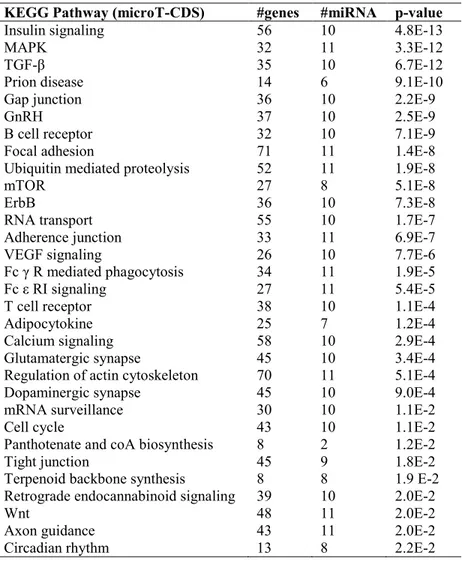
The potential combinatorial effect of miRNAs, in regulation of biochemical pathways, was studied by an enrichment analysis performed through DIANA-miRPath v.2.0 (Vlachos et al., 2012). This bioinformatic tool calculates the probability for each miRNA to be significantly associated to a KEGG (Kyoto Encyclopedia of Genes and Genomes) pathway. DIANA-miRPath, given a list of microRNA, goes back to target genes by means of two different algorithms: miRTarBase (Hsu et al., 2011) and microT-CDS (Reczko et al., 2012). The algorithm miRTarBase gives as output experimentally verified targets; whereas microT-CDS predicts extra targets based on matching of microRNA in coding sequences (CDS) of mRNA. Therefore, the software performs and enrichment analysis of KEGG pathways regulated by miRNAs. Pathways predicted with microT-CDS are listed along with p-values, number of genes and miRNAs. We, hereby, report a literature based search along with predicted “TarBase” pathways, in order to corroborate prediction results.
Results
The databases HMDD and miR2Disease did not generate any miRNA as entry associated to either glaucoma or AMD. Thus we have analyzed the current literature about annotated miRNA involved in glaucoma and AMD (Tables 1 and 2), data have been matched with miRNA involved in AD and represented as a Venn diagram (Fig. 1). We found 8 deregulated miRNAs in glaucoma, 9 and 23 in AMD and AD, respectively. One miRNA is
35
commonly deregulated in glaucoma and AMD (23a). Two miRNA (29a, miR-29b) are common to glaucoma and AD, and four miRNAs where identified to be commonly deregulated in AMD and AD (miR-9, miR-31, miR-21, miR-34a, miR-146a). The match of the miRNA common to glaucoma and the other two neurodegenerative diseases (AMD and AD) did not generate any output.
Hence, we proceeded with prediction of miRNA from genes retrieved from gene association studies in glaucoma and AMD. Analysis of gene association studies are reported in Tables 3 and 4 for glaucoma and AMD, respectively. Predicted miRNAs, that target the reported genes, have been matched with annotated miRNA associated to AD. We found an increased number of common miRNA between glaucoma, AMD and AD. In particular, 88 predicted miRNA are common to glaucoma and AMD; 19 are common to glaucoma and AD, 9 are common to AMD and AD. Indeed predicted miRNA common to the three neurodegenerative diseases are 9 (107, 137, 146a, 181c, miR-197, miR-21, miR-22, miR-590, miR-9).
Pathways identified through the algorithm microT-CDS are shown in Table 5. Pathways involved in autoimmunity, BRB and BBB integrity, and trophism of nervous systems are represented. Within such pathways there is the Erb B signaling, that seems to have a role in axonal regeneration (Evangelopoulos et al., 2009; Leinster et al., 2013; Tsai et al., 2010), has shown high number of involved genes, good p-value (7.3 E-8), and 10 regulatory miRNA (miR-107, miR-137, miR-146a-5p, miR-197-3p, miR-181-5p, miR-21-3p, miR-22-3p, miR-22-5p, miR-590-miR-22-3p, miR-590-5p). Those results corroborate the neurodegenerative etiology of glaucoma common to AMD and AD. Interestingly the microT-CDS algorithm identies for glaucoma, AMD and AD the VEGF signaling pathway (p-value 7.7 E-6, 26 target genes, 10 miRNAs).
Therefore, we predicted the association of these 9 common miRNA to KEGG pathways through TarBase annotation; those results are represented in Fig. 2. The most representative KEGG pathways (higher number of associated miRNA and best p-values) are: “apoptosis”, “cytokine-cytokine receptor interaction”, “Toll-like receptor signaling pathway”, “NF-kappa B”, “HIF-1” and “neurotrophins”. Incidentally, other pathways are also (well) represented and experimentally identified such, TGF-β, hypoxia inducible factor-1, chemokine signaling, and the pathways related to cell cycle regulation (e.g. p53) (Fig. 2).
36
The prediction of involved pathways, associated to predicted miRNA common to glaucoma, AMD and AD seems to be supported by literature as it follows.
Apoptosis
Even if apoptosis triggering factors are not well characterized, programmed cell death is widely recognized to have a pathological role in neurodegenerations (Yuan and Yankner, 2000) such as AD, whose the most recognized cause is Aβ-induced neurotoxicity. Although elevated intraocular pressure IOP is the most recognized risk factor of glaucoma, retinal ganglion cells death in glaucoma related to apoptotic events might be due to factors besides the mechanical damage, (Qu et al., 2010) such as Aβ toxicity (Guo et al., 2007) and aging (Buckingham et al., 2008). The hypothesis of neuroprotective therapies (Baltmr et al., 2010) along with development of early diagnosis methods for glaucoma are growing. In fact, Normando et al. have developed an early non-invasive diagnostic method to detect in-vivo apoptotic retinal ganglion cells, this method was useful in several preclinical models of glaucoma and a phase I clinical trial has been scheduled to start soon (Normando et al., 2013). The role of apoptosis in AMD is not straightforward to describe. Recently, pharmacological inhibition, knockdown and gene knockout (KO) of c-Jun N-terminal kinase JNK1 has been reported to reduce apoptosis and choroidal neovascularization (CNV) in AMD animal model (Du et al., 2013). However results are strictly dependent from animal model and type of AMD (dry, wet or geographic macular atrophy), because of apoptosis could be beneficial to control vessels growth in wet AMD (Kaplan et al., 1999). Furthermore, it has been reported, through a bioinformatic analysis of gene expression profiles, that the apoptotic pathway was up-regulated in geographic atrophy and in CNV but not in other AMD types, where cell death might be related to necrosis or autophagy (Makarev et al., 2014).
Cytokine-Cytokine receptor interaction
It is well-known that increases in the production of pro-inflammatory cytokines, primarily including TNF-α, are hallmarks of inflammation in glaucomatous neurodegenerative process, although a cause-effect relationship remains to be validated (Yang et al., 2011). TNF-α through the binding of TNFR1, a death receptor, exhibits important links to glial
37
activation response, mediation of RGCs death, and inflammatory processes during the neurodegenerative injury in glaucoma (Tezel, 2008). Among inflammatory cytokines, TNF- has been found to have a role in mediating differential cell response to glaucomatous injury. RGCs underwent to TNF- mediated apoptosis whereas activated glial cell survived. Kinase activity is crucial for the establishment of a balance between cell death signaling and cell survival. In particular, increased phospho-JNK and phospho-p38 have been found to shift the balance toward RGCs apoptotic cell-death; whereas sustained ERK signaling in glial cells has accounted to cell survival. HSPs expression (heat shock proteins) along to ERK, MAPKs and NF-kb signaling and have shifted the TNF- balance toward cells survival (Tezel, 2008).
Although the brain is an immune privileged site, pro-inflammatory cytokines and their interaction with corresponding receptors have been found to mediate inflammation in ocular neurodegenerative disease as well as in AD (Gebicke-Haerter et al., 2001; Janelsins et al., 2008; Pocock and Liddle, 2001; Vukic et al., 2009). However, whether the inflammation is a consequence of AD or has a role in initiation and triggering of the disease it has not been univocally stated.
It has been found a link between apoptosis and cytokines; certain caspases, effectors of apoptotic pathways, have been associated to immune-mediated inflammation and caspase activation is associated to maturation of inflammatory cytokines (Keller et al., 2008). Caspase 8 and caspase 3/7 have been found to be up-regulated in frontal cortex of AD patients; and the inhibition of this caspases has been found to be beneficial, hindering microglia activation (Burguillos et al., 2011). Cytokines are also known to promote matrix metalloproteinases expression, in particular MMP-9, which have a role in parenchymal infiltration promoting BBB (Vandooren et al., 2014) and BRB breakdown (Abu El-Asrar et al., 1998; Brown et al., 1994; Grant et al., 1998; Grieshaber and Flammer, 2007; Plantner et al., 1998; Steen et al., 1998). Inflammation has been hypothesized to affect the incidence and progression of AMD, along with oxidative stress and endothelial dysfunction, thus several inflammation markers have been recently evaluated in an epidemiological study (Klein et al., 2014). In this study inflammatory markers and one marker of endothelial dysfunction were found to be modestly associated with the 20-year cumulative incidence of early AMD. Those data may provide a hint to the inflammatory etiology of AMD, however whether inflammation is a cause of a consequence of AMD has not been yet established. Interestingly Aβ, found in drusen of AMD patients, has shown pro-inflammatory action in
38
in-vivo models of AMD (Liu et al., 2013). Furthermore TNF- and other inflammatory cytokines have been found to influence VEGF expression and vascularization in human retinal pigmented epithelial cells (Nagineni et al., 2012).
Toll-like receptor signaling pathway
Toll-like receptors (TLRs) trigger innate inflammatory response through activation of NFkB-mediated expression of cytokines, i.e TNF-, IL-1β, IL-8 and CCL5 (van Noort and Bsibsi, 2009). TLRs exogenous ligands are mainly conserved molecules derived from bacteria; whereas endogenous ligands are molecules released from damaged cells and are called damage-associated molecular patterns (DAMPs) (i.e. HMGB1 and HSPs) (Takeuchi and Akira, 2010). TLRs appeared to be expressed in neuronal cells in condition were no foreign invaders are obvious, such as CNS of Alzheimer patients (Walter et al., 2007) and in case of multiple sclerosis (Bsibsi et al., 2002). As reported previously activation of caspases 8 and 3/7, that are activated in AD and Parkinson patients brains, has been mediated by inflammation and this signaling has been initiated by activation of TLR4 receptor. TLR4 is activated mainly by non-microbial ligands, i.e. DAMPs. TLR4 has shown a role in JNK induced neuronal apoptosis mediated by Aβ and the lipid peroxidation product 4-hydroxynonenal (HNE). In fact, Aβ and HNE were found to induce TLR4 expression in cortical neuronal cells sensitizing neurons to apoptotic cell death (Tang et al., 2008). The role of TLR4 in a animal model of acute glaucoma has been recently elucidated (Chi et al., 2014). In particular, elevated IOP increased expression of TLR4 and initiated the expression of caspase-8 and NLRP1/NLRP3 inflammasome along with production of IL-β, thus characterizing the involvement of innate immunity in a non-microbial stimuli such as IOP-induced ischemia. In the described study TLR4 inhibition attenuated RGC apoptotic death.
Several gene-association studies did not confirm an association of TLRs to AMD (Despriet et al., 2008; Edwards et al., 2008; Klein et al., 2010). However in 2011 Kleinman has found that siRNA (21 nt long) led to RPE degeneration through activation of TLR3 and related caspase 3 activation, developing a animal model of geographic atrophy. Shorter, 16 nt, siRNA did not elicited such cytotoxicity (Kleinman et al., 2012). Those results led to a further small epidemiological study, where Chinese patients with wet AMD showed increased TLR2/3 expression and reactivity in peripheral blood monuclear cells (PBMCs)
39
in comparison to control patients. At least this last study might provide hints for the validation of toll-like receptors as biomarker. The role of TLRs in macular degenerations has been further investigated for geographic atrophy by the group of Jayakrishna Ambati from University of Kentucky. In particular, the microRNA processing enzyme DICER1 was reduced in the RPE of GA eyes leading to abundance of Alu RNA transcripts which led to RPE death with mechanism that is, however, independent from TLRs (Kim et al., 2014). However, inhibition of TLR4 receptor has been found to be beneficial in a transgenic model of retinal degeneration (Abca4-/- and Rdh8-/- mice) (Kohno et al., 2013).
NF-kB pathway
Nuclear factor kappa-light-chain-enhancer of activated B cells (NF-kB) is a protein complex, which controls DNA transcription. Genotoxic, inflammatory and oxidative stresses activate NF-kB and aberrant NF-kB signaling has been documented in numerous age-related diseases including neurodegeneration and diabetes (Tilstra et al., 2011). The most potent activators of NF-kB are TLRs, as well as TNF- and IL-1. NF-kB promotes inflammation through transcription of inflammatory mediators. Inflammatory response, mediated by Aβ, has been shown to be dependent from NF-kB activation (Combs et al., 2001), and activation of microglia by Aβ has been shown to sustain NF-kB activation leading to neuronal cell death (Chen et al., 2005). However, whether NF-kB inhibition would be detrimental or beneficial to cell survival is not well established; because NF-kB belongs to the TNF- signaling, where many effectors proteins play a role in cell death/survival balance. As reported by Yang X. in 2011 (Yang et al., 2011), an extended proteomic analysis on specimens of glaucomatous eyes showed that NF-kB pathway was activated. NF-kB plays an essential role as key regulator of neuronal survival programs in TNF- (Beg and Baltimore, 1996), however it should be stated that NF-kB functions are strictly dependent on cell-specific factors and unbalanced activation of different subunit complexes (Hayden and Ghosh, 2008). Besides that, the beneficial activation of NF-kB on RGC survival along with neuroprotection mediated by pigmented-epithelium-derived-factor PEDF has been recently reported by Unterlauft et al 2014 (Unterlauft et al., 2014). NF-kB translocation to nucleus, has been reported in several AMD models such as retinal iron-induced oxidative stress (Rodriguez Diez et al., 2013) and laser-iron-induced CNV (Lu et al., 2014). In the iron induced oxidative model of retinal degeneration, NF-kB translocation
40
was modulated by cytosolic PLA2 (cPLA2) and by secretory PLA2 (sPLA2). In that model it has been found that sPLA2 acts as both a pro-inflammatory and anti-inflammatory enzyme, respectively inducing NF-kB and decreasing the activity of COX-2. NF-kB activation was found in the laser-induced CNV; inhibition of IKK2 and relative inhibition of traslocation of NF-kB led to reduced CNV. Although, inhibition of NF-kB was generally associated to cell apoptosis induced by TNF-, the ARPE-19 cells of the mentioned study did not undergo to apoptosis.
A recent study shows that human ARPE-19 cells underwent to cytotoxicity upon treatment with lysosome destabilizers, which are able to activate NLRP3 inflammasome via the NF-kB pathway (Tseng et al., 2013). NLRP3 inflammasome has been found to be expressed in retina of AMD and GA patients but not in control eyes. Thus inflammasome activation along with NF-kB seems to have a pathological role in macular degenerations. The presented data about the involvement of kB in macular degeneration suggests that NF-kB has a mainly a detrimental pro-inflammatory action; however more information are needed e.g from different animal models of AMD along with focused experimental protocols.
HIF-1 pathway
The hypoxia-inducible transcription factor (HIF) complex is a heterodimer composed of a constitutive HIF-1 subunit and a inducible subunit HIF-1, both subunits belong to the family of aryl hydrocarbon receptor nuclear translocators (ARNT). Under normoxia, HIF-1 undergoes to proteasomal degradation, because of it is hydroxylated at specific proline residues leading to immediate ubiquitination. HIF-1 hydroxylation is mediated by prolyl hydroxylases PHDs, which require Fe(II) as cofactor. On the contrary, under hypoxia, HIF-1 hydroxylation is suppressed and HIF-HIF-1 can accumulate and traslocate into the nucleus where it binds to HIF-1 regulating transcription of several genes (Weidemann and Johnson, 2008). HIF-1 regulates genes involved in anaerobic metabolism (glucose transporters GLUT1/3, glycolysis enzymes), in angiogenesis (VEGF) and pH regulation (carbonic anhydrase) (Yee Koh et al., 2008). HIF-1 has been found to regulate pro-apoptotic genes, however this regulation depends mainly on the type of injured tissues (Wang et al., 2009). .Despite its name, HIF-1 is induced not only in response to reduced
41
oxygen availability but also by other stimulants, such as nitric oxide, or various growth factors(Kanehisa et al., 2014). The molecular mechanism that link hypoxia to neurodegeneration, e.g in AD, is not well clear. Hypoxic/ischemic events up-regulate the amyloid precursor protein (APP); leading to an increase of APP, that is neuroprotective, but also to accumulation of A which is neurotoxic. In addition, the stabilization of the complex HIF-1 by means of inhibition of PHDs activity, through iron chelation, has been shown to reduce progression of disease in AD patients (Ogunshola and Antoniou, 2009). Furthermore, the HIF induction and signaling pathways were found to be down-regulated with aging (Chavez and LaManna, 2003). In glaucoma vascular abnormalities and altered blood flow at the optic nerve head (vascular hypothesis of glaucoma) have been claimed to lead to RGCs death due to local hypoxia similarly to AD (Flammer et al., 2002). Thus, the HIF1 is thought to be involved in the pathology of glaucoma; increased presence of HIF-1 was found in glaucomatous eyes and this protein was found to be co-localized in regions of visual field defects (Tezel and Wax, 2004). In addition, it was recently observed in a animal model of glaucoma (optic nerve axotomy), that inhibition of oxidative stress led to decreasing expression of HIF-1 and increased RGCs survival. Although, retinal ischemia does not seem to have a role in subretinal neovascularizations such as CNV, HIF-1 has been found to be stabilized and promote expression of VEGF in AMD models. HIF-1 stabilization in AMD likely occurs under oxidative stress condition that affect RPE and photoreceptors. In fact, ROS (reactive oxygen species) were found to inhibit PHDs (Campochiaro, 2013).
NT pathway
Neurotrophins (NT) are a family of proteins that regulate the growth, survival and morphology of the populations of neurons in the adult brain. Damage related to many neurodegenerative diseases are developed through a malfunction of neurotrophins pathway. Four different families of neurotrophins have been characterized so far: Nerve growth factor (NG); Brain-derived neurotrophic factor (BDNF ); neurotrophin 3; neurotrophin 4. Two types NT receptors have been identified: tyrosine kinase (Trk) and p 75 neurotrophin (p75NTR). Activation of Trk receptor leads to a series of intracellular signaling cascades that include MAPK, PI-3 kinase, and PLC, mediating signals of positive type as the enhanced survival and growth. On the contrary, p75NTR would mediate cell survival and cell death
42
by a number of studies. However, early studies did not show an decreased level of NGF in AD brains. The involvement of NGF in AD is likely to be related to the impaired retrograde transport of NGF. Furthermore, the correlation between BDNF deficiency and AD is to be confirmed, because of BDNF mRNA and protein levels are essentially poorly expressed in the cortex and hippocampus of patient with AD (Allen et al., 2013). The neuroprotective role of NGF in experimental models of glaucoma was extensively investigated (Roberti et al., 2014). Lambiase et al. in 1997 (Lambiase et al., 1997) showed that retro-ocular administration of NGF led to reduced RGCs loss in an animal model of glaucoma. In another study, it was shown that NGF eye drops (200 ug/ml) were effective in promoting RGCs survival in a rat model of glaucoma (Sposato et al., 2009). However, the transport mechanism of NGF from ocular surface to posterior chamber of the eye it is not clear. The protective role of NT and NT receptor in glaucoma was supported by a recent study: brimonidine an IOP lowering drug (2 adrenergic receptor agonist) was found to lead to neuroprotection by increasing the expression of Trk in a animal model (optic nerve injury) (Fujita et al., 2013). In AMD, neurodegeneration occurs at photoreceptors after sustained insult to RPE due to chronic inflammation and muller cells active gliosis has been implicated in secretion of pro-inflammatorry mediators. It was found in a inducible transgenic animal model of AMD (muller cell ablation), but not in control animals, that pro-neurotrophin/P75NTR pathway is activated leading to increased inflammation (Shen et
al., 2014). A recent work reports miRNA profiling in the same animal model of AMD described above (Chung et al., 2015). The miRNAs that are deregulated in this animal model were indentified to regulate NT pathways along with others such as p53, Jak-stat and chemokine signaling pathways.
Discussion
There is an emerging interest in find a sort of fil rouge between ocular and CNS neurodegenerative diseases. Anatomically and developmentally the eye is considered a projection of CNS. The retina is characterized by layers of differentiated neurons where RGCs collect information from the outermost layer. Although RGCs have distinct morphology, they bear similar characteristics of CNS neurons such as cell body dendrites and axon. RGCs axons form the optic nerve, that is not a peripheral nerve since it is covered with myelin and as well as CNS fibers is enclosed by the three meningeal layers
43
(Berson, 2008). Furthermore, along to similarities between the blood brain barrier (BBB) and the blood retinal barrier (BRB), the eye, as an immune-privileged site and it shares with CNS other closely related characteristics (London et al., 2013). Several CNS neurodegenerative diseases, such as AD (Sivak, 2013), present ocular manifestations. Moreover, efforts are currently undertaken in order to use non-invasive approaches to be used by ophthalmologists for AD diagnosis [ClinicalTrials.gov, ID NCT01555827]. In this view, the characterization of some ocular disorders as neurodegenerative disease is not only challenging but also promising in searching of elucidation of common pathological mechanisms, novel diagnostic and therapeutic approaches.
The idea that neurodegenerative disease can be considered an RNA disorder, (Johnson et al., 2012) related to miRNA deregulation, is growing. Furthermore, deregulation of miRNA might be connected to aging and susceptibility to cellular stress (Emde and Hornstein, 2014). Several laboratories have shown significant changes in expression of some miRNAs in the brain of AD patients. Down-regulation of these miRNAs is believed to contribute to increased production and accumulation of amyloid- in these brains.
A therapeutic approach aimed at dysregulated miRNA is promising; however, so far only one miRNA-directed therapy (antagomir) has completed a phase II clinical trial (ClinicalTrials.gov NCT01200420) for treatment of HCV. Furthermore efforts are ongoing for identification of miRNA associated to several diseases, and several trials are registered in order to identify miRNA in different diseases (ClinicalTrials.gov). Identification and validation of deregulated miRNA in neurodegenerative diseases might help in early diagnosis and monitoring of progression. In this view, development of miRNA as biomarker is promising but also challenging because of not all miRNA are circulating in plasma or at least cerebrospinal fluid (CSF) allowing analysis of cell-free samples.
Given the relationship between glaucoma and AD, we hypothesize that in glaucoma, the retina and optic nerve experience changes in miRNA expression similar to those reported in the brain of AD patients. The observation of changes in expression of specific miRNAs associated with glaucoma should be useful in elucidating the pathogenic mechanisms involved in the loss of RGCs and could identify novel therapeutic targets.
Furthermore, it is noteworthy that Aβ has been found also in drusen, that are extracellular deposits in retina of AMD patients. AMD represents one of the leading cause of blindness among the elderly people. In addition, Aβ has been shown to have a role in AMD in
44
mediation of oxidative stress, inflammation and angiogenesis. In addition, other aggregated proteins have been found either in drusen or Aβ plaques of AD patients, thus AMD could be considered a conformational disease where misfolded proteins mediate neurodegenerative process. (Dentchev et al., 2003; Isas et al., 2010; Johnson et al., 2002). Hence, we included in our bioinformatic study gene association miRNA, either experimentally identified or hereby predicted, involved in AMD. Our bioinformatic approach was also aimed at study the link between neurodegenerative ocular diseases, such as glaucoma and AMD, and CNS neurodegenerative disease such as AD
Signs of irreversible neurodegeneration in glaucoma, AMD and AD, are usually evident at least a decade after onset of disease; thus early diagnosis to start effective therapy against neurodegenerative process is an urgent need. Identification of deregulated miRNA and associated pathways common to glaucoma, AMD and AD might help in the challenging search of biomarkers and therapeutic strategies. In consideration that no or few information report miRNA associated to glaucoma and AMD, whereas there are several studies about deregulated miRNA in AD, we looked at miRNA common to the three diseases by means of bioinformatic methods for prediction miRNAs. Starting from literature search, no miRNA were found to be deregulated in glaucoma, AMD and AD. Prediction of involved miRNAs, allowed the identification of nine, within 236, miRNAs deregulated in glaucoma, as well as in AD and AMD. In order to find a support to link these neurodegenerative diseases, we looked to pathways associated to those nine identified miRNA. We have found interesting results by accessing to DIANA-miRPath. Prediction of pathways with microT-CDS would support the common neurodegenerative etiology of glaucoma, AMD and AD; besides it is intriguing to determine which is the primum movens that leads to neurodegenerative process. The VEGF pathway, interestingly, was found to be represented by miRNA common to glaucoma, AD and AMD; thus supporting the vascular hypothesis of glaucoma and AD.
The results about predicted pathways with miRTarBase annotation would help in the identification of the possible primum movens. Those pathways, corroborated by the current independent literature, are: apoptosis, cytokine-cytokine receptor interaction, toll-like receptor, NF-kB, HIF-1 and neurotrophin pathway.
45
Furthermore, most of those pathways are linked to each other and are mainly indicative of an inflammatory response. As regards the predicted pathways associated to putative miRNA common to glaucoma, AMD and AD, we have not information about the direction toward each pathway is deregulated (up-regulated or down-regulated). However this data should be considered as hints of mechanistic information to further investigations.
Inflammation could be the governing principle in glaucoma, AMD and AD. However, as extensively reported in the present study, whether the inflammation is the cause or the consequence or the triggering factor of neurodegenerative process has not been already clarified.
Identification of inflammation as a therapeutic target of neurodegenerative process is critical, because inflammation is a physiological process that can become pathological. It has been stated that sustained tissue para-inflammation (graded state between basal and inflammatory tissue conditions), would lead to chronic inflammatory disease in case of conditions not present in early evolution of humans: high-caloric food intake, low levels of physical activity, exposure to xenobiotic toxic agents and old age (Medzhitov, 2008). Thus neurodegenerative process, with putative inflammatory priming, might be consequence of aging and environmental factors.
Hereby, we report our efforts in search of miRNA associated to glaucoma, as potential biomarkers or therapeutic targets. We hypothesized, based on prediction of miRNA and associated biochemical pathways, that inflammation is one possible therapeutic target common to the three neurodegenerative diseases (glaucoma, AMD and AD). In conclusion, the findings hereby obtained provide a valuable hint to assess deregulation of specific miRNA, as potential biomarkers and therapeutic targets, in glaucoma and other neurodegenerative diseases by means of preclinical and clinical studies.
46
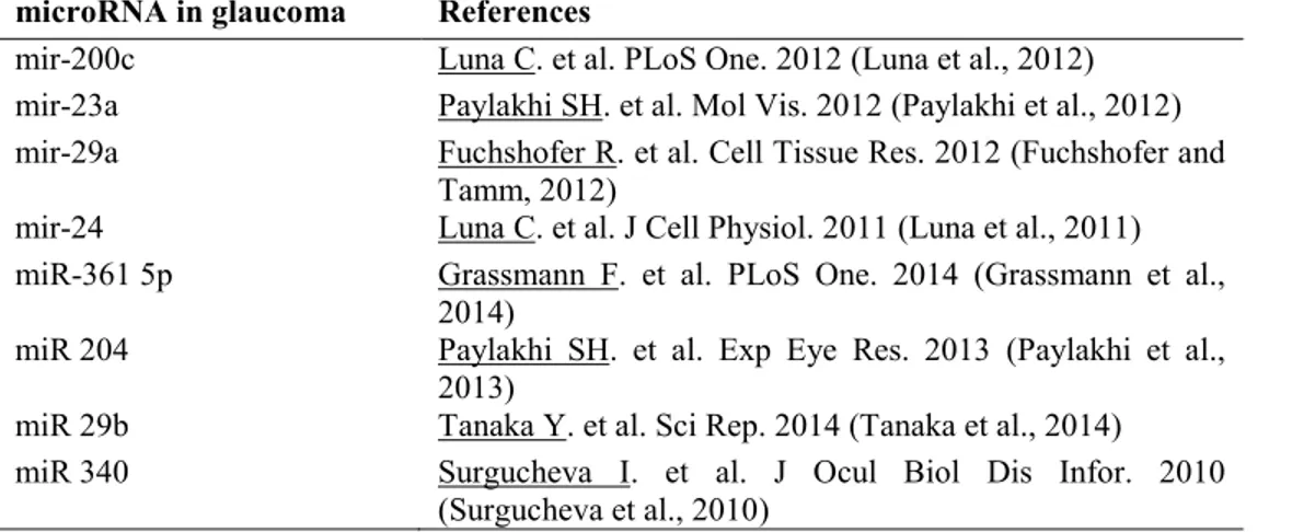
Table 1. Validated microRNA in glaucoma
microRNA in glaucoma References
mir-200c Luna C. et al. PLoS One. 2012 (Luna et al., 2012) mir-23a Paylakhi SH. et al. Mol Vis. 2012 (Paylakhi et al., 2012) mir-29a Fuchshofer R. et al. Cell Tissue Res. 2012 (Fuchshofer and
Tamm, 2012)
mir-24 Luna C. et al. J Cell Physiol. 2011 (Luna et al., 2011) miR-361 5p Grassmann F. et al. PLoS One. 2014 (Grassmann et al.,
2014)
miR 204 Paylakhi SH. et al. Exp Eye Res. 2013 (Paylakhi et al., 2013)
miR 29b Tanaka Y. et al. Sci Rep. 2014 (Tanaka et al., 2014) miR 340 Surgucheva I. et al. J Ocul Biol Dis Infor. 2010