1
UNIVERSITA’ DEGLI STUDI DI CATANIA
FACOLTA’ DI MEDICINA E CHIRURGIASection of Endocrinology, Andrology and Internal Medicine,
and Andrological, Human Reproduction and Biotechnology Sciences, Department of Internal Medicine and Systemic Diseases
Dottorato di ricerca in “Scienze Andrologiche, della Riproduzione Umana e Biotecnologie” XXIII Ciclo
Dott.ssa Glenda Scandura
The impact of APE/Ref-1 on hypoxia regulated genes;
potential applications for cancer
Tesi di Dottorato
Anno Accademico 2010-2011 Correlatore e Tutor: Chiar.mo Prof. R. D’Agata
2
INTRODUCTION Mechanisms of Signal Transduction in Hypoxia
Tumor hypoxia
Hypoxia plays critical roles in the pathobiology of heart disease, cancer, stroke, and chronic lung disease, which are responsible for 60% of deaths in the United States (Semenza GL, 2011).
Oxygen (O2) is carried in the blood by haemoglobin, and the affinity of haemoglobin for O2 is affected by a number of physiological variables. The most important of these are raised partial pressure of carbon dioxide (PCO2), decreased pH (acidity), raised temperature and increased concentration of the organic phosphate, 2,3-diphosphoglycerate (2,3-DPG).
2,3- DPG is a by-product of erythrocyte metabolism, which competes with O2 for binding sites on haemoglobin. All of the aforementioned decrease the affinity of haemoglobin for O2, thereby facilitating the delivery of O2 to the tissues. (Berne RM et al., 1993; Ganong WF, 1999)
Tissue hypoxia occurs when there is an inadequate supply of O2 that compromises normal biological processes in the cell (Hockel M et al. 2001; Harris AL, 2002). This stressful microenvironment is a hallmark of solid tumours, meaning that O2 delivery to the respiring cancer cells is reduced or abolished. Most tumours larger than 1 mm3 in volume contain regions of hypoxia as a result of the disordered blood vessel structure and increased diffusion distances found in tumours. In addition, hypoxia can be caused by low haemoglobin levels in the blood due to tumour-associated and therapy-induced anaemia, which further compromises the O2-carrying capacity of the blood (Vaupel P et al., 2001; Dachs GU and Tozer GM, 2000).
Causative mechanism
Hypoxia can be caused by a number of factors, such as 1) low O2 partial pressure (O2 tension) in arterial blood due to, e.g., pulmonary diseases or high altitude (hypoxemic hypoxia);
3
2) reduced ability of blood to carry O2 as a result of anemia, methemoglobin formation, or carbon monoxide poisoning (anemic hypoxia); 3) reduced tissue perfusion, generalized or local (circulatory or ischemic hypoxia); 4) deterioration of the diffusion geometry, e.g., increased diffusion distances, concurrent versus countercurrent blood flow within microvessels (diffusional hypoxia); or 5) inability of cells to use O2 because of intoxication, as in cyanide poisoning (histotoxic or cytotoxic hypoxia). Because of finely tuned regulatory processes, increases in tissue O2 consumption are generally matched by an increase in blood flow and, therefore, do not usually lead to hypoxia unless the system regulating blood flow fails to meet the increased O2 demand of the tissue in question. Biochemists usually define hypoxia as O2-limited electron transport (Boyer PD et al., 1977). Physiologists and clinicians define hypoxia as a state of reduced O2 availability or decreased O2 partial pressures below critical thresholds, thus restricting or even abolishing the function of organs, tissues, or cells (Honig CR, 1988; Zander R, Vaupel P., 1985; Glossary on respiration and gas exchange, 1973).
Anoxia describes the state where no O2 is detected in the tissue (O2 partial pressure that means 0 mm of mercury [mmHg]). In solid tumors, oxygen delivery to the respiring neoplastic and stromal cells is frequently reduced or even abolished by deteriorating diffusion geometry, severe structural abnormalities of tumor microvessels, and disturbed microcirculation (Vaupel P. et al., 1985). In addition, anemia and the formation of methemoglobin or carboxyhemoglobin reduce the blood‘s capacity to transport O2. As a result, areas with very low (down to zero) oxygen partial pressures exist in solid tumors, occurring either acutely or chronically. These microregions of very low or zero O2 partial pressures are heterogeneously distributed within the tumor mass and may be located adjacent to regions with normal O2 partial pressures. In contrast to normal tissue, neoplastic tissue can no longer fulfill physiologic functions. Thus, tumor hypoxia cannot be defined by functional deficits, although areas of necrosis, which are often found in tumor tissue on microscopic examination, indicate the loss of vital cellular functions. (Hockel M, Vaupel P., 2001)
4
When an unrestricted supply of oxygen is available, for most tumors, the rate of O2 consumption (respiration rate) and adenosine triphosphate (ATP) production is comparable to that found in the corresponding normal tissue, despite the deregulated organization of cells in malignant tumors. To maintain a sufficient energy supply for membrane transport systems and synthesis of chemical compounds, an adequate supply of O2 is required.
In hypoxia, the mitochondrial O2 consumption rate and ATP production are reduced, which hinders inter alia active transport in tumor cells. Specifically, major effects of the reduced production of ATP are 1) collapse of Na+ and K+ gradients, 2) depolarization of membranes, 3) cellular uptake of Cl−, 4) cell swelling, 5) increased cytosolic Ca2+ concentration, and finally, 6) decreased cytosolic pH, resulting in intracellular acidosis in tumor cells. (Hockel M, Vaupel P., 2001)
Otto Warburg was the first to note that solid tumors showed accelerated glycolysis (glucose→2 lactate) and reduced oxygen consumption, prompting him to suggest that respiration in cancer cells was impaired in some manner (O. Warburg, 1930). Once mitochondria were identified as the source of cellular respiration, Warburg's subsequent studies on isolated mouse ascites cells (O. Warburg, 1956) convinced him that cancer cells possessed defective mitochondria and that the accelerated rate of glycolysis was a compensatory response to maintain ATP synthesis (the Warburg effect).
Just as this theory possessed its detractors in Warburg's era (O. Warburg, 1956), there continues to be vigorous debate about the origins of this aerobic glycolytic phenotype in cancer cells. In fact, some cancer cells grown under normoxic conditions show no evidence of a Warburg effect, with an energy metabolism dominated by oxidative phosphorylation. Such cancer cells may demonstrate elevated glycolytic rates but only in response to hypoxia in the microenvironment (i.e., a Pasteur effect) (X.L. Zu and M. Guppy, 2004). Nonetheless, many cancers show a high glycolytic rate/low mitochondria rate even under normoxia (X.L. Zu and M. Guppy, 2004), and it remains unclear whether these differences in metabolic poise (glycolytic versus oxidative) are specific to a cancer type, specific cell lines, or growth context.
5
According to the definition given above, hypoxia is present in tumors when the O2 partial pressure falls below a critical value causing the O2 consumption rate or ATP production rate of a cell or a tissue to decrease progressively. On the basis of experimental results from isolated xenografted human breast cancer tissue (Vaupel P et al., 1987; Kallinowski F et al., 1989), tumor tissue hypoxia with reduced O2 consumption rates is expected when the O2 partial pressure in the blood at the venous end of the capillaries (end-capillary blood) falls below 45–50 mmHg. This critical threshold, however, has been validated only under the following boundary conditions: a tumor blood flow rate of 1 mL/g per minute, a hemoglobin concentration of 140 g/L and an arterial O2 partial pressure of 90–100 mmHg. Reducing the perfusion rate to 0.3 mL/g per minute yields a hypoxic tissue fraction of approximately 20% (Groebe K., 1999). When the hemoglobin concentration falls below 100 g/L or the normal O2 content of arterial blood decreases (hypoxemia), the relative proportion of hypoxic tissue substantially increases in the experimental tumor system described.
On a global tissue level, the critical O2 partial pressure in tumors, below which the detrimental changes associated with reduced O2 consumption have been observed, is 8–10 mmHg. Measurements of the microregional distributions of ATP by quantitative bioluminescence and photon imaging in rodent tumors have shown that the concentration of ATP is relatively constant (1.0–1.8 mM) as long as an adequate supply of oxygen (i.e., comparable to that of normal tissues or organs) can be maintained (Vaupel PW, 1994; Schaefer C et al, 1992).
In FSaII murine fibrosarcomas growing subcutaneously in mice, relatively constant ATP levels were present as long as the median O2 partial pressure was 10 mmHg or higher (Vaupel P et al, 1994). Similar results were obtained in rat tumors when the global ATP content was evaluated with highperformance liquid chromatography (Kruger W, et al, 1991; Vaupel P, 1992). Median O2 partial pressures of approximately 10 mmHg thus appear to represent a critical threshold for energy metabolism in FSaII tumors. At higher median O2 tensions, the levels of ATP, phosphomonoesters and total inorganic phosphate were relatively constant, coinciding with intracellular alkalosis or neutrality and a stable ATP/inorganic phosphate ratio, energy charge, and phosphorylation potential. Median O2 partial pressures of less than 10 mmHg result in
6
intracellular acidosis, ATP depletion, a drop in the energy supply and increasing levels of inorganic phosphate.
Oxidative phosphorylation for ATP formation will continue to a cellular O2 partial pressure of 0.5–10 mmHg (Marshall RS et al., 1986; Starlinger H et Lubbers DW, 1972; Froese G., 1962; Robiolio M et al, 1989). Certainly, the threshold O2 partial pressure below which oxidative phosphorylation ceases is dependent on the cell line investigated and its respiratory capacity, the type of medium and substrate chosen, the temperature and pH of the suspending medium and even the type and accuracy of the setup used to measure O2 consumption rates. Mitochondrial oxidative phosphorylation is limited at O2 partial pressures of less than approximately 0.5 mmHg (Honig CR 1988; Robiolio M et al 1989). Above this threshold, mitochondria should function physiologically. Again, this critical threshold depends on the actual substrate supply, on the pH of the suspending medium, and on the technique used to measure O2. Cytochromes aa3 and c in ascites cells require O2 partial pressures of greater than 0.02–0.07 mmHg (Honig CR 1988; Wilson DF et al. 1988; Chance B et al., 1973) to maintain respiration. At O2 partial pressures above this range, cytochromes are fully oxidized. Spectrophotometric measurements on living and rapidly deep-frozen tissues indicate that the same is true in vivo. From this rather rudimentary summary of critical O2 partial pressures for metabolic hypoxia, there does not appear to be a single hypoxic threshold that is generally applicable. Hypoxic thresholds range from 45–50 mmHg in end-capillary blood to 0.02 mmHg in cytochromes. Furthermore, such data on hypoxic thresholds in a given tissue do not take into consideration the existence of severe heterogeneities even on a microscopic level related to variable O2 demands and O2 supply.
Hypoxia inducible genes
Solid tumours with hypoxic regions have a poorer prognosis than their well-oxygenated counterparts, independent of treatment (Hockel M et al., 1993). This is a consequence of the genetic characteristics of viable hypoxic tumour cells, which enable survival under hypoxic conditions, invariably resulting in a more aggressive tumour phenotype. Biological pathways that are regulated by hypoxia-inducible genes, usually under the control of the transcription factor
7
hypoxia-inducible factor (HIF-1), include apoptosis, cell cycle arrest, angiogenesis, glycolysis and pH regulation, some of which may affect chemotherapy resistance (Harris AL, 2002; Maxwell PH et al., 1997; Carmeliet P et al., 1998)
HIF-1 (Hypoxia-Inducible Factor)
Selection of cells under hypoxia reduced the rate of oxygen consumption and increased the levels of HIF-1α (J. Bourdeau-Heller and T.D. Oberley, 2007).
HIF-1 is a hypoxia-regulated transcription factor, which modulates the expression of numerous hypoxia-inducible genes. It is a heterodimer consisting of a HIF-1α and HIF-1ß subunit, 120 and 80-kDa, respectively (Wang GL et Semenza GL, 1995). Both subunits contain a basic-helixloop- helix motif and a Per arnt Sim (PAS) proteinprotein interaction domain (Wang GL et al, 1995). The transcription factor is activated during dimerisation of HIF-1α and HIF-1 ß. HIF-1b is also known as aryl hydrocarbon receptor nuclear translocator (ARNT) and is constitutively expressed (Jiang BH et al., 1996).
Under normoxic conditions, HIF-1α is essentially undetectable due to its rapid degradation by the ubiquitin-proteasome system (Salceda S et Caro J, 1997), which is mediated by the von Hippel-Lindau (VHL) tumour suppressor protein (Maxwell PH et al. 1999). This explains why all HIF-1 dependent genes are upregulated when VHL is mutated or lost. A family of prolyl hydroxylase enzymes regulates the binding of VHL to HIF-1α by hydroxylating key proline residues on the HIF-1α protein, which is then ubiquitylated and targeted to the proteasome for rapid destruction (Salceda S et Caro J, 1997; Ivan M et al. 2001; Jaakola P et al., 2001). The regulatory activity of HIF-1 is therefore determined by the stability of the HIF-1α protein, (Huang LE et al., 1996) which is stabilised by hypoxia through an O2-dependent degradation domain, rapidly accumulating following exposure to hypoxic conditions (Huang LE et al., 1996; Huang LE et al., 1998). The mechanism by which cells sense O2 tension is currently unknown, although there is some evidence that it is mediated by an iron binding site(s) in the HIF-1α protein. Lu et al. (Lu H et al., 2002) recently provided evidence that lactate and pyruvate also stimulate the accumulation of HIF-1α, independently of hypoxia.
8
Once the complex is formed, it binds to a 256 base-pair enhancer region called the hypoxia-response element (HRE) in a hypoxia-sensitive target gene such as erythropoietin (Epo), thus activating it (Semenza GL et al., 1991). The association and dissociation of HIF-1 from the HRE is extremely rapid, with the half-life for both processes being less than one minute (Wang GL and Semenza G, 1993). As well as Epo, HIF-1 also binds to HREs in genes such as vascular endothelial growth factor (VEGF) and glucose transporter-1 (GLUT-1) leading to angiogenesis and glycolysis (Shweiki D et al., 1992; Levy AP et al., 1996; Bashan N et al. 1992), and plays a role in p53 accumulation (Graeber TG et al., 1994), Ras pathway stimulation (Mazure NM et al., 1997), nitric oxide synthase (NOS) expression (Melillo G et al., 1997) and multi-drug resistance (MDR) gene expression (Comerford KM et al., 1992).
Hypoxic responses are also mediated by HIF-2, a heterodimer composed of HIF-1β and 2α (a paralogue of 1α that is also regulated by oxygen-dependent hydroxylation). HIF-1α is present in all nucleated cells of all metazoan species, whereas HIF-2α expression is restricted to certain cell types within vertebrate species and plays an important role in both erythropoiesis and vascularization. (Patel SA and Simon MC, 2008)
9
Fig1:At normoxia with ample oxygen available these enzymes directly modify the HIF-alpha proteins and keep them inactive. One group of these oxygen sensing enzymes, the prolyl hydroxylases (PHDs), modify distinct proline residues in the HIF proteins at normoxia resulting in the recruitment of the Von Hippel Lindau protein (pVHL), polyubuiquitylation and rapid proteosomal degradation of the HIF-alpha proteins. A second enzyme, an asparaginyl hydroxylase called FIH-1 that was first characterised by our laboratory, also modifies the HIF proteins at normoxia. This modification represses their transcriptional activity by preventing the interaction with transcriptional coactivators such as CBP/p300. When oxygen is limiting both prolyl and asparaginyl hydroxylases are unable to modify the HIFs, resulting in stable, transcriptionally active HIFs activating their target genes in response to hypoxia
10 Pancreatic cancer
Pancreatic cancer remains one of the most lethal of all solid tumours of the gastrointestinal tract. It is characterized by late diagnosis, aggressive local invasion, early metastasis and resistance to chemoradiotherapy (Duffy JP et al., 2003). Pancreatic cancers account for only 2% of all newly diagnosed cancers in the USA each year, but 5% of all cancer deaths (Miller BA et al., 1996). Fewer than 20% of all pancreatic cancers are amenable to surgical resection at presentation and even after surgery with curative intent the 5-year survival rate is poor at 15% (Knaebel H et al., 2005) In addition to being nearly uniformly fatal, pancreatic cancer significantly reduces quality of life of many terminal patients because of symptoms such as pain, fatigue, jaundice, malnutrition, haemorrhage and gastric outlet obstruction. (McKenna S and Eatock M, 2003; Cascinu S et al., 1999)
The pancreas is a compound gland that consists of two functionally and morphologically distinct cell populations derived from the endoderm. The exocrine pancreas consists of enzyme secreting acinar cells arranged into clusters at the end of the ducts. Mature duct cells actively secrete bicarbonate and mucins, as well as having a more mundane plumbing function of draining acinar digestive enzymes towards the duodenum (Slack, 1995).
The endocrine compartment of the pancreas comprises five different hormone-secreting cell types: the glucagon-secreting α-cell, insulin-secreting β-cell, somatostatin-releasing δ-cell, ghrelin-producing ε-cell, and finally the pancreatic polypeptide-secreting PP-cells. All of these hormones are involved in regulating nutrient metabolism and glucose homeostasis.
The endocrine cells aggregate to form the islets of Langerhans, which are intermingled with blood vessels, neurons, and a mesodermally-derived stromal component. The intimate interaction between endocrine and vascular cells regulates hormone release, establishing a fine-tuned glucose homeostasis in the body (Slack, 1995; Prado et al., 2004).
Over the past few years, our knowledge of the pathogenesis of pancreatic cancer has advanced significantly because of a rapid increase in our understanding of the molecular biology of it. Like many other malignant diseases, pancreatic cancer results from the accumulation of
11
inherent and acquired genetic and epigenetic alterations. The multigenic nature of most pancreatic cancers is reflected by abnormalities of three broad classifications of genes: oncogenes, tumor suppressor genes and genomic maintenance genes (Sohn TA and Yeo CJ, 2000; Sakorafas GH and Tsiotos GG, 2001). Accumulated alterations of such genes are believed to occur over a predictable time course. Based on the understanding of the histological and molecular genetic profiles of pancreatic cancer, investigators have developed a progression model that describes pancreatic ductal carcinogenesis: the pancreatic ductal epithelium progresses from normal epithelium to increasing grades of pancreatic intraepithelial neoplasia to invasive cancer (Hruban RH, 2000) The majority of pancreatic cancers occur sporadically and have been fairly well characterized at the genetic level. Pancreatic cancer pathogenesis is apparently involved in the activation of several oncogenes and/or inactivation of various tumor suppressor genes. (Sohn TA and Yeo CJ, 2000; Kern SE, 2000)
Since the identification of the first notable genetic alteration of the K-ras oncogene, there has been an explosion in our understanding of pancreatic cancer genetics (Sohn TA and Yeo CJ, 2000; Kern SE, 2000). For examples, more than 85% of pancreatic cancers have an activating point mutation in the K-ras gene at a very early stage of development (Almoguera C et al., 1988). Also, the tumor suppressor gene p16 is inactivated in about 95% of pancreatic cancers, and inactivation typically occurs late in pancreatic carcinogenesis. TP53, a well-characterized tumor suppressor gene located on chromosome 17p, is the second most frequently inactivated gene (Xie K et al., 2006)
Pancreatic cancers are hypoxic tumors that respond poorly to existing chemotherapeutic agents and radiation (Duffy JP et al., 2003). Pancreatic cancer cells overexpress many families of growth factors and their receptors, including epidermal growth factor (EGF), vascular endothelial growth factor (VEGF), fibroblast growth factor (FGF) and its receptor and platelet-derived growth factor (PDGF), as well as many cytokines, such as transforming growth factor (TGF)-b, tumor necrosis factor-a, interleukin (IL)-1, IL-6 and IL-8, which enhances mitogenesis. Pancreatic cancer also exhibits loss of responsiveness to various growth-inhibitory signals, such as members of the TGF-b family.
12
NFkB and HIF-1α have been identified as leading drivers of cell growth in pancreatic cancer; both are under APE1/Ref-1 redox signaling control which is the focus of our studies (Tell G et al., 2009; Luo M et al., 2008; Bapat A et al., 2009). It was previously showed that APE1/Ref-1 is upregulated in human pancreatic cancer cells and modulation of its redox activity blocks the proliferation and migration of pancreatic cancer cells (Zou GM et Maitra A., 2008; Jiang Y et al., 2010) and pancreatic cancer-associated endothelial cells (PCEC) in vitro (Zou GM et al.,2009)
The cell line used in this project was PaCa2, adenocarcinoma cell lines.
Prostate cancer
Prostate cancer (PCa) is the third most common tumor type in men. The appearance of this neoplasia is linked to age. In the European Union, PCa is directly responsible for the death of 3% of men and 10% of cancer deaths.
The incidence of PCa has risen in recent years, primarily due to the significant increase in life expectancy, and secondly because of the introduction of the determination of serum PSA levels in PCa screening, raising the diagnostic in the preclinical phase. In Spain, the epidemiological situation of PCa is not significantly different from the rest of Europe. Every year, some 13,300 new cases are diagnosed (13.6% of tumors among Spanish men), with survival at 5 years around 65%, with an average age of death of 75 years. (López-Abente G et al., 2004)
Histologically, PCa is constituted of a heterogeneous mixture of cells, mainly epithelial and stromal. (Nelson WG et al., 2003) This process begins with a dysplasia that starts as a proliferative inflammatory atrophy (PIA), progressing to prostatic intraepithelial neoplasia (PIN), and in some cases it leads to a carcinoma. There is evidence to suggest that one of the triggers of tumorogenesis could be a prostate inflammation due to infectious agents or ingestion of carcinogens. In parallel, some cells accumulate genetic alterations that, along with the androgenic signaling, stimulate the growth and proliferation of the tumor. (Taichman RS et al., 2007)
13
up being lethal, and others that are relatively indolent, (Taichman RS et al., 2007) which, to start with raise the problem of how to distinguish some tumors from others and the manner of best clinical approach in each case. Currently, serum PSA levels provide highly organ-specif information, but little disease-specific. Thus, both in benign prostatic hyperplasia and prostatitis, serum increases of this biomarker are produced, but many patients with localized PCa also have PSA values that overlap with those of healthy subjects, resulting in a gray area of difficult interpretation of the range between 4 and 10ng/ml. (Balk SP et al., 2003) Moreover, numerous studies suggest that PCa is overdiagnosed in 30-50% of the cases, that is, not all the patients with an elevated PSA have a prostate tumor. After the diagnosis, the main prognostic factor is the Gleason score, which consists of assigning a grade of 1-5 in descending differentiation to each of the two main foci of the tumor. The sum of both values is the score. Although this parameter is the gold standard in the clinical management of PCa, it presents certain problems: first, the determination is made on tissue obtained from a prostate biopsy, a surgical procedure that has certain comorbidity, particularly significant in elderly patients; besides, this score suffers from interpretive variation. (Evans AJ et al., 2008)
In the prognosis of the disease, the lack of a reliable method capable of determining the time at which the prostate tumor will become hormone-resistant is problematic, because from here on, the patient‘s prognosis worsens and bone metastases, for which currently only palliative treatment is available, often occur. (Msaouel P et al., 2008)
For all this, it is very important to identify new biomarkers that represent useful tools in the diagnosis and clinical management of PCa. These markers should be determinable by objective, quantitative and mechanism-specific techniques, and as far as possible, they should be accessible by noninvasive methods.
PC-3 and DU145 human prostate cancer cell lines are the "classical" cell lines of prostatic cancer. (Abate-Shen C. and Shen M.M., 2000) PC3 cells have high metastatic potential compared to DU145 cells which have a moderate metastatic potential. (Abate-Shen, C. and Shen, M.M., 2002) PC3 cell lines were originally derived from advanced androgen independent bone metastasis metastasized prostate cancer. PC3 have low testosterone-5-alpha reductase activity and express PSA.
14 APE1/Ref1
Apurinic/apyrimidinic endonuclease/redox effector factor (APE1/Ref-1) is a protein with multifunctional roles in cells impacting on a wide variety of important cellular functions. It acts on apurinic/apyrimidinic (AP) sites in DNA as a major member of the base excision repair (BER) pathway, is involved in oxidative DNA damage repair and stimulates the DNA binding activity of AP-1 (Fos, Jun) proteins, as well as nuclear factor-κB (NF-κB), polyoma virus enhancer-binding protein 2 (PEBP2), early growth response-1 (Egr-1), Myb, members of the ATF/CREB family, HIF-1α (hypoxia inducible factor-1α), HIF 2α (HIF-like factor), Pax-5, and Pax-8 (Y. Akamatsu et al., 1997; M. Ema et al., 1999; L.E. Huang et al., 1996; R.P. Huang, E.D. Adamson et al., 1993; D. Lando, et al., 2000; S. Xanthoudakis et T. Curran, 1992; S. Xanthoudakis et al. 1992; K.S. Yao et al., 1994).
The DNA binding activity of these latter proteins is sensitive to reduction-oxidation (redox).
APE1/Ref-1, which is the major AP-1 redox activity in cells, represents a novel redox component of signal transduction processes that regulate eukaryotic gene expression. Recent developments also have implicated APE1/Ref-1 as a major controlling factor for p53 activity through redox dependent and independent mechanisms, (C. Gaiddon et al., 1999; L. Jayaraman et al., 1997). APE1/Ref-1 has been shown to be closely linked to apoptosis (KA Robertson et al., 1997) and altered levels or cellular location of APE1/Ref-1 have been found in some cancers, including ovarian, cervical, prostate and germ cell tumors (MR Kelley et al., 2000; MR Kelley et al., 1998; DH Moore et al., 2000; Y. Xu et al., 1997). Therefore, APE1/Ref-1 appears to form a unique link between the DNA BER pathway, cancer, transcription factor regulation, oxidative signaling, and cell-cycle control. (Fig.2; Evans AR, 2000)
APE1/Ref-1 genes, proteins, and structure
AP endonucleases are classified into two families according to their homology to E. coli endonucleases: exonuclease III (xth) and endonuclease IV (nfo). The first family of AP endonucleases derives from organisms across several phyla including, exonuclease III (E. coli),
15
Exo A (Steptococcus pneumoniae), Rrp 1 (Drosophila melanogaster), Arp (Arabidopsis
thaliana), Apn2 (S. cerevisiae), APEX (mouse), BAP1 (bovine), rAPE (rat), chAPE1 (hamster),
and Ape1/Ref-1 (humans; previously referred to as HAP1 and APEX1). These enzymes exhibit strong AP hydrolytic activity and 3′-diesterase activity with APE1/Ref-1 having the highest 5′-endonuclease rate, but lowest 3′-diesterase activity. Most of the proteins do not exhibit 3′–5′-exonuclease activity, the exceptions, to date, are 3′–5′-exonuclease III and APEX (Demple B. et al.,1991; Seki S. et al., 1991) Typically, the exonuclease III family of endonucleases accounts for approximately 95% of the repair activity in the organism. However, Apn1 comprises approximately 90% of the repair activity in S. cerevisiae, and it, along with endonuclease IV (E.
coli), Spapn1 (S. pombe), CeApn1 (C. elegans) are major members of the second family of
endonucleases, the endonuclease IV family.
The DNA repair activity of endonucleases resides in the C-terminal region, and between APE1/Ref-1 and the prokaryotic homologues, 25–40% sequence identity is apparent (Barzilay G. and I D Hickson ,1995). In contrast, there is a high degree of homology among mammalian AP endonucleases, suggesting these proteins are very closely related to one another. For example, the homology of deduced amino acid sequences between bovine and human is 93% (Robson CN and ID Hickson, 1991) mouse and human is 94% (Seki S. et al., 1991); rat and human is 85% (Wilson T.M. et al., 1994) and hamster and human 92% (Purohit S. and Arenaz P., 1999). In all cases, the C-terminus functions in repair activities, whereas the role of the N-terminal region is less well understood. In the case of Rrp 1, the N-terminal domain may be involved in Mg2+-, ATP-dependent renaturation of single-stranded DNA (Barzilay G., Hickson ID, 1995) whereas Arp and APE1/Ref-1, the N-terminal domain is essential for redox control of other proteins (Babiychuk E. et al, 1994; Xanthoudakis S. et al, 1994). Presumably, the N-terminus in all the mammalian homologs exhibits redox activity since they share a great deal of homology to APE1/Ref-1. Furthermore, mouse, rat, and human all contain a cysteine at position 65 (Wilson T.M. et al., 1994), a residue thought to be important for redox activity (Walker L.J. et al, 1993).
The gene encoding the APE1/Ref-1 protein maps to chromosome 14 bands q11.2–12 in the human genome (Harrison L. et al., 1992; Robson C.N. et al, 1992). The APE1/Ref-1 protein is modest in size; it is 318 amino acids in length and ~37 kDa. It contains two distinct domains. The
16
N-terminal domain contains the nuclear localization sequence (residues 1–36) (Robbins J et al, 1991); and is essential for redox activity while the endonuclease activity resides in the C-terminal region (Xanthoudakis S. et al, 1994). It was believed previously that the domains could be separated without disrupting their individual activities, however, recent studies using deletional analysis, demonstrate some overlap in the functional domains. Endonuclease activity requires residues between 61 and 80 and all the C-terminus (Izumi T. and Mitra S. ,1998) and redox activity requires residues 43–93 (Jayaraman L. et al, 1997)
APE1/Ref-1 is a globular α/β protein consisting of two domains each of which is made up of a six-stranded β-sheets surrounded by α helices. The protein forms a four-layered α/β sandwich that resembles the folds of exonuclease III and DNase I (Gorman M.A. et al.,1997; Xanthoudakis S. et al, 1994). Structural analysis reveals a single active site in APE1/Ref-1 for DNA repair activity (Gorman M.A. et al., 1997). The important residues for substrate recognition and catalysis have been determined by site-directed mutagenic studies.
17
Fig.2. Multifunctional activities of the human AP endonuclease. Ape1/Ref-1 is a multifunctional protein involved in BER, transcription factor regulation, and oxidative signaling. In DNA BER, it functions as an AP endonuclease. It is also involved in the activation of transcription factors such as p53, AP-1, 1α, and HIF-2α (HLF). This activation can be through redox-dependent and/or redox-independent mechanisms.
DNA Repair Function of APE1/Ref-1
Multiple oxidative DNA damage such as strand breaks, base loss, and base modifications are caused by reactive oxygen species (ROS) that are generated endogenously or due to environmental stress (Ames BN et al., 1993; Breen AP et Murphy JA, 1995). Nearly all oxidized forms of DNA bases (as well as methylated or inappropriate bases) are repaired via the BER
18
pathway which is initiated with excision of the damaged base by a DNA glycosylase to generate AP site (Hazra TK et al., 1993; Krokan HE et al., 1997; Mitra S. et al., 2002). APE1/Ref-1, the second enzyme in the BER pathway, then hydrolyzes the phosphodiester backbone immediately 5' to an AP site to produce 3'OH group and 5' deoxyribose-5-phosphate (Demple B et Harrison L, 1994; Doetsch PW et Cunningham RP, 1990). Following removal of this blocking group via dRP lyase activity of DNA polymerase ß repair DNA synthesis, followed by DNA ligase action restores genome integrity (Sobol RW and Wilson SH, 2001). Oxidized base-specific DNA glycosylases have intrinsic AP lyase activity and cleaves the DNA strand 3' to the AP site (Hazra TK et al, 2003; Krokan HE et al., 1997). The resulting 3' blocking group is removed by APE1/Ref-1 (or in some cases polynucleotide kinases) in the next step of repair (Chen DS et al., 1991; Whitehouse CJ et al., 2001). APE1/Ref-1‘s 3' phophodiesterase activity is also involved in repairing DNA single-strand breaks with 3' blocking group directly generated by ROS (Izumi T et al., 2000). Unrepaired AP sites also lead to DNA strand breaks, apoptosis, and increases cytotoxicity (Loeb LA and Preston BD, 1986). Thus, the DNA repair function of APE1/Ref-1 protects the cell from both endogenous and exogenous DNA damage. All APEs have dual activities as an endonuclease and a 3'phosphodiesterase (Demple B and Harrison L, 1994; Doetsch PW and Cunningham RP, 1990). However, mammalian APE1‘s endonuclease activity is quite strong relative to its 3'exonuclease/phosphodiesterase activity (Chen DS et al., 1991; Demple B and Harrison L, 1994; Wiederhold L et al., 2004).
APE1/Ref-1 also coordinates BER as an assembly factor by interacting with downstream BER protein such as DNA polymerase ß, X-ray cross-complementing-1 (XRCC1), proliferating nuclear antigen (PCNA), and flap endonucelase (FEN1) (Dianova II et al., 2001; Fan J and Wilson DM, 2005; Izumi T et al., 2003). A recent study shows that Bcl2, an anti-apoptotic protein, directly interacts with APE1/Ref-1 and inhibits AP site repair by downregulating AP-endonuclease activity of APE1/Ref-1 (Zhao J et al., 2008). Exposure of lung cancer cells to the DNA damaging agent promotes Bcl2 accumulation and association with APE1/Ref-1 in the nucleus (Zhao J et al., 2008).
19 Regulation of APE1/Ref-1 Expression
Although APE1/Ref-1 is ubiquitously expressed in cells and tissues, its expression and subcellular localization level appear to be cell-type specific (Kakolyris S, et al., 1998; Tell G et al., 2005). APE1/Ref-1 is regulated at both transcriptional and post-transcriptional levels. Expression of APE1/Ref-1 in mouse NIH3T3 cells was found to be cell cycle dependent with the highest level of APE1/Ref-1 in early or middle S-phase, pointing to a particular function of APE1/Ref-1 in this phase of cell cycle (Fung H et al., 2001). The effects of ROS on APE1/Ref-1 induction have been extensively studied. It has been shown that hydrogen peroxide (H2O2) and hypochlorous acid (HOCl) acts as inducers of the APE1/Ref-1 gene (Grosch S et al., 1998; Ramana CV et al., 1998). Subsequently, several in vivo and in vitro studies confirmed APE1 gene activation by oxidative stress (Grosch S and Kaina B, 1999; Pines A et al., 2005). This observation is of particular interest, because H2O2 and HOCl are endogenously formed during inflammatory response of macrophages and lymphocytes. Endogenous ROS may elevate the level of DNA damage which then signals an increase in APE1/Ref-1 level, thus enhancing the BER capacity. Indeed, induction of APE1/Ref-1 was found to be accompanied by an adaptive response of cells to the cytotoxic and clastogenic activity of oxidative agents, indicating its physiological relevance of the phenomenon (Fritz G et al., 2003; Grosch S et al., 1998; , Ramana CV et al.,1998 ).
Induction of oxidative stress was shown to be involved in the enhanced nuclear translocation of thioredoxin (TRX) and APE1/Ref-1 and augmentation of the APE1/NF-κB complex formation in the parenchyma cells of injured lung (Gorbunov NV et al., 2007). In many cell types, ROS-mediated activation of APE1/Ref-1 involves two steps.
In the first step, APE1/Ref-1 translocates from the cytoplasm to the nucleus. In B-lymphocytes and thyroid cells, such translocation is fairly rapid, within an hour, whereas in HeLa and other cells the process takes many hours (Ramana CV et al., 1998; Tell G et al., 2000; Tell G et al., 2009). The second step involves de novo protein synthesis via transcriptional activation of the APE1/Ref-1 promoter, because various agents that block transcription or protein synthesis, also abolish induction of APE1/Ref-1 (Ramana CV et al., 1998). Additionally, APE1/Ref-1 induction is associated with an increase in AP-endonuclease activity and cells resistance to cytotoxic effect
20
of H2O2, methyl methane sulphonate (MMS), bleomycin, and γ-radiation (Fritz G et al., 2003; Grosch S et al., 1998; Ramana CV et al., 1998).
Transiently overexpressed APE1/Ref-1 protects cells against genotoxicity and cell killing provoked by ROS (Fritz G et al., 2003). However, whether protection against ROS-induced cell killing by APE1/Ref-1 is due to of its repair or transcriptional regulatory functions or both is still unknown.
Other external stimuli such as hormones and cytokines modulate APE1/Ref-1 expression. Thyrotropin (TSH) induces APE1/Ref-1 expression in thyroid cells (Asai T et al., 1996; Tell G et al., 2001; Tell G et al., 2000). Similarly, human chorionic gonadotropin has been demonstrated to enhance APE1/Ref-1 mRNA synthesis in murine Leydig cells (Suzuki S et al., 1998). IL-2-dependent APE1/Ref-1 upregulation has also been demonstrated in a murine Pro-B cell line (Yan M et al., 2000). Interestingly, Helicobacter pylori induced IL-8 activation in gastric epithelial cells was found to be dependent on APE1/Ref-1 (O‘Hara AM et al., 2006). Another recent study demonstrated that ATP-mediated purinergic receptor activation upregulates APE1 expression in human tumor thyroid cell line (Pines A et al., 2005).
Another factor that modulates APE1/Ref-1 expression is hypoxia, which mimics oxygen tension that is encountered by cells in tissues in vivo. Hypoxia induces APE1/Ref- mRNA and protein levels in HT29 cells (Yao KS et al., 2004). Elevation of APE1/Ref-1 steady-state mRNA levels is an early event following hypoxia, and persists after restoration of cells to normoxia (Yao KS et al., 2004). Nuclear run-on analysis demonstrated that induction of transcription is responsible for elevation of APE1/Ref-1 mRNA (Yao KS et al., 2004). Changes in APE1/Ref-1 expression in response to hypoxia was correlated with its requirement for enhanced AP-1 binding following hypoxia via redox activation (Yao KS et al., 2004). However, another possible role for prolonged expression of APE1/Ref-1 following hypoxia relates to DNA repair function that remains to be elucidated. Although it is not known whether hypoxia-inducible factors (HIFs) bind specifically to the APE1/Ref-1 promoter or enhancer, APE1/Ref-1 regulates HIF-1α functions in vivo (Ema M et al., 1999, Huang LE et al., 1996). APE1/Ref-1 up-regulation significantly potentiates hypoxia-induced expression of a reporter construct containing the HIF-1α-binding site
21
(Ema M et al., 1999). Moreover, Ema et al. (Ema M et al., 1999) and Carrero et al. (Carrero P et al., 2000) showed that APE1/Ref-1 is critical to linking coactivator proteins, CBP/p300 and SRC-1 to HIF-SRC-1α. In contrast, Hall et al. showed that hypoxia downregulates APE1/Ref-1 protein level in both calf pulmonary artery endothelial (CPAEC) and human umbilical vein endothelial (HUVEC) cells (Hall JL et al., 2001). Such hypoxia- induced decrease of APE1/Ref-1 was associated with significant induction of apoptosis in CPAEC and HUVEC cells (Hall JL et al., 2001). Thus, APE1/Ref-1 downregulation may be permissive in promoting apoptosis in endothelial cells in response to hypoxia. Indeed, APE1/Ref-1 overexpression was shown to protect CPAEC cells from hypoxia-induced apoptosis (Hall JL et al., 2001).
Recently, it has been shown that soy isoflavones downregulate expression of APE1/Ref-1 in PC3 prostate cancer cells (Raffoul JJ et al., 2007). Moreover, pretreatment with soy isoflavones inhibits radiation-induced APE1/Ref-1 expression and activation of NF-κB . Although the mechanism by which soy isoflavones down-regulates APE1/Ref-1 expression is not known, downregulation of APE1/Ref-1 and inhibition of NF-κB activation by soy isoflavones was shown to inhibit tumor growth in vivo (Raffoul JJ et al., 2007).
Regulation of transcription factors
In 1992, Xanthoudakis and Curran (Xanthoudakis S. and Curran T., 1992) identified APE1/Ref-1 as an important redox activator of the DNA binding of transcription factors Fos and Jun, subunits of activator protein 1 (AP-1) (Xanthoudakis S. and Curran T., 1992). It was discovered, through mutational analysis, that the conserved cysteine residue located in the DNA binding domain of Fos and Jun was essential for the APE1/Ref-1-mediated activation of AP-1 (Ait–Si–Ali S et al., 1998; Xanthoudakis S. and Curran T., 1992). APE1/Ref-1 has been shown to activate numerous transcription factors and facilitate their DNA binding via the reduction of a cysteine residue.
Redox regulation of cellular functions occurs as a consequence of the so-called ―redox-cellular status,‖ which is the result of a balance between the activity of antioxidant enzymatic cell systems (such as GSH/GSSG, superoxide dismutase, catalase, peroxidases, glutathione
22
peroxidases, etc.) and the amount of reactive oxygen species (ROS) such as superoxide anion (O2•), hydrogen peroxide (H2O2), and hydroxyl radical (•OH) (Tell et al., 2005)
These last molecules can be produced in several ways: as byproducts of respiration, thus being associated with cell proliferation rate; by external noxious agents, such as ionizing radiation (Wilson DF et al., 1988); during pathological states in activated neutrophils (Nakamura H et al., 1997) and as ―second messengers‖ produced by intracellular enzymatic systems, such as NADPH oxidase regulated by the ubiquitous small GTPase Rac1 (Deshpande SS et al., 2000; Droge W, 2002; Gorlach A et al.,2000). It therefore represents a useful tuning device for intracellular signal transduction, as is the case in cascades induced by cytokines, such as tumor necrosis factor α or interleukin (IL) - γ (Nakamura H et al., 1997).
This redox regulation ultimately affects gene expression. Recently, a great body of experimental evidence suggested that these outcomes are achieved through modulation of TFs activity. Up to now, several TFs containing specific Cys residues have been demonstrated to be the target of redox regulation. APE1/Ref-1 has been identified as a protein capable of nuclear redox activity, inducing the DNA-binding activity of several TFs, such as AP-1 (Xanthoudakis S. et al, 1992), NF-κB (Nishi T et al., 2002), Myb (Xanthoudakis S. and Curran T., 1992), PEBP-2 (Y. Akamatsu et al., 1997), HIF-2α (M. Ema et al., 1999), NF-Y (Nakshatri H et al., 1996), Egr-1 (Huang R.P. and Adamson E.D., 1993), HIF-1α (Huang LE et al., 1996), ATF/CREB family (Xanthoudakis S. and Curran T., 1992), p53 (C. Gaiddon et al., 1999), Pax proteins (Cao X et al., 2002; Tell G et al., 1998; Tell G et al., 2000). It accomplishes this through the control of the redox state of Cys residues located in the DNA-binding domains or within regulatory regions, such as the transactivation domain of the thyroid-specific transcription factor 1 (i.e., TTF-1) of the TFs themselves (Tell G et al., 2002). In order to properly bind specific DNA target sequences, these TFs require that critical Cys residues are in the reduced state. Therefore, by maintaining these cysteines in the reduced state, APE/Ref-1 provides a redox-dependent mechanism for regulation of target gene expression. APE/Ref-1 contains two cysteine residues located within the active domain (Cys65 and Cys93), and previous studies show that Cys65 should be the redox-active site of the protein by using recombinant protein (Walker LJ et al., 1993). In agreement with the molecular model describing redox regulation exerted by APE1/Ref-1, Cys65 should interact with the sensitive cysteine residues within the DNA-binding domains of TFs. (Tell, 2005)
23
HIF-1α and HIF-2α (or HLF) are transcription factors induced by hypoxia. Upon induction, these proteins form a heterodimer with an Ah receptor nuclear translocator (Arnt), translocate to the nucleus, and transcriptionally activate a variety of genes such as erythropoietin, vascular endothelial growth factor, glycolytic enzymes, and inducible nitric oxide synthase, among others. (Halterman M.W. and Federoff. H.J., 1999; G.L.Semenza, 1995)
Although there is 48% homology between HIF-2α and HIF-1α, APE1/Ref-1 exerts differential redox control of these proteins. DNA binding is redox dependent for HIF-2α, but not for HIF-1α (D. Lando et al., 2000). Furthermore, adding to the complexity of redox regulation by APE1/Ref-1 are the more recent data showing that APE1/Ref-1 is important for the transactivation activities for both HIF-1α and HIF-2α (Carrero P et al., 2000; Lando D. et al., 2000). APE1/Ref-1 reduces the N-terminus of HIF-2α and thereby, stimulates DNA binding. Conversely, the N-terminal region of HIF-1α binds DNA without APE1/Ref-1. This discrepancy is apparently owing to a difference in one amino acid. HIF-1α contains a serine residue, whereas HIF-2α has a cysteine. Mammalian two-hybrid assays indicate that APE1/Ref-1 interacts with HIF-2α N-terminal region, but not the HIF-1α N-terminus. Interestingly, mutating the serine residue to a cysteine converts HIF-1α from a redox-resistant DNA binding transcription factor to redox-sensitive one (D. Lando et al., 2000). Co-transfection experiments in HeLa cells using antisense APE1/Ref-1
and a luciferase reporter gene construct show that antisense APE1/Ref-1 RNA reduces the ability of HIF-2α to function as a transcription factor; a decrease from 25- to 8-fold in luciferase expression is reported. In these same experiments, the transcription activity of HIF-1α is also attenuated; suggesting that transcriptional activation by HIF-1α, is under redox control possibly through interaction of APE1/Ref-1 in the C-terminus (D. Lando et al., 2000)
The C-terminal domain of HIF-1α and HIF-2α contains the transactivation domain that interacts with the co activators CREB binding protein (CBP) and SRC-1, a family member of 160 kDa co-activator proteins, to augment HIF-1α mediated transcriptional regulation under hypoxic conditions (Carrero P et al., 2000; M. Ema et al., 1999).In co-transfection experiments, Ema et al. (M. Ema et al., 1999) show that the C-terminal domain of HIF-1α and HIF-2α required CBP for transcriptional activation of reporter gene constructs. The interaction between CBP and the C-terminus likely occurs through a redox mechanism because a C-terminal cysteine to serine mutation abolishes the interaction between these proteins (M. Ema et al., 1999). Moreover,
24
protein/protein interactions between the C-terminal domain and CBP are enhanced in yeast two-hybrid assay by TRX or APE1/Ref-1 (M. Ema et al., 1999) and over-expression of APE1/Ref-1
or TRX enhances the transcriptional activation of the C-terminus of HIF-1α and HIF-2α. These data suggest that a cysteine residue in HIF-1α C-terminus is reduced by APE1/Ref-1 or TRX, which enhances its interaction with CPB and, consequently, transcriptional activity. Overall, it appears that APE1/Ref-1 promotes transcriptional activation by two independent redox mechanisms: by stimulating DNA binding directly and indirectly by enhancing the transactivation activities for HIF-2α and HIF-1α. APE1/Ref-1 stimulates the transcriptional activity of numerous transcription factors that have physiological functions as diverse as cell cycle control, apoptosis, angiogenesis, cellular growth, cellular differentiation, neuronal excitation, hematopoiesis and development.
Consequently, APE1/Ref-1 is a pivotal signaling factor involved in coordinating the cellular adaptation to a wide array of environmental stimuli.
APE1/Ref-1 and cancer
Whether relationships exist between APE1/Ref-1 levels and cancerous tissue is of enormous importance, not only for understanding the role and mechanism, APE1/Ref-1 may play in the initiation and development of various cancers, but also for developing diagnostic markers for early detection of cancers.
Several investigators have initiated studies to evaluate the role APE1/Ref-1 plays in cancer and results, thus far, are promising. In breast cancer tissue, there are no differences between abnormal tissue and normal tissue in terms of their ability to repair abasic sites, suggesting that DNA repair by BER is not a pathological factor in breast cancer (O. Rossi et al., 2000). There are, however, some cancers where differential patterns of APE1/Ref-1 expression have emerged. In cervical, prostate, human pancreatic cancer cells (Fishel ML et al., 2011) and epithelial ovarian cancers,
APE1/Ref-1 protein levels are dramatically elevated compared to normal tissues (M.R. Kelley et al., 2000; D.H. Moore et al., 2000; Y. Xu, et al., 1997). Additionally, increased APE1/Ref-1
levels are observed in pediatric rhabdomyosarcomas (B. Thompson et al., 2000), and germ cell tumors (M.R. Kelley et al., 1998). In other types of cancer, APE1/Ref-1 expression levels are
25
unchanged, but the cellular localization differs between normal and cancerous tissue. For example, in normal colonic tissue APE1/Ref-1 is nuclear in the crypts where cells are undifferentiated and cytoplasmic in the differentiated surface epithelium. In cancer cells, the pattern is not nuclear-restricted; nuclear and cytoplasmic localization is common in colorectal adenomas and carcinomas (S. Kakolyris et al., 1997). Additionally, epithelial ovarian cancers display nuclear and cytoplasmic staining with cytoplasmic localization predominating, while normal tissue exhibits nuclear localization exclusively (D.H. Moore et al., 2000). The altered patterns of expression, particularly where APE1/Ref-1 is elevated, need to be further characterized on a mechanistic level to understand the meaning of altered expression patterns and its relationship to cell line studies.
Through both the redox and DNA repair functions APE1/Ref-1 supports cancer cell proliferation, and elevated expression levels have been shown to correlate to poor patient prognosis. (Evans AR et al., 2000; Tell G et al., 2005; Izumi T et al., 2005) APE1/Ref-1 is overexpressed in a number of cancers, where increased levels of DNA repair leads to resistance against DNA damaging agents, and increased redox activity is expected to enhance replication through redox cycling of transcription factors. Therefore APE1/Ref-1 represents an interesting therapeutic target in different mechanistic contexts. Inhibitors of the BER function of APE1/Ref-1 can be utilized as a complementary treatment option for those encountering resistance to DNA-damaging agents. Alternatively, inhibition of the redox function of APE1/Ref-1 might interfere with regulation of transcription and alter a number of stress-induced responses of cancer cells. Recent data indicates that blocking the repair function of APE1/Ref-1 leads to cell death, while redox activity inhibition leads to decreased cell growth and cytostatic effects. (Luo M et al., 2008) Additionally, recent data indicates that blocking Ape1 redox function blocks angiogenesis. (Luo M et al., 2000; Zou GM et al., 2008; Zou GM, Maitra A, 2008) Small molecule inhibitors of the redox function can also serve as tools to separate the two functions of APE1/Ref-1 without the lethality of knocking out APE1/Ref-1 completely. (Jiang Y et al., 2009)The design of inhibitors targeting the redox function of APE1/Ref-1 is hindered by a lack of information regarding the redox active site. Mutation analysis has shown that cysteine 65 is necessary for redox activity; however, in every crystal structure C65 is buried, suggesting that a conformational
26
change might be required to present the relevant redox-active structure. (Georgiadis MM et al., 2008) Furthermore, there is only one known compound in the literature that has been shown to inhibit the redox function of APE1/Ref-1 (Evans AR et al., 2000) To provide structural insight into potential inhibitor specificity for the redox active site, a series of benzoquinones and naphthoquinones has been synthesized based on the structure of (E)-3-(5,6-dimethoxy-3-methyl-1,4-dioxocyclohexa-2,5-dienyl)-2-nonylpropenoic acid (E3330), a known inhibitor of the redox function of APE1/Ref-1. (Evans AR et al., 2000)
Analogs with improved physicochemical and binding profiles also have the potential to provide crystallographic data when complexed with the protein to elucidate the structure of the redox active site.
27 RNA interference
Epigenetic regulation of gene expression is a heritable change in gene expression that cannot be explained by changes in gene sequence. It can result in repression or activation of gene, referred to as gene silencing or gene activation, respectively (Vaucheret et al. 2001). During the 1990s, a number of gene-silencing phenomena that occurred at the posttranscriptional level were discovered in plants, fungi, animals and ciliates, introducing the concept of post-transcriptional gene silencing (PTGS) or RNA silencing. (Baulcombe 2000; Matzke et al. 2001) PTGS results in the specific degradation of a population of homologous RNAs. PTGS was first observed after introduction of an extra copy of an endogenous gene (or of the corresponding cDNA under the control of an exogenous promoter) into plants (Napoli et al. 1990; Smith et al. 1990; Van der Krol et al. 1990). Because RNAs encoded by both transgenes and homologous endogenous gene(s) were degraded, the phenomenon was originally called co-suppression. A similar phenomenon in the fungus Neurospora crassa was named quelling (Romano and Macino 1992; Cogoni et al. 1996). Fire et al. (1998) identified a related mechanism, RNA interference (RNAi) in animals. RNAi results in a specific degradation of endogenous RNA in the presence of homologous dsRNA either locally injected or transcribed from an inverted-repeat transgene (Tavernarakis et al. 2000; Vaucheret et al. 2001). They applied single-stranded antisense RNA and double- stranded RNA in their experiments. To their surprise, it was found that dsRNA was more effective at producing interference than either strand individually. After injection into an adult Caenorhabditis elegans, single-stranded antisense RNA had a modest effect in diminishing specific gene expression whereas double-stranded mixtures caused potent and specific interference (Fire et al. 1998; Zou and Yoder 2005).
RNAi is a multistep process involves the generation of small interfering RNAs (siRNAs) in vivo through the action of the RNase III endonuclease ‗Dicer‘. The resulting 21 to 23-nucleotide (nt) siRNAs mediate degradation of their complementary RNA (Shi 2003; Zou and Yoder 2005). Hamilton et al. (2002) have now discovered second category of siRNAs, long siRNAs (25 nt), distinguishable by size from 21-22-nt siRNAs class they had previously found (Hamilton and Baulcombe 1999; L. Timmons, H. Tabara, C. Mello and A. Fire 2002 Systemic
28
RNAi. Midwest Worm Meeting). Unlike the 21-22-nt siRNAs, long siRNAs do not participate in PTGS (Hamilton et al. 2002). ARGONAUTE4 and long siRNAs direct chromatin modifications, including histone methylation (Zilberman et al. 2003).
Mechanism of RNAi
RNAi, which can cause the degradation of virtually any RNA, involves a simple mechanism. Long dsRNA is processed to short interfering RNAs (siRNAs) by the action of a dsRNA-specific endonuclease known as Dicer (Bernstein et al. 2001; Hammond et al. 2000). The resultant siRNAs are 21 to 24 nt in length, are double stranded, and have 3′ overhangs of 2 nt (Stevenson 2004).
Exogenous synthetic siRNAs or endogenous expressed siRNAs can also be incorporated into the RNA-induced silencing complex (RISC), thereby bypassing the requirement for dsRNA processing by Dicer. siRNAs are incorporated into the multiprotein RISC. A helicase in RISC unwinds the duplex siRNA, which then pairs by means of its unwound antisense strand to messenger RNAs (mRNAs) that bear a high degree of sequence complementarity to the siRNA (Stevenson 2004). Cleavage of the target mRNA begins at a single site 10 nt upstream of the 5′-most residue of the siRNA-target mRNA duplex (Elbashir et al. 2001). Although the composition of RISC is not completely known, it includes members of the Argonaute family (Hammond et al. 2001) that have been implicated in processes directing post- transcriptional silencing (Stevenson 2004). Argonaute proteins were first implicated in RNAi when the RNAi-deficient 1 (rde-1) gene was identified in a large-scale genetic screen for proteins required for RNAi in C. elegans (Tabara et al. 1999; Zamore 2006). Argonaute proteins are essential components of the RNAi machinery that associate with distinct classes of small RNAs to exert their effector functions. One branch of the Argonaute family, the PIWI subfamily of proteins, form complexes with Piwi-interacting RNAs (piRNAs) and are essential for restricting the activity of transposons in the germ line. Argonaute proteins are associated with small interfering RNAs (siRNAs) or microRNAs (miRNAs), and silence gene expression by either siRNA guided cleavage of the target mRNA transcript, or by miRNA-mediated post-transcriptional repression involving both translational inhibition and/or mRNA degradation. In Drosophila there are three PIWI proteins
29
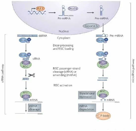
and two proteins of the argonaute family, AGO1 and AGO2. Genetic and biochemical evidence has demonstrated functional specialization in fly AGO proteins, with AGO1 binding to miRNAs and AGO2 being associated with siRNA-mediated-gene silencing. Functional specialization extends to the biogenesis pathways associated with these small RNAs; miRNAs are processed from endogenous hairpin precursors by cleavage events involving the RNaseIII enzymes Drosha and Dicer1 (Dcr-1) with its partner loquacious (Loqs). siRNAs loaded into AGO2 are processed from long dsRNAs by Dicer2 (Dcr-2) and its partner R2D2, but until recently only siRNAs from exogenous long dsRNAs had been reported in flies and mammals (Rivas 2008). There are two small RNAs in the RNAi pathway: small interfering RNAs (siRNAs) and microRNAs (miRNAs) that are generated via processing of longer dsRNA and stem loop precursors (Novina et al. 2002; Yin and Wan 2002; Tijsterman and Plasterk 2004). Dicer enzymes play a critical role in the formation of these two effectors of RNAi (Tijsterman and Plasterk 2004). They can cleave long dsRNAs and stem-loop precursors into siRNAs and miRNAs in an ATP-dependent manner, respectively (Tan and Yin 2005). (Fig 3, de Fougerolles A ,2007)
The biogenesis of miRNAs is a multistep process (Kim 2005). A primary miRNA transcript (pri-miRNA) (Lee et al. 2002), which is frequently synthesized from intronic regions of protein-coding RNA polymerase II transcripts (Cai et al. 2004; Lee et al. 2004), is first processed by a protein complex containing the double-strand specific ribonuclease Drosha in the nucleus to produce a hairpin intermediate of 70nt (Lee et al. 2003). This precursor miRNA (pre-miRNA) is subsequently transported by exportin-5/RanGTP (Lund et al. 2004; Yi et al. 2003) to the cytoplasm where it is cleaved by another dsRNA specific ribonuclease, Dicer, (Bernstein et al. 2001; Hutvagner et al. 2001) into miRNA duplexes. After strand separation of the duplexes, the mature single-stranded miRNA is incorporated into an RNA-induced silencing complex (RISC)-like ribonucleoprotein particle (miRNP) (Hutvagner et al. 2001; Martinez et al. 2002a; Tang 2005; Yekta et al. 2004; Weiler et al. 2006)
RNAi has several applications in biomedical research, immune system and health care such as treatment for HIV, viral hepatitis, cardiovascular and cerebrovascular diseases, metabolic disease, neurodegenerative disorders and cancer.
30
Fig 3: RNA interference (RNAi) pathways are guided by small RNAs that include small interfering RNA (siRNA) and microRNAs (miRNAs). The siRNA pathway begins with cleavage of long double-stranded RNA (dsRNA) by the Dicer enzyme complex into siRNA. These siRNAs are incorporated into Argonaute 2 (AGO2) and the RNAi-induced silencing complex (RISC). The siRNA guide strand recognizes target sites to direct mRNA cleavage (carried out by the catalytic domain of AGO2). The microRNA pathway begins with endogenously encoded primary microRNA transcripts (pri-miRNAs) that are transcribed by RNA polymerase II (Pol II) and are processed by the Drosha enzyme complex to yield precursor miRNAs (pre-miRNAs). These precursors are then exported to the cytoplasm by exportin 5 and subsequently bind to the Dicer enzyme complex, which processes the pre-miRNA for loading onto the AGO2–RISC complex. The mature miRNA recognizes target sites (typically in the 3'-UTR) in the mRNA, leading to direct translational inhibition. Binding of miRNA to target mRNA may also lead to mRNA target degradation in processing (P)-bodies.
31
Application of RNAi in biomedical research and health care
RNAi is being used for a variety of purposes including biomedical research and health care (Gupta 2006) and has begun to produce a paradigm shift in the process of drug discovery (Hannon and Rossi 2004). In order to meet this objective, dsRNA molecules have been designed for silencing of specific genes in humans and animals. Such silencing RNA molecules are introduced into the cell to facilitate activation of the RNAi machinery. This method has already become an important research tool in biomedicine. Several recent publications show successful gene silencing in human cells and experimental animals. For instance, a gene causing high blood cholesterol levels was shown to be silenced by treating animals with silencing RNA. Plans are also underway to develop silencing RNA as a treatment for cardiovascular diseases, cancer, endocrine disorders, and virus infections (Gupta 2006), such as those caused by the hepatitis C virus (HCV) and the human immunodeficiency virus (HIV) (Hannon and Rossi 2004).
Cancer is a genetic disease in which mutational and/or epigenetic changes in a genome lead to stepwise deregulation of cell proliferation and cell death mechanisms (Weiler et al. 2006). RNAi is being explored as a way to inhibit the expression of genes involved in oncogenesis.
Pancreatic and colon carcinomas, in which RAS genes are often mutated, provide an example of the use of RNA silencing in treating cancers. In many cases, the RAS oncogenes contain point mutations that differ by a single-base mutation from their normal counterparts. The use of retroviral vectors to introduce interfering RNAs specific for an oncogenic variant of K-RAS (called K-K-RASV12) reduces the level of K-K-RASV12 transcripts and effects a loss of anchorage-independent growth and tumourigenicity (Brummelkamp et al. 2002; Wilda et al. 2002). Studies of these kind provide proof of concept for RNAi-based strategies aimed at reversing tumourigenesis. A major factor confounding cancer treatment is resistance to chemotherapeutic agents. The siRNAs have been used to decrease the drug resistance of cells in vitro by inhibiting the expression of MDR1, a multidrug transporter with a major role in multidrug resistance (Nieth et al. 2003).
32
pathogenesis (Weiler et al. 2006). For example, deletions or mutations in genes that code for miRNA tumour suppressors might lead to loss of a miRNA or miRNA cluster, and thereby contribute to inappropriate stabilization of oncogenes (McManus 2003; Gong et al. 2005). The results of a recent large-scale miRNA study suggest that 50% of miRNA genes are frequently located in cancer-associated genomic regions or fragile sites (Calin et al. 2004). The genes encoding mir-15 and mir-16 are located at chromosome 13q14, a region that is deleted in the majority of B-cell chronic lymphocytic leukaemias (B- CELL) (Calin et al. 2002), and in other cancers such as mantle cell lymphoma and prostate cancer (Stilgenbauer et al. 1998). Interestingly, none of the protein-coding genes in this region were found to cause B-CLL (Migliazza et al. 2000), suggesting that mir-15 and mir-16 may possibly function as tumour suppressors.
MiRNAs, miR-143 and miR-145, display significant downregulation in colonic adenocarcinoma samples compared to matched normal mucosa tissues (Michael et al. 2003). Putative mRNA targets of these miRNAs include several genes that have been implicated in oncogenesis such as RAF1 kinase, G-protein 7 and tumour-suppressing subfragment candidate 1, although molecular interaction of these genes with their putative miRNA counterparts in vivo remains to be proven (Weiler et al. 2006).
MicroRNAs as robust diagnostic and prognostic biomarkers
MiRNAs are excellent biomarkers for the diagnosis and prognosis of cancer. Due to their gene regulation activities, the potential for using miRNA in cancer therapy is evident. So-called anti-miRNA oligonucleotides (AMOs), which are designed to be complementary to oncogenic miRNAs, are able to specifically inhibit miRNA activity in tumours. On the other hand, overexpression of miRNAs that act as tumour suppressors might also be beneficial for anticancer therapy. MicroRNAs provide not only promising therapy approaches for cancer, but also for many other diseases like virus infections or cardiovascular diseases, in which they are also involved as gene regulators. While the understanding for the gene regulation driven by miRNAs is under extensive research focus, the knowledge about the mechanisms regulating the gene
33
expression of the miRNAs themselves still needs to be broadened. Amongst others, miRNAs are thought to be controlled by epigenetic mechanisms not only due to their tissue and tumour specific expression patterns. As a matter of fact, several miRNAs have shown to be regulated by DNA methylation. (S. A.A. et al, 2010) Treating human bladder cancer cells with demethylating agents, Saito et al. (2006) have shown that 5% of the human miRNAs became upregulated more than three-fold. The strongest effect was seen in miR-127, whose corresponding gene was found to be embedded in a CpG island. After epigenetic reactivation of miR-127, one of its target genes, the proto-oncogene BCL6, became downregulated, leading to the assumption that miR-127 acts as a tumour suppressor gene. In cases like these, an epigenetic anticancer therapy becomes feasible (Lange and Stahler 2009).