S. Lazzi &L. Leoncini &M. Lucioni &D. Novero &S. Pileri &M. Ponzoni &E. Sabattini &C. Tripodo &A. Zamò &
M. Paulli8,14&L. Ruco1,15
Received: 24 January 2019 / Revised: 15 July 2019 / Accepted: 26 July 2019 / Published online: 6 August 2019
Abstract
An accurate diagnosis of clinically distinct subgroups of aggressive mature B cell lymphomas is crucial for the choice of proper treatment. Presently, precise recognition of these disorders relies on the combination of morphological, immunophenotypical, and cytogenetic/molecular features. The diagnostic workup in such situations implies the application of costly and time-consuming analyses, which are not always required, since an intensified treatment option is reasonably reserved to fit patients. The Italian Group of Haematopathology proposes herein a practical algorithm for the diagnosis of aggressive mature B cell lymphomas based on a stepwise approach, aimed to select cases deserving molecular analysis, in order to optimize time and resources still assuring the optimal management for any patient.
Keywords HGBL . Double hit . DLBCL . Diagnosis . FISH . MYC
Introduction
Diffuse large B cell lymphomas not otherwise specified (DLBCL NOS) represent a spectrum of malignancies associ-ated with diversified clinical outcomes. Characterization of molecular features of clinical importance, such as the cell of origin (COO) and the rearrangements of MYC, BCL2, and BCL6 genes, has been incorporated as a new requirement in the revised World Health Organization (WHO) classification of tumors of hematopoietic and lymphoid tissues [1].
Gene expression profiling (GEP) or surrogated immu-nohistochemical algorithms allow subclassification of DLBCL NOS mainly into the germinal center (GCB) and the activated (ABC) or non-GCB types based on
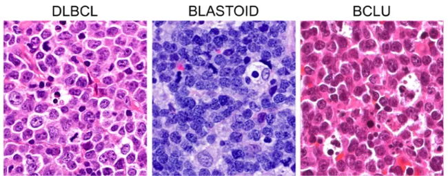
the cell of origin, with ABC lymphomas displaying poorer prognosis than GCB ones [2]. pt?>Fluorescence in situ hybridization (FISH) is required to distinguish among high-grade B cell lymphomas with double or triple hit rearrangement (HGBL DH/TH), high-grade B cell lymphomas not otherwise specified (HGBL NOS), and DLBCL NOS. HGBL DH/TH are aggressive mature B cell lymphomas with variable morphology, ranging from pleomorphic large cells to medium-sized cells with features intermediate between DLBCL and Burkitt lym-phoma (BCLU), to blastoid cells (Fig. 1), where FISH analyses identify MYC gene rearrangement in association with BCL2 and/or BCL6 gene rearrangements (Fig. 2). Notably, HGBL DH/TH account for approximately 5% of all cases with DLBCL morphology and generally have a low complete response rate with R-CHOP that advises for more intensive chemotherapy regimens [1, 3]. HGBL NOS includes cases with neoplastic B cells hav-ing either blastoid morphology or histopathological fea-tures intermediate between DLBCL and Burkitt Lymphoma (BCLU) that do not carry a double or a triple rearrangement. Recently, gene expression signatures
M. Paulli and L. Ruco contributed equally to this work.
This article is part of the Topical Collection on Quality in Pathology * Arianna Di Napoli
Extended author information available on the last page of the article # The Author(s) 2019
and mutational profiles identified high-risk patients with DLBCL comprising double hit lymphomas [4].
GEP and FISH studies, which are the elective tech-nologies for the definition of the COO, of gene rear-rangements and of high-risk lymphomas are expensive, time consuming, and not available in all laboratories. To date, no guidelines are available driving both patholo-gists and clinicians in the selection of aggressive mature B cell lymphomas deserving molecular analyses in a cost-effective management of the patients. The use of immunohistochemistry, cheaper and widely applicable, as surrogate tool for the assessment of the COO and the presence of MYC gene rearrangements in DLBCLs have been proposed [5–8]. However, this approach bears intrinsic limitations. Although HGBL-DH overexpress MYC and BCL2 proteins in most instances, they only account for a small proportion of the so-called double
expressors DLBCL (DE DLBCL). In addition, HGBL carrying MYC and BCL2 gene rearrangements almost exclusively belong to the GCB category, while the ma-jority of DE DLBCL fall into the non-GCB group [3, 8]. Of note, nearly 20% of GCB DLBCL carrying MYC rearrangement do not express MYC protein [8].
On the other hand, molecular subtyping all DLBCLs to identify all HGBL DH/TH may be unnecessary, since treat-ment choice is also driven by patient’s age, comorbidities, and performance status, with dose-intense treatment options being usually reserved to fit and young (age < 60 years) patients.
The purpose of this article is to propose a stepwise, work-ing algorithm aimed at the rationalization of the diagnostic efforts in aggressive mature B cell lymphomas. The attempt is to provide minimal required criteria to select cases deserv-ing FISH analysis, in order to save time and resources still assuring the optimal management for any patient.
Fig. 1 Morphological features of aggressive mature B cell lymphomas. In DLBCL, the cells are pleomorphic with centroblastic and/or immunoblastic features. Blastoid cells are medium-sized cells with a fine chromatin pattern and inconspicuous nucleoli. Cases with features
overlapping between BL and DLBCL (BCLU) show medium-sized cells, less monomorphic than in classical BL, with multiple paracentrally located nucleoli and frequent starry sky pattern
Fig. 2 Interphase FISH showing rearrangements of MYC, BCL2, and BCL6 genes using dual color break-apart (BA) probes. MYC/IGH translocation is detected using a dual color dual fusion FISH probe
biopsies might not be fully informative to render an accurate diagnosis of lymphoma, excisional lymph node biopsies should be favored whenever possible.
Diagnostic workflow
An initial diagnosis of an aggressive mature B cell lym-phoma should incorporate the assessment of cytological and immunohistochemical features, including the COO, and the percentage of MYC- and BCL2-expressing cells. Whenever a B cell lymphoma with a DLBCL morphology displays either a GCB COO and/or a double expression of MYC and BCL2 proteins (in more than 40% and 50% of neoplastic cells, respectively) (DE DLBCL), analysis of MYC, BCL2, and BCL6 gene rearrangements by FISH is indicated in order to rule out the possibility of a HGBL DH/TH. Although there is no complete agreement about the percentage of MYC protein-expressing cells that accu-rately predicts the presence of MYC gene rearrangement [6, 9,10], the cutoff value of 70% has been recently reported to be reproducible among different centers and of clinical value in identifying patients with a worse prognosis [7].
Before proceeding with FISH analysis, in DLBCL cases, it is highly recommended to discuss upfront with the referring hematologist the results of immunohistochemical screening in order to verify patient’s fitness and potential eligibility to un-dergo intensified therapy for HGBL DH/TH.
On the contrary, FISH analyses should be performed in any case of:
a. B cell lymphomas with blastoid morphology, with the exclusion of TdT+ lymphoblastic lymphoma or cyclin D1+ pleomorphic/blastoid mantle cell lymphoma (Fig.3). In these cases, FISH analysis for MYC, BCL2, and BCL6 genes allows classification of the malignancy as HGBL DH/TH or HGBL NOS. CCDN1 translocations should also be investigated to rule out cyclin D1-expressing DLBCL.
Immunohistochemistry
Aggressive mature B cell lymphomas should express B cell–associated antigens (e.g., CD20, CD19, CD79a) and lack cyclin D1. In the case of a cyclin D1+ large B cell lymphoma, immunohistochemistry for CD5, SOX11, and FISH analysis with a CCND1 break-apart probe must be performed in order to rule out a pleomorphic/blastoid mantle cell lymphoma [1]. The use of CD5 is also en-couraged to identify de novo CD5+ DLBCLs, which might display an unfavorable outcome [11].
The COO of DLBCL can be investigated by gene expression profiling (GEP) or, alternatively, by immuno-histochemistry (IHC) following algorithms suggested by the 2017 WHO Classification [1]. Among these, the most popular is Hans algorithm, which splits DLBCLs in germinal center (GCB) and non-germinal center (non-GCB) type based on the expression of CD10, BCL6, and IRF4/MUM1 proteins [12]. Its output shows reason-able correlation with the GEP, although some cases of DLBCL GCB type are misclassified as non-GCB type by IHC [5]. In addition to its role in discriminating different DLBCL prognostic subgroups (non-GCB carry-ing worse prognosis in comparison with GCB type), determination of COO might help in identifying those cases potentially harboring rearrangements of MYC, BCL2, and BCL6. Indeed, almost all the HGBL DH/ TH fall within the GCB subtype with less than 1% of ABC harboring MYC and BCL2 and 2% MYC and BCL6 rearrangements [3].
Immunohistochemical investigation of MYC and BCL2 protein expression in DLBCL is highly recom-mended since overexpression of these proteins is asso-ciated with shorter survival [10, 13, 14]. Moreover, HGBL DH without MYC or BCL2 overexpression dis-play a more favorable outcome than double expressor HGBL DH [7, 10, 13, 14]. Cutoff values for MYC and BCL2 that have been significantly associated with survival are 40% and 50%, respectively (independently of the intensity of the staining) [9]. Whenever the IHC
staining is not homogeneously distributed across the section, the percentage of positive cells should be cal-culated as the average, and the occurrence of hot spots with MYC > 70% should be reported. A high percentage of MYC+ cells is more likely to be associated with MYC translocation [6, 7, 9]. Some pathologists have advocated the use of Ki67 staining, although the prolif-erative fraction is variable in HGBL DH/TH and it can-not be considered a reliable marker for screening pa-tients that require FISH [1, 10].
FISH analysis
Rearrangements of MYC, BCL2, and BCL6 genes are gen-erally assessed using break-apart probes. Since the definition of HGBL DH/TH requires the presence of MYC rearrange-ment, this could be investigated first, followed by BCL2 and BCL6 gene analyses in MYC rearranged cases. The use of dual color dual fusion IGH-MYC probes in addition to MYC break-apart probes (Fig. 2) increases the sensitivity of detec-tion [14]. Furthermore, the definidetec-tion of the partner gene
(IGH or non-IGH) of MYC translocation could be clinically relevant, although this issue is still debated [9, 14, 15]. By an administrative standpoint, FISH analysis could be request-ed as an additional investigation either by the clinician or by the pathologist according to local rules.
Concluding remarks
In conclusion, we believe that the application of the proposed workflow could represent a useful strategy to rationalize the procedures and optimize the resources, speeding up the diag-nosis of aggressive mature B cell lymphomas and allowing the more appropriate treatment option for each patient.
Acknowledgments Di A, Napoli, Remotti D, and Ruco L are contribu-tors on behalf of Lazio Group of Haematopathologist composed by: Anemona Lucia, Bakacs Arianna, Bianchi Antonella, Cancellario Francesca, Caruso Lia, De Vito Rita, Di Cristofano Claudio, Di Napoli Arianna, Fratoni Stefano, Giacobbi Erica, Giordano Carla, Gomes Enzo Vito, Licci-Stefano, Ruco Luigi, Macciomei-M. Cristina, Marino Mirella, Monardo-Francesca, Pescarmona Edoardo, Ramieri Maria Teresa, Remotti Daniele, Riminucci Mara, Stella Francesca, Taccogna Silvia, Uccini Stefania.
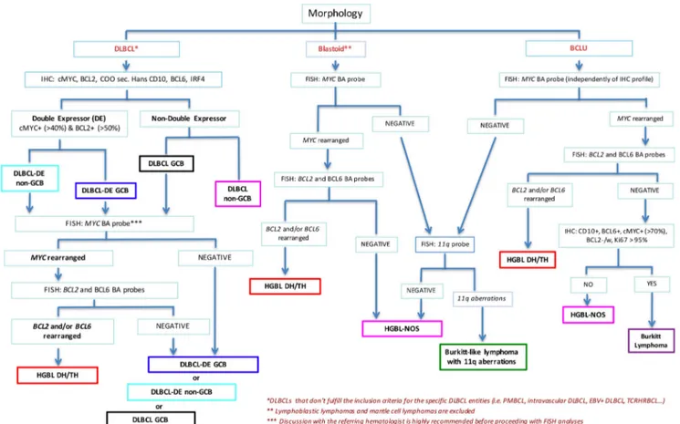
Fig. 3 Diagnostic workflow for the diagnosis of aggressive mature B cell lymphomas . The workflow applies to DLBCLs that don’t fulfill the inclusion criteria for the specific DLBCL entities (i.e. primary mediastinal B cell lymphoma (PMBCL), intravascular DLBCL, EBV+ DLBCL, T cell rich histiocyte rich B cell lymphoma (TCRHRBCL), etc
(*) and to blastoid lymphomas excluding lymphoblastic lymphomas and mantle cell lymphomas (**). In DLBCL discussion with the referring hematologist is highly recommended before proceeding with FISH analyses (***).
Conflict of interest The authors declare that they have no conflict of interest.
Open AccessThis article is distributed under the terms of the Creative C o m m o n s A t t r i b u t i o n 4 . 0 I n t e r n a t i o n a l L i c e n s e ( h t t p : / / creativecommons.org/licenses/by/4.0/), which permits unrestricted use, distribution, and reproduction in any medium, provided you give appro-priate credit to the original author(s) and the source, provide a link to the Creative Commons license, and indicate if changes were made.
References
1. Swerdlow SH, Campo E, Harris NL et al (2017) WHO classifica-tion of tumours of haematopoietic and lymphoid tissues. In: World Health Organization Classification of Tumours. Revised, 4th edn. International Agency of Research on Cancer, Lyon
2. Alizadeh AA, Eisen MB, Davis RE et al (2000) Distinct types of diffuse large B-cell lymphoma identified by gene expression pro-filing. Nature 03(6769):503–511
3. Sesques P, Johnson NA (2017) Approach to the diagnosis and treat-ment of high-grade B-cell lymphomas with MYC and BCL2 and/or BCL6 rearrangements. Blood 129(3):280–288
4. Chan WC (2019 Jan 20) Using gene expression profiling to move beyond MYC/BCL2 rearrangements in high-grade lymphoma. J Clin Oncol 37(3):175–177
5. Gutiérrez-García G, Cardesa-Salzmann T, Climent F et al (2011) Gene-expression profiling and not immunophenotypic algorithms predicts prognosis in patients with diffuse large B-cell lymphoma treated with immunochemotherapy. Blood 117(18):4836–4843 6. Green TM, Nielsen O, de Stricker K, Xu-Monette ZY, Young KH,
Møller MB (2012) High levels of nuclear MYC protein predict the presence of MYC rearrangement in diffuse large B-cell lymphoma. Am J Surg Pathol 236(4):612–619
7. Ambrosio MR, Lazzi S, Lo Bello G et al (2019) MYC protein expression scoring and its impact on the prognosis of aggressive B-cell lymphoma patients. Haematologica 104(1):e25–e28
Meyer PN, Lenz G, Wright G, Rimsza LM, Valentino C, Brunhoeber P, Grogan TM, Braziel RM, Cook JR, Tubbs RR, Weisenburger DD, Campo E, Rosenwald A, Ott G, Delabie J, Holcroft C, Jaffe ES, Staudt LM, Gascoyne RD (2012) Concurrent expression of MYC and BCL2 in diffuse large B-cell lymphoma treated with rituximab plus cyclophosphamide, doxoru-bicin, vincristine, and prednisone. J Clin Oncol 30(28):3452–3459 11. Xu-Monette ZY, Tu M, Jabbar KJ, Cao X, Tzankov A, Visco C, Nagarajan L, Cai Q, Montes-Moreno S, An Y, Dybkaer K, Chiu A, Orazi A, Zu Y, Bhagat G, Richards KL, Hsi ED, Choi WW, van Krieken J, Huh J, Ponzoni M, Ferreri AJ, Zhao X, Møller MB, Farnen JP, Winter JN, Piris MA, Miranda RN, Medeiros LJ, Young KH (2015) Clinical and biological significance of de novo CD5+ diffuse large B-cell lymphoma in Western countries. Oncotarget 6(8):5615–5633
12. Hans CP, Weisenburger DD, Greiner TC, Gascoyne RD, Delabie J, Ott G, Müller-Hermelink HK, Campo E, Braziel RM, Jaffe ES, Pan Z, Farinha P, Smith LM, Falini B, Banham AH, Rosenwald A, Staudt LM, Connors JM, Armitage JO, Chan WC (2004) Confirmation of the molecular classification of diffuse large B-cell lymphoma by immunohistochemistry using a tissue microarray. Blood 103(1):275–282
13. Green TM, Young KH, Visco C, Xu-Monette ZY, Orazi A, Go RS, Nielsen O, Gadeberg OV, Mourits-Andersen T, Frederiksen M, Pedersen LM, Møller MB (2012) Immunohistochemical double-hit score is a strong predictor of outcome in patients with diffuse large B-cell lymphoma treated with rituximab plus cyclophospha-mide, doxorubicin, vincristine, and prednisone. J Clin Oncol 30(28):3460–3467
14. Muñoz-Mármol AM, Sanz C, Tapia G, Marginet R, Ariza A, Mate JL (2013) MYC status determination in aggressive B-cell lympho-ma: the impact of FISH probe selection. Histopathology 63(3):418– 424
15. McPhail ED, Maurer MJ, Macon WR et al (2018) Inferior survival in high-grade B-cell lymphoma with MYC and BCL2 and/or BCL6 rearrangements is not associated with MYC/IG gene rearrange-ments. Haematologica 103(11):1899–1907
Publisher’s note Springer Nature remains neutral with regard to jurisdictional claims in published maps and institutional affiliations.
Affiliations
Arianna Di Napoli1 &D. Remotti2&C. Agostinelli3&M. R. Ambrosio4&S. Ascani5&A. Carbone6&F. Facchetti7&
S. Lazzi4&L. Leoncini4&M. Lucioni8&D. Novero9&S. Pileri10&M. Ponzoni11&E. Sabattini3&C. Tripodo12,13&A. Zamò9&
1
Pathology Unit, Department of Clinical and Molecular Medicine, Sant’Andrea Hospital, Sapienza University, Via di Grottarossa 1035, 00189 Rome, Italy
2
Pathology Unit, San Camillo-Forlanini Hospital, Rome, Italy 3
Hematopathology Unit, S. Orsola University Hospital, Bologna, Italy
4
Pathology Unit, Department of Medical Biotechnology, University of Siena, Siena, Italy
5 Pathology Unit, Ospedale di Terni, University of Perugia, Terni, Italy 6
Department of Pathology, Centro di Riferimento Oncologico di Aviano, Istituto di Ricovero e Cura a Carattere Scientifico, Aviano, Italy
7
Pathology Section, Department of Molecular and Translational Medicine, University of Brescia, Brescia, Italy
8
Pathology Unit, University of Pavia and Fondazione IRCCS San Matteo Policlinico, Pavia, Italy
9 Department of Oncology, University of Turin and Pathology Unit, AOU Città della Salute e della Scienza, Turin, Italy
10
Division of Haematopathology, European Institute of Oncology, Milan, Italy
11
Ateneo Vita-Salute, Pathology Unit, IRCCS San Raffaele Scientific Institute, Milan, Italy
12 Tumor Immunology Unit, Department of Health Sciences, University of Palermo, Palermo, Italy
13
Tumor and Microenvironment Histopathology Unit, the FIRC Institute of Molecular Oncology (IFOM), Milan, Italy 14
Italian Group of Haematopathology (GIE), Rome, Italy 15 Pathology Board of the Italian Lymphoma Foundation (FIL),