A photoactivatable Pt
anticancer complex conjugated to the
RNA ligand guanidinoneomycin
Evyenia Shaili,
[b]Marta Fernández-Giménez,
[a]Savina Rodríguez-Astor,
[a]Albert Gandioso,
[a]Lluís
Sandín,
[a]Carlos García-Vélez,
[a]Anna Massaguer,
[d]Guy J. Clarkson,
[b]Julie A. Woods,
[c]Peter J.
Sadler,*
[b]and Vicente Marchán*
[a]Dedication ((optional))
Abstract: A photoactivatable PtIV complex, trans,trans,trans-[Pt(N3)2(OH)(succ)(py)2] (succ = succinylate, py = pyridine), has been conjugated to guanidinoneomycin to study the effect of this guanidinum-rich compound on the photoactivation, intracellular accumulation and phototoxicity of the pro-drug. Surprisingly, trifluoroacetic acid treatment causes the replacement of an azido ligand and the axial hydroxide ligand by trifluoroacetate, as shown by NMR, MS and X-ray crystallography. Photoactivation of the Pt-guanidinoneomycin conjugate in the presence of 5’-GMP led to the formation of trans-[Pt(N3)(py)2(5’-GMP)]+, as does the parent PtIV complex. Binding of the PtII photoproduct {PtN3(py)2}+ to guanine nucleobases in a short single-stranded oligonucleotide was also observed. Finally, cellular uptake studies showed that guanidinoneomycin conjugation improves the intracellular accumulation of the PtIV pro-drug in two cancer cell lines, particularly in SK-MEL-28 cells. Notably, the higher phototoxicity of the conjugate in SK-MEL-28 cells than in DU-145 cells suggests a degree of selectivity towards the malignant melanoma cell line.
Introduction
Platinum-based anticancer drugs like cisplatin (1 in Scheme 1) and its second-generation derivatives (carboplatin and oxaliplatin) are amongst the most widely used antitumour agents in chemotherapeutic regimes in the clinic.[1] However, the dose that can be administered to patients is usually limited by the appearance of severe toxic side-effects, including nephrotoxicity, neurotoxicity, ototoxicity, nausea and vomiting. Moreover, the scope of the application of these metallodrugs is frequently limited by intrinsic or acquired resistance. In this context, photoactivation can be used to improve the therapeutic efficacy of metal-based chemotherapeutics by triggering drug release from selective delivery systems or through activating a pro-drug
at a desired time and place and with dosage control.[2] Taking into account that the effect of the anticancer drug will be limited to a specific irradiated area, this promising approach is expected to increase drug efficacy and reduce toxic side-effects provided that the surrounding normal tissues will not be damaged. Photoactivatable platinum(IV) pro-drugs are attractive compounds for cancer treatment since they benefit from the advantages of classical PtIV complexes (e.g. higher stability in biological media, aqueous solubility and the possibility of oral administration),[3] and of the use of light to control the release of PtII active species by light-promoted reduction.[4] In recent years, we have developed photoactivatable trans-dihydroxido PtIV diazido anticancer complexes with trans dia(m)mine ligands.[5] These complexes are inert and nontoxic in a biological environment in the dark, but upon light irradiation, they are selectively activated and become potently cytotoxic towards a number of cancer cell lines. Replacement of one or two NH3 ligands with pyridine in trans,trans,trans-[Pt(N3)2(OH)2(NH3)2] leads to higher photocytotoxicity and visible-light activation. This is an important feature for clinical applications because visible light penetrates more deeply than UVA. Indeed, trans,trans,trans-[Pt(N3)2(OH)2(py)2] (2 in Scheme 1A) can be activated over a range of wavelengths and is highly active against a range of cancer cell lines using low doses of visible light, including cisplatin-resistant A2780 human ovarian carcinoma cells.[6] These features together with its high stability in solution and towards gluthathione result in complex 2 being a promising lead compound for developing new photoactivatable PtIV pro-drugs.[7] The fact that these complexes platinate DNA and produce lesions that are distinctly different from those generated by cisplatin offers a potential for new mechanisms of action and non-cross-resistance with existing therapies.[8] In recent years, the approach of using UV and/or visible light to activate anticancer metal-based pro-drugs has been extended to other metals,[9] including Ru, Re, Rh and Ir. Representative examples are ruthenium(II) arene complexes ([(η6 -p-cym)Ru(bpm)(py)]2+) and RuII polypyridyl complexes in which the active species are photo-released by dissociation of Ru-pyridine or Ru-thioether bonds. Very recently, ruthenium(II)[10] and rhenium(I)[11] complexes have been masked with caging groups to control their activation by UV irradiation.
In view of the potential of photoactivatable metallodrugs for cancer treatment, it is desirable to optimise their pharmacological properties (e.g. aqueous solubility and cell uptake). Moreover, the application of the so-called targeted strategies to these complexes has enormous potential for developing innovative highly-selective metal-based anticancer drugs.[12] In this context, we demonstrated the feasibility of
[a] M. Fernández-Giménez, S. Rodríguez-Astor, A. Gandioso, L. Sandín, C. García-Vélez, Dr. V. Marchán
Departament de Química Orgànica and IBUB Universitat de Barcelona
Martí i Franquès 1-11, E-08028, Barcelona, Spain E-mail: [email protected]
[b] Dr. E. Shaili, G. J. Clarkson, Prof. P. J. Sadler Department of Chemistry
University of Warwick
Warwick, CV4 7AL, Coventry, UK E-mail: [email protected] [c] Dr. J. A. Woods
Photobiology Unit, Department of Dermatology Ninewells Hospital
Dundee, DD1 9SY, UK [d] Dr. A. Massaguer
Departament de Biologia Universitat de Girona
Campus Montilivi, E-17071, Girona, Spain
Supporting information for this article is given via a link at the end of the document.((Please delete this text if not appropriate))
conjugating photoactivatable RuII arene complexes to receptor-binding peptides which could then undergo photo-induced reactions with DNA.[9b] This work provided the first example of a potential Ru-based anticancer agent with a dual mechanism of selectivity. In the case of PtIV pro-drugs, derivatization of the detachable axial positions with carrier molecules can also be used to improve their pharmacological properties, such as cellular uptake and tumour selectivity,[13] as recently found by us by conjugating classical[13e] and photoactivatable[13f] PtIV complexes to RGD-containing peptides. Upconversion nanoparticles have also been used as drug carriers of photoactivated PtIV complexes.[14] Importantly, the loss of (axial) ligands upon reduction, either light-promoted (in photoactivatable PtIV complexes)[6,7,13f,14] or by intracellular reducing agents such as gluthathione or ascorbate (in classical PtIV complexes),[13,15] can cause the concomitant loss of the carrier molecule (see Scheme 1B), which would not interfere with the ultimate mode of action of the active PtII species (e.g. DNA/RNA interaction) or the potential production of reactive species (in the case of photoactivatable PtIV complexes).[6b] Guanidinoglycosides, which are obtained by replacing the amine functions of natural aminoglycoside antibiotics with guanidinium groups,[16] are known to be taken up more efficiently by eukaryotic cells compared to their aminoglycoside precursors.[17] This feature has been exploited in the case of guanidinoneomycin, since it is able to transport large bioactive cargos into cells in a selective proteoglycan-dependent manner.[18] Recently, we described the conjugation of a cytotoxic ruthenium(II) arene complex to neomycin and to its guanidinylated derivative.[19] Ruthenium accumulation studies confirmed that guanidinylation enhanced cellular uptake of the ruthenium complex, and more importantly, that the cytotoxic activity was very dependent on the nature of the cell line, being higher in cancer than in healthy cells. This can be attributed to differences between the expression level and/or in the composition of proteoglycan receptors on the cell membrane surface.
Based on these precedents and on the promising biological activity of trans,trans,trans-[Pt(N3)2(OH)2(py)2], now we explore the use of guanidinoneomycin as a carrier to improve the pharmacological properties of photoactivatable PtIV complexes. Such a strategy aims to contribute to the development in the near future of new RNA-targeted photoactivatable platinum anticancer complexes by using ligands which are selective for RNA. In this work, we have conjugated trans,trans,trans-[Pt(N3)2(OH)(succ)(py)2] (succ = succinylate) to guanidinoneomycin (Scheme 1A), and studied the effect of conjugation on the photoactivation of the PtIV pro-drug in the presence of 5’-GMP as a nucleobase model, as well as in the photo-induced reactions with a synthetic oligonucleotide. The phototoxicity of the compounds towards cancer cell lines in the presence of blue light and cellular uptake by ICP-MS were also investigated.
Scheme 1. a) Structure of cisplatin (1), trans,trans,trans-[Pt(N3)2(OH)2(py)2] (2),
trans,trans,trans-[Pt(N3)2(OH)(succ)(py)2] (3), guanidinoneomycin and of the
PtIV
-guanidinoneomycin conjugate. The cationic structure of guanidinoneomycin is shown since guanidinium groups are expected to be protonated under physiological conditions. b) Schematic representation of the photodissociation process and of two representative PtII adducts with a nucleic acid duplex.
Results and Discussion
Synthesis and characterization of photoactivatable Pt-guanidinoneomycin conjugates
Following previous studies on the derivatization of PtIV complexes with RGD-containing peptides,[13e,13f] we utilized trans,trans,trans-[Pt(N3)2(OH)(succ)(py)2][20] (3, Scheme 1) and attached it to a suitably-protected guanidinoneomycin derivative via the formation of an amide bond. Photoactivation was expected to cause the loss of the guanidinoglycoside carrier from the axial position and to generate an active PtII species (Scheme 1B).
Since the guanidinium groups, which are protonated under physiological conditions, are important for cell uptake of the compounds and for interaction with polyanionic nucleic acid targets, we conjugated complex 3 at the 5’’-OH function of guanidinoneomycin.[21] This primary hydroxyl group can be regioselectively converted to an amino function. As shown in Scheme 2, the 5’’-amino-5’’-deoxy-Boc-protected guanidinoneomycin derivative (6) was prepared from the natural aminoglycoside neomycin B in a six-step synthetic route following previously reported procedures,[21] although some modifications were required for the last two steps. Firstly, after acid treatment (30% TFA in DCM) of 5’’-azide-5’’-deoxy-Boc-protected neomycin intermediate (4), guanidinylation with N,N’-di-Boc-N”-triflylguanidine afforded a complex mixture of compounds that were difficult to separate by column chromatography. However, the use of N,N’-di-Boc-1H-pyrazole-1-carboxamidine,[22] another guanidinylating reagent employed for the conversion of amino functions into bis-Boc-protected guanidinium groups, afforded the desired intermediate (5) in 64% yield after column chromatography. To our surprise, reduction of the 5’’-azide group by Staudinger reaction did not afford the expected compound as reported elsewhere[21] but the iminophosphorane intermediate, which was very stable even in the presence of acidified water. Finally, reduction of the azide
function by catalytic hydrogenation afforded the expected guanidinoneomycin derivative 6 after column chromatography (40% yield).
Scheme 2. Synthesis of the photoactivatable PtIV-guanidinoneomycin conjugates 8 and 9. Although the trifluoroacetate salt of the conjugates was obtained, the neutral structures are shown.
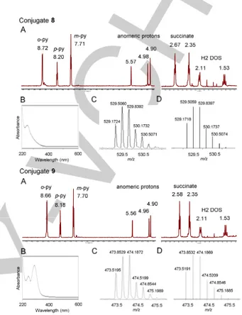
The next step involved the covalent attachment of the PtIV complex to the guanidinoneomycin derivative. First, complex 3 was activated with HATU and DIPEA in anhydrous DMF for 2 min, and then allowed to react with 6 for 2 h at room temperature. The expected Boc-protected Pt-guanidinoneomycin conjugate (7) was isolated by column chromatography (yield: 89%). High-resolution ESI MS analysis afforded m/z values that were consistent with the calculated values of the charged species ([M+2H]2+ and [M+3H]3+) and with the expected isotopic distribution of platinum. Then, compound 7 was treated with a 1:1 mixture of TFA/DCM for 2 h at room temperature to deprotect the guanidinium groups. Reversed-phase HPLC analysis showed a main peak (Rt = 15.7 min; see Figure S1 in the Supporting Information) that was isolated and analyzed by HR ESI MS. To our surprise, m/z values ([M+2H]2+ and [M+3H]3+) were not consistent with the expected values for the mass of the target conjugate (9 in Scheme 2), but instead with the formation of a modified Pt-guanidinoneomycin conjugate (8 in Scheme 2) in which the expected hydroxido ligand in the axial position of the platinum complex as well as one of the two azido ligands (Figure 1 and Figure S2 in the Supporting Information) had been replaced by trifluoroacetate ligands. The same product was obtained when the reaction was repeated without isolating intermediate 7 by column chromatography, but by carrying out TFA deprotection at the level of the crude product. The absence of the characteristic band around 310 nm in the UV-vis spectra of conjugate 8 (Figure 1) supports the modification of the Pt moiety during the acidic treatment, particularly the loss of one azide, as evident by comparing the spectra of parent complexes 26a and 320 (see the UV-vis spectra in Figure 2) and conjugate 9 (Figure 1). Conjugate 8 was fully characterized by 1D 1H NMR spectroscopy and 2D COSY and TOCSY experiments. As shown in Figure 1, diagnostic signals from the platinum complex
(pyridine ligands and succinate) and from guanidinoneomycin glycoside moiety (anomeric protons from the three sugars and H2 protons from the 2-deoxystreptamine ring) confirmed the covalent attachment of both moieties.
Figure 1. Characterization data for conjugates 8 (top) and 9 (bottom). (A)
Expanded regions of the 1H NMR spectra in D2O and (B) UV-vis spectra of the
compounds. Expanded ESI mass spectrum of the molecular peak ([M+3H]3+
), experimental (C) and calculated (D).
These unexpected results led us to evaluate the stability of photoactivatable PtIV complexes towards TFA. First, trans,trans,trans-[Pt(N3)2(OH)2(py)2] (2) was allowed to react with TFA/DCM 1:1 for 2 h at room temperature. Characterization by ESI MS, NMR spectroscopy (1H, 13C, 195Pt and 19F) and X-ray crystallography revealed the formation of a new complex, trans,trans,trans-[Pt(N3)2(CF3COO)2(py)2)] (10), in which both hydroxido ligands in the axial positions were replaced by trifluoroacetate (Figure 2A and Figures S11-S12). In this case, the two azido ligands were retained by the platinum center. Trans,trans,trans-[Pt(N3)2(OH)(succ)(py)2] (3) was also transformed into a new complex upon TFA treatment. This was characterized by MS, NMR and X-ray crystallography as trans,trans,trans-[Pt(N3)(CF3COO)(succH)(CF3COO)(py)2)] (11) (succH = 3-carboxypropanoate) (Figure 2B). In this complex, the single axial hydroxido ligand was replaced by trifluoroacetate, thereby reproducing the behavior found with 2. Interestingly, one of the two azido ligands was lost and replaced by trifluoroacetate during the acid treatment, which is in good agreement with the formation of the modified Pt-guanidinoneomycin conjugate 8. As shown in Figure 2B, the UV-vis spectrum of complex 11 is very
similar to that of conjugate 8 (see Figure 1), and clearly different from that of the platinum complexes containing the two azido ligands (2, 3 and 10). In this case, the two signals in the 19F NMR spectrum of 11 (Figure S13) correspond to the two trifluoroacetate ligands (δ= -76.1 and -76.2 ppm).
A b s o rb a n c e A b s o rb a n c e A b s o rb a n c e A b s o rb a n c e
Figure 2. The contrasting reactivity of complexes 2 (a) and 3 (b) with excess
TFA. The PtIV
dihydroxido complex (2) retains both the equatorial azido ligands, whereas in the monocarboxylato complex (3) one azide is also replaced by trifluoroacetate. The absence of one azide is reflected in the UV-vis spectra, where the band at ca. 314 nm is lost.
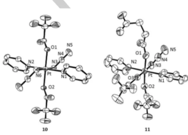
Furthermore, crystals suitable for X-ray diffraction studies were obtained for both modified complexes (10 and 11; see Figure 3, Table 1 and Table S1 in the Supporting Information). Complex 10 crystallized in the trigonal space group R-3 with 9 molecules in the unit cell whereas complex 11 crystallized in monoclinic P21/c space group with 8 molecules in the unit cell. The asymmetric unit of 11 contains two PtIV complexes, labeled A and B (Table 1), which form an H-bonded dimer via the carboxylic acid groups (see Figure S10). The bond distances between PtIV and pyridine nitrogen atoms (Pt-N1 and Pt-N2) have no significant differences between the two complexes and they fall within the range of other PtIV-diazido complexes with pyridine ligands. Similarly,[20] the Pt-N3 bond lengths are within the expected range[23] but interestingly are significantly longer in complex 10 than in 11, which might lead to an increased lability of the azido group. The axial Pt-O bonds (Pt-O2) from the trifluoroacetate group have no significant differences between 10 and 11A, whereas in 11B are longer. The Pt-O bond lengths from the succinate group resemble other PtIV-carboxylate bonds.[20] When compared to the limited examples reported for other PtIV-trifluoroacetate complexes, ranging from 1.979-2.014 Å,[24] complex 11 has longer distances (Pt-O2, O3: 2.013-2.061 Å).
The introduction of trifluoroacetate ligands into PtIV complexes has been explored by Gibson et al.[25] However in their examples, the synthesis involved reaction with trifluoroacetic anhydride, whereas herein the hydroxido ligand is replaced directly in the presence of excess of trifluoroacetic acid. Previous work has demonstrated that the pKa of the axial hydroxide of dia(m)mine PtIV-diazido-dihydroxo complexes is ca. 3.4, suggesting that in the presence of concentrated TFA solution, the hydroxide ligand can become protonated, thus facilitating its substitution by TFA.[26] Although the trifluoroacetate ligands can confer interesting and beneficial properties (e.g. increased uptake and cytotoxicity, more facile reduction), the complexes reported in
this work were found to be insoluble in aqueous media, especially complex 10, where both axial ligands are replaced with trifluoroacetate. The replacement of one equatorial ligand with trifluoroacetate in complex 11 is an interesting outcome and the first example of a ground-state substitution of an azide in PtIV-diazido complexes, as the previously reported examples were species formed upon photosubstitution. Rationalization of the differing behaviour of parent complexes 2 and 3 is not straightforward, as the X-ray structures[20] do not show any significant difference in the lengths of Pt-N3 bonds which might suggest a weaker bond in the ground state. The neighboring dangling terminal carboxylic acid in the case of 3 might play a role in the mechanism of substitution of the equatorial azide, for example by promoting its dissociation as N3H, and a subsequent substitution by trifluoroacetate.
Figure 3. ORTEP diagrams for the X-ray crystal structures of 10 and 11. The
ellipsoids are set to 50% probability level. Labelling of the atoms does not represent the numbering in the cif files (CCDC 1060450-1060451) but is used to facilitate comparison between the two complexes.
Table 1. Selected bond distances and angles for complexes 10 and 11.
Bond (Å) Angle (o) 10 11(A) 11(B)
Pt–N1 N1–Pt–N2 2.0419(18)/ 180.0 2.033(5)/ 178.9(3) 2.027(8)/178.9(4) Pt–N2 N3–Pt–N6 or N3–Pt–O3 2.0419(18)/ 180.0 2.029(6)/ 179.3(2) 2.031(9)/170.4(8) Pt–N3 O1–Pt–O2 2.0558(19)/180.0 2.010(7)/ 176.31(19) 2.015(3)/176.3(3) Pt–N6 Pt–N3–N4 2.0558(19)/ 115.51(15) NA/ 115.3(6) NA/ 118(2) Pt–O1 N3–N4–N5 2.0093(15)/175.6(2) 1.995(5)/ 173.6(9) 2.006(7)/168(4) Pt–O2 - 2.0093(15) 2.012(5) 2.061(7) Pt–O3 - NA 2.049(5) 2.044(7)
Taking into account the unexpected reactivity of photoactivated PtIV complexes based on the parent trans,trans,trans-[Pt(N3)2(OH)2(py)2] (2), an inverse approach was explored for the synthesis of the Pt-guanidinoneomycin 9 (Scheme 2). The guanidinoneomycin derivative 6 was first deprotected with TFA/DCM 1:1, and the resulting trifluoroacetate salt of 5’’-amino-5’’-deoxy-guanidinoneomycin was allowed to react with complex 3 by using HATU as activating reagent in the presence of DIPEA. Reversed-phase HPLC analysis of the reaction crude showed
the presence of three main peaks (Figure S5) that were isolated and analyzed by HR ESI-MS. The product with lower retention time (Rt =12.0 min) was characterized by MS, 1H NMR and UV-vis as the expected Pt-guanidinoneomycin conjugate (9), which was obtained as a white solid after purification by HPLC and lyophilization (yield: 29%). The peaks with higher retention time were characterized by MS as conjugates incorporating two Pt moieties. Despite the lower nucleophilicity of guanidinium compared with an amino function, the use of a potent amide-forming reagent accounts for this result. As shown in Figure 1, the UV-vis spectrum of conjugate 9 was similar to that of the parent Pt complexes 2[6a] and 3[20] (Figure 2) and HR ESI MS analysis afforded m/z values that were consistent with the calculated value of the charged species ([M+2H]2+ and [M+3H]3+) and with the expected isotopic mass distribution patterns of platinum (see Figures 1 and S6). Although the 1H NMR spectrum of conjugate 9 was almost identical to that of 8 (Figure 1 and S7), chemical shifts for the protons close to the platinum center (e.g. the ortho protons in pyridine ligands and the methylene of the succinate) were slightly shifted to lower fields in the modified conjugate. This is in agreement with the electron-withdrawing character of the two trifluoroacetate ligands coordinated to the Pt center in conjugate 8.
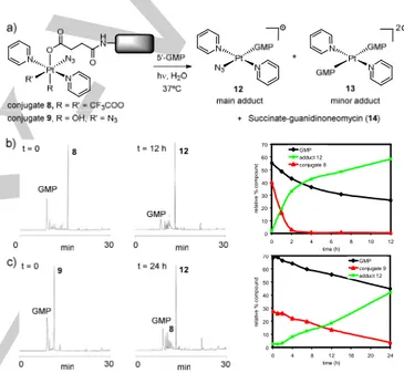
Photo-induced reactions with 5’-guanosine monophosphate. Next we studied the photoactivation of conjugate 9 to determine how the derivatization of the axial position of trans,trans,trans-[Pt(N3)2(OH)2(py)2] with guanidinoneomycin affects its photochemical properties and the type of photoadducts formed with nucleic acids. Modified conjugate 8 was investigated to determine whether it could still be photoactivated to give PtII photoproducts despite the presence of only one azido ligand. Electron transfer from two azido ligands to PtIV can generate PtII and azidyl radicals.[6b] If only one azide is present, and this is retained in the PtII photoproduct, then the two electrons required for the reduction of PtIV need to be donated by other ligands such as the trifluoroacetate or the carboxylate. In situ photo-induced reactions were carried out by irradiating an aqueous solution of the conjugate (35 µM) with visible light at 310 K in the presence of 5’-guanosine monophosphate, 5’-GMP (2 mol equiv), as a simple model for nucleic acid binding and monitored by reversed-phase HPLC (see Figure S15). As shown in Figure 4, irradiation led to a decrease in the concentration of conjugate 9 and of 5’-GMP, and the appearance of a major product that was isolated and characterized by HR ESI-MS as the PtII adduct trans-[Pt(N3)(py)2(5’-GMP)]+ (12) (Figure S16). Traces of the adduct trans-[Pt(py)2(5’-GMP)2]2+ (13) were also detected by MS-HPLC (GMP is considered neutral in the formulae). The photorelease of the succinate-derivative of guanidinoneomycin (14) was confirmed by MS-HPLC since absorption of this compound in the UV region is too small to be detected. These results agree with those found previously for the parent complex (2)[6a] or its succinate derivative (3),[13f,20] and demonstrate that the covalent attachment of guanidinoneomycin does not modify the photoactivation properties of the platinum complex and the reactivity of the photoproducts for guanine nucleobase. It is interesting to note that several intermediate compounds were
formed during the irradiation process (see Figure S15 in the Supporting Information) that evolved into the main adduct 12. To our surprise, the modified conjugate 8 was also photoactivated, but at a much faster rate. As shown in Figure 4, adduct 12 was again the major compound identified in the reaction mixture, thereby reproducing the results obtained with 9. These results prompted us to evaluate the stability of both compounds in the dark in the presence of 5’-GMP. After incubation for 18 h at 310 K, HPLC analysis revealed that the peak for conjugate 9 was unaltered, whereas that of conjugate 8 was reduced by 25% leading to the formation of adduct 12 (Figure S17). This seems to indicate that trifluoroacetylation of the axial position together with the replacement of an azido ligand by trifluoroacetate in the platinum coordination sphere led to a complex with low stability that could be thermally activated as well. 0 10 20 30 40 50 60 70 0 4 8 12 16 20 24 time (h) re la tiv e % c o m p o u n d GMP conjugate 9 adduct 12 0 10 20 30 40 50 60 70 0 2 4 6 8 10 12 time (h) re la ti v e % c o m p o u n d GMP adduct 12 conjugate 8
Figure 4. Photo-induced reactions of PtIV-guanidinoneomycin conjugates with 5’-GMP under visible light irradiation. A 1 M aqueous solution of NaNO2 was
used to filter out the UV and ensure the appropriate wavelength range (> 400 nm). (A) Schematic representation of the photoproducts, (B) reversed-phase HPLC traces for the in situ reaction between 5’-GMP and conjugate 8, and (C) conjugate 9 at t= 0 and after 12 or 24 h of irradiation at 310 K, respectively, and representation of the distribution of products with time.
Photo-induced reactions with 5´dCATGGCT
Once the reactivity of Pt-guanidinoneomycin conjugates with the model 5’-GMP under visible light irradiation had been demonstrated, we investigated the ability of the most stable conjugate (9) to platinate a synthetic oligodeoxynucleotide sequence. We selected 5´dCATGGCT as a simple model since it contains two consecutive guanine nucleobases which are the preferred GG target sequence of cisplatin. Moreover, it has been used previously as a nucleic acid model to study the interaction with conjugates between a dicarba analogue of octreotide and a dichloridoplatinum(II) complex or a photoactivated ruthenium(II) arene complex.[9b]
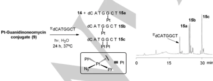
First, a mixture of conjugate 9 and 5´dCATGGCT (4:1 mol ratio) was irradiated with visible light (λ > 400 nm) at 310 K for 24 h. According to previous studies with 5’-GMP (see Figure 4C), conjugate 9 was expected to be completely photoactivated within this time. Reversed-phase HPLC analysis showed the presence of three major peaks with higher retention times than the parent oligonucleotide (Rt = 24.2, 24.5 and 30.0 min; relative ratio 1:1.1:1.6, respectively; see Figure 5), which were isolated and characterized by MALDI-TOF MS (see Figure S19 in the Supporting Information). The two products with similar hydrophobicity correspond to isomeric platinated DNA adducts in which a single platinum fragment, {PtN3(py)2}+, was coordinately bound to the DNA strand (15a-b) (Figure 5). The most hydrophobic compound was characterized as a DNA adduct incorporating two {PtN3(py)2}+ fragments (15c). In addition, several minor compounds were identified by MS, such as oligonucleotide adducts with a single {Pt(py)2}2+ fragment (Rt = 20.8 min) or with two different platinum fragments, {PtN3(py)2}+ and {Pt(py)2}2+, (Rt = 24.9 min). Enzymatic digestion with 5´- and 3´-exonucleases (bovine spleen and snake venom phosphodiesterases, respectively) in combination with MALDI-TOF MS analysis confirmed the position of the platinum fragments in the three major compounds.[27,28] As expected from the known preference of PtII complexes for nucleobases, platination occurred at the two guanines in the oligonucleotide sequence: a single {PtN3(py)2}+ was bound to 3’G in 15a or to 5’G in 15b, whereas the two platinum moieties were bound to both guanines in 15c.
As a control, the reaction between 5´dCATGGCT and trans,trans,trans-[Pt(N3)2(OH)(succ)(py)2] (3) (2 mol equiv) was also studied (Figure S20). The same major adducts were formed, as inferred by HPLC and MS, which confirms again that the photoactivation of 3 or its guanidinoneomycin conjugate 9 lead to the same PtII photoproducts that react in a similar way with the model oligonucleotide sequence. These results are in good agreement with photoreactions carried out with 9 in the presence of 5’-GMP since in that case the major photoproduct was trans-[Pt(N3)(py)2(5’-GMP)]+ (12) which retained the two pyridines and one azide as well. Although guanidinoneomycin conjugation may modify the final nucleic acid target of the photoactivated PtIV complex, the formation of similar photoadducts with complex DNA or RNA structures would be expected.
Figure 5. In situ photo-induced reactions of conjugate 9 with 5’dCATGGCT. A 1 M aqueous solution of NaNO2 was used to filter out the UV and ensure the
appropriate wavelength range (> 400 nm). Schematic representation of the photoadducts and reversed-phase HPLC traces after 24 h of irradiation at 310 K.
Cellular uptake and phototoxicity studies.
The antiproliferative potency of metallodrugs depends not only on their structures but also on cellular uptake.[29] In the case of the Pt-guanidinoneomycin conjugate, the guanidinium-rich carrier is likely to have a major influence on the cell accumulation of the photoactivated PtIV pro-drug. Highly positively-charged compounds like guanidinoglycosides might have higher uptake efficiencies than typical carriers based on poly-arginine peptides.[17,30] For this purpose, human malignant melanoma cells (SK-MEL-28) and human prostate carcinoma cells (DU-145) were exposed to 10 µM Pt-guanidinoneomycin conjugate (9) in the dark for 1 h. The two parent complexes, trans,trans,trans-[Pt(N3)2(OH)2(py)2] (2) and trans,trans,trans-[Pt(N3)2(OH)(succ)(py)2] (3) were also included in the study for reference purposes. The intracellular levels of platinum were quantified by inductively-coupled plasma mass spectrometry (ICP-MS) using 196Pt detection.
0 50 100 150 200 250 300
Complex 2 Complex 3 Conjugate 9
p m o l P t / 1 0 6ce ll s DU-145 SK-MEL-28
Figure 6. Cell accumulation of platinum in SK-MEL-28 and DU-145 cells after
exposure to complexes 2- 3 and conjugate 9 (10 µM, dark, 310 K, 1 h). The platinum content is related to the cell number. Errors bars represent the standard deviation of three replicates + SD.
As shown in Figure 6, the accumulation of platinum after exposure of SK-MEL-28 cells to Pt-guanidinoneomycin conjugate (9) (276.2 + 14.3 pmol Pt/106 cells) was about 4-fold higher than that of the two parent complexes (72.6 + 1.9 pmol Pt/106 cells for 2 and 75.4 + 9.6 pmol Pt/106 cells for 3). These results confirm the beneficial effect of guanidinoneomycin conjugation on the intracellular accumulation of the PtIV pro-drug. Notably, the accumulation of conjugate 9 was considerably lower (about 8-fold) in DU-145 cells compared with SK-MEL-28 cells, which suggests a preference for the malignant melanoma cell line. Such differences in intracellular accumulation dependent on the cell type were also found with a Ru-guanidinoneomycin conjugate[19] and can be attributed to differences between SK-MEL-28 and DU-145 cells in the expression level and/or in the composition of negatively charged cell-surface proteoglycans.[18,31] Despite the reduced uptake of the three compounds in DU-145 cells compared with SK-MEL-28 cells, it is interesting to note that the accumulation of conjugate 9 (36.2 + 7.1 pmol Pt/106 cells) was still higher than that of complex 2 (about 1.4-fold; 26.2 + 2.4 pmol Pt/106 cells) and 3 (about 2.3-fold; 15.9 + 2.7 pmol Pt/106 cells), which again
highlights the positive effect of guanidinoneomycin on the uptake of the photoactivated platinum pro-drug.
Finally, we studied the phototoxicity of the Pt-guanidinoneomycin conjugate 9 and the control complexes 2 and 3 upon irradiation with visible light (λmax= 420 nm, 5 J/cm2) in SK-MEL-28 and DU-145 cells. The photoactivated dose-dependent inhibition of cell viability is summarised in Table 2, and the cytotoxicity plots are shown in Figure S21 (see the Supporting Information). As shown in Table 2, both the conjugate 9 and the parent succinylated complex 3 showed the same phototoxicity towards SK-MEL-28 cells (IC50 = 15.5 µM), which was slightly lower than that of complex 2 (IC50 = 10.2 µM). The phototoxicity of the three compounds was reduced in DU-145 cells compared with SK-MEL-28 cells: complex 3 (IC50 = 20 µM) more phototoxic than 2 or 9 (IC50 = 43 and > 48 µM, respectively). The higher sensitivity of SK-MEL-28 cells to this family of photoactivated PtIV pro-drugs is particularly relevant since the malignant melanoma cell line is known to be resistant to many anticancer drugs, including cisplatin.[32] Again, the fact that the phototoxic activity of the PtIV complex towards SK-MEL-28 cells was maintained upon conjugation of 3 with guanidinoneomycin but reduced in DU-145 cells, seems to suggest some selectivity for malignant melanoma cells.
Table 2. IC50 values of 5 J/cm 2
visible light irradiated human DU-145 and SK-MEL-28 cells pretreated with trans,trans,trans-[Pt(N3)2(OH)2(py)2] (2),
trans,trans,trans-[Pt(N3)2(OH)(succ)(py)2] (3) and the Pt-guanidinoneomycin
conjugate (9). Cell
line SK-MEL-28 DU-145
Com poun d IC50 [a] (µM) (95% CI) Viability ± SE at [b]MAD (%) IC50 [a] (µM) (95% CI) Viability ± SE at [b]MAD (%) 2 10.2 (7.9-13.0) 74.2 ± 9.2 43.2 (33.0-56.6) 62.4 ± 13.8 3 15.5 (10.2-23.6) 130.2 ± 12.9 20.0 (14.7-27.3) 89.3 ± 4.5 9 15.5 (12.1-19.7) 99.4 ± 12.9 >48 NA 86.0 ± 4.3
[a] IC50 is defined as the concentration of compound that inhibits dye uptake by
50%. The lowest value indicates the highest toxicity to cells. [b] MAD: the viability of the sham-irradiated cells at the maximum administered dose.
As shown in Table 2, a higher phototoxicity was found in the melanoma cancer cells that accumulated a higher amount of each compound compared with prostate carcinoma cells. However, to our surprise, the phototoxicity of the Pt-guanidinoneomycin conjugate (9) towards SK-MEL-28 cells was not increased with respect the parent complexes despite the considerably higher intracellular accumulation (about 4-fold). The existence of a different mechanism of action for the parent Pt complexes and the guanidinoneomycin conjugate could account for this result. In fact, guanidinoneomycin cannot be considered a simple passive carrier that improves cellular uptake and accumulation of the platinum pro-drug, but an active molecule that could modify the final target of the photoactivated PtIV pro-drug. Hence, after internalization and accumulation inside the target cancer cells, the Pt-guanidinoneomycin
conjugate could interact preferentially with RNA molecules based on the high binding affinity and selectivity of aminoglycosides and their guanidinylated derivatives for RNA compared with DNA.[33] This would facilitate RNA platination by photoreleased cytotoxic PtII species from the RNA-bound conjugate and, consequently, modify the photocytotoxic activity of the parent PtIV pro-drug.
Conclusions
In summary, we report the synthesis and characterization of a conjugate (compound 9) between a photoactivated platinum(IV) pro-drug, trans,trans,trans-[Pt(N3)2(OH)2(py)2] (2), and guanidinoneomycin, a known RNA-binding ligand. The aim was to use this polycationic compound to promote the intracellular accumulation and targeting of a phototoxic platinum pro-drug. Photoactivatable PtIV complexes offer the possibility of a spatial and temporal control on the release of PtII-based cytotoxic species upon visible light irradiation, and the guanidinoneomycin vector can potentially promote platination of RNA over DNA, which would result in chemotherapeutic agents with a novel mechanism of action.
First, we discovered the unexpected reactivity of trans-diazido PtIV complexes towards trifluoroacetic acid. Low-spin 5d6 PtIV complexes are classically inert towards ligand substitution, which has been our previous experience with these diazido complexes (in the dark).[5-7] We observed particularly the replacement of an azido ligand by trifluoroacetate and trifluoroacetylation of the free axial position in trans,trans,trans-[Pt(N3)2(OH)(succ)(py)2] (3), as inferred by NMR, MS and X-ray crystallography. Despite the reduced stability of a Pt-guanidinoneomycin conjugate (8) containing this modified PtIV complex, visible light irradiation in the presence of 5’-GMP led to the formation of the adduct trans-[Pt(N3)(py)2(5’-GMP)]+ (12), a similar pathway to that followed by the parent complexes 2 and 3. This result opens the door to the design of new mono azido-containing photoactivatable PtIV complexes that might be used as chemotherapeutic agents. Photoactivation of the unmodified Pt-guanidinoneomycin conjugate 9 in the presence of a short single-stranded oligonucleotide led to the platination of guanine with {PtN3(py)2}+, mirroring the result observed upon photoactivation of 9 in the presence of 5’-GMP at which adduct 12 was formed. Notably, guanidinoneomycin conjugation improved the intracellular accumulation of the PtIV pro-drug in two cancer cell lines, particularly in SK-MEL-28 melanoma cells (about 4-fold), although the phototoxic activity was similar to that of the parent complex 3. Interestingly, the phototoxicity of conjugate 9 was reduced in DU-145 human prostate cells, which points to a degree of selectivity towards melanoma cancer cell lines.
In view of the high biological relevance of RNA as a drug target and its ability to be platinated by PtII complexes such as cisplatin and derivatives,[21,34] selective light-triggered RNA platination by photoactivatable PtIV pro-drugs could offer new opportunities to treat human diseases such as cancer. Work is in progress to
further investigate the effect of guanidinoneomycin conjugation on the biological activity of such photoactivated PtIV pro-drugs.
Experimental Section
Materials and Methods. Unless otherwise stated, common chemicals
and solvents (HPLC grade or reagent grade quality) were purchased from commercial sources and used without further purification. Peptide grade DMF was from Scharlau. Milli-Q water was directly obtained from a Milli-Q system equipped with a 5000-Da ultrafiltration cartridge.
NMR spectra were recorded at 298 K on a Varian Mercury 400 MHz, Bruker AV-400 MHz or Bruker 500 MHz spectrometers, using deuterated
solvents. Tetramethylsilane (TMS) was used as an internal reference (δ 0
ppm) for 1H spectra recorded in CDCl3 and the residual signal of the
solvent (δ 77.16 ppm) for 13C spectra. For CD3OD, acetone-d6, DMSO-d6
or D2O, the residual signal of the solvent was used as a reference. 195Pt
NMR was referenced with K2PtCl6 (D2O) set to 0 ppm.
High-resolution MALDI-TOF mass spectra were recorded on a 4800 Plus MALDI-TOF/TOF spectrometer (Applied Biosystems) in the positive mode, using 2,4-dihydroxybenzoic acid as a matrix. ESI mass spectra (ESI-MS) were recorded on a Micromass ZQ instrument with single quadrupole detector coupled to an HPLC. High-resolution electrospray mass spectra (HR ESI MS) were obtained on an Agilent 1100 LC/MS-TOF or Agilent 6130 single Quad instrument.
Analytical reversed-phase HPLC analyses were carried out on a Jupiter
Proteo column (250x4.6 mm, 4 µm, flow rate: 1 mL/min), using linear
gradients of 0.045% TFA in H2O (solvent A) and 0.036% TFA in ACN
(solvent B). In some cases, small-scale purification was carried out using the same column. Large-scale purification was carried out on a Jupiter
Proteo semipreparative column (250 x 10 mm, 10 µm, flow rate: 3
mL/min), using linear gradients of 0.1% TFA in H2O (solvent A) and 0.1%
TFA in ACN (solvent B). After several runs, pure fractions were combined and lyophilized.
Suitable single crystals of C14H10F6N8O4Pt (10) and C18H15F6N5O8Pt (11)
were selected and mounted on a glass fibre with Fromblin oil and placed on an Oxford Diffraction Xcalibur Gemini diffractometer with a Ruby CCD area detector. The crystals were kept at 150(2) K during data collection.
Using Olex2,[35] the structures were solved with the Superflip[36] structure
solution program using Charge Flipping and refined with the ShelXL[37]
refinement package using Least Squares minimisation. In the Pt1
complex of 11, the CF3 group was modelled as disordered by a small
rotation of the CF3 group. This refined to an occupancy of 9:1. In the Pt2
complex, both trifluoroacetate ligands were modeled as disordered over two positions related by a rotation about the bound carboxylate oxygen – carboxylate carbon bond (so each disordered molecule shares the bound oxygen of the major component). The disorder of the two trifluoroacetate ligands was linked to avoid steric clashes of the disordered components. The disorder was linked to a free variable. The occupancy of trifluoroacetates F49C and O50-F52C to minor components O47-F49F and O50-F52F (as labelled in the cif files) was 54:46. The azide ligand on Pt2 complex modeled as disordered about two positions related by a small shift in the position of attachment of the azide to Pt2. The occupancy of the two components was linked to a free variable that refined to an occupancy of 59:41. All minor components were refined isotropically. The Pt2-N3 bond length as well as the Pt-N4 and N3-N4-N5 angles, as depicted in Table 1 were calculated by taking the average of the two azide positions.
All the syntheses and purifications were carried out in the dark with minimal light exposure.
Synthesis and characterization of guanidinoneomycin derivatives 6 and 7.
5’’-Azide-5’’-deoxy-Boc-protected guanidinoneomycin derivative (5).
5’’-azide-5’’-deoxy-Boc-protected neomycin derivative (4)[21] (144 mg,
0.13 mmol) was dissolved in a 1:1 (v/v) mixture of TFA/DCM (16 mL) and allowed to react for 45 min at RT. After evaporation in vacuo, several co-evaporations with toluene and DCM were carried out to remove TFA completely. The white solid residue was dissolved in MeOH (4 mL) and TEA was added (1.62 mL). After addition of N,N’-di-Boc-1H-pyrazole-1-carboxamidine (331 mg, 1.07 mmol), the reaction mixture was stirred at RT for 60 h. Once the reaction reached completion (TLC and MS analysis), the solvent was removed in vacuo. Purification by silica gel flash-column chromatography (gradient: 0-8 % of MeOH in DCM)
afforded the desired product as a white solid (175 mg, 64%). Rf (5%
MeOH in DCM): 0.45; ESI MS, positive mode: m/z 2093.52 (calcd mass
for C89H154N21O36 [M+H]+: 2093.09), m/z 1047.15 (calcd mass for
C89H155N21O36 [M+2H]2+: 1047.05).
5’’-Amino-5’’-deoxy-Boc-protected guanidinoneomycin derivative (6). To a solution of compound 5 (182 mg, 0.086 mmol) in MeOH (1 mL),
Pd/C (10 wt % on activated carbon, 46 mg, 0.04 mmol) was added. The mixture was stirred under an atmosphere of hydrogen for 17 h at RT. The catalyst was removed by filtration through Celite, and the filtrate was concentrated in vacuo. Purification by silica gel flash-column chromatography (gradient: 0-10 % of MeOH in DCM) afforded the
desired product as a white solid (71 mg, 40%). Rf (5% MeOH in DCM):
0.40; ESI MS, positive mode: m/z 2066.85 (calcd mass for C89H156N19O36
[M+H]+: 2067.09), m/z 1033.90 (calcd mass for C
89H157N19O36 [M+2H]2+:
1034.05).
Synthesis and characterization of conjugates 8 and 9.
Boc-protected Pt-guanidinoneomycin conjugate (7). A solution of
trans,trans,trans-[Pt(N3)2(OH)(succ)(py)2] (6.11 mg, 10.7 µmol) and
DIPEA (25 µL, 143 µmol) in freshly N2-bubbled anhydrous DMF (500 µL)
was added under an Ar atmosphere to an Eppendorf tube containing solid HATU (3.5 mg, 9.2 µmol). After stirring for 3 min under Ar, the resulting yellow mixture was added to a solution of the Boc-protected amino derivative of guanidinoneomycin 6 (15.1 mg, 7.3 µmol) in anhydrous DMF (500 µL) and DIPEA (15 µL, 86 µmol). The reaction mixture was stirred for 2 h at RT under an Ar atmosphere. After
evaporation in vacuo, purification by silica gel flash-column
chromatography (gradient: 0-8.5 % of MeOH in DCM) afforded the
desired product as a white solid (17 mg, 89%). Characterization: Rf (5%
MeOH in DCM): 0.75; HR ESI-MS, positive mode: m/z 1310.5886 (calcd
mass for C103H171N27O40Pt [M+2H]2+: 1310.5906), m/z 874.0611 (calcd
mass for C103H172N27O40Pt [M+3H]3+: 874.0628).
Pt-guanidinoneomycin conjugate (8). Boc-protected conjugate 7 (5 mg,
2 µmol) was dissolved in a 1:1 (v/v) mixture of TFA/DCM and stirred for 2 h at room temperature protected from light. The reaction mixture was concentrated in vacuo and after several co-evaporations with toluene and DCM, the crude was dissolved in water and lyophilized. Purification by analytical HPLC afforded the trifluoroacetate salt of conjugate 8 as a
yellow solid (1.5 mg, 34%). Characterization: Rt= 15.7 min (analytical
gradient: 0 to 100% B in 30 min); HR ESI MS, positive mode: m/z 793.7551 (calcd mass for C47H74F6N24O19Pt [M+2H]2+: 793.7552), m/z
529.5060 (calcd mass for C47H75F6N24O19Pt [M+3H]3+: 529.5059); 1H
NMR (500 MHz, D2O), δ (ppm): 8.72 (4H, Hortho py, d, J = 6.0 Hz), 8.20
(2H, Hpara py, t, J = 7.5 Hz), 7.71 (4H, Hmeta py, t, J = 6.7 Hz), 5.57 (1H, d,
J = 4.0 Hz), 4.98 (1H, d, J = 2.0 Hz), 4.90 (1H, br s), 4.19 (1H, m), 4.12 (1H, t, J = 5.5 Hz), 4.01 (1H, t, J = 2.7 Hz), 3.98 (1H, t, J = 6.7 Hz), 3.83 (1H, q, J = 5.5 Hz), 3.67–3.58 (4H, m), 3.51–3.44 (5H, m), 3.40–3.28 (8H, m), 3.22 (1H, m), 2.67 (2H, t, J = 6.7 Hz), 2.35 (2H, t, J = 6.7 Hz), 2.11 (1H, dt, J = 12.0 Hz), 1.53 (1H, q, J = 12.0 Hz). 19F NMR (376 MHz, D 2O),
δ (ppm): -76.1 ppm (6F, br). The 195Pt NMR could not be recorded due to
sample limitation.
Pt-guanidinoneomycin conjugate (9). Compound 6 (4 mg, 2 µmol) was
deprotected with TFA/DCM 1:1 (1 mL) for 2 h at RT. The reaction mixture was evaporated in vacuo and after several co-evaporations with toluene
and DCM, the trifluoroacetate salt of 5’’-amino-5’’-deoxy-guanidinoneomycin was obtained. On the other hand, a solution of
complex 3 (1.8 mg, 3.1 µmol) and DIPEA (2 µL, 11.4 µmol) in freshly N2
-bubbled anhydrous DMF (100 µL) was added under an Ar atmosphere to an Eppendorf tube containing solid HATU (1.0 mg, 2.6 µmol). After stirring for 3 min under Ar, the resulting yellow mixture was added to a solution of the deprotected 5’’-amino-5’’-deoxy-guanidinoneomycin in anhydrous DMF (100 µL) and DIPEA (3 µL, 17 µmol). The reaction mixture was stirred for 2 h at RT under an Ar atmosphere. After evaporation in vacuo, the crude was dissolved in water and lyophilized. Purification by analytical HPLC afforded the trifluoroacetate salt of conjugate 9 as a yellow solid (1.20 mg, 29%). The synthesis was repeated two more times to get enough sample for cellular uptake and
phototoxicity studies. Characterization: Rt = 12.0 min (analytical gradient:
0 to 100% B in 30 min); HR ESI MS, positive mode: m/z 710.2765 (calcd
mass for C43H75N27O16Pt [M+2H]2+: 710.2760), m/z 473.8529 (calcd
mass for C43H76N27O16Pt [M+3H]3+: 473.8531); 1H NMR (500 MHz, D2O),
δ (ppm): 8.66 (4H, Hortho py, d, J = 5.5 Hz), 8.18 (2H, Hpara py, t, J = 8.0
Hz), 7.70 (4H, Hmeta py, t, J = 7.0 Hz), 5.56 (1H, d, J = 3.0 Hz), 4.96 (1H,
d, J = 2.0 Hz), 4.90 (1H, br s), 4.20 (1H, m), 4.13 (1H, t, J = 5.5 Hz), 4.02 (1H, t, J = 3.0 Hz), 3.99 (1H, m), 3.84 (1H, q, J = 5.5 Hz), 3.67–3.57 (5H, m), 3.50–3.42 (4H, m), 3.39–3.26 (8H, m), 3.22 (1H, m), 2.58 (2H, t, J = 7.0 Hz), 2.35 (2H, t, J = 7.0 Hz), 2.11 (1H, dt, J = 12.5 Hz), 1.53 (1H, q, J
= 12.5 Hz). The 195Pt NMR could not be recorded due to sample
limitation.
Reactivity of complexes 2 and 3 with TFA; synthesis and characterization of complexes 10 and 11.
trans,trans,trans-[Pt(N3)2(CF3COO)2(py)2] (10)
Complex 2 (trans,trans,trans-Pt(N3)2(OH)2(pyr)2][6a] (0.04 g, 0.085 mmol)
was dissolved in a 1:1 mixture of TFA/anhydrous DCM (1 mL) and allowed to react for 2 h at room temperature protected from light. The solvent was evaporated and the crude solid was purified via silica gel chromatography, using DCM as an eluent, which afforded complex 10 as a yellow solid (22 mg, 39%). Crystals suitable for X-ray diffraction were formed by the slow evaporation of a concentrated solution of 10 in DCM at room temperature overnight. Complex 10 has extremely low solubility
in water (0.40 µM in 10% DMSO and 0.25 µM in 5% DMSO as calculated
by ICP-MS analysis). Characterization: Rf (DCM): 0.40; HR ESI MS,
positive mode: m/z 686.0268 (calcd mass for C14H10F6N8NaO4Pt
[M+Na]+: 686.0270); 1H NMR (500 MHz, CD 3OD), δ (ppm): 8.94 (4H, Hortho, dd, 3J1H1H = 5.7 Hz, 3J195Pt1H = 25.2 Hz), 8.30 (2H, Hpara, t, 3J1H1H = 7.4 Hz,), 7.90 (4H, Hmeta, t, 3J1H1H = 7.0 Hz); 195Pt NMR (107 MHz, CD3OD), δ (ppm): 1233; 19F NMR (376 MHz, CD3OD), δ (ppm): -76.0 (6F, d, 4J 195Pt19F = 5.1 Hz); 13C NMR (125 MHz, CD3OD), δ (ppm): 150.8
(Cortho), 144.4 (Cpara), 128.2 (Cmeta, 3J195Pt13C= 12.5 Hz). The tertiary
trifluoroacetate carbons were not observed after 16384 scans.
trans,trans,trans-[Pt(N3)(CF3COO)(succ)(CF3COO)(py)2)] (11)
Complex 3 (trans,trans,trans-Pt(N3)2(OH)(succ)(pyr)2]20 (0.023 g, 0.040
mmol) was dissolved in a 1:1 mixture of TFA/anhydrous DCM (560 µL)
and allowed to react for 2 h at room temperature protected from light. The solvent was then removed and purification was carried out via silica gel chromatography using as an eluent a 9:1 (v/v) mixture of DCM/MeOH. The pure product was dissolved in the minimum amount of DCM and hexane was added to induce the precipitation. Complex 11 was obtained as a yellow solid (14 mg, 48%). Crystals suitable for X-ray diffraction were formed upon the slow evaporation of a concentrated solution of 11 in 10% MeOH/ 90% DCM at room temperature. The complex is soluble in a variety of solvents (e.g. diethyl ether, ethanol, methanol, acetone) although when the sample was left standing in methanol for 24 days,
decomposition was observed (25% by NMR). Characterization: Rf
(DCM/MeOH 9:1): 0.25; HR ESI MS, positive mode: m/z 761.0365 (calcd
mass for C18H15F6N5NaO8Pt [M+Na]+: 761.0366); 1H NMR (400 MHz,
acetone-d6), δ (ppm): 8.9 (4H, Hortho, dd, 3J1H1H = 5.9 Hz, 3J195Pt1H = 12.0
Hz), 8.4 (2H, Hpara, t, 3J1H1H = 7.4 Hz), 7.9 (4H, Hmeta, t, 3J1H1H = 7.7 Hz),
2.6 (2H, Hsucc, t, 3J1H1H = 6.9 Hz), 2.5 (2H, Hsucc, t, 3J1H1H = 6.9 Hz); 195Pt
NMR (107 MHz, CD3OD), δ (ppm): 1611; 19F NMR (376 MHz, CD3OD), δ
(ppm): -76.1 ppm (3F, d, 4J
195Pt19F = 3.0 Hz, TFA axial or equatorial),
-76.2 ppm (3F, d, 4J
195Pt19F = 4.2 Hz, TFA axial or equatorial); 13C NMR
(125 MHz, CD3OD), δ (ppm): 150.0 (Cortho), 144.0 (Cpara), 127.8 (Cmeta, 3J
195Pt13C= 12.4 Hz), 177.9 (-C=O, Csucc), 176.1 (-C=O, Csucc), 31.2 (-CH2-,
Csucc), 30.9 (-CH2-, Csucc). The tertiary trifluoroacetate carbons were not
observed after 16384 scans.
Photoreactions with 5’-guanosine monophosphate. The required
volume of an aqueous solution of conjugate 8 or 9 (20 nmol) was mixed with the required volume of an aqueous solution of 5’-GMP (2 mol equiv). The solutions were 35 µM in the conjugates. Platination reactions were carried out at 310 K in a 0.1 cm path-length quartz cuvette under visible light irradiation. The light source was a Philips Belgium A3 Master HPI-T
Plus 100W visible lamp and a 1 M aqueous solution of NaNO2 was used
as a filter to cut off the UV-light and ensure the appropriate wavelength range (> 400 nm). The evolution of the reactions was monitored by
reversed-phase HPLC on a Jupiter Proteo column (250x4.6 mm, 4 µm,
flow rate: 1 mL/min), using linear gradients of 0.045% TFA in H2O
(solvent A) and 0.036% TFA in ACN (solvent B). Platinum adducts were isolated after several HPLC runs by using analytical separation
conditions and characterized by HR ESI-MS. Adduct
trans-[Pt(N3)(py)2(5’-GMP)]+ (12). HR ESI MS, positive mode: m/z 758.1142
(calcd mass for C20H24N10O8PPt [M]+: 758.1164). Adduct
trans-[Pt(py)2(5’-GMP)2]2+ (13). HR ESI MS, positive mode: m/z 1078.1575
(calcd mass for C30H37N12O16P2Pt [M-H]+: 1078.1573).
Photoreactions with 5´dCATGGCT. A) Reactions with single-stranded
oligonucleotide. A solution (300 µL) of 5´dCATGGCT (7.5 nmol) in 10 mM
phosphate buffer, pH = 6.8, was added either over lyophilized conjugate
9 (4 mol equiv) or over complex 3 (2 mol equiv). The resulting solutions
(25 µM in the oligonucleotide) were transferred into quartz cuvettes and irradiated under visible light for 24 h at 310 K as indicated above. B) Analysis and characterization of the platinum adducts: The evolution of
the reactions was monitored by reversed-phase HPLC on a Jupiter C18
column (250 x 4.6 mm, 5 µm, flow rate: 1 mL/min, detection wavelength:
260 nm), using linear gradients of aqueous triethylammonium acetate
(0.05 M) (solvent A) and ACN/H2O 1:1 (solvent B). Platinum adducts
were isolated after several HPLC runs by using analytical separation conditions. HR MALDI-TOF MS analysis was carried out in the positive or
negative mode using 2,4,6-trihidroxyacetophenone matrix with
ammonium citrate as an additive. Enzymatic digestions with 5’- and 3’-exonucleases (bovine spleen and snake venom phosphodiesterases,
respectively) were performed as previously described.[27,28] B.1) Adduct
5´dCATGGCT-{PtN
3(py)2}+ 15a: Rt= 25.1 min (gradient: 0 to 50% B in 40
min). MALDI-TOF-MS, positive mode: m/z 2490.6 (calcd mass for
C78H97N30O41P6Pt [M]+: 2490.45). MALDI-TOF-MS, negative mode, after
digestion with snake venom phosphodiesterase: m/z 1894.8 (-pCpT)
(calcd mass for C59H70N25O28P4Pt [M-2H]-: 1895.34). MALDI-TOF-MS,
negative mode, after digestion with bovine spleen phosphodiesterase:
m/z 1581.9 (-CpApTp) (calcd mass for C49H58N20O23P3Pt [M-2H]-:
1582.28), m/z 1252.9 (-CpApTpGp) (calcd mass for C39H46N15O17P2Pt
[M-2H]-: 1253.23). B.2) Adduct 5´dCATGGCT-{PtN
3(py)2}+ 15b: Rt= 25.5
min (gradient: 0 to 50% B in 40 min). MALDI-TOF-MS, positive mode:
m/z 2490.5 (calcd mass for C78H97N30O41P6Pt [M]+: 2490.45).
MALDI-TOF-MS, negative mode, after digestion with snake venom
phosphodiesterase: m/z 1565.8 (-pGpCpT) (calcd mass for
C49H58N20O22P3Pt [M-2H]-: 1566.29). MALDI-TOF-MS, negative mode,
after digestion with bovine spleen phosphodiesterase: m/z 1885.8 (-CpAp) (calcd mass for C59H71N22O30P4Pt [M-2H]-: 1886.33), m/z 1581.9
(-CpApTp) (calcd mass for C49H58N20O23P3Pt [M-2H]-: 1582.28). B.3)
Adduct 5´dCATGGCT-{PtN
3(py)2}22+ 15c: Rt= 31.0 min (gradient: 0 to
50% B in 40 min). MALDI-TOF-MS, positive mode: m/z 2884.5 (calcd mass for C88H106N35O41P6Pt2 [M-H]+: 2884.50), m/z 2490.5 (calcd mass
for C78H97N30O41P6Pt [M-{PtN3(py)2}]+: 2490.45). MALDI-TOF-MS,
m/z 2289.8 (-pCpT) (calcd mass for C69H79N30O28P4Pt2 [M-3H]-: 2289.39),
m/z 1894.8 (-pCpT) (calcd mass for C59H70N25O28P4Pt [M-{PtN3(py)2
}-2H]-: 1895.34). MALDI-TOF-MS, negative mode, after digestion with
bovine spleen phosphodiesterase: m/z 2279.4 (-CpAp) (calcd mass for C69H80N27O30P4Pt2 [M-3H]-: 2280.38), m/z 1885.8 (-CpAp) (calcd mass
for C59H71N22O30P4Pt [M-{PtN3(py)2}-2H]-: 1886.33), m/z 1581.8
(-CpApTp) (calcd mass for C49H58N20O23P3Pt [M-{PtN3(py)2}-2H]-: 1582.28).
Phototoxicity studies. Cell culture media and other chemicals were
obtained from Sigma-Aldrich Ltd (Poole, UK). Disposable sterile cell culture plastics were obtained from Greiner Bio-One (Cambridge, UK). All procedures were carried out in a specially adapted photobiology laboratory with ambient light levels measured below 1 lux (Solatell, UK). Phototoxicity was determined according to the OECD 432 guideline with some modification as described below. DU-145 human prostate carcinoma cells and SK-MEL-28 human melanoma cells were obtained from the European Collection of Cell Cultures (Porton Down, UK) and maintained in DMEM containing 10% (v/v) foetal calf serum according to instructions. Cells were mycoplasma free and maintained in
antibiotic-free conditions in a humidified atmosphere of 5% CO2/95% air. For
experiments, cells were seeded at a density of 6-7 x 10⁴cells/cm2 in
96-well plates. Compounds were prepared immediately before use in optically clear Earle’ Balanced Salt Solution (EBSS) and filter sterilized. The compounds were incubated with the cells for 60 min prior to irradiation. Irradiations were immediately performed in optically-clear medium and experiments were controlled for light, complex, and handling.
Visible light (5 J/cm2) was delivered by a bank of TL03 fluorescent tubes
(λmax: 420 nm) with wavelengths shorter than 400 nm blocked by filtering.
The irradiation time was 60-70 min. Irradiance was measured with a Gigahertz Optik meter calibrated to the source using a spectroradiometer
(Bentham Instruments Ltd, UK; mean irradiance 1.3 mW/cm2 ± 0.1).
Sham-irradiated cells were treated identically and in parallel with irradiated cells, except that photons were blocked. The viability of DU-145 cells irradiated with visible light was 102.5 ± 6.7%; and of SK-MEL-28 cells was 101.3 ± 6.9%.
Phototoxicity was determined by neutral red dye uptake 24 h after irradiation. The absorbance of the neutral red dye was read at 540 nm in a Synergy™ 2 plate reader. The concentration of compound required to
inhibit dye uptake by 50% (IC50 value) was calculated using non-linear
regression from the log-transformed cytotoxicity curves normalised to untreated cells (Graphpad Prism v.6). Goodness of fit was determined by
the 95% confidence interval of the IC50 value, and the R2 value. The
results represent the mean and error of at 2/3 independent experiments performed in triplicate.
Platinum accumulation in cancer cells. For platinum cellular uptake
studies, about 1.0 x10⁶ SK-MEL-28 and DU-145 cells were plated in 100
mm Petri dishes and allowed to attach for 24 h. Next, the plates were
exposed to trans,trans,trans-[Pt(N3)2(OH)2(py)2] (2),
trans,trans,trans-[Pt(N3)2(OH)(succ)(py)2] (3) or to the PtIV-guanidinoneomycin conjugate
(9) at a 10 µM concentration. Additional plates were incubated with
medium alone as negative control. After 1 h of incubation in the dark at 310 K, the cells were rinsed three times with cold PBS and harvested by trypsinization. The number of cells in each sample was counted manually in a haemocytometer using the trypan blue dye exclusion test. Then the cells were centrifuged to obtain the whole cell pellet for ICP-MS analysis. All experiments were conducted in triplicate.
ICP-MS analysis. The whole cell pellets were dissolved in 500 µL of
concentrated 72% v/v nitric acid, and the samples were then transferred into wheaton v-vials (Sigma-Aldrich) and heated in an oven at 373 K for 18 h. The vials were then allowed to cool, and each cellular sample solution was transferred into a volumetric tube and combined with washings with Milli-Q water (1.5 mL). Digested samples were diluted 5
times with Milli-Q to obtain a final HNO3 concentration of approximately
3.6% v/v. Platinum content was analyzed on an ICP-MS Perkin Elmer
Elan 6000 series instrument at the Centres Científics i Tecnològics of the Universitat de Barcelona. The solvent used for all ICP-MS experiments
was Milli-Q water with 1% HNO3. The platinum standard (High-Purity
Standards, 1000 µg/mL + 5 µg/mL in 5% HNO3) was diluted with 1%
HNO3 to 20 ppb. Platinum standards were freshly prepared in Milli-Q
water with 1% HNO3 before each experiment. The concentrations used
for the calibration curve were in all cases 0, 0.2, 0.4, 1, and 2 ppb. The
isotope detected was 196Pt and readings were made in triplicate.
Rhodium was added as an internal standard at a concentration of 10 ppb in all samples.
Acknowledgements
This work was supported by funds from the Spanish Ministerio de Economía y Competitividad (grants CTQ2010-21567-C02-01-02, CTQ2014-52658-R and the RNAREG project, grant CSD2009-00080), the Generalitat de Catalunya (2009SGR-208 and the Xarxa de Referència de Biotecnologia), the ERC (grant 247450), EPSRC (EP/F034210/1) and EPSRC (MOAC Doctoral Training Centre, EP/F500378/1). We acknowledge helpful assistance of Dr. Mª Antonia Molins and Dr. Francisco Cárdenas (NMR), Dr Maite Romero (ICP-MS) and Dr. Irene Fernández and Laura Ortiz (MS) from Centres Científics i Tecnològics of the University of Barcelona.
Keywords: anticancer agents • platinum • photoactivation • DNA binding • guanidinoneomycin
[1] a) P. J. Dyson, G. Sava, Dalton Trans. 2006, 1929-1933; b) L. Kelland,
Nat Rev Cancer 2007, 7, 573-584; c) N. J. Wheate, S. Walker, G. E.
Craig, R. Oun, Dalton Trans. 2010, 39, 8113-8127; d) B. W. Harper, A. M. Krause-Heuer, M. P. Grant, M. Manohar, K. B. Garbutcheon-Singh, J. R. Aldrich-Wright, Chem. Eur. J. 2010, 16, 7064-7077; e) S. Dasari, P. Bernard Tchounwou, Eur. J. Pharmacol. 2014, 740, 364-378. [2] a) N. J. Farrer, L. Salassa, P. J. Sadler, Dalton Trans. 2009,
10690-10701; b) D. Crespy, K. Landfester, U. S. Schubert, A. Schiller, Chem.
Commun. 2010, 46, 6651-6662; c) U. Schatzschneider, Eur. J. Inorg. Chem. 2010, 10, 1451-1467; d) E. C. Glazer, Isr. J. Chem. 2013, 53,
391-400.
[3] a) M. D. Hall, H. R. Mellor, R. Callaghan, T. W. Hambley, J. Med. Chem.
2007, 50, 3403-3411; b) J. J. Wilson, S. J. Lippard, Chem. Rev. 2014,
114, 4470-4495; c) E. Wexselblatt, D. Gibson, J. Inorg. Biochem. 2012, 117, 220-229; d) E. Gabano, M. Ravera, D. Osella, Dalton Trans. 2014, 43, 9813-9820.
[4] a) S. J. Berners-Price, Angew. Chem. Int. Ed. 2011, 50, 804-805; b) E. Shaili, Sci. Prog. 2014, 97, 20-40.
[5] F. S. Mackay, J. A. Woods, P. Heringova, J. Kasparkova, A. M. Pizarro, S. A. Moggach, S. Parsons, V. Brabec, P. J. Sadler, Proc. Nat. Acad.
Sci. U.S.A. 2007, 104, 20743-20748.
[6] a) N. J. Farrer, J. A. Woods, L. Salassa, Y. Zhao, K. S. Robinson, G. Clarkson, F. S. Mackay, P. J. Sadler, Angew. Chem. Int. Ed. 2010, 49, 8905-8908; b) J. S. Butler, J. A. Woods, N. J. Farrer, M. E. Newton, P. J. Sadler, J. Am. Chem. Soc. 2012, 134, 16508-16511.
[7] a) Y. Zhao, J. A. Woods, N. J. Farrer, K. S. Robinson, J. Pracharova, J. Kasparkova, O. Novakova, H. Li, L. Salassa, A. M. Pizarro, G. J. Clarkson, L. Song, V. Brabec, P. J. Sadler, Chem. Eur. J. 2013, 19, 9578-9591; b) Y. Zhao, N. J. Farrer, H. Li, J. S. Butler, R. J. McQuitty, A. Habtemariam, F. Wang, P. J. Sadler, Angew. Chem. Int. Ed. 2013, 52, 13633-13637; c) A. M. Pizarro, R. J. McQuitty, F. S. Mackay, Y. Zhao, J. A. Woods, P. J. Sadler, ChemMedChem 2014, 9, 1169-1175.