Università Politecnica delle Marche
Biomolecular Sciences XXXI cycle
Doctoral thesis
Characterization of archaeal protein
aIF5A: a multifunctional translation
factor
Alice Romagnoli
Supervisor: Prof.ssa Anna La Teana
2018
2
Abstract
The protein synthesis machinery includes protein factors that are highly conserved throughout evolution. Among these are EF-P in Bacteria and a/eIF5A in Archaea and Eukarya. Both, eIF5A and EF-P, are translation elongation factors which perform the essential task to rescue ribosomes from stalling during the synthesis of proteins bearing particular sequences such as polyproline stretches. Indispensable for this action is the characteristic and unique post-translation modification: hypusination in eIF5A and β-lysinylation in EF-P, which occurs in both proteins at a corresponding residue located in the N-terminal domain.
In Eukarya, a specific lysine is modified in two enzymatic reactions catalyzed by deoxyhypusine synthase (DHS), that forms an intermediate called deoxyhypusine, and by deoxyhypusine hydroxylase (DOHH) which converts the intermediate into hypusine. However, the hypusination pathway in Archaea remains obscure because, despite the presence of the hypusinated protein, homology search of archaeal genomes indicates that only the first enzyme, DHS, is present. aIF5A genes are present in all archaeal genomes sequenced to date but information concerning the protein and its function are still fragmentary. To fill this gap, we have undertaken a structural and functional characterization of aIF5A and DHS from the crenarchaeal model organism Sulfolobus solfataricus.
The present work shows that aIF5A in S. solfataricus is hypusinated, like its eukaryal counterpart. Moreover, the recombinant protein is a monomer in solution and forms a very stable complex with deoxyhypusine synthase (DHS), the first enzyme of the hypusination pathway. The enzyme forms a tetramer in solution and is able to modify its substrate in vitro resulting in deoxyhypusinated aIF5A, as in Eukarya. Thus, the first step towards hypusination in Sso appears to be conserved and similar to the eukaryotic one.
Concerning aIF5A function, our data confirm an evolutionary conserved role of aIF5A as a translation factor, but they also suggest the hypothesis of a multitasking protein. We provide evidence that aIF5A in fact is endowed with an RNA-binding activity as well as an RNA degrading activity. We speculate that these two conflicting properties might be regulated by the post-translational modification status of the protein (hypusinated vs non-hypusinated) and/or by its interaction with different protein partners.
3
Table of contents
1. Introduction
... 51.1 Protein synthesis in Archaea ... 6
1.2 The eukaryotic translation factor eIF5A ... 9
1.2.1 Structure and post-translational modification ... 9
1.2.2 Function of eIF5A in translation ... 13
1.2.3 Other functions of eIF5A and implication in pathological cellular process ... 17
1.3 The bacterial translation factor EF-P ... 18
1.3.1 Structure and post-translational modification ... 18
1.3.2 Function of EF-P ... 23
1.4 The archaeal translation factor aIF5A ... 25
1.4.1 Structural features ... 25
1.4.2 Post-translational modification of aIF5A ... 27
2. Materials and Methods
... 302.1 Purification of recombinant N-His-aIF5A from E. coli ... 31
2.2 Purification of recombinant proteins N-His-aIF5A and aIF5A-C-His from S. solfataricus ... 31
2.3 Purification of native aIF5A from S. solfataricus ... 32
2.4 Liquid chromatography – mass spectrometry (LC-MSMS analysis) ... 33
2.5 Purification of N-His-DHS and DHS C-His from E.coli ... 34
2.6 Small-angle X-ray scattering (SAXS) experiments ... 34
2.7 SAXS data analysis ... 35
2.8 Size-exclusion molecular chromatography ... 35
2.8.1 Size-exclusion molecular chromatography of aIF5A... 35
2.8.2 Size-exclusion molecular chromatography of DHS-aIF5A complex ... 36
2.9 Non-denaturing gel elecrophoresis ... 36
2.10 In vitro hypusination assay ... 37
2.11 Preparation of S. solfataricus cell lysate ... 39
2.12 Analysis of aIF5A levels under different growth conditions ... 39
2.13 Preparation of salt-washed ribosomes ... 40
4
2.15 Interaction of aIF5A with ribosomes ... 41
2.16 Western Blot analysis ... 41
2.17 Sucrose gradients ... 42
2.18 Immunoprecipitation (IP) of native aIF5A from Sulfolobus solfataricus lysate and RNA extraction ... 43
2.19 Reverse transcriptase-PCR assay ... 44
2.20 In vitro transcription ... 46
2.21 5’-end-radiolabelling of 2508sh mRNA and ncRNA 98 ... 46
2.22 In vitro RNA degradation assay ... 47
2.23 Electrophoretic mobility shift assay (EMSA) ... 47
3. Results
... 493.1 Sulfolobus solfataricus native aIF5A is hypusinated ... 50
3.2 Purification of recombinant aIF5A in E. coli and in S. solfataricus ... 54
3.3 Production and purification of Sulfolobus solfataricus deoxyhypusine synthase (DHS) in E. coli ... 55
3.4 Structural characterization of aIF5A and DHS ... 57
3.4.1 N-His-aIF5A is a monomer in solution and is indipendent of the hypusine residue ... 57
3.4.2 DHS-C-His is a tetramer as the eukaryotic DHS ... 64
3.5 Complex formation between aIF5A and DHS ... 65
3.6 Sso DHS performs the deoxyhypusine sythesis in vitro ... 69
3.7 Sulfolobus solfataricus aIF5A is a multifunctional protein ... 70
3.7.1 aIF5A is involved in translation process ... 70
3.7.2 A specific subset of RNA molecules are in vivo associated with Sso aIF5A ... 74
3.7.3 Sso aIF5A exhibits in vitro mRNA ribonucleolytic activity, which does not require hypusination ... 78
3.7.4 Sso aIF5A shows endonucleolytic activity ... 80
3.7.5 Hypusine-dependent ncRNA binding of Sso aIF5A ... 82
3.8 Sso aIF5A is expressed throughout the growth curve ... 83
4. Discussion
... 855
6
1.1 Protein synthesis in Archaea
The ‘Central Dogma’ of biology, proposed in 1958 by Francis Crick, states that the instructions contained in the DNA are first copied into a molecule of RNA and then converted into a functional protein product. A key step in the dogma is the process of translation, in which ribosomes translate the information, converting a nucleotide sequence into an aminoacid sequence, representing, therefore, the link between genotype and phenotype.
Protein sythesis is one of the most complex cellular processes in all domains of life for number of components and molecular interactions, and the one using up most of cell’s resources. In addition, it is also one of the most conserved, in fact a translational machinery was already present in the last universal common ancestor (LUCA) of life forms.
Each of the three primary domains has developed variations of some step of translation: initiation is the main rate-limiting step and the principal target of translational regulation, moreover, together with termination and ribosome recycling, has specific features in Archaea, Bacteria and Eukarya (Londei, 2007). Elongation phase is instead essentially invariant in all cells.
In the last twenty years, many efforts have been undertaken to highlight the protein synthesis mechanism and to characterize all the different components involved in different steps, expecially for translational initiation, whose central problem is the recognition of the mRNA start codon and the setting of the correct reading frame.
Most of the differences in the mechanism of translational initiation between eukaryotes and prokaryotes can be attributed to the different structure of their mRNAs. Prokaryotic mRNAs are usually polycistronic, unmodified at their 5’ and 3’ ends and, in most cases, contain the Shine-Delgarno motifs (SD) that allow the direct interaction of the small ribosomal subunits with the mRNA molecules through a sequence, in 16S rRNA, complementary to SD motif in the mRNA (called anti-SD). On the other hand, eukaryotic mRNAs are monocistronic, modified by capping at the 5’ end and by polyadenylation at 3’ end, and do not contain any
cis-acting ribosome binding motifs. In Eukarya, ribosomes bind the 5’-end of the transcript
with the help of translation factors, start a scanning in 3’ directions of mRNA and when the initiation codon is encountered, a codon/anti-codon interaction is established.
7
In Archaea informations about translational machinery are very limited. The early steps in protein synthesis are probably similar to the bacterial ones, but two distint mechanisms seem to exist (Benelli et al, 2003): one is based on mRNA/ribosome recognition through SD/anti-SD interaction and operates with polycistronic mRNAs, the other is used for monocistronic mRNAs that are devoid of 5’-UnTraslated Region (UTR). This type of mRNA, called leaderless, cannot contact directly the ribosome, but only when a codon-anticodon interactions, which requires the presence of tRNAi, has occurred. In some archaeal species, such as halophiles and extreme thermophiles of Crenarcheota branch, mRNA leaderless are very abuntant (72% in Haloferax volcanii and 69% in Sulfolobus solfataricus). This latter mechanism has been proposed to be the evolutionary oldest mechanism of initiation, since leaderless transcripts occur in all three domains of life, and they can be translated in vitro by all different types of ribosomes (Grill et al, 2000).
The overall size of archaeal ribosomes is similar to the bacterial one, in both cases, 30S and 50S subunits form mature 70S particles, and these contain the same rRNAs molecules: 5S, 16S and 23S. Similar resistance patterns to antibiotics targeting protein sythesis also outlines a similarity between archaeal and bacterial ribosomes (Benelli and Londei, 2009). By contrast, Archaea and Eukaryotes share a greater complexity in ribosome proteins composition. Ribosomal proteins in Bacteria are 57, in Archaea 68 and in Eukarya 78, among which 33 r-proteins are present only in Archaea and Eucaryotes, with high homology (Lecompte et al, 2002).
Besides, ribosomes must associate with specialized proteins called translation factors, which perform important functions during the translation process. Similar to ribosomal proteins, more translation factors are shared between Archaea and Eukaryotes. Translation initiation factors (IFs) help ribosomes to select start codon, interact with tRNAi and, in eukaryotes, select the cap on 5’ end of mRNA. Bacteria only use three monomeric proteins: IF1, IF2, IF3; whereas eukaryotes have at least ten IFs, several of which are large multimeric complexs and completely unrelated in term of sequence homology with bacterial IFs. Even in this, Archaea seems to be closer to eukaryotes, despite the fact that they are prokaryotes. Six IFs have been identified to date in Archaea, but, as mentioned before, information about translation in these organisms is still very limited, and it is possible that additional IF proteins will be discovered in future. Nowadays we know that these six proteins are homologs to
8
eukaryotes IFs, and no factor is shared only by Bacteria and Archaea, underlining the evolutionary closeness of the Archaea and the Eukarya.
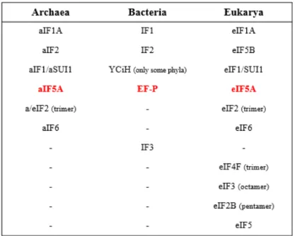
A list of IFs is presented in table 1:
Table 1 Initiation factors in the primary domains (modified from Londei, 2005).
The probable structure of the full archaeal pre-initiation complex, including small 30S subunit, aIF1, aIF1A, Met-tRNAi, mRNA, and αßγ-aIF2 was proposed by Cryo-EM analysis (figure 1).
Fig.1 Cryo-EM maps of archaeal initiation complex (Coureux et al, 2016). The full archaeal initiation complex
includes 30S from Pyrococcus abyssi (Pa-30S), shown in pale yellow, initiation factors Pa-aIF1 (in violet), Pa-aIF1A (in orange), Pa-aIF2 composed of three subunits α (in cyan), ß (in blue), γ (in green). Met-tRNAi from E. coli is shown in
9
The trimeric aIF2, like its eukaryotic counterpart eIF2, interacts with Met-tRNAi and stimulates its binding to the 30S subunits, in a GTP-dependent reaction (Pedullà et al, 2005). More recently it was discovered that the γ-subunit of aIF2 is able to bind the 5’ end of mRNAs, protecting them from degradation, displaying a dual function of this factors in S.
solfataricus (Hasenӧhrl et al, 2008). aIF1(aSUI1), homolog with eIF1, bind 30S subunits
and facilitates the interaction of the tRNAi and mRNA to the ribosome (Hasenӧhrl et al, 2006). aIF1A is a homologue of eukaryal eIF1A, but its functions still remain elusive. The translation factor IF6 is present in Archaea and in the Eukarya, but is not found in Bacteria. Benelli and co-workers (2009) demostrated that aIF6 in thermophilic S.
solfataricus binds specifically the large 50S subunit of ribosome preventing the formation
of 70S. They also proved that aIF6 is over-expressed under stress conditions, therefore its probable function is also to modulate protein synthesis under unfavourable circumstances. Last archaeal IF in Table 1 is the subject of this doctoral thesis: the archeal translational factor aIF5A. This factor is universal conserved and homologous between Eukarya and Archaea and with an orthologue, EF-P, in Prokaryotes.
1.2 The eukaryotic translation factor eIF5A
1.2.1 Structure and post-translational modification
The eukaryotic translation factor eIF5A is a small acidic protein of 17 kDa, highly conserved in all three primary domains, with homologues in Archaea (aIF5A) and in Bacteria (EF-P). This protein is essential in Eukarya and Archaea but not in Bacteria (Gäbel et al, 2013; Schnier et al, 1991; Park et al, 2010; Zhang et al, 2018; Balibar et al, 2013). It was initially identified from fractionated rabbit reticulocyte lysates as an initiation translation factor in the 1970s (Kemper et al, 1976; Benne et al, 1978).
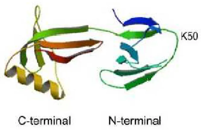
The three- dimensional structure of human eIF5A shows that this protein is mainly composed of ß-sheets. As shown in figure 2, it consists in two different domains: a basic N-terminal domain is folded in an SH3-like barrel, found in other proteins related to translation, while the C-terminal domain harbors an OB-fold (oligonucleotide-binding fold), a five-stranded beta-barrel known to bind nucleic acids and typical of other translational machinery components, like eIF1A, eIF2α and other ribosomal proteins (Dias et al, 2013).
10
Fig. 2 Crystal structure of human eIF5A protein (PDB ID code 3cpf) (Park et al, 2010)
This eIF5A protein shows high amino acids conservation in all eukaryotes, particularly in an exposed loop linking ß3 and ß4 strands in N-terminal domain, in which a special lysine residue (K50 in human and K51 in yeast eIF5A) is post-translational modified in hypusine residue.
Fig.3 Conservation of the amino acid eIF5A sequence in Eukarya. The human sequence is shown. The degree of
conservation is indicated by color coding: red, 100% identity; dark orange to yellow, conservative replacements with >80 to >50% sequence identity; white, no significant sequence identity (Wolff et al, 2007)
The amino acid sequence surrounding this modification site (SKTGKHGHAK) is found in more than 100 different species in eukaryotes, and is a very basic and hydrophilic portion (Park et al, 2010).
Hypusine was discovered by Shiba et al. (1971) from bovine brain extract during their search for new amine components. Its name derived from its two components: hydroxyputrescine and lysine. About 10 years later it was discovered that hypusine is present exclusively in the translational factor eIF5A, being added in a post-translational modification process. Until today eIF5A is still the only known hypusinated protein. This unique modification pathway
11
is achieved in two enzymatic steps (Figure 4): in the first step deoxyhypusine synthase (DHS) tranfers an aminobutyl group from polyamine spermidine to one specific lysine residue (K51 in yeast) of eIF5A precursor, catalyzing the formation of an intermediate, deoxyhypusine [Nε-(4-amino-butyl)-lysine] residue. DHS depends on NAD+ as coenzyme.
This intermediate is subsequently hydroxylated by deoxyhypusine hydroxylase (DOHH) to form mature and active eIF5A with hypusine residue (Park et al, 2010; Wolff et al, 2007).
Fig.4 Hypusine biosynthesis in eIF5A (Park, 2006).
Regarding the quaternary structure of eIF5A, the protein was shown to exist as a homodimer both in solution and in vivo. In a study of 2009, Gentz and coworkers demostrated that formation of the eIF5A dimerin S. cerevisiae requires the presence of the hypusine residue as well as the presence of cellular RNA molecules (Gentz et al, 2009). More recent experiments always performed in yeast, showed that eIF5A forms dimers, both in vitro and
in vivo, in an RNA-dependent manner, but regardless of the presence of the hypusine residue.
This oligomer conformation was also confirmed by SAXS analysis which showed that the eIF5A dimer is L-shaped resembling the bacterial EF-P and superimposable with the tertiary structure of a tRNA (Dias et al, 2013).
12
eIF5A and its hypusine modification are essential for eukaryotes and besides eIF5A, also the enzymes DHS and DOHH are highly conserved in the entire eukaryotic kingdom (Wolff et
al, 2007).
Two or more eIF5A isoforms have been identified in Eukarya (Jenkins et al, 2001). In yeast
S. cerevisiae there are two eIF5A genes, TIF51A and TIF51B, regulated by oxygen with TIF51A mainly expressed under aerobic condition while TIF51B only under anaerobic
conditions (Schnier et al, 1991). These two genes were found to perform the same function in yeast and gene inactivation of one gene or both rendered S. cerevisiae not viable, underling the essential nature of eIF5A. The eIF5A-depleted cells showed a phenotype characteristic of G1 arrest (Kang and Hershey, 1994). On the contrary, in higher multicellular eukaryotes like C. elegans, the two eIF5A genes, IFF-2 and IFF-1, code for two isoforms (eIF5A1 and eIF5A2 respectively) with different functions and tissue localization. eIF5A1 is required in soma for somatic tissue growth and organization, while eIF5A2 is required in germline and gametogenesis (Hanazawa et al, 2004).
The essential role of hypusine and the ezymes involved in this modification, were also studied in yeast, through the costruction of eIF5A-1 mutant (K51R), in which the specific lysine 51 was substituted with arginine. DHS enzyme, is unable to modify this eIF5A mutant in S. cerevisiae strains, making this yeast strain unfit to grow (Schnier et al, 1991).
DHS structure was characterized in human through crystal structure of recombinant enzyme in complex with NAD+, available since 1998 (Liao et al, 1998). Human DHS is a
homotetramer composed of four identical subunits of 40 kDa and its active sites are located at the interface between dimers, providing the binding site for NAD+. There is a single DHS
gene in yeast and in most eukaryotes. The role of deoxyhypusine synthase was demostrated by gene distruption in yeast and with knock-out studies in mice. In both cases it was established the essential requirement for DHS, for growth and survival of eukaryotes, from yeast to mammals.
Like eIF5A and DHS, also DOHH is highly conserved in the eukaryotic kingdom. DOHH exists as a product of a single gene which in human is a 32 kDa protein. Sequence alignment of DOHH proteins reveals that this enzyme belongs to a HEAT-repeat-containing proteins family. It is composed of a symmetrical super helical structure with 8 helical hairpins and requires oxygen and iron for his activity, harboring in his active center a nonheme diirion site. Recently the crystal structure of human DOHH has also been published (Han et al.,
13
2015). Curiously, DOHH activity is only essential in higher eukaryotes, such as mammals, but not in yeast. In mammalian cells metal-chelating inhibitors of DOHH cause G1 cycle arrest and growth inhibition (Hanauske-Abel et al, 1994). Moreover, inactivation of DOHH is also lethal in C. elegans and D. melanogaster (Spradling et al, 1999; Maeda et al, 2001; Patel et al, 2009). In contrast, in yeast S. cerevisiae the DOHH gene is not essential, even though native eIF5A exists as hypusine-modified form. This may explain the recent discovery in protozoan parasite Trichomonas vaginalis, in which dohh gene is lacking but eIF5A protein is completely hyspusinated. Researchers demonstrate in this study that TvDHS is able to perform both DHS and DOHH reactions, because in its tetrameric structure that resemble the human DHS, the enzyme has also HEAT-motifs typical of DOHH enzyme, allowing to perform also hydroxylase reactions (Quintas-Granados et al, 2015).
In addition to the hypusine post-translational modification, it is known that yeast eIF5A is also phosphorylated on the Ser2 residue of the protein (Klier et al, 1993), but mutation studies reveal that phosphorylation of eIF5A does not appear to be indispensable for protein’s function, unlike its hypusine modification.
1.2.2 Function of eIF5A in translation
eIF5A was originally purified from ribosomes of reticulocyte lysate in 1978, suggesting immediately an involvement in protein translation. At the beginning it was hypothesized that eIF5A stimulate the methionyl-puromycin synthesis, indicative of a role in the formation of the first peptide bond (Benne et al, 1978). In the years, biochemical and molecular genetic studies of yeast eIF5A provided new information about the function of this translation factor. Examining the impact of eIF5A depletion in yeast, a pronounced defect in total protein synthesis was observed, unlike the moderate inhibition previously reported (Kang and Hershey, 1994; Saini et al, 2009). Moreover, in other studies it was demonstrated that eIF5A fisically interacts with structural components of translation machinery. Proteins co-purifying with recombinant tagged-eIF5A were identified in S. cerevisiae and among these a 60S ribosomal protein P0, 40S ribosomal protein S5 and translation elongation factor 2 (eEF2) were found. These interactions appeared to be hypusine-dependent, since mutation of lysine 51 to arginine in the site that undergoes hypusination, decreased intensity of the interaction just described, especially for eEF2, as loss of interplay between eIF5A/eEIF2 was observed
14
(Zanelli et al, 2006). Recently it was proved that these two translation factors display a negative cooperativity on binding to the ribosome (Rossi et al, 2016).
Interaction ofeIF5A with ribosomes was also confirmed by the work of Jao and Chen (2006), in which they provided a list of eIF5A-tagged interacting partners. 14 out of 19 proteins are ribosomal proteins from either 40S or 60S, the other proteins include translation factor 1A (eIF1A) and two chaperonine. They suggested that eIF5A binds 80S ribosome in active translation and this interaction requires RNA and hypusine modification. Using endogenous yeast eIF5A, Jao et al (2006) reached the same conclusions.
A further evidence for the involvement of eIF5A in translation elongation was obtained by polysome profile analysis. This technique is used mainly to assess translation function in yeast, with cycloheximide treatment that freezes ongoing translation elongation and extracts are then separated on sucrose gradients. Analyses of polysome profile of temperature-sensitive eIF5A mutants showed an increase of polysome/monosome ratio at the restrictive temperature, that could be explained with a block of translation elongation (Zanelli et al, 2006), the same pattern expected for polysome profile of wild type cells after sordarin treatment, an elongation factor 2 inhibitor (Saini et al, 2009), supporting once again a role of eIF5A in elongation.
An important scientific milestone for the function of eIF5A in translation was reached in 2013 when Gutierrez and coworkers used in vivo and in vitro assays to unveil that eIF5A plays a crucial role in translation of specific proteins containing consecutive polyproline residue (Gutierrez et al, 2013), consistent with the reports of the ortholog EF-P. A set of dual-luciferase reporter constructs in which 5’ Renilla luciferase and 3’ firefly luciferase open reading frames (ORFs) are separated by sequences encoding ten consecutive codons for each of twenty amino acids, were introduced into wild type and eIF5A temperature-sensitive eIF5A mutant strains of yeast. Monitoring of all 20 luciferase reporter constructs revealed that only those containing proline codons were specifically impaired in eIF5A mutant. In vitro analysis confirmed this result, as only those peptides containing at least three consecutive proline residues showed absolute dependence on eIF5A. Ribosomes encountering these polyproline motifs stall, they are unable to proceed with translation and need to be rescued by eIF5A.
The reason for this, resides in the fact that not all peptide bonds are indeed formed with equal efficiency, as certain amino acids are poor donors or acceptors. Proline is an imino acid with
15
its side-chain cyclized onto the backbone nitrogen. The constrained geometry of the cyclic proline side chain makes this amino acid both a poor acceptor as well as a poor donor in the peptydil transferase reaction, therefore consecutive proline stretches causes the ribosome to stall.
Structural studies of eIF5A bound to the ribosome helped to understand how eIF5A could contribute to rescuing ribosomes stalled during translation. A model was proposed in 2016 by Schmidt with cryo-electron microscopy (Schmidt et al, 2016) and subsequently by Melnikov with crystal structure of eIF5A bound to 80S S. cerevisiae ribosome (Melnikov et
al, 2016), in which eIF5A is located between the P and the E sites, interacting with tRNAs,
rRNA and ribosomal proteins. As shown in figure 5B, domain I of eIF5A contacts 25S rRNA nucleotides located in helices H74 and H93, while domain II contacts ribosomal protein L1 and L42 and charged hypusine interacts A76 of the CCA-end of peptidyl-tRNA in PTC of the ribosome, stabilizing and restricting the position of the polyprolines in the P-site. Thus, eIF5A facilitates the transfer of the nascent chain from P to A-site.
Fig.5 Cryo-EM structure of eIF5A bound to the yeast 80S ribosome (A) Cryo-EM map of eIF5A-80S complex, in
which it is shown the eIF5A binding site (B) Molecular model for the interaction of domain I and II to the ribosome component. (Schmidt et al, 2016).
16
Using a proteomic approach, the frequency of polyproline motifs in all three primary domains of life was determined and it was found that the frequency of PPP or PPG containing proteins increases with organism complexity, in E. coli the frequency is 5.8%, in S.
cerevisiae is 9,7% while in H. sapiens is 33,5%. This could explain why eIF5A is essential
in all eukaryotes contrarly to EF-P of bacteria (Mandal et al, 2014), and the number of polyproline triplets increases considerably during the evolution (Figure 6).
Fig.6 The frequency of PPP and PPG motifs in different organisms. The histogram shows the frequency of Pro-pro-pro
and pro-pro-gly per 104 amino acid residues (Turpaev, 2018).
However, this view has been slightly changed in a recent study in which ribosome profiling of eIF5A depletion strain in yeast showed that many other tri-peptide motifs cause ribosome stalling. Interestingly, many of these motifs contain not only polyproline, like previously reported, but a combination of aspartic acid, glycine and other amino acids and few of these contain even no proline at all. Moreover, eIF5A depletion impacts in a defect of global elongation rate, while EF-P loss does not (Schuller et al, 2017). These findings led to hypotize that eIF5A is involved in a global translation elongation and support a model in which eIF5A is a general elongation factor, in contrast of the ortholog EF-P, which is much more specialized in translation of proteins containing proline stretches.
In the same study Schuller et al. also demostrated a role of eIF5A in translation termination, greatly expanding the role of this essential translation factor. Using the same eIF5A-depletion strain, they proved, through in vivo ribosome profiling experiments, that the stop
17
codon peak is 12-fold higher compared to wild-type and ribosome occupancy in 3’-UTR is increased, consistent with a translation termination defect, while previous studies of this group showed no such effect in E. coli cells lacking EF-P. They also showed using an in
vitro translation system, that hypusinated eIF5A stimulates an eRF1-mediated peptide
hydrolysis of peptidyl-tRNA by 17-fold compared to wild type, and hypusine is critical for this activity, as the rate of peptidyl-tRNA relase has a minor effect (4-fold). Taken together, this data provided support to the idea that eIF5A plays a crucial role in translation termination by stimutating the activity of eRF1.
1.2.3 Other functions of eIF5A and implication in pathological cellular
process
In addition to its role as a translation factor, the eukaryal eIF5A has been implicated into a variety of cellular processes, like mRNA decay (Zuk and Jacobson 1998), cell cycle progression (Hanauske-Abel et al, 1994), apoptosis (Caraglia et al, 2013), cell polarity (Chatterjee et al, 2006; Zanelli e Valentini 2005), retroviral and protozoan infection (Hoque
et al, 2009, Olsen and Connor, 2017) and stress response (Gosslau et al, 2009).
Studies on the immunodeficiency virus (HIV) reveal that eIF5A interacts with viral mRNAs, mediating the translocation of these mRNAs from the nucleus to the cytoplasm, and these interactions are mediated by a stable binding of the hypusinated protein with specific mRNA (Liu et al, 1997). Using SELEX technique (systematic evolution of ligands by exponential enrichment) it was shown that mRNAs bound by eIF5A, isolated by co-immunoprecipitation with FLAG-tagged eIF5A, shared a conserved motifs UAACCA and AAAUGU (Xu and Chen, 2001). They speculated that eIF5A-RNA interaction is due to some structural elements in RNA molecules, like hairpins and internal loops, suggesting that eukaryotic translation factor 5A is also a specific hypusine-dependent RNA binding-protein. This notion finds support in the analysis of the structure of eIF5A C-terminal domain, which, as described above, has an OB-fold typical of nucleid acid binding-protein. In particular this portion of the protein is negatively charged and shares a structural similarity with the RNA chaperone CspA.
There is also evidence that eIF5A is a key protein in the pathogenicity of different diseases, such as diabetes, malaria, HIV-1 infections and human cancer (Kaiser et al, 2012). Several
18
studies in these last years own the implication of eIF5A in many tumor types as a main topic. In particular, it has been shown that the two isoforms of human eIF5A display different functions and expression patterns. eIF5A-1 protein is constitutively expressed in all tissues, whereas eIF5A-2 is not normally detectable, but is over-expressed in certain human ovarian cancer tissues and colorectal (Clement et al, 2003), human hepatocarcinoma cell lines and mouse embryonic livers (Lee et al, 2010) and this upregulation of the protein caused cellular transformation. Based on these findings eIF5A-2 is proposed to be an oncogene. In this scenario, the hypusine modification seems to have an important task. Several studies indicate that hypusination of eIF5A is required for tumor maintenance and disease progression and the inhibition of hypusine prevents tumorogenesis (Nakanishi and Cleveland, 2016). Hence research in this field is moving towards the identification of therapeutic compounds targeting eIF5A protein and the hypusination pathway (hypusine, DHS, DOHH), which could be considered as new drugs and, combined with the other therapies, used to treat cancer patients.
1.3 The bacterial translation factor EF-P
1.3.1 Structure and post-translational modification
EF-P, isolated by Glick and Ganoza in 1975, is a small protein of 21 kDa which exibits a three-domain structure (I, II, III), with an overall L shape remindful of a tRNA molecule (figure 7). The N-terminal part consists of domain I and II, forming one arm of the L, whereas the other arm is formed by domain II and III, the latter domain forming the C-terminus of EF-P. Domains II and III are similar to the OB-fold motif (oligonucleotide-binding fold) observed in E. coli cold shock protein, polyribonucleotide nucleotidyltransferase, transcription factor Rho, and other proteins known to bind DNA or RNA molecules. However, EF-P is an acidic protein (PI= 4.6) and most of the overall surface is negatively charged, especially domain II and III presents most of the negatively charged aminoacids (Glu-76, Glu-78, Asp-84, Glu 89, Glu-106 for domain II and Asp-134, Glu-154, Glu-166, Glu-169 for domain III are conserved).
19
Fig.7 Comparison of crystal structure of EF-P from T. thermophilius (PDB ID code 1UEB) and tRNA (Lassak et al, 2016).
As shown in figure 8, Domain I of EF-P and the N-terminal domain of eIF5A show the same fold, as do domain II of EF-P and C-terminal domain of eIF5A. The third domain is an exclusive of EF-P and has high structural homology with domain II: this suggest that this additional domain in EF-P arose as a result of a duplication event (Lassak et al, 2016). Crystal structure of T. thermophilus of EF-P (PDB ID code 1UEB) demostrate that the angle formed by the two arms of EF-P is around 95°C and notably, the interfaces between domain I-II and II-III are composed of hydrophobic chains with high surface complementarities. This proves that the L shape of this translational factor is due to a native conformation of the protein instead of an artifact in crystal packing (Hanawa-Suetsugu et al, 2004). Comparison of this structure with the other X-ray results of the bacterial EF-P available on PDB, coming from P. aeruginosa (Choi and Choe, 2011) and Clostridium thermocellum (PDB ID code 1YBY), denotes a high structural flexibility (>60° rotation) of domain I respect to domains II and III, which remain relatively rigid.
20
Fig.8 Comparison of eukaryal eIF5A and bacterial EF-P structure. (Dever et al, 2014).
The alignment of the bacterial EF-P sequence with eukaryal eIF5A shows that in the N-terminal domain, in the region corresponding to the eukaryal hypusinated Lys50 residue, there is the highest conservation of aminoacids, although in E. coli DHS and DOHH are absent (Park et al, 2011). In some species including Thermus thermophilus, lysine residue is replaced by arginine. Several experiments proved that also EF-P is subjected to a post-translational modification, although not in all bacteria. This modification, studied in E. coli and Salmonella sp, is called ß-lysylation and occurs in three steps and requires the enzymes YjeK, YjeA and YfcM (Navarre et al, 2010; Yanagisawa et al, 2010; Peil et al, 2012). In a first step the enzyme YjeK (called also EpmB), a lysine-2,3-aminomutase, converts a free S-α-Lysine to R-ß-Lysine (Behshad et al, 2006). The second enzyme involved in this modification is a paralog of the lysyl-tRNA synthetases (YjeA, also known as EpmA) and transfers an R-ß-Lysine portion to the є-amino group of a specific lysine, using ATP. Based on this hypothesis, the crystal structure of E. coli YjeA in complex with EF-P was solved in 2010, demostrating lysinylation of Lys 34 (Yanagisawa et al, 2010). Moreover, in a previous experiment, Aoki et al. revealed that native EF-P purified from E. coli had an extra mass of +144 Da at Lys34 (Aoki et al, 2008). Sole lysinylation is predicted to increase the mass of
21
EF-P of only +128 Da. This discrepancy was resolved when Peil et al (2012) demostrated that another step of modification occurrs, performed by YfcM (EpmC). They proved by MS-analysis that native EF-P from cells lacking YfcM showed a decrease of 16 Da in mass, indicating that this third enzyme complete the modification patway of EF-P in E. coli (Peil
et al, 2012). YfcM recognizes EF-P only in its modified form and hydroxylates the
lysyl-lysine residue.
22
Although hydroxylation is the last modification step also in eukaryotes, there is no evolutionary correlation between hydroxylases involved in hypusination of eIF5A and lysinylation of EF-P.
This modification system has been identified only in about one quarter of all bacterial genomes (EpmA and EpmB are found in about 26% of bacteria while EpmC is restricted almost excusively to γ-proteobacterial genomes), but ß-lysinylation is necessary for EF-P function in species that contain this modification, such as E. coli and Salmonella sp. Based on the importance of modification in this species, Lassak and coworkers speculated that lysinylation is not the only modified mechanism present in Bacteria (Lassak et al, 2015). As mentioned above, some species are characterized by a conserved arginine residue in EF-P (Arg32) at the position equivalent to Lys34 found in E. coli.
Using γ-proteobacterium Shewanella oneidensis as a model organism they described rhamnosyl modification pathway, in which 2’-deoxy-thymidine-ß-L-rhamnose is attached on the arginine 34 of EF-P by a glycosyltransferase (EarP). Also, this modification produced a mass shift of +146 Da in MS-analysis in EF-P -Arg32 modification pathway, consistent with a deoxyhexose formation. Members that assess EF-P activation via EarP also comprise notable pathogenic bacteria as Neisseria sp., Bordetella pertussis and Pseudomonas
aeruginosa, and deletion of efp or earP genes lead to a lack of pathogenicity in these
organisms. In P. aeruginosa mutant strains are unable to kill human cells. (Lassak et al, 2015).
23
Anyway, it is important to mention that both modification in EF-P and the enzymes involved in these pathways were identified in only about 35% of the availabe bacterial genomes. The hypothesis is that there are probably alternative bacterial strategies of modification not identified yet.
1.3.2 Function of EF-P
EF-P is coded by only one gene, efp, and is universally conserved in Bacteria. In contrast to what was reported for eIF5A, P is not an essential protein. It was demonstrated that EF-P is dispensable in Escherichia coli and EF-Pseudomonas aeruginosa (Balibar et al, 2013). The EF-P protein was initially identified as an elongation factor, as was predominantly isolated from post-ribosomal supernatant (90%). According to this model EF-P stimulates peptide bond formation during translation elongation (Glick et al, 1975). Afterwards, some studies speculated on the hypotesis that EF-P enhances peptidyl-transferase activity in specific aminoacyl-tRNAs, such as those charged or with relative wide size of amino acids side chain (Glick et al, 1979; Ganoza and Aoki, 2000). The next important scientific breakthrough for EF-P was in 2009 when a crystal structure of Thermus thermophilus EF-P bound to 70S ribosome (Fig.6) was described (Blaha et al, 2009). Despite its resemblance to a tRNA, EF-P bound to the 70S ribosome between the EF-P and E sites, adjacent to the EF-P-site tRNA and interacts with both ribosomal subunits in the interface between 30S and 50S. Domain I and II of EF-P, which are superimposable to N-terminal and C-terminal of a/eIF5A, bind next to the acceptor stem and D-loop of tRNA in P site; EF-P domain III, which is absent in a/eIF5A, interacts with fMet-tRNA near its anticodon stem-loop, close to 30S subunit. EF-P domain II has also connection with L1 ribosomal protein, that is structurally and functionally conserved among all organisms, involved in tRNA translocation and release. In domain I a loop contacts the CCA-end of tRNA, maybe orienting the amino acyl arm optimally for peptide bond formation.
24
Fig.11 Crystal structure of EF-P bound to the ribosome. (A) E-site (shown in red) and P- site (shown in green) tRNAs
bound to 70S. (B) EF-P (shown in shades of magenta to indicate different domain of the protein (I, II, III) and P-site tRNA binding in the 70S ribosome. (C) Same as (A) but with ribosomal protein L1 (shown in gold). (D) L1 movement,
due to the presence of EF-P. (Blaha et al, 2009)
Next important insights for the function of EF-P were provided by studies of the eukaryotic eIF5A, that was demostrated in 2009 to have a role during translation elongation.
Mutant of EF-P, YjeA, YjeK cause the accumulation of polysomes (Bullwinkle et al, 2013) and these bacteria exibit pleiotropic phenotypes, correlated with specific cellular process, such as virulence, cell mobility, stress conditions resistance. These results were consistence with a function of EF-P in translation elongation, in particular it could be involved in
25
traslation of specific mRNAs, as described for eIF5A. In fact, recent studies in ribosome profiling of E. coli demostrated that EF-P is important in peptidyl bond formation between two proline motifs. In Bacteria, lacking EF-P or the enzyme for lysinylation, ribosomes stop at these motifs and are unable to procede with the translation. Levels of pausing at Pro-Pro varied depending on precedent and subsequent codon: PPW, PPN, PPD, PPP, GPP, DPP, APP induced very strong pauses whereas PPI, PPL, PPM, PPY, PPF exhibited no pausing. As previously described for the eukaryal counterpart, this phenomenon is due to the geometry of the cyclic proline side chain and EF-P function is to stabilize the CCA-end of the peptidyl-tRNA. More recently, the same poly-proline specific function for eIF5A has been demostrated in yeast by Gutierrez and coworkers (2013), suggesting that the hypusine residue directly interact the CCA-end of tRNA in PTC. The same mechanism can be proposed for EF-P in that 30% of bacteric species harboring the Lys/Arg 34. However, the number of proteins containing polyP, PPG or other stalling motifs is higher in eukaryotes and this could explain the fact that eIF5A is essential while bacterial EF-P is not.
Many questions about EF-P, still remain open, and to answer these questions further functional and structural studies are needed.
1.4 The archaeal translation factor aIF5A
1.4.1 Structural features
Archaea contains an eIF5A homolog protein and EF-P ortholog, termed archaeal initiation factor 5A (aIF5A), that shows strong structural and functional similarity to the eukaryotic and bacterial factors. This protein was isolated in 1992 from the thermophilic archaeabacterium Sulfolobus acidocaldarius as a small protein of 15 kDa containing a specific post-translational modification of a lysine residue, called hypusine, as described for eukaryotes (Bartig et al, 1992).
Concerning the structure, the crystal analysis of aIF5A from hyperthermophililc archaebacterium Pyrococcus horikoshii highlights the resemblance of the archaeal protein to the eukaryatic one.
Pho-aIF5A is predominantly composed of ß-strands and comprises two distinct domains
(Fig.12A), an N-terminal domain which includes residues 1-69 and has a SH3-like barrel, consisting of a 310-helix (α1) and a six-ß-stranded (ß1 ß6) anti-parallel ß-sheets. This
26
domain also includes a long hairpin loop called L1, that comprises residues 33-41, which hold a specific lysine (Lys37), that is supposed to be modified into hypusine, as it happens in eukaryotes. The C-terminal domain is composed of residues 72-138 and has an OB-fold (oligonucleotide binding fold) motif, comprising two short α-helices (α2- α3) and five stranded anti parallel ß-sheets (ß7 ß11). These two domains are connected by a short linker of only two residues (70-71). Moreover, between the N- and C-terminal domain the basic residues create a positively charged surface, and also the L1 hairpin with hypusine modification is positive. On the contrary C-domain is mostly negatively charged and these difference in electrostatic potential of the protein led to the speculation that aIF5A can act as a bimodular protein capable of interacting with nucleic acid (especially RNA molecules) through his positive portion and conversely the C-terminal domain most negatively charged interact with proteins (Yao et al, 2003).
Fig. 12 (A) Representation of translation initiation factor 5A from P. horikoshii (Yao et al, 2003) (B) Comparison of crystal structure of aIF5A with EF-P (Park et al, 2010).
27
Comparing them to EF-P, eIF5A and aIF5A seem to share more similarity since both are lacking an additional third domain present in EF-P and have the same post translational modification (Figure 13A and B), though eukaryotic protein has an alpha-helix in C-terminal domain not present in aIF5A.
But if we consider the amino acids sequence between aIF5A and domain I and II of EF-P, identity between aIF5A/EF-P is greater than between eIF5A/EF-P (Aoki et al, 2008). The archaeal and bacterial structures are superimposable as shown in figure 12B, while eukaryal and bacterial proteins have similar overall shape, as eIF5A is a dimer in solution and resemble a L-shape protein exactly like EF-P (Figure 13 C and D). These structural similarities suggest that there are probably a structural and functional conservation of these translation factors throughout the evolution.
Fig.13 Structural insights into eIF5A, aIF5A and EF-P (Lassak et al, 2016) Comparison of crystal structure of (A) P.
horikoshii aIF5A (PDB ID code 1IZ6), (B) human eIF5A (PDB ID code 3CPF) and (C) T. thernophilus EF-P (PDB ID code 1UEB). (D) Dimerization model of eIF5AHYp in solution from S. cerevisiae obtained by SAXS data (Dias et al,
2013).
1.4.2 Post-translational modification of aIF5A
Very little is known about the translation factor aIF5A in the archaeal kingdom. Like eIF5A, archaeal aIF5A is essential: the gene was found to be essential in Haloferax volcanii (Gäbel
et al, 2013), in Sulfolobus acidocaldarius (La Teana and Albers, unpublished data) and more
recently in Sulfolobus islandicus (Zhang et al, 2018).
Hypusine was identified since 1989 in some aerobic archaea such as Sulfolobus
acidocaldarius, Halobacterium cutirubrum and Thermoplasma acidophilum (Schumann et al, 1990).
28
A year later a subsequent study revealed that the distribution of hypusine in the kingdom is heterogenous. Several species, strictly anaerobic thermophilic in the archaeal kingdom, contain deoxyhypusine instead of hypusine, the intermediate of eIF5A in eukaryotic process, such as Thermoproteus tenax, Pyrodietium oecultum and Desulfurolobus ambivalens (Bartig
et al, 1990). Interesting, Thermoproteales sp. own also hypusine, suggesting that
deoxyhypusine could be an intermediate of hypusine modification as in eukaryotes, but probably through an alternative hydroxylation pathway, since these anaerobic organisms are unable to perform an oxygenation reaction.
Although the mechanism of hypusine modification is widely studied in eukaryotes, the hypusination pathway and the role of aIF5A in Archaea are poorly understood. aIF5A is present in all archaeal genomes sequenced so far and also the DHS homologs are present in all sequenced archaeal genomes but no DOHH homologs has been identified in any archaeal genomes or proteomes to date (Park et al, 2006), raising a question about how hypusine residue is synthesized in certain archaeal family like Sulfolobaceae. Archaeal DHS is found to be essential as the eukaryal counterpart, because growth inhibition by N1
-Guanyl-1,7-diaminoheptane (GC7), an efficient DHS-inhibitor in eukaryotes, was demostrated in four archaeal species (S. acidocaldarius, S. solfataricus, Halobacterium haloboum and Haloferax
mediterranei) (Jansson et al, 2000). These findings suggest that the first step of modification
in Archaea is conserved and deoxyhypusine could be synthesized by similar mechanism as described in eukaryotes. GC7 competes with spermidine for the binding to DHS in eukaryotes; this compound associates with the enzyme at the same site of spermidine binding (Jakus et al, 1993), thus preventing the transfer of N-butilic-group of spermidine to lysine. The authors suggested that the presence of unmodified aIF5A, unable to synthetize a subset of proteins involved in cell cycle progression, lead to arrest of growth of these organisms at the end of the D period (G2).
A recent analysis of euryarchaeota H. volcanii aIF5A reveals that the protein is deoxyhypusinated and provides important information about the mechanism of this type of modification (Prunetti et al, 2016). First of all, a mass spectrometry analysis of Hfx-aIF5A proved for the first time that is modified with deoxyhypusine at the conserved lysine 36 (in 99% cases) and no hypusine is detected. Secondly, the inefficiency of GC7 on the H. volcanii growth and the absence of intracellular spermidine in this organism, suggest a different pathway and lead researchers to hypotesize a model in with agmatine, an essential polyamine
29
mainly present in halophiles, could be the substrate for the deoxyhypusine modification, differing from the canonical eukaryotic pathway.
Moreover, they characterized for the first time the archaeal DHS enzyme as a tetramer complex of 148 kDa, finding in agreement with the structural information of eukaryotic DHS.
However, this alternative modification reaction could not be a common feature of all Archaea or all narrow group to which Haloferax belongs, as DHS activity of another euryarchaea was explored as a control. In fact, Prunetti et al. showed for the first time an in
vitro activity of T. kodakarensis DHS similar to eukaryotic enzyme: the transfer of the
4-aminobutyl group from spermidine to Tko-aIF5A. The modified aIF5A was analyzed by mass spectrometry demostrating the presence of deoxyhypusine residue on the lysine 42. These findings pave the way for futher studies, necessary to elucidate hypusination pathway of translational factor aIF5A in Archaea, that could change from organism to organism. As mentioned at the beginning, the function of aIF5A in Archaea is still poorly characterized and despite this protein seems to be similar to the eukaryal one, an involvment in protein synthesis has never been demostrated yet.
Wagner and coworkers showed for the first time an involvment of aIF5A from
Halobacterium sp. in RNA-mediated processes, showing an RNA cleavage activity, which
has not been reported for eukaryal eIF5A, and an RNA binding capacity. This latter activity required the hypusinated form of the protein, as hypusine residue is necessary to stabilize the RNA-aIF5A complex. Conversely, RNA degradation mediated by aIF5A does not require the hypusination of the protein, and the author indicated that this cleavage preferentially occurrs between adenine and cytosine nucleobases, within single strand regions (Wagner et al, 2007). Anyway, how these two activities are regulated is still obscure and furtherwork is necessary to clarify these functions.
30
31
2.1 Purification of recombinant N-His-aIF5A from E. coli
The recombinant protein for Sulfolobus solfataricus aIF5A (ORF 0970) was expressed in E.
coli and purified with a protocol described in Bassani, PhD thesis (2017).
Briefly, aIF5A gene was inserted into the plasmid pETM11 (kindly provided by Dr. Roberto Spurio, University of Camerino), to obtain a construct encoding a protein with six histidine residues at its N-terminal end. The recombinant plasmid was used to transform E. coli Rosetta (DE3) /pLysS cells. The resulting cells were grown at 37°C until they reached an optical density (OD) of 0.7 at 600 nm, then the production of the recombinant fusion protein was induced by addition of 0.5 mM IPTG (Isopropil-β-D-1-tiogalattopiranoside). After 3 hours cells were collected, resuspended in 20 ml of cold lysis buffer (50 mM Tris HCl pH 7.4, 150 mM NaCl, 15 mM Imidazole, 1 mM PMSF(Phenylmethanesulfonyl fluoride), 10 mM ß-mercaptoethanol, 0.1% Triton X-100, 25 µg/ml lysozyme) and incubated for 30 min on ice. After lysis by sonication, the cell lysate was centrifuged at 25000 g for 30 min at 4°C and the fusion protein purified by affinity chromatography on Ni-NTA Agarose resin (Quiagen). Binding of the N-His-aIF5A to the resin was carried out overnight on a rotating wheel at 4°C. The beads were washed with 30 ml of Wash Buffer (50 mM Tris HCl pH 7.4, 500 mM NaCl, 40 mM Imidazole) and eluted in 2 ml Elution Buffer (50 mM Tris HCl pH 7.4, 150 mM NaCl, 250 mM Imidazole). The eluate was dialyzed overnight at 4°C against Dialysis Buffer (50 mM Tris HCl pH 7.4, 150 mM KCl, 5% glycerol).
The concentration of aIF5A was determined by Bradford assay and the purity was assessed by SDS-PAGE (Sodium Dodecyl Sulphate - PolyAcrylamide Gel Electrophoresis), followed
by Coomassie-blu staining. Aliquots of the protein were stored at -80°C.
2.2 Purification of recombinant protein aIF5A-C-His from S.
solfataricus
Construction of plasmids for transformation of S. solfataricus PH1-16 cells is described in Bassani, PhD thesis (2017).
PH1-16 (pMJ05(ptf55)-aIF5A-C-His) strain was grown at 75°C in Brock’s medium (composed of Brock’s salts and supplemented with 0.2% NZamine, 0.2% sucrose, pH 3.0). At an A600 nm of 0.8, the cells were harvested and lysed by sonication. Fusion proteins were
32
purified by affinity chromatography on Ni-NTA resin, as described for N-His-aIF5A from
E. coli.
2.3 Purification of native aIF5A from S. solfataricus
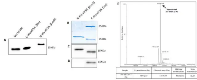
Purification of native aIF5A was carried out essentially as described in Bartig et al (1992), with some modifications. Affinity chromatography was performed at 4°C, all other purification steps were done at room temperature. The post-ribosomal supernatant (S100) obtained form 20 g of cell paste was subjected to affinity chromatography on Cibacron blue Sepharose 6-fast flow (GE Healthcare, 50 ml bed volume, 25 x 2.5cm, 20 ml/h), equilibrated in Buffer A (20 mM Tris HCl pH 7.5, 0.1 mM EDTA, 150 mM KCl). The column was developed stepwise with Buffer A, Buffer A containing 6 mM spermine, again Buffer A, and finally Buffer A containing 1.5 M NaCl. 10 ml fractions from each step were collected and 10 µl from each fraction were tested by Dot-Blot (Biorad) using polyclonal anti-aIF5A antibody (Eurogentec), available in our lab. aIF5A positive fractions were pooled and concentrated using a Centricon 3KDa Amicon® Ultra centrifugal filter device. The following two chromatographic steps were performed using a FPLC Akta Purifier apparatus (GE Pharmacia) at room temperature. The concentrated protein pool was subjected to gel filtration chromatography on a Superose 12 column (GE Healthcare, 24 ml bed volume, 30 x 1.0 cm, 0.5 ml/min), equilibrated in Buffer A. Fractions of 0.5 ml were eluted with the same buffer and 2 µl from each fraction were tested by Dot-blot. aIF5A positive fractions were concentrated using Centricon 3 KDa (Amicon) and the buffer was changed to Buffer B (20 mM Tris HCl pH 9, 0.1 mM EDTA, 150 mM KCl). The sample was loaded on a Mono Q HR 5/5 column Pharmacia (1ml bed volume, 0.5 ml/min), equilibrated with Buffer B, and eluted employing a linear gradient of 0.05-1 M KCl. 0.5ml fractions were collected and 2 µl from each fraction were analyzed by Dot-blot. aIF5A positive fractions were concentrated and dialysed against aIF5A buffer (50 mM Tris HCl pH 7.5, 150 mM KCl).
Bradford analysis and SDS-PAGE were performed to determine protein concentration while Western Blot with anti-hypusine (Merk Millipore) antibody and LC-MSMS analysis were performed to identify the nature of protein modification.
33
2.4 Liquid chromatography – mass spectrometry (LC-MSMS
analysis)
The purified native aIF5A was loaded on a Mini-PROTEAN TGX Stain-Free Precast Gel (BioRad), and the protein band was excised and analyzed by LC–MS/MS analysis. The gel band was digested with trypsin as described (Mair et al, 2015). After digestion the peptide solution was desalted on a custom-made C18 stagetip (Rappsilber et al, 2007). Tryptic digests were separated on an Ultimate 3000 RSLC nano-flow chromatography system (Thermo Fisher Scientific), using a pre-column for sample loading (PepMapAcclaim C18, 2 cm × 0.1 mm, 5 μm) and a C18 analytical column (PepMapAcclaim C18, 50 cm × 0.75 mm, 2 μm, Dionex-Thermo-Fisher Scientific), applying a linear gradient from 2 to 35% solvent B (80% acetonitrile, 0.1% formic acid; solvent A 0.1% formic acid) at a flow rate of 230 nl min−1 for 60 min. The eluting peptides were analyzed on a Q Exactive HFX Orbitrap mass spectrometer, equipped with a Proxeon nanospray source (Thermo Fisher Scientific). The data-dependent mode survey scans were obtained in a mass range of 375–1500 m/z with lock mass on, at a resolution of 60.000 at 200 m/z and an AGC target value of 3E6. The 10 most intense ions were selected with an isolation width of 1.6 Da, fragmented in the HCD cell at 27% collision energy, and the spectra were recorded at a target value of 1E5 and a resolution of 30000. Peptides with a charge of +1 were excluded from fragmentation, the peptide match and exclude isotope features were enabled and selected precursors were dynamically excluded from repeated sampling for 15 s. The raw data were processed with MaxQuant software package (version 1.6.0.16, http://www.maxqu ant.org) (Cox and Mann 2008) by searching against the sequence of aIF5A in the background of the Sulfolobus
solfataricus Uniprot (http://www.unipr ot.org) and sequences of common contaminants,
with tryptic specificity allowing 2 missed cleavages. Carbamidomethylation was set as fixed modification, oxidation of methionine, N-terminal protein acetylation, hypusine and deoxyhypusine on lysines as variable modifications, all other parameters were set to default. Results were filtered at a protein and peptide false discovery rate of 1%. The protein list was further filtered for a minimum of 2 unique and razor peptides. Peptide hits returned by MaxQuant were manually validated.
34
2.5 Purification of N-His-DHS and DHS C-His from E. coli
The gene for S. solfataricus Deoxyhypusine synthase (DHS) (ORF Sso0967) was amplified by PCR using 100 ng genomic DNA of S. solfataricus P2, with two pairs of oligonucleotides for cloning the gene in two different plasmids:
1. forward 5’-GCGGCCATGGTAAATAGAGAGGAC-3’ (NcoI restriction site)
2. reverse 5’-CCGGGATCCTTAGCTTAATAAAGACG-3’ (BamHI restriction site)
3. forvard 5'-AAAAGCATGCGCATAAATAGAGAGGACTTGTTAAAAAACCC-3'
(SphI restriction site)
4. reverse 5'-AAAAGGATCCGCTTAATAAAGACGCGGCCAAAATAGG-3'
(BamHI restriction site)
ORF Sso0967 amplified with primers 1 and 2 was cloned in pETM11 for expression of N-terminal His-tagged DHS, while primers 3 and 4 were used for cloning of aDHS in the pQE-70 plasmid (Quiagen) which adds a C-terminal His-tag to the recombinant protein.
N-His-aDHS and aDHS-C-His were expressed in E. coli BL21(DE3) and purified with the same protocol described for aIF5A.
2.6 Small-angle X-ray scattering (SAXS) experiments
The recombinant Sso N-His-aIF5A and DHS-C-His proteins were produced from E. coli and purified by affinity chromatography as described above. The two proteins were, subsequently, subjected to SAXS experiments in order to obtain structural details about the overhall shape of the proteins and their conformation on solution at high temperature, and also to verify a formation of a stable complex between aIF5A and DHS.
The measurements were carried out at the Diamond Light Source (Harwell Science & Innovation Campus, Didcot, UK) synchrotron SAXS beamline BL21 with monochromatic beam. We have measured SAXS curves of the aIF5A protein, in 20 mM Tris HCl pH 7.7 and 2 % glycerol, at five different protein concentration (0.5, 1, 2, 5 and 10 mg/ml) with 2 different KCl amounts (60 and 1000 mM). Data have been recorded in the capillary heater at step of 5°C from 20°C to 63°C, the maximum temperature achievable by the apparatus. The range of the scattering vector moduls q ranged from 0.01 to 0.37 Å-1. Sso DHS protein in 20 mM Tris HCl pH 6.8, 120 mM KCl, 2% glycerol, were measured at 63°C at a concentration of 4 mg/ml, alone and supplemented with 2 mM NAD+ and 2 mM spemidine.
35
To assessed formation of a complex between aIF5A and DHS, scattering was recorded with the two proteins mixed in a molar ratio of 4:1, in the absence and in the presence of all the other components of the modification reaction, 2 mM NAD+ and 2 mM spermidine. Buffer
scattering was recorded before each sample scattering.
All protein preparations were centrifuged for 10 min at 14000 g before the measurements, to remove aggregates or particles, and stored at 4°C. During the analysis the samples were kept at room temperature.
2.7 SAXS data analysis
All analysis of SAXS data were performed in collaboration with the biophysics group of Polytechnic University of Marche (Ancona), coordinated by Prof. Francesco Spinozzi. The radius of gyration (Rg) and molecular weight of the aIF5A protein was determined by the Guinier equation and with a model that combines Guinier and Porod analysis (fit Guinier-Porod). For curves construction the Gnuplot program was used, editing the scripts with
Emacs program.
A Kratky plot is obtained by plotting scattering intensity as vs. This representation divides out the decay of the scattering, making certain other features more evident.
The QUAFIT software was used to determine the tertiary structure of the protein (Spinozzi and Beltramini, 2012), from the analysis of SAXS curve of aIF5A 10 mg/ml, 60 mM KCl, 63°C.
2.8 Size-exclusion molecular chromatography
2.8.1 Size-exclusion molecular chromatography of aIF5A
The recombinat Sso proteins N-His-aIF5A from E. coli and aIF5A-C-His from S.
solfataricus, were loaded on a 24 ml Superdex 75 10/300 GL Tricorn column (GE Life
Science). The molecular mass standard proteins (Sigma-Aldrich) BSA (Bovine serum
albumin) (67KDa) and myoglobin (17KDa), each at a concentration of 1 mg/ml, were used to calibrate the column. The void volume of column was calculated using Blue Dextran 2000. The chromatographic bufferwas subjected to filtration and contained 50 mM Tris HCl pH 7.4, 150 mM KCl. The column was equilibrated with 3 volumes of buffer, than 0.5 mg/ml
36
of different aIF5A preparations, pre-heated at 65°C for 10 min and centrifuged at 10000g for 10 min, were separated on a gel-filtration column using 1.5 volumes of buffer (35 ml), with 0.4 ml/min of flow rate. Elution fractions (500 µl) were collected and those containing protein selected according to their absorbance at 280 or 254 nm after which the proteins were analyzed by SDS-PAGE and visualized with Western Blot, using anti-aIF5A antibodies.
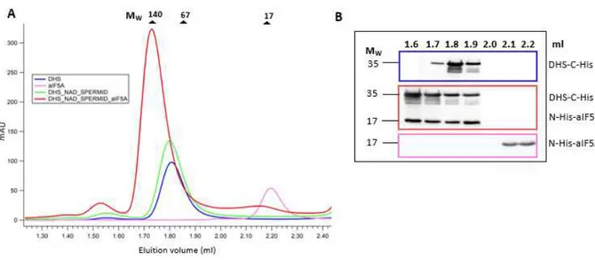
2.8.2 Size-exclusion molecular chromatography of DHS-aIF5A complex
A 3 ml Superdex 200 increase 5/150 GL Tricorn column (GE Life Science) was used to identify complex formation between aIF5A and DHS.N-His-aIF5A and DHS-C-His were purified from E. coli as described above. Both recombinant proteins were used in 50 µl enzymatic assays containing a reaction buffer (20 mM Tris HCl pH 7.4, 150 mM KCl), 50 µg of DHS-C-His, 25 µg of N-His-aIF5A, 2 mM NAD+, 2 mM spermidine. Reactions were also performed with aIF5A alone (20 µg), DHS
alone (20 µg), DHS (25 µg) with 2 mM NAD+ and 2 mM spermidine in the same buffer. All
reactions were incubated at 65°C for 30 min, and then loaded onto a gel filtration column, set at 65°C and pre-equilibrated with 9 ml of reaction buffer.
BSA (67 kDa), lactate-dehydrogenase (140 kDa) and myoglobin (17 kDa) were used as mass standard proteins, each at a concentration of 1 mg/ml. The void volume of the column was calculated using Blue Dextran 2000.
Elutions were carried out with 8ml of reaction buffer at 0.4 ml/min flow rate.
Elution fractions (400 µl) were collected and analyzed by SDS-PAGE followed by Western Blot, using anti-histidine antibodies, to appreciate complex formation between aIF5A and DHS.
2.9 Non-denaturing gel elecrophoresis
Mixtures of purified recombinant Sso proteins N-His-aIF5A (5 µg, 300 pmol) and deoxyhypusine synthase DHS-C-His (10 µg, 80 pmol) from E. coli in a molar ratio of 1:4 (enzyme/substrate), in the presence or absence of 1 mM NAD+ and 1 mM spermidine, were
incubated in 200 mM glycine/ NaOH pH 8.2 at 65°C for 30 min. After addition of native gel sample buffer 6x (187.5 mM Tris HCl pH 8.8, 75% glycerol, 3% bromophenolblue), the
37
proteins were separated under non-denaturing conditions on 12% polyacrilamide gel using following receipt:
• for a 10 ml native PAGE separating gel
• for a 5 ml native PAGE stacking gel
Gel was run in 25 mM Tris base, 192 mM glycine running buffer at 120 V for 2 hours, at 4°C.
Proteins separation was determined through Comassie Brilliant Blue staining. To actually prove complex formation between aIF5A and DHS, the corresponding band of the complex, detected by staining with Comassie, was excised from the native gel, cut into fine pieces, incubated with an equal volume of Sample Buffer 1X (62.5 mM Tris HCl pH 6.8, 2% SDS, 10% Glycerol, 5% ß-mercaptoethanol, 0.002% bromophenolblue) and boiled for 10 min at 98°C. Sample was loaded on a 15% SDS-PAGE and the two band of 5A and DHS were visualized by Comassie Blue staining.
2.10 In vitro hypusination assay
The Deoxyhypusine Synthase activity was tested in vitro using different amount of the recombinant protein, purified from E. coli, after pre-incubation for 10 min at 65°C. 200 and
38
1200 pmol of DHS-C-His were incubated in presence of 400 pmol of recombinant N-His-aIF5A purified from E. coli, 2 mM spermidine, 2 mM NAD+, 2 mM MgCl
2, 50 mM
glycine/NaOH pH 9.4, 150 mM KCl and 1 mM DTT in a final volume of 30 µl. The reaction mixture was incubated for 2 h at 65°C. An eventual increase in mass of N-His-aIF5A after the reaction was monitored by LC-MS. The recombinat protein N-His-aIF5A produced in E.
coli and, therefore, unmodified, was analyzed as a control.
High performance liquid chromatography was performed on a Dionex Ultimate 3000 HPLC (Thermo Fisher Scientific) system configured with the Chromeleon 6.0 software (Thermo Fisher Scientific).
Proteins were reduced in 100 mM DTT for 30 minutes at room temperature and then separated on an Aeris Widepore C4 column (3.6 μm particle size, dimensions 2.1 x 150 mm, Phenomenex) running a six minutes step gradient from 10% up to 70% acetonitrile in 0.1% formic acid.
39
The working temperature was set at 50°C and the flow rate at 300 μl/min. The LC-system was coupled online to the quadrupole-time of flight-mass spectrometer Synapt G2-Si (Waters), operated via the MassLynx V 4.1 software package, using a Z Spray ESI source (Waters). Mass spectra were acquired in the m/z range from 500-2000, at a scan rate of 1 sec and the mass spectrometer was calibrated with a MS spectrum of [Glu1] -Fibrinopeptide B human (Glu-Fib) solution.
Acquired data were analyzed with the MaxEnt algorithm to reconstruct the uncharged average protein mass.
2.11 Preparation of S. solfataricus cell lysate
Sulfolobus solfataricus P2 culture was aerobically grown in Brock’s medium (Brock et al,
1972), at 75°C and pH 3, with supplement of 0.2% NZ-amine and 0.2% sucrose.
Cells at exponential phase of growth (0.8 OD600) were harvested and pelleted. S. solfataricus
cells extract was prepared according to Benelli and Londei (2007) starting from 20 g (wet weight from 20 L of culture) of frozen cells. These were distrupted by grinding, with a sterile, cold mortar on ice, adding 40 g of alumina (2 g alumina each g of cells). Cells were resuspended in 20 ml of Extraction Buffer (Tris-HCl pH 7.4 20 mM, MgAc 10 mM, NH4Cl
40 mM, ß-mercaptoethanol 6 mM), and centrifuged at 15300 x g for 30 min at 4°C.
The supernatant was centrifuged at 15300 x g for 45 min at 4°C, and crude cell extract S30 was collected. S30 was than ultracentrifuged with Beckman Type 55.2 Ti rotor at 30000 rpm (100000 g) for 16 h at 4°C. Resulting pellet was ribosomal fraction and the supernatant was cytoplamic fraction, called S100, rich in proteins and cytoplasmatic factor.
S30 and S100 concentration was determined by Bradford.
2.12 Analysis of aIF5A levels under different growth conditions
The S. solfataricus cells were aerobically cultivated at 75°C pH 3, as previously described. Cell growth was monitored measuring A600nm until late exponential growth phase (34 h of
growth). When culture reached different densities, 15 ml of culture at 0.2 OD (8h), 0.4 OD (18h), 0.8 OD (24h), 1.3 OD (26h), 1.8 OD (30h) and 1.82 (34h), were harvested by centrifugation at 4000 rpm for 20 min at 4°C. Pellets were resuspended with Extraction