The ecology and diversity of
benthic deep-sea fungi
PhD Dissertation
Department of Life and Environmental Sciences Marine Biology and Ecology
XXXI Cycle
Candidate Tutor
Giulio Barone Professor Antonio Dell’Anno
Co-Tutor
Index Chapter 1
Fugal ecology and diversity in benthic deep-sea ecosystems 1
Microbial component in the deep sea 2
Diversity and ecology of fungi in different benthic deep-sea habitats and ecosystems 3
1. Hypersaline anoxic basins (DHABs) 3
2. Cold seeps 6
3. Hydrothermal Vents 9
4. Deep-sea sediments 14
5. Deep-sea sub-surface sediments 17
Conclusions and perspectives 20
References 22
Chapter 2
Benthic deep-sea fungi in submarine canyons of the Mediterranean Sea 28
Introduction 29
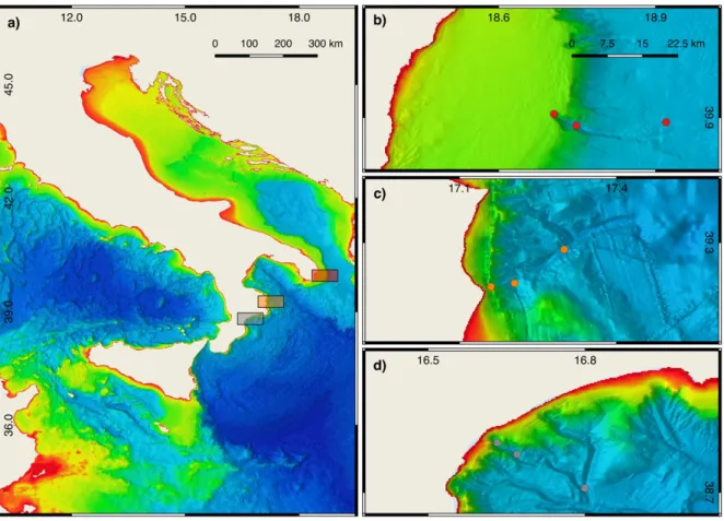
Materials and methods 30
Study area and sampling design 30
Quantity and biochemical composition of organic matter 31
Fungal biomass 32
DNA extraction and purification for molecular analysis 33
Quantitative real-time PCR of fungal 18S rRNA gene sequences 33
Fungal diversity 34
Statistical analyses 34
Results and discussion 35
References 43
Supplementary materials—Benthic deep-sea fungi in submarine canyons of the Mediterranean Sea 48
Chapter 3
Fungal abundance and diversity in the sediments of the deepest ecosystem on Earth 51
Introduction 52
Material and methods 53
Study site and sampling 53
Environmental and trophic conditions 54
DNA extraction and purification for molecular analysis 55
Fungal abundance 55
ITS1 sequencing 56
Statistical analyses 56
Results and discussion 57
Fungal relative abundance 57
Fungal diversity and assemblage composition 59
Conclusions 62
Chapter 4
Patterns and drivers of benthic deep-sea fungal abundance and diversity across oceans and
eco-regions 66
Introduction 67
Material and methods 68
Study areas and sampling strategy 68
Environmental and trophic conditions 69
DNA extraction and purification for molecular analysis 70
Quantitative real-time PCR of fungal 18S rRNA gene 70
Fungal diversity and assemblage composition 71
Statistical analyses 71
Results and discussion 72
Patterns and drivers of fungal abundance 72
Fungal diversity and assemblage composition 76
Conclusions 83
References 84
Chapter 5
Fungal abundance and diversity in deep-sea sediments of the Ross Sea (Southern Ocean) 88
Introduction 89
Material and methods 90
Study site and sampling strategy 90
Sedimentary organic matter 91
DNA extraction and purification for molecular analysis 91
Fungal abundance 91
Fungal diversity based on ITS1 sequencing 92
Statistical analyses 92
Results and discussion 93
Conclusions 97
References 98
Chapter 6
Chapter 1
Fugal ecology and diversity in benthic deep-sea ecosystems
Abstract: Benthic deep-sea ecosystems host unique and diverse habitats which are
characterised by highly diverse biological components. In this review, I analysed the available literature information dealing with fungal diversity in benthic deep-sea habitat and ecosystems worldwide to provide insights into their ecology. Available information based on the use of culture-dependent and -independent approaches indicates that fungi are a diverse and ubiquitous component of the microbial food web in benthic deep-sea ecosystems. Present findings also highlight that benthic deep-sea fungi could be actively involved in critical biogeochemical and ecological processes. Despite this, their quantitative relevance, diversity and ecological role are still far to be elucidated. In particular, while a considerable number of studies have described the diversity of fungi in different benthic deep-sea habitats, their ecological significance and the factors influencing abundance and diversity patterns are almost neglected. Moreover, the different methodologies adopted in the different investigations considerably limits a proper comparison. Thus, this review highlights the need of broad spatial scale investigations thorough standardised methodologies for the assessment of abundance and diversity of benthic deep-sea fungi in order to improve our understanding on their ecological significance and factors influencing their distribution in the largest biome on Earth.
Microbial component in the deep sea
The deep sea accounts for about 95% of oceans volume, and it is the largest and least explored biome of the Earth (Corinaldesi 2015). The deep sea hosts very diverse habitats and incredibly diverse assemblages (Jørgensen and Boetius 2007), where microbes, principally prokaryotes, account for a significant fraction of the total biomass and are critical players of biogeochemical cycles (Azam and Malfatti 2007, Falkowski et al. 2008, Danovaro et al. 2015). However, the Fungi are not yet considered in any model concerning benthic deep-sea ecosystem functioning model.
Acting as decomposers of organic matter, mutualists, or parasites and pathogens, fungi drive carbon cycling and food web dynamics in terrestrial ecosystems. In particular, as decomposers, fungi are known to produce a vast array of organic compounds and enzymes able to decompose even the most recalcitrant fraction of natural and human-made organic materials (Clipson et al., 2006). Fungi are ubiquitous components in marine ecosystems and reported by both culture-dependent and -independent approaches from many of the most remote and extreme deep-sea habitats including hypersaline anoxic basins (Alexander et al. 2009, Bernhard et al. 2014), cold seeps (Nagano and Nagahama 2012, Wang et al. 2014), hydrothermal vents (Gadanho and Sampaio 2005, Burgaud et al. 2009) and superficial and subsurface sediments (Singh et al. 2012, Rédou et al. 2014). Interestingly, the majority of these organisms is genetically similar or identical to known terrestrial taxa (Takishita et al. 2006, Lai et al. 2007, Singh et al. 2011, Zhang et al. 2015).
Recent findings, reporting metabolic activity and capability to thrive at "deep-sea" conditions, highlights an incredible capability of fungi to cope with a variety of environmental conditions. These studies then concluded that fungi might occupy an essential role in benthic deep-sea biogeochemical processes (Singh et al. 2010, Burgaud et al. 2015). Moreover, more than 5 million species of fungi are predicted to be present in the entire Biosphere, but only 5 % have been described (Hawksworth 2001, Blackwell 2011). A large number of unknown fungal sequences has been reported especially from deep-sea habitats highlighting that the majority of fungal diversity is yet to be described (Corinaldesi et al. 2017).
Because of fungal contribution to biogeochemical cycles in terrestrial habitats, marine and marine-derived fungi might play a yet unrecognised role in benthic deep-sea ecosystems processes (Cathrine and Raghukumar 2009, Burgaud et al. 2009, Jebaraj et al. 2010). Increasing evidence indicates that fungi are not only a diversified component of benthic deep-sea microbial assemblages but are also metabolically active, fungal ability to live under deep-sea conditions, and molecular signatures of active metabolisms has been demonstrated (Burgaud et al. 2013, Zhang et al. 2013, Ciobanu et al. 2014, Pachiadaki et al. 2016). Nevertheless, the majority of studies on fungi have focussed on the exploration of their diversity, rather than to investigate their ecological role. In this review, we compile available information on fungal diversity patterns to provide insights into fungal ecology in different benthic deep-sea habitats and ecosystems.
Diversity and ecology of fungi in different benthic deep-sea habitats and
ecosystems
1. Hypersaline anoxic basins (DHABs)
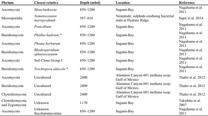
DHABs are typically formed at depths greater than 3’000 m below sea level. The high density of the hypersaline water hampers the mixing with the overlying oxygenated seawater (Figure 1). The combination of high salinity, high density, high hydrostatic pressure, the absence of light, and anoxia, makes these basins some of the most extreme habitats on Earth. Moreover, redox boundaries occurring within short vertical distances are zones of intense biogeochemical cycling, involving key elements such as carbon, nitrogen, sulphur, and hydrogen as well as iron and manganese. These habitats are exciting environments to obtain new insights into the microbial diversity and ecosystem functioning and have already extended our knowledge of the environmental factors that define the limits of life (Eder et al. 1999, 2001, Hallsworth et al. 2007, Edgcomb et al. 2016).
Figure 1. Deep Sea Hypersaline Anoxic Basin in the Gulf of Mexico, image credit BBC.
Although most of the studies focused on prokaryotes, eukaryotic ribosomal DNA analyses, as well as microscopic images, revealed the presence of unexpected micro-eukaryotic communities thriving in DHABs (Alexander et al. 2009, Stock et al. 2012, Edgcomb and Bernhard 2013, Bernhard et al. 2014). The eukaryotic communities identified in several DHABs are characterised by a variety of taxa including, ciliates, alveolates, stramenopiles and fungi, suggesting that even under high salinity and permanent anoxic conditions eukaryotic life is not only possible but even diverse (Table 1).
Table 1. Fungal diversity in deep-sea hypersaline anoxic basins. Stars correspond to taxa characterised by large fruiting bodies and strictly terrestrial.
Phylum Closest relative Depth (mbsl) Location Reference
Ascomycota Acremonium 850 Thuwal Wang et al. 2014
Ascomycota Aspergillus 3500; 3258 L'Atalante Upper halocline; Thetis brine Alexander et al. 2009; Stock et al. 2012
Ascomycota Candida 3258 Thetis brine Stock et al. 2012
Ascomycota Cladosporium 3258 Thetis brine Stock et al. 2012 Cryptomycota LKM11 3500 L'Atalante Lower Halocline; L'Atalante Upper halocline Alexander et al. 2009 Basidiomycota Lycoperdon * 3500 L'Atalante Upper halocline Alexander et al. 2009 Basidiomycota Malassezia 3258 Thetis interface; Bannock interface; Thetis brine; Thetis Lower halocline Edgcomb et al. 2009; Stock et al. 2012
Basidiomycota Malasseziomycetes 3582
Discovery Upper Halocline; L'Atalante Lower Halocline; L'Atalante Upper halocline; Urania Halocline
Bernhard et al. 2014
Basidiomycota Microbotryomycetes 3430 L'Atalante Lower Halocline Bernhard et al. 2014 Ascomycota Penicillium 3500 L'Atalante Upper halocline Alexander et al. 2009 Basidiomycota Rhodosporidium 3258 Thetis brine Stock et al. 2012
Basidiomycota Rhodotorula 3500 L'Atalante Upper halocline; Thetis brine; Thetis interface Alexander et al. 2009; Stock et al. 2012
Ascomycota Sarocladium 850 Thuwal Wang et al. 2014
Ascomycota Schizosaccharomyces Bannock interface; Oxygenated deep-sea water Edgcomb et al. 2010; Edgcomb et al. 2011
Ascomycota Sordaria 3500 L'Atalante Upper halocline Alexander et al. 2009 Basidiomycota Unknown Agaricomycetes * 3258 Thetis brine; Thetis interface Stock et al. 2012 Basidiomycota Unknown Tremellomycets * 3258 Thetis brine Stock et al. 2012
Among eukaryotes, molecular investigations of environmental SSU rDNA have identified fungi as the most diverse taxonomic group in the brine of the Thetis basin (Stock et al. 2012). Some of the retrieved fungal taxa were closely related to described species, widely distributed in the deep sea, such as Rhodotorula, Cladosporium and Aspergillus genera. However, the majority of molecular signatures were only distantly related to described species and potentially representing novel taxa even at high taxonomic level. Also, the hypoxic/suboxic brine layer near the Thuwal cold seeps in the Red Sea is characterised by abundant fungal signatures mostly affiliating with Acremonium genus which include several saprophytic species (Wang et al. 2014). The study also succeeded in isolating fungal cultures similar or
identical to the Sarocladium strictum and Acremonium sp. previously identified from the seafloor of the Arabian Sea (Jebaraj et al. 2010, Wang et al. 2014). While Thetis and Thuwal DHABs were characterised by abundant fungal genera belonging to Ascomycota (Stock et al. 2012, Wang et al. 2014), rRNA analyses of some Mediterranean DHABs (i.e. Discovery, Urania, L'Atalante) showed a large share of reads affiliating to Basidiomycota (Bernhard et al. 2014).
Furthermore, Bernhard and colleagues (2014) were also able to visualise fungal hyphae within sediments, including the lower halocline samples. Concerning fungal molecular diversity, they found the Malassezia signature to account for a significant proportion of OTUs in the lower haloclines of both Discovery and L' Atalante. Interestingly, Malassezia can be found almost everywhere on Earth (Amend 2014) and Malassezia clones retrieved from Discovery and L’Atalante (Bernhard et al. 2014) were also found in the anoxic lower halocline water layer of the Thetis basin (Stock et al. 2012), the anoxic marine Cariaco Basin (Edgcomb et al. 2011b), the anoxic fjord Saanich Inlet (Orsi et al. 2012) and deep sub-surface marine sediments of Peru Margin (Edgcomb et al. 2009, 2011a). However, this species of Malassezia remain uncultured, and it is not possible to infer much about on its ecology. Nevertheless, the diversity of habitats in which Malassezia-like organisms are found suggests this group may have a variety of trophic strategies ranging from saprotrophy to biotrophy (Amend 2014).
The majority of the studies regarding DHAB eukaryotic communities aimed to identify species able to live in such hostile habitats. However, there is almost no information regarding the drivers of fungal diversity and abundance, thus limiting our understanding of their ecology. The overall phylotype richness in hypersaline systems is, however, lower than in the surrounding seawater suggesting that only a few taxa can thrive in such harsh environmental conditions (Behnke et al. 2006). Nonetheless, molecular diversity surveys sometimes identified fungal to account for small but significant fractions of total microbial diversity (Stoeck et al. 2006). Moreover, SSU rRNA and microscopic observations have detected fungi up to 360‰ salinity (Edgcomb et al. 2009, Alexander et al. 2009, Stock et al. 2012). Some authors suggested that fungi might be a significant member of microbial communities (Jebaraj et al. 2010), and that their signatures in DHABs lower haloclines where organic detritus accumulate might indicate that fungi may be involved in organic matter degradation (Pachiadaki et al.
are not only present but also a metabolically active component of benthic microbial communities in DHABs systems (Bernhard et al. 2014, Edgcomb et al. 2016). In particular, the Urania middle halocline and Discovery lower halocline had high metabolic potential, with most transcripts affiliating with Malasseziomycetes, Dothideomycetes and Microbotryomycetes as well as to Aspergillus and Penicillium genera and various yeasts (Bernhard et al. 2014, Edgcomb et al. 2016). The quantitative relevance of transcripts belonging to saprophytic fungi in the Discovery and Urania basin (Edgcomb et al., 2016), further support their crucial role in carbon cycling. While these findings shed light on the potential ecological role and diversity of fungal communities in Urania and Discovery, L'Atalante had rare or absent fungal transcripts suggesting that the elevated salinity of the lower halocline may represent a barrier for most fungi (Edgcomb et al., 2016). There is also some evidence that competition can occur among members of the microbial community (Edgcomb et al., 2017). For example, expression of antibiotic production or resistance genes, including fusaric acid produced by the genus
Fusarium has been reported in the Discovery upper and lower halocline and the Urania middle
halocline (Edgcomb et al., 2017). Indeed, while fungi might not be abundant or hyperdiverse, they could be involved in both biological and carbon and nutrient dynamics in some of the most adverse ecosystem on Earth.
2. Cold seeps
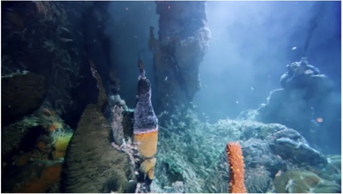
Cold seeps are geologically diverse reducing environments (Boetius and Wenzhöfer 2013) located near to tectonically active and passive continental margin (German et al. 2011). Generally, seep systems originate where reduced chemicals such as methane and sulphide subsurface interstitial fluids, resulting from biogenic or thermogenic origins, are released as a result of tectonic activity, differential compaction of organic-rich sediments, gas hydrate dissociation and subsurface salt migration (Figure 2). Here, chemosynthetic production provides nutrition to a variety of benthic and planktonic heterotrophic species which depends on the oxidation of reduced of sulphur and methane by microorganisms (Levin et al. 2016) while hard and biotic substrates produced by prokaryotic-driven carbonate precipitation, provide critical habitat for several chemosynthetic and heterotrophic organisms (Levin et al. 2016). When seepage ceases, these areas may still host a few species characteristic of seep habitats, but attract new assemblages including cold-water corals (Cordes et al. 2008).
In the last three decades, extensive research has been carried out to investigate the diversity of bacterial, archaeal and metazoan assemblages living in cold seeps ecosystems (Levin et al. 2016). However, studies on the micro-eukaryotic component account for only a small number of studies. Nevertheless, these studies have revealed that a vast diversity of microbial eukaryotes including novel lineages and endemism which could be found in these habitats, including fungi (Table 2).
Table 2. Fungal diversity in deep-sea cold seeps. Stars correspond to taxa characterised by large fruiting bodies and strictly terrestrial.
Phylum Closest relative Depth (mbsl) Location Reference
Other Basal Clone Group I 850–1200 Sagami-Bay Nagahama et al. 2011
Ascomycota Candida 1170; 850–1200 Sagami Bay; Sagami-Bay
Takishita et al. 2007; Nagahama et al. 2011 Ascomycota Cladosporium 850–1200 Sagami-Bay Nagahama et al. 2011 Ascomycota Coniothyrium 850–1200 Sagami-Bay Nagahama et al. 2011
Basidiomycota Cryptococcus curvatus 9cm bsf; 650; 1170 Kuroshima Knoll; Sagami Bay)
Takishita et al. 2006; Takishita et al. 2007 Ascomycota DSF-group1 2400 Alaminos Canyon 601 methane seep (Gulf of Mexico) Thaler et al. 2012 Basidiomycota Entorrhiza fineranae 850–1200 Sagami-Bay Nagahama et al. 2011
Ascomycota Geomyces 850–1200 Sagami-Bay Nagahama et al. 2011
Ascomycota Hypocrea 850–1200 Sagami-Bay Nagahama et al. 2011
Ascomycota Kluyveromyces nonfermentans 850–1200 Sagami-Bay Nagahama et al. 2011
Cryptomycota LKM11 850–1200 Sagami-Bay Nagahama et al. 2011
Basidiomycota Lopharia mirabilis* 850–1200 Sagami-Bay Nagahama et al. 2011 Basidiomycota Malassezia restricta 850–1200 Sagami-Bay Nagahama et al. 2011
Figure 2. Cold seep assemblages. Okeanos methane seep (image credit NOAA) and Carbonate pavement (image credit MARUM).
Table 2. Continued.
Phylum Closest relative Depth (mbsl) Location Reference
Ascomycota Metschnikowia 850–1200 Sagami-Bay Nagahama et al. 2011 Microsporidia Nematocenator marisprofundi 587–810 Nematode; sulphide-oxidising bacterial mats at Hydrate Ridge Sapir et al. 2014
Ascomycota Penicillium 850–1200 Sagami-Bay Nagahama et al. 2011
Basidiomycota Phallus hadriani * 850–1200 Sagami-Bay Nagahama et al. 2011 Ascomycota Phoma herbarum 850–1200 Sagami-Bay Nagahama et al. 2011 Basidiomycota Rhodosporidium sphaerocarpum 850–1200 Sagami-Bay Nagahama et al. 2011 Ascomycota Soil Clone Group I 850–1200 Sagami-Bay Nagahama et al. 2011 Basidiomycota Trechispora alnicola * 850–1200 Sagami-Bay Nagahama et al. 2011 Ascomycota Uncultured 2400 Alaminos Canyon 601 methane seep; Gulf of Mexico Thaler et al. 2012 Basidiomycota Uncultured 2400 Alaminos Canyon 601 methane seep; Gulf of Mexico Thaler et al. 2012 Chytridiomycota Uncultured 2400 Alaminos Canyon 601 methane seep; Gulf of Mexico Thaler et al. 2012 Chytridiomycota
and Zygomycota Unknown 1170 Sagami Bay
Takishita et al. 2007
Ascomycota Unknown Saccharomycotina 850–1200 Sagami-Bay Nagahama et al. 2011
Initial studies on fungal diversity in cold-seeps suggested that overall fungal diversity was low and the saprophytic basidiomycetous yeast Cryptococcus curvatus as the dominant eukaryotic component (Takishita et al. 2006). Consistently, microscopic observation provided further evidence of the quantitative importance of yeast-like cells (Takishita et al. 2007). Similarly, C.
curvatus was dominant also in the sediments of a deep-sea methane cold seep (1170 mbsl) in
Sagami Bay, Japan (Takishita et al. 2007). However, later studies identified a wider array of fungal diversity where Candida and phylotypes related to Chytridiomycota and Zygomycota with no affiliation with any sequence of known fungal species were also observed (Takishita et al. 2007). In addition, fungal-specific PCR-based analysis of environmental DNA in the deep-sea methane cold-seep area in Sagami-Bay (depth 850–1200m) identified 35 phylotypes, including 12 early diverging clones and 23 phylotypes within Dikarya (Nagahama et al. 2011) in which Penicillium chrysogenum was the dominant phylotypes (Lai et al. 2007, Bass et al. 2007). Similarly, a total of 39 fungal sequences were recovered from deep-sea methane seeps (2400 mbsl) at Alaminos Canyon in the Gulf of Mexico (Thaler et al. 2012). Consistently, Ascomycota accounted for the majority of recovered sequences, followed by Basidiomycota, and Chytridiomycota and the number of recovered sequences was highest at the sediment redox boundary where the most intense carbon and nutrient cycling take place (Thaler et al. 2012).
Authors suggest that fungi might contribute in a significant way to several processes within seep systems. Several fungal sequences retrieved from seep systems are known to have important degradation capabilities, while others, such as Rhodosporidium sphaerocarpum and
M. restricta, are known to establish parasitic relationships with a broad set of organisms
(López-García et al. 2007, Lai et al. 2007, Bass et al. 2007, Le Calvez et al. 2009). For example, many species of the order Saccharomycetales have a fermentative metabolism and the commonly retrieved Candida genus can grow under strictly anaerobic conditions consistent to those encountered in seep systems (Nagahama et al. 2011). Other taxa belonging to
Penicillium, Phoma, Cladosporium and Geomyces genera possess enzymatic activity for
lignocellulosic degradation (Junghanns et al. 2009) and perhaps involved in the degradation of recalcitrant compounds such as lignin and other carbohydrates (Damare et al. 2006b).
Cryptococccus and its teleomorphs have been found to be common across various oceanic
regions, and C. curvatus is a known opportunistic pathogen of animals, including humans, and has been suggested to be a possible pathogen of seeps animals as well (Takishita et al. 2006).
Malassezia is known causative agents of skin diseases in mammals and invertebrates and also
soil nematodes (Amend 2014). Therefore, it has been suggested that Malassezia species retrieved from seeps and other deep-sea systems might be associated with small marine invertebrates, such as nematodes or polychaetes (Nagahama et al. 2011, Amend 2014). However, the ecological relevance of fungi in biological and biogeochemical dynamics are still unclear and not often sufficiently addressed.
A particular exception is portrayed by the microsporidia Nematocenator marisprofundi. This genus was found infecting benthic nematodes at the Hydrate Ridge methane seeps site (Sapir et al. 2014). The infection was described as species-specific and temporally and spatially stable indicating an ecologically consistent host-parasite interaction with potentially significant consequences on nematodes population dynamics (Sapir et al. 2014). Supporting the authors’ conclusions, Microsporidia fungi have been shown by ultrastructural and molecular approaches to be true parasites of several hosts in various marine habitats (Ardila-Garcia and Fast 2012).
3. Hydrothermal Vents
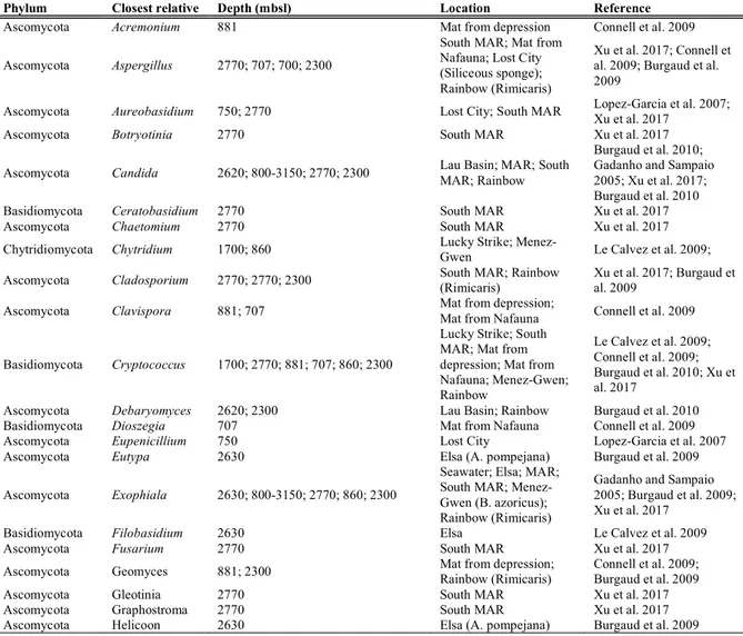
Hydrothermal vents are highly productive ecosystems, and by hosting giant tubeworms, large mussel and clam beds, and dense shrimp and crab aggregations are also among the most diverse
of geological settings such as ridges, seamounts, back-arc basins, margins and trenches, where they originate from the outflow of chemically altered seawater, circulating from the ridge flanks through the crust near shallow magmatic intrusions at elevated temperature and pressure (Levin et al. 2016). Here, hard and biotic substrates produced at vents are used by benthic fauna for attachment, shelter and food, while microbial communities by chemosynthetic production provide nutrition to a variety of benthic and planktonic heterotrophic species (Figure 3; Levin et al. 2016).
Probably owing to their diversity and centrality in biogeochemical cycles, extensive research has been carried out to explore hydrothermal vent communities. Fungi have been reported to have an essential role in C cycling in terrestrial vent systems (Pang and Mitchell 2005, Le Calvez et al. 2009) and can contribute to weathering processes of silicates and volcanic rocks (Etienne and Dupont 2002).
Active seamounts and ridges offer several low- and high-temperature hydrothermal habitats that are characterised by fluids with high contents of reduced metals such as Fe (II) and Mn(II) (Statham et al. 2005). In these environments, rich fungal communities have been reported in the actively growing Fe-oxide mats and basalt rock surfaces from the active volcano of the Vailulu'u Seamount (Connell et al. 2009). Connell and colleagues (2009) found that yeasts and yeast-like species were dominant, and fungal assemblage compositions significantly differed between in mat rather than rock substrates. Yeasts and filamentous fungi, including new species, have also been retrieved associated with hydrothermal water and sediment samples (Burgaud et al. 2009, 2010; Table 3), and strikingly, a significant portion of fungi has been recovered from hydrothermal shrimps and mussels (Burgaud et al. 2011). Notably, among the isolated fungi, Aspergillus sydowii, the causal agent of disease among sea fan corals (Soler-Hurtado et al. 2016), was detected suggesting possible interactions with cold-water corals.
Figure 3. Mid Atlantic Ridge hydrothermal vent systems, image credit BBC.
Furthermore, Burgaud et al. (2009) retrieved fungi of the Chaetothyriales order from deep-sea mussel samples. This order has been associated with diseases of deep-sea mussels (Van Dover et al. 2007). These results confirmed earlier findings which suggest that the deep-sea hydrothermal systems of the Mid-Atlantic Ridge are characterised by the presence of abundant yeast community (Gadanho and Sampaio 2005). Again, molecular investigations targeting the SSU rRNA gene sequences identified many unknown sequences, even at high taxonomic levels in the Chytridiomycota, Ascomycota, and Basidiomycota phyla (Xu et al. 2017). Supporting previous findings regarding Chytridiomycota phylotypes affiliating to species that have not been described (Le Calvez et al. 2009).
Table 3. Fungal diversity in deep-sea hydrothermal vents. Stars correspond to taxa characterised by large fruiting bodies and strictly terrestrial.
Phylum Closest relative Depth (mbsl) Location Reference
Ascomycota Acremonium 881 Mat from depression Connell et al. 2009
Ascomycota Aspergillus 2770; 707; 700; 2300
South MAR; Mat from Nafauna; Lost City (Siliceous sponge); Rainbow (Rimicaris)
Xu et al. 2017; Connell et al. 2009; Burgaud et al. 2009
Ascomycota Aureobasidium 750; 2770 Lost City; South MAR Lopez-Garcia et al. 2007; Xu et al. 2017
Ascomycota Botryotinia 2770 South MAR Xu et al. 2017
Ascomycota Candida 2620; 800-3150; 2770; 2300 Lau Basin; MAR; South MAR; Rainbow
Burgaud et al. 2010; Gadanho and Sampaio 2005; Xu et al. 2017; Burgaud et al. 2010
Basidiomycota Ceratobasidium 2770 South MAR Xu et al. 2017
Ascomycota Chaetomium 2770 South MAR Xu et al. 2017
Chytridiomycota Chytridium 1700; 860 Lucky Strike; Menez-Gwen Le Calvez et al. 2009; Ascomycota Cladosporium 2770; 2770; 2300 South MAR; Rainbow (Rimicaris) Xu et al. 2017; Burgaud et al. 2009 Ascomycota Clavispora 881; 707 Mat from depression; Mat from Nafauna Connell et al. 2009
Basidiomycota Cryptococcus 1700; 2770; 881; 707; 860; 2300
Lucky Strike; South MAR; Mat from depression; Mat from Nafauna; Menez-Gwen; Rainbow Le Calvez et al. 2009; Connell et al. 2009; Burgaud et al. 2010; Xu et al. 2017
Ascomycota Debaryomyces 2620; 2300 Lau Basin; Rainbow Burgaud et al. 2010 Basidiomycota Dioszegia 707 Mat from Nafauna Connell et al. 2009
Ascomycota Eupenicillium 750 Lost City Lopez-Garcia et al. 2007
Ascomycota Eutypa 2630 Elsa (A. pompejana) Burgaud et al. 2009
Ascomycota Exophiala 2630; 800-3150; 2770; 860; 2300
Seawater; Elsa; MAR; South MAR; Menez-Gwen (B. azoricus); Rainbow (Rimicaris)
Gadanho and Sampaio 2005; Burgaud et al. 2009; Xu et al. 2017
Basidiomycota Filobasidium 2630 Elsa Le Calvez et al. 2009
Ascomycota Fusarium 2770 South MAR Xu et al. 2017
Ascomycota Geomyces 881; 2300 Mat from depression; Rainbow (Rimicaris) Connell et al. 2009; Burgaud et al. 2009
Ascomycota Gleotinia 2770 South MAR Xu et al. 2017
Ascomycota Graphostroma 2770 South MAR Xu et al. 2017
Table 3. Continued.
Phylum Closest relative Depth (mbsl) Location Reference
Ascomycota Helotiales 2630; 860; 2300; 2300; 2300
Elsa (A. pompejana); Menez-Gwen (B. azoricus); Rainbow (Rimicaris); Rainbow (B. azoricus commensal worm); Rainbow (B. azoricus) Burgaud et al. 2009;
Ascomycota Hexophiala 2630 Elsa Le Calvez et al. 2009
Ascomycota Hortaea 2300 Rainbow Burgaud et al. 2010
Basidiomycota Malassezia 750 Lost City Lopez-Garcia et al. 2007
Ascomycota Meyerozyma 2770 South MAR Xu et al. 2017
Basidiomycota Micropsalliota * 2770 South MAR Xu et al. 2017
Ascomycota Penicillium 2770 South MAR Xu et al. 2017
Ascomycota Phaeotheca 2300 Rainbow Burgaud et al. 2010
Ascomycota Phialemonium 2770 South MAR Xu et al. 2017
Ascomycota Phialophora 2770 South MAR Xu et al. 2017
Ascomycota Phoma 2770 South MAR Xu et al. 2017
Ascomycota Pichia 700; 800-3150; 881; 707 Lost City; MAR; Mat from depression; Mat from Nafauna
Burgaud et al. 2010; Gadanho and Sampaio 2005; Connell et al. 2009
Ascomycota Ramichloridium 2770 South MAR Xu et al. 2017
Basidiomycota Rhodosporidium 800-3150; 881; 2300 MAR; Mat from depression; Rainbow
Gadanho and Sampaio 2005; Connell et al. 2009; Burgaud et al. 2010
Basidiomycota Rhodotorula 1667; 800-3150; 2770; 2300
Exposure experiment; MAR; South MAR; Rainbow
Gadanho and Sampaio 2005; Connell et al. 2009; Burgaud et al. 2010; Xu et al. 2017 Ascomycota Scolecobasidium 2300 Rainbow (Coral) Burgaud et al. 2009 Basidiomycota Sporidiobolus 707 Mat from Nafauna Connell et al. 2009 Basidiomycota Sporobolomyces 2770; 2300 South MAR; Rainbow Burgaud et al. 2010 Xu et al. 2017;
Ascomycota Stachybotrys 2770 South MAR Xu et al. 2017
Basidiomycota Tilletiopsis 2630; 2770 Elsa (Riftia); South MAR Burgaud et al. 2009; Xu et al. 2017 Ascomycota Toxicocladosporium 2770 South MAR Xu et al. 2017
Basidiomycota Trichosporon 800-3150; 2770 MAR; South MAR Gadanho and Sampaio 2005; Xu et al. 2017 Basidiomycota Unknown Agaricomycotina 1700; 860 Lucky Strike; Menez-Gwen Le Calvez et al. 2009 Ascomycota Unknown Chaetothyriales 1990 Mussel hydrothermal vent Hill Van Dover et al. 2006
Ascomycota Unknown Dothideomycetes 2630; 1700; 860
Elsa (A. pompejana); Lucky Strike; Menez-Gwen
Burgaud et al. 2009; Le Calvez et al. 2009
Ascomycota Unknown Eurotiales 2300 Rainbow Burgaud et al. 2009 Basidiomycota Tilletiopsis 2630; 2770 Elsa (Riftia); South MAR Burgaud et al. 2009; Xu et al. 2017 Ascomycota Toxicocladosporium 2770 South MAR Xu et al. 2017
Basidiomycota Trichosporon 800-3150; 2770 MAR; South MAR Gadanho and Sampaio 2005; Xu et al. 2017 Basidiomycota Unknown Agaricomycotina 1700; 860 Lucky Strike; Menez-Gwen Le Calvez et al. 2009 Ascomycota Unknown Chaetothyriales 1990 Mussel hydrothermal vent Hill Van Dover et al. 2006
Ascomycota Unknown Dothideomycetes 2630; 1700; 860
Elsa (A. pompejana); Lucky Strike; Menez-Gwen
Burgaud et al. 2009; Le Calvez et al. 2009
Ascomycota Unknown Eurotiales 2300 Rainbow Burgaud et al. 2009 Ascomycota Unknown Hypocreales 860; 3650
Menez-Gwen (B. azoricus); TAG (Rimicaris)
Burgaud et al. 2009 Basidiomycota Unknown Sebacina * 2770 South MAR Xu et al. 2017 Ascomycota Unknown Sordariomycetes 2770 South MAR Xu et al. 2017 Basidiomycota Unknown Tomentella 2770 South MAR Xu et al. 2017
Although we are getting a glimpse of the diversity of fungal communities at hydrothermal vents we still largely ignore their ecological relevance. Several yeast species retrieved from hydrothermal sites are capable of producing siderophores to acquire and utilise Fe (III), and
Rhodotorula graminis is an Mn (II)-oxidizing fungus and might be involved in iron and
manganese cycling. However, no study investigated such processes in deep-sea hydrothermal vents where fungi can scavenge iron and other metals by several pathways and may be functionally relevant in their biogeochemical cycling (Connell et al. 2009).
Fungi are known parasites of a multitude of organisms, and parasitic infections play significant roles in population dynamics. In hydrothermal vents, fungi have been found associated with benthic fauna. For example, at the Mussel Hill hydrothermal vent in Fiji Basin, a Capronia-like black yeast (order Chaetothyriales) was reported to induce a hemocytic immune response in the mussels Bathymodiolus brevior with tissue deterioration (Van Dover et al. 2007). Although it is not clear if the fungal infections were secondary or facilitated by concurrent infections of other pathogens, the identification of fungi in otherwise healthy individuals makes it clear that the fungus is not a strict saprophyte. The described infection and the high prevalence of the fungus within the population, progressive and pervasive connective tissue degradation, together with decreased volume of bacteriocytes and symbiotic bacteria associated with the black-body stage of the disease of the mussel, suggest that massive mussel mortality were imminent within the Mussel Hill vent field (Van Dover et al. 2007).
Furthermore, yeasts and filamentous fungi retrieved from hydrothermal vent fauna suggest that fungi can benefit from nutrients as in the case of those isolated from gill chambers of shrimps or have a role in the decomposition of organic material entrapped in mussel byssi (Burgaud et al. 2009, 2010). Supporting pathogenic associations, Scolecobasidium sp.– the causative agent of necrotic patches on different corals–was found associated to deep-sea corals, near Rainbow hydrothermal vent site, indicating that this halophilic fungus could be implicated in deep-sea coral diseases. Also, the pathogenic genus Exophiala sp. Was repeatedly reported from sediments (Gadanho and Sampaio 2006, Burgaud et al. 2009, Xu et al. 2017) and alongside nematodes (Bhadury et al. 2009).
4. Deep-sea sediments
Benthic deep-sea ecosystems are represented mainly by soft sediments characterised by the presence of highly diverse taxa belonging to prokaryotes and metazoans (i.e. meiofaunal and macrofaunal organisms; Corinaldesi 2015). Metazoan life is generally present only in the top 20-50 cm of the sediment, whereas prokaryotes are present even in the deep biosphere (i.e. several hundred meters below the sediment surface; Orcutt et al. 2013).
One of the first reports of fungi in deep-sea sediments is dated back to the ‘60s (Roth 1964). Since then, several known and new fungal taxa have been found in different deep-sea sedimentary habitats (Nagahama et al. 2003). However, despite these reports, the number of studies addressing fungal diversity and ecology in deep-sea sediments are still relatively scarce compared to other benthic components.
Concerning diversity, Ascomycota and Basidiomycota represent the most abundant groups (Singh et al. 2011, Zhang et al. 2016; Table 4). Although Basidiomycota has been claimed to account for a relatively low diversity, different methodological approaches have provided contrasting results. For example, multiple-primer amplification and sequencing of sediment samples of the Pacific Ocean retrieved seven basidiomycete classes and only five Ascomycete classes along with two uncultured fungal groups affiliating with uncultured soil fungal taxa and uncultured Zygomycetes (Xu et al. 2016). Moreover, a large number of sequences with low similarity to public databases even at high taxonomic levels has been reported (Xu et al. 2014).
Figure 4. Abyssal plane, image credit BBC.
Table 4. Fungal diversity from deep-sea sediments
Phylum Closest relative Depth (mbsl) Location Reference
Ascomycota Acremonium 1190-1589 Okinawa Zhang et al. 2016
Ascomycota Aspergillus 5000; 5017-6986; 1190-1589 CIB; Pacific; Okinawa Singh et al. 2010; Xu et al. 2014; Zhang et al. 2016 Ascomycota Aureobasidium 5017-6986 Pacific Xu et al. 2014
Basidiomycota Auricularia 5017-6986 Pacific Xu et al. 2014
Ascomycota Bionectriaceae 5000 CIB Singh et al. 2010
Ascomycota Candida 1200-10000; 5000; 5017-6986 Pacific; CIB; Pacific Nagano et al. 2010; Singh et al. 2010; Xu et al. 2014 Ascomycota Capnodium coffeae 5000 CIB Singh et al. 2010
Ascomycota Chaetomium 5017-6986; 1190-1589 Pacific; Okinawa Xu et al. 2014; Zhang et al. 2016 Ascomycota Cladosporium 5017-6986 Pacific Xu et al. 2014
Ascomycota Coniellab 1190-1589 Okinawa Zhang et al. 2016 Ascomycota Corynespora 1190-1589 Okinawa Zhang et al. 2016 Basidiomycota Cryptococcus 5017-6986 Pacific Xu et al. 2014 Ascomycota Debaryomyces yamadae 5000 CIB Singh et al. 2010 Ascomycota Emericella 1190-1589 Okinawa Zhang et al. 2016
Ascomycota Exophiala 1190-1589; 5017-6986 Okinawa; Pacific Xu et al. 2014; Zhang et al. 2016 Ascomycota Faurelina 1190-1589 Okinawa Zhang et al. 2016
Ascomycota Fusarium 5017-6986; 1190-1589 Pacific; Okinawa Xu et al. 2014; Zhang et al. 2016 Ascomycota Gloeotinia 1200-10000; 5017-6986 Pacific; Pacific Nagano et al. 2010; Xu et al. 2014
Ascomycota Hortaea 5000 CIB Singh et al. 2010
Ascomycota Lecanicillium 5017-6986 Pacific Xu et al. 2014 Ascomycota Lecythophora 1190-1589 Okinawa Zhang et al. 2016 Ascomycota Leptosphaeria 1190-1589 Okinawa Zhang et al. 2016 Ascomycota Lophiostoma 1190-1589 Okinawa Zhang et al. 2016
Basidiomycota Malassezia 5000; 5017-6986 CIB; Pacific Singh et al. 2010; Xu et al. 2014 Ascomycota Meyerozyma 5017-6986 Pacific Xu et al. 2014
Ascomycota Microdochium 1190-1589 Okinawa Zhang et al. 2016 Ascomycota Minimidochium 1190-1589 Okinawa Zhang et al. 2016 Zygomycota Mortierella 1190-1589 Okinawa Zhang et al. 2016 Ascomycota Myriogenospora 1190-1589 Okinawa Zhang et al. 2016 Ascomycota Myrothecium 1190-1589 Okinawa Zhang et al. 2016 Ascomycota Nectria 1190-1589 Okinawa Zhang et al. 2016
Ascomycota Nodulisporium 5000 CIB Singh et al. 2010
Ascomycota Paecilomyces 5017-6986; 1190-1589 Pacific; Okinawa Xu et al. 2014; Zhang et al. 2016 Ascomycota Penicillium 1200-10000; 5017-6986; 1190-1589 Pacific; Pacific; Okinawa Nagano et al. 2010; Xu et al. 2014; Zhang et al. 2016 Ascomycota Periconia sp. 5017-6986 Pacific Xu et al. 2014
Chytridiomycota Phlyctochytrium 1190-1589 Okinawa Zhang et al. 2016
Ascomycota Phoma 5000; 1190-1589 CIB; Okinawa Singh et al. 2010; Zhang et al. 2016
Ascomycota Pichia 5000 CIB Singh et al. 2010
Ascomycota Plectosphaerella 1190-1589 Okinawa Zhang et al. 2016 Basidiomycota Pleurotus * 5017-6986 Pacific Xu et al. 2014 Ascomycota Podospora 1190-1589 Okinawa Zhang et al. 2016 Ascomycota Preussia 1190-1589 Okinawa Zhang et al. 2016 Ascomycota Pseudogymnoascus 1190-1589 Okinawa Zhang et al. 2016 Ascomycota Ramichloridium 5017-6986 Pacific Xu et al. 2014 Basidiomycota Rhodosporidium 5000 CIB Singh et al. 2010 Basidiomycota Russula * 1190-1589 Okinawa Zhang et al. 2016
Ascomycota Sagenomella 5000 CIB Singh et al. 2010
Basidiomycota Schizophyllum * 1190-1589 Okinawa Zhang et al. 2016 Ascomycota Simplicillium 5017-6986 Pacific Xu et al. 2014 Ascomycota Stemphylium 1190-1589 Okinawa Zhang et al. 2016 Ascomycota Stenella musicola 5000 CIB Singh et al. 2010 Ascomycota Thelebolus 1190-1589 Okinawa Zhang et al. 2016 Basidiomycota Trametes * 5017-6986 Pacific Xu et al. 2014 Ascomycota Trichocladium 1190-1589 Okinawa Zhang et al. 2016
Table 4. Continued
Phylum Closest relative Depth (mbsl) Location Reference Ascomycota Ulospora bilgramii 5000 CIB Singh et al. 2010 Basidiomycota Unknow Polyporales 5017-6986 Pacific Xu et al. 2014
Ascomycota Unknown 5000 CIB Singh et al. 2010
Basidiomycota Unknown 5000 CIB Singh et al. 2010
Ascomycota Unknown 1190-1589 Okinawa Zhang et al. 2016 Basidiomycota Unknown Agaricomycetes 1190-1589 Okinawa Zhang et al. 2016 Ascomycota Unknown Dothideomycetes 5000 CIB Singh et al. 2010 Ascomycota Unknown Dothideomycetes 1190-1589 Okinawa Zhang et al. 2016 Ascomycota Unknown Eurotiomycetes 1190-1589 Okinawa Zhang et al. 2016 Ascomycota Unknown Leotiomycetes 1190-1589 Okinawa Zhang et al. 2016 Basidiomycota Unknown Lycoperdaceae * 5017-6986 Pacific Xu et al. 2014 Ascomycota Unknown Pezizomycetes 1190-1589 Okinawa Zhang et al. 2016 Ascomycota Unknown Pleosporales 5017-6986 Pacific Xu et al. 2014 Ascomycota Unknown Sordariomycetes 1190-1589 Okinawa Zhang et al. 2016 Basidiomycota Unknown Thelephoraceae * 5017-6986 Pacific Xu et al. 2014 Basidiomycota Unknown Trechisporales * 5017-6986 Pacific Xu et al. 2014 Basidiomycota Unknown Tremellomycetes 1190-1589 Okinawa Zhang et al. 2016 Basidiomycota Wallemia 5000 CIB Singh et al. 2010 Ascomycota Xylaria * 5017-6986 Pacific Xu et al. 2014
Fungi in deep-sea sediments are thought to be involved in carbon cycling and symbioses. For instance, fungal hyphae have been observed to grow under high hydrostatic pressure forming various stages of accretion of particles around them, leading to the formation of aggregates withholding of humic material, carbohydrate, and proteins (Damare and Raghukumar 2008). Such evidence suggests that fungi in deep-sea sediments may be involved in humic aggregate formation as it happens in terrestrial habitats, indicating indeed that carbon sequestration process and benthic deep-sea food web needs to be further investigated. Furthermore, several genera including Rhodotorula, Rhodosporidium, Malassezia, Trichosporon and
Sterigmatomyces comprehend several pathogens and parasites also of marine animals such as
bivalves, nematodes and other organisms (Singh et al. 2011, Lin et al. 2012). For example,
Paecilomyces and Myrothecium species were repeatedly found while surveying nematode
molecular diversity, indicating a possible interaction between fungi and nematodes (Bhadury et al. 2009, 2011). Therefore, fungi can have a profound effect on biogeochemical processes and food web dynamics in sedimentary habitats as in soil.
Interestingly, fungal-nematodes relationships in benthic deep-sea ecosystems are neglected. However, because the critical ecological role of nematodes in these environments (Danovaro et al. 2008), the fungus-nematode interactions should be further investigated to understand the magnitude and direction of such relationship. For example, the co-amplified Paecilomyces and
Myrothecium are known to include nematicidal species and often used as pest control.
5. Deep-sea sub-surface sediments
While debate continues whether Archaea or Bacteria predominate in sub-surface sediments (Schippers and Neretin 2006, Smith et al. 2011, Jorgensen 2011, Orcutt et al. 2013), fungi remain understudied despite they have been identified from a few centimetres below the seafloor (Damare et al. 2006a) down to 1740 m below the sediment surface (Ciobanu et al. 2014).
Fungi have been reported to dominate over other eukaryotic groups in sub-surface sediments collected in different oceanographic regions (Edgcomb et al. 2011a, Ciobanu et al. 2014; Table 5). Although some study did not specifically target fungal diversity, DNA- and RNA-based clone libraries suggest that Basidiomycetes and Ustilaginusmycets were the dominant fungal taxa and that fungal diversity decrease with increasing depth into the sediment. Tremellomycetes, Sordariomycetes and Eurotiomycetes were abundant at shallow depths while Saccharomycetes were detected even at depths between 630 and 1’365 m while deeper layers were dominated by Wallemiomycetes, Microbotryomycetes and Tremellomycetes (order Filobasidiales) (Ciobanu et al. 2014) indicating that depth within the sediment could be responsible for selection con community structuring processes. Similarly, DNA and RNA fungal signatures within subseafloor sediments of the Canterbury basin (New Zealand) by 454 pyrotag sequencing with fungal-specific primer set confirmed that a large number of OTUs belonging to Basidiomycota yeasts. This study also revealed low but unexpected fungal diversity along the sediment depth with some taxa occurring only at specific depths and others that were found in all samples. For example, Leptosphaerulina was found only at 346 mbsf while Pleurostomophora and Exophiala were only detected at 583 mbsf. These genera were correlated with high porosity and high organic carbon concentration occurring in the upper layers of the sediment. On the contrary, M. guillermondii, which also represented an abundant OTU, was detected throughout all the sediment layer using universal eukaryotic primer sets,
abundant OTUs and its relative abundance has been found to be positively related with the concentration of methane supporting earlier evidence indicating Cryptococcus as the dominant taxon in methane seeps (Takishita et al. 2006).
Table 5. Fungal diversity in deep-sea subsurface sediments
Phylum Closest relative Depth (mbsl) Depth (mbsf) Location Reference Ascomycota Acremonium 344 4-1884 Canterbury Basin Redou et al. 2015 Ascomycota Aspergillus n.d. 4-1884; 12-31-40 Canterbury Suruga Bay Basin; Redou et al. 2015; Nagano et al. 2016 Ascomycota Batcheloromyces 344 346-1740 Canterbury Basin Redou et al. 2014 Basidiomycota Bjerkandera * 344 4-1884 Canterbury Basin Redou et al. 2015 Basidiomycota Bullera 344 4-1884 Canterbury Basin Redou et al. 2015
Ascomycota Cadophora n.d. 3 Suruga Bay Nagano et al. 2016
Ascomycota Cladophialophora 344 4-1884 Canterbury Basin Redou et al. 2015 Ascomycota Cladosporium 344 4-1884 Canterbury Basin Redou et al. 2015 Ascomycota Cordyceps 344 4-1884 Canterbury Basin Redou et al. 2015 Basidiomycota Cryptococcus 344 346–1740 Canterbury Basin Redou et al. 2014 Ascomycota Cyberlindnera 344 346–1740 Canterbury Basin Redou et al. 2014 Basidiomycota Elmerina * 344 346–1740 Canterbury Basin Redou et al. 2014 Ascomycota Eurotium 344 4-1884 Canterbury Basin Redou et al. 2015 Ascomycota Exophiala 344 4-1884 Canterbury Basin Redou et al. 2014; Redou et al. 2015 Basidiomycota Filobasidium 344 346–1740 Canterbury Basin Redou et al. 2014 Ascomycota Fusarium 344 4-1884 Canterbury Basin Redou et al. 2014; Redou et al. 2015 Ascomycota Galactomyces 344 346–1740 Canterbury Basin Redou et al. 2014 Ascomycota Leptosphaerulina 344 346–1740 Canterbury Basin Redou et al. 2014 Basidiomycota Leucosporidiella 344 346–1740 Canterbury Basin Redou et al. 2014 Basidiomycota Malassezia 344 346–1740 Canterbury Basin Redou et al. 2014 Ascomycota Meyerozyma 344 4-1884 Canterbury Basin Redou et al. 2014; Redou et al. 2015 Ascomycota Microascus 344 4-1884 Canterbury Basin Redou et al. 2015 Ascomycota Oidiodendron 344 4-1884 Canterbury Basin Redou et al. 2015 Ascomycota Paecilomyces 344; n.d. 4-1884; 12 Suruga Bay Redou et al. 2015; Nagano
et al. 2016
Ascomycota Penicillium 344 4-1884 Canterbury Basin Redou et al. 2014; Redou et al. 2015 Ascomycota Phialophora 344 4-1884 Canterbury Basin Redou et al. 2015
Ascomycota Pichia n.d. 3-12 Suruga Bay Nagano et al. 2016
Ascomycota Pleurostomophora 344 346-1740 Canterbury Basin Redou et al. 2014 Ascomycota Purpureocillium 344 4-1884 Canterbury Basin Redou et al. 2015 Ascomycota Rhinocladiella 344 346-1740 Canterbury Basin Redou et al. 2014 Basidiomycota Rhodosporidium 344 346-1740 Canterbury Basin Redou et al. 2014 Basidiomycota Rhodotorula 344 4-1884 Canterbury Basin Redou et al. 2014; Redou et al. 2015 Ascomycota Sarocladium 344 4-1884 Canterbury Basin Redou et al. 2015 Basidiomycota Sistotrema * 344 4-1884 Canterbury Basin Redou et al. 2015 Basidiomycota Trametes * 344 4-1884 Canterbury Basin Redou et al. 2015 Basidiomycota Tremella * 344 346-1740 Canterbury Basin Redou et al. 2014 Ascomycota Trichoderma 344 346-1740 Canterbury Basin Redou et al. 2014 Basidiomycota Trichosporon 344 346-1740 Canterbury Basin Redou et al. 2014 Ascomycota Unknown Chaetothyriales 344 346-1740 Canterbury Basin Redou et al. 2014 Basidiomycota Wallemia muriae 344 346-1740 Canterbury Basin Redou et al. 2014
Information on diversity of fungi in the deep biosphere obtained through DNA analysis does not allow to infer on their ecological significance since fungal sequences may belong to extracellular DNA not associated with living biomass (Coolen et al. 2006, Boere et al. 2011, Kouduka et al. 2017) or to fungi in dormant stage. The majority of subsurface microbes are on
physiological standby, with only a small fraction actually active (Jorgensen 2011). However, meta-transcriptomic analyses instead suggest the presence of an active fungal community (Pachiadaki et al. 2016). Fungal-Associated transcripts for metabolic and cellular processes, cell and membrane functions, and catalytic activities have been observed in several sediment samples (Pachiadaki et al. 2016). Furthermore, fungal communities at comparable depths at the two geographically separated locations appear dominated by distinct taxa, and different gene expression. These results suggest that different organic content and quality and location might shape different fungal communities (Pachiadaki et al. 2016). Also, microscopic analysis of Canterbury Basin sediment samples from 4 and 403 mbsf revealed the presence of fungal structure and conidiogenesis (Pachiadaki et al. 2016).
Culture-dependent methods on sediment samples down to 344 mbsl in the Canterbury Basin (New Zealand) allowed the recovery of both filamentous and yeast forms, and molecular investigations have allowed identifying 21 genera belonging to Ascomycota and Basidiomycota and one uncultured Agaromycetes (Rédou et al. 2015). The majority of taxa affiliated with Rhodotorula and Meyrozyma. In particular, the red yeast Rhodotorula
mucilaginosa is widely distributed, and it has also been isolated from deep-sea sediments and
basaltic crust (Singh et al. 2010). This study also indicated a negative relationship between diversity and increasing depth with 25 different species within the uppermost samples (4 to 37 mbsf) and only eight different species between 137 and 1884 mbsf (Rédou et al. 2015). Similarly, culturable fungal communities were investigated in marine subsurface sediment from 3 to 40 mbsl in Suruga Bay (Nagano et al. 2016). Although occurrence and diversity of culturable fungi were extremely low, fungi were successfully cultured from 5 out of 15 samples, and Aspergillus, Pichia and Cadophora represented the most common genera. Species found in these studies have a high potential as saprotrophs and can be competitors of prokaryotic communities for the limited resource available in this environment. For example, the red yeast Rhodotorula mucilaginosa and Fusarium oxysporum, reported as the most abundant taxon in the Canterbury Basin sediment, are widely distributed organisms, even in deep-sea sediments and basaltic crust (Singh et al. 2010, Smith et al. 2011, Rédou et al. 2015). In particular, Fusarium oxysporium has been shown to grow under deep-sea marine conditions Burgaud et al. (2013) and to have important denitrifying capabilities (Cathrine and
hypotheses are also supported by transcriptomic analyses which revealed that fungi although occurring in lower abundance than prokaryotes, possess the ability to degrade a variety of organic substrates in deep-subseafloor sediments (Orsi et al. 2013a).
Supporting the suggested metabolic activities of fungi in such harsh environments, fungal diversity has been related to organic matter availability (Ciobanu et al. 2014, Rédou et al. 2015). For example, Leptosphaerulina chartarum, Fusarium solani, Trichoderma sp.,
Cyberlindnera jadinii and in particular Cryptococcus curvatus were found strictly related to
methane contents. Consistently, C. curvatus is the dominant taxa in methane seeps systems (Takishita et al. 2006). Trichosporon mucoides, Malassezia pachydermatis, Meyerozyma
guilliermondii, Pleurostomophora richardsiae, Exophiala dermatitidis were only found in
sediment samples with a low organic carbon concentration (Rédou et al. 2014). These heterotrophic fungi have also been retrieved from other deep sediments in which they have been demonstrated to be active members of microbial communities (Orsi et al. 2013b, Pachiadaki et al. 2016).
Overall, studies on micro-eukaryotic communities in subsurface sediments indicate that fungi are not only diverse but also viable. Indeed, these results suggest that fungi represent a ubiquitous component of benthic deep-sea ecosystems likely with major implications in C and nutrient cycling (Ciobanu et al. 2014).
Conclusions and perspectives
Fungi have been retrieved from any benthic deep-sea ecosystem investigated so far, but their ecology and the drivers influencing their abundance and diversity are still far to be elucidated. From the available information, it appears that the overall fungal diversity in benthic deep-sea ecosystems is relatively low, but some studies pointed out that fungi might often be the dominant micro-eukaryotic component. Concerning fungal diversity, Ascomycota and Basidiomycota often account for the largest share of reads while other phyla are scarce or absent. However, results on fungal diversity are frequently inconsistent with the literature likely resulting from differences in DNA isolation, primer selection and sequencing techniques, highlighting the need to standardise assessment protocols to compare fungal diversity more effectively (Zhang et al. 2016). For instance, high-throughput sequencing approach, the most successful and most widely adopted sequencing platform, had been widely applied in
investigating fungal diversity of many environmental samples, such as soil, plants and marine corals, it was rarely applied in exploring the fungal diversity of deep-sea sediment samples (Zhang et al. 2016). Furthermore, despite the use of fungal-specific primers revealed the presence of diverse fungal phylotypes, the specific primers designed for amplification and sequencing of fungal 18S, ITS or 28S rDNA regions might be biased towards specific fungal taxonomic groups (Singh et al. 2012). For instance, deep-sea sediment samples from the Pacific Ocean provided different results when different primer sets were employed (Xu et al. 2016). At the OTU level, the ITS resulted in 18 OTUs while, the 18S rDNA primer pairs returned 44 different OTUs and the 28S rDNA primers pair 78 OTUs, including 41 OTUs. At the same time, community structure changed based on the primer set used. Moreover, all studies highlighted that a significant fraction of sequences fails to match public database even at a high taxonomic level, suggesting the presence of taxa to be identified yet. Primer set selection and limited match of the retrieved sequences to public databases can be one of the main reasons for the low diversity described.
There is evidence that environmental conditions may have a role in shaping fungal assemblage composition. For instance, patterns of fungal diversity in marine ecosystems suggest that fungal communities can depend on the amount and quality of organic carbon (Tisthammer et al. 2016). However, information on this issue is still too limited to conclude, especially for benthic deep-sea ecosystems. Moreover, the quantitative relevance and ecological role of fungi in benthic deep-sea ecosystems are still largely unknown. Also, several fungal signatures have been observed during nematodes DNA barcoding providing further evidence that some fungi can be involved in parasitic associations with nematodes (Troemel et al. 2008, Ardila-Garcia and Fast 2012). Such findings suggest that fungi might also have an essential role in controlling population and community dynamics of metazoans (i.e. nematodes) inhabiting benthic deep-sea ecosystems (Vanreusel et al. 2010), but evidence regarding the involvement of fungi in pathogenic or parasitic associations is rare or absent in benthic deep-sea ecosystems. Overall, there is an urgent need to improve our knowledge of factors influencing the abundance and diversity of fungi in different benthic deep-sea habitats and ecosystems for a better understanding of the functioning of the largest and less explore biome on Earth.
References
Alexander, E., A. Stock, H.-W. Breiner, A. Behnke, J. Bunge, M. M. Yakimov, and T. Stoeck. 2009. Microbial eukaryotes in the hypersaline anoxic L'Atalante deep-sea basin. Environmental Microbiology 11:360–381. Amend, A. S. 2014. From Dandruff to Deep-Sea Vents: Malassezia-like Fungi Are Ecologically Hyper-diverse.
PLoS Pathogens 10:e1004277–4.
Ardila-Garcia, A. M., and N. M. Fast. 2012. Microsporidian Infection in a Free-Living Marine Nematode. Eukaryotic Cell 11:1544–1551.
Azam, F., and F. Malfatti. 2007. Microbial structuring of marine ecosystems. Nature Reviews Microbiology 5:782–791.
Bass, D., A. Howe, N. Brown, H. Barton, M. Demidova, H. Michelle, L. Li, H. Sanders, S. C. Watkinson, S. Willcock, and T. A. Richards. 2007. Yeast forms dominate fungal diversity in the deep oceans. Proceedings of the Royal Society B: Biological Sciences 274:3069–3077.
Behnke, A., J. Bunge, K. Barger, H. W. Breiner, V. Alla, and T. Stoeck. 2006. Microeukaryote Community Patterns along an O2/H2S Gradient in a Supersulfidic Anoxic Fjord (Framvaren, Norway). Applied and Environmental Microbiology 72:3626–3636.
Bernhard, J. M., K. Kormas, M. G. Pachiadaki, E. Rocke, D. J. Beaudoin, C. Morrison, P. T. Visscher, A. Cobban, V. R. Starczak, and V. P. Edgcomb. 2014. Benthic protists and fungi of Mediterranean deep hypsersaline anoxic basin redoxcline sediments. Frontiers in Microbiology 5:360.
Bhadury, P., H. Bik, J. D. Lambshead, M. C. Austen, G. R. Smerdon, and A. D. Rogers. 2011. Molecular Diversity of Fungal Phylotypes Co-Amplified Alongside Nematodes from Coastal and Deep-Sea Marine Environments. PLOS ONE 6:e26445–8.
Bhadury, P., J. D. Lambshead, M. C. Austen, G. R. Smerdon, and A. D. Rogers. 2011. Molecular diversity of fungal phylotypes co-amplified alongside nematodes from coastal and deep-sea marine environments. PLOS ONE 6:e26445–8.
Bhadury, P., P. D. Bridge, M. C. Austen, D. T. Bilton, and G. R. Smerdon. 2009. Detection of fungal 18S rRNA sequences in conjunction with marine nematode 18S rRNA amplicons. Aquatic Biology 5:149–155. Bik, H. M., D. L. Porazinska, S. Creer, J. G. Caporaso, R. Knight, and W. K. Thomas. 2012. Sequencing our way
towards understanding global eukaryotic biodiversity. Trends in Ecology & Evolution 27:234–244. Blackwell, M. 2011. The Fungi: 1, 2, 3 ... 5.1 million species? American Journal of Botany 98:426–438.
Boere, A. C., W. I. C. Rijpstra, G. J. De Lange, J. S. Sinninghe Damsté, and M. J. L. Coolen. 2011. Preservation potential of ancient plankton DNA in Pleistocene marine sediments. Geobiology 9:377–393.
Boetius, A., and F. Wenzhöfer. 2013. Seafloor oxygen consumption fuelled by methane from cold seeps. Nature Publishing Group 6:725–734.
Burgaud, G., D. Arzur, J. P. Sampaio, and G. Barbier. 2011. Candida oceani sp. nov., a novel yeast isolated from a Mid-Atlantic Ridge hydrothermal vent (−2300 meters). Antonie van Leeuwenhoek 100:75–82.
Burgaud, G., D. Arzur, L. Durand, M.-A. Cambon-Bonavita, and G. Barbier. 2010. Marine culturable yeasts in deep-sea hydrothermal vents: species richness and association with fauna. FEMS Microbiology Ecology 73:121–133.
Burgaud, G., N. T. M. Hué, D. Arzur, M. Coton, J.-M. Perrier-Cornet, M. Jebbar, and G. Barbier. 2015. Effects of hydrostatic pressure on yeasts isolated from deep-sea hydrothermal vents. Research in Microbiology 166:700–709.
Burgaud, G., S. Woehlke, V. Rédou, W. Orsi, D. J. Beaudoin, G. Barbier, J. F. Biddle, and V. P. Edgcomb. 2013. Deciphering the presence and activity of fungal communities in marine sediments using a model estuarine system. Aquatic Microbial Ecology 70:45–62.
Burgaud, G., T. Le Calvez, D. Arzur, P. Vandenkoornhuyse, and G. Barbier. 2009. Diversity of culturable marine filamentous fungi from deep-sea hydrothermal vents. Environmental Microbiology 11:1588–1600.
Cathrine, S. J., and C. Raghukumar. 2009. Anaerobic denitrification in fungi from the coastal marine sediments off Goa, India. Mycological Research 113:100–109.
Ciobanu, M.-C., G. Burgaud, A. Dufresne, A. Breuker, V. R. E. dou, S. Ben Maamar, F. E. D. E. R. Gaboyer, O. Vandenabeele-Trambouze, J. S. Lipp, A. Schippers, P. Vandenkoornhuyse, G. Barbier, M. Jebbar, A. Godfroy, and K. Alain. 2014. Microorganisms persist at record depths in the subseafloor of the Canterbury Basin 8:1370–1380.
Clipson, N., Otte, M., Landy, E., 2006. Biogeochemical roles of fungi in the marine and estuarine habitats. In: Gadd, G.M. (Ed.), Fungi in Biogeochemical Cycles. Cambridge University Press, New York, pp. 436– 461.
Connell, L., A. Barrett, A. Templeton, and H. Staudigel. 2009. Fungal Diversity Associated with an Active Deep Sea Volcano: Vailulu'u Seamount, Samoa. Geomicrobiology Journal 26:597–605.
Coolen, M. J. L., A. Boere, B. Abbas, M. Baas, S. G. Wakenham, and J. S. Sinninghe Damsté. 2006. Ancient DNA derived from alkenone-biosynthesizing haptophytes and other algae in Holocene sediments from the Black Sea. Paleoceanography 21:n/a–n/a.
Cordes, E. E., M. P. McGinley, E. L. Podowski, E. L. Becker, S. Lessard-Pilon, S. T. Viada, and C. R. Fisher. 2008. Coral communities of the deep Gulf of Mexico. Deep Sea Research Part I: Oceanographic Research Papers 55:777–787.
Corinaldesi, C. 2015. New perspectives in benthic deep-sea microbial ecology. Frontiers in Marine Science 2:731–12.
Damare, S., and C. Raghukumar. 2008. Fungi and macroaggregation in deep-sea sediments. Microbial Ecology 56:168–177.
Damare, S., C. Raghukumar, and S. Raghukumar. 2006a. Fungi in deep-sea sediments of the Central Indian Basin. Deep Sea Research Part I: Oceanographic Research Papers 53:14–27.
Damare, S., C. Raghukumar, U. D. Muraleedharan, and S. Raghukumar. 2006b. Deep-sea fungi as a source of alkaline and cold-tolerant proteases. Enzyme and Microbial Technology 39:172–181.
Danovaro, R., C. Corinaldesi, E. Rastelli, and A. Dell'Anno. 2015. Towards a better quantitative assessment of the relevance of deep-sea viruses, Bacteria and Archaea in the functioning of the ocean seafloor. Aquatic Microbial Ecology 75:81–90.
Danovaro, R., C. Gambi, A. Dell’Anno, C. Corinaldesi, S. Fraschetti, A. Vanreusel, M. Vincx, and A. J. Gooday. 2008. Exponential Decline of Deep-Sea Ecosystem Functioning Linked to Benthic Biodiversity Loss. Current Biology 18:1–8.
Eder, W., L. L. Jahnke, M. Schmidt, and R. Huber. 2001. Microbial Diversity of the Brine-Seawater Interface of the Kebrit Deep, Red Sea, Studied via 16S rRNA Gene Sequences and Cultivation Methods. Applied and Environmental Microbiology 67:3077–3085.
Eder, W., W. Ludwig, and R. Huber. 1999. Novel 16S rRNA gene sequences retrieved from highly saline brine sediments of kebrit deep, red Sea. Archives of Microbiology 172:213–218.
Edgcomb, V. P., and J. M. Bernhard. 2013. Heterotrophic protists in hypersaline microbial mats and deep hypersaline basin water columns. Life (Basel, Switzerland) 3:346–362.
Edgcomb, V. P., D. J. Beaudoin, R. Gast, J. F. Biddle, and A. Teske. 2011a. Marine subsurface eukaryotes: the fungal majority. Environmental Microbiology 13:172–183.
Edgcomb, V. P., M. G. Pachiadaki, P. Mara, K. A. Kormas, E. R. Leadbetter, and J. M. Bernhard. 2016. Gene expression profiling of microbial activities and interactions in sediments under haloclines of E. Mediterranean deep hypersaline anoxic basins. The ISME Journal 10:2643–2657.
Edgcomb, V. P., W. Orsi, C. Leslin, S. S. Epstein, J. Bunge, S. Jeon, M. M. Yakimov, A. Behnke, and T. Stoeck. 2009. Protistan community patterns within the brine and halocline of deep hypersaline anoxic basins in the eastern Mediterranean Sea. Extremophiles 13:151–167.