Familial Vesicoureteral Reflux: Testing Replication of
Linkage in Seven New Multigenerational Kindreds
Simone Sanna-Cherchi,*
†Adam Reese,*
‡Terry Hensle,
‡Gianluca Caridi,
§Claudia Izzi,
储You Yeun Kim,* Anita Konka,*, Luisa Murer,
¶Francesco Scolari,
储Roberto Ravazzolo,**
††Gian Marco Ghiggeri,
§Ali G. Gharavi*
*Department of Medicine, Division of Nephrology, Columbia University College of Physicians and Surgeons, New York,
New York;
†Department of Clinical Medicine, Nephrology, and Health Science, University of Parma, Italy;
‡Department
of Urology, Columbia University College of Physicians and Surgeons, New York, New York;
§Laboratory on
Pathophysiology of Uremia, G. Gaslini Institute, Genoa, Italy;
储Division and Chair of Nephrology, Spedali Civili,
University of Brescia, Italy;
¶Department of Pediatrics, University of Padua, Italy; **Laboratory of Molecular Genetics,
G. Gaslini Institute, and
††Department of Pediatrics and CEBR, University of Genoa, Italy
Vesicoureteral reflux (VUR) (OMIM %193000), a common cause of childhood renal failure, is strongly influenced by hereditary factors. Familial VUR most closely conforms to autosomal-dominant inheritance, but because of variable pen-etrance and expressivity, large multigenerational pedigrees tractable to linkage analysis have been difficult to ascertain. A single genome-wide study of familial VUR has demonstrated linkage to chromosome 1p13, with 78% locus heterogeneity. Previous studies in humans have also suggested loci on chromosomes 6p21, 10q26, and 19q13, whereas mutations in ROBO2 were recently reported in some patients with VUR. Replication of these studies was attempted in seven previously unde-scribed families from Italy and the United States. Simulation studies, assuming 50% locus heterogeneity, showed that these kindreds had 85% power to replicate linkage and 53% power to achieve genome-wide significance at candidate intervals. Thirty-five markers on chromosomes 1p13, 3p12, 6p21, 10q26, and 19q13 were genotyped and analysis of linkage under a variety of models was performed. Parametric analysis excluded linkage to all candidate loci under genetic homogeneity; moreover, the data did not support statistically significant linkage under models of locus heterogeneity. Similarly, nonpara-metric, allele-sharing analysis did not reveal any evidence of linkage at any of the loci tested. Thus, despite sufficient power, linkage of familial VUR to previously reported candidate intervals could not be replicated. These data demonstrate substantial genetic heterogeneity of VUR and suggest that mapping strategies relying on a large number of kindreds or single “loaded” pedigrees will be most effective to achieve replication or detection of linkage.
J Am Soc Nephrol 16: 1781–1787, 2005. doi: 10.1681/ASN.2004121034
P
rimary vesicoureteral reflux (VUR) (Online Mendelian Inheritance in Man [OMIM] %193000) occurs in ap-proximately 1% of infants and is caused by malfunction of the vesicoureteral junction, resulting in retrograde flow of urine from the bladder into the kidneys (1– 4). Reflux nephrop-athy, the renal disease associated with VUR, accounts for up to 25% of cases of ESRD in children (5). The biologic basis for this disorder has not yet been defined. VUR presents in association with other urogenital anomalies such as renal aplasia, multi-cystic dysplastic kidney, duplicated ureters, or ureteropelvic junction (UPJ) obstruction (6,7), suggesting a common patho-genic link between these disorders. In addition, increased sib-ling recurrence rates (30% to 50%) as well as parent– offspring transmission strongly implicate hereditary factors in thedevel-opment of VUR (8,9). The severity of VUR varies between affected individuals in a family, and the abnormalities often improve or can entirely resolve with age, making complete ascertainment of affected family members difficult. Moreover, because the diagnosis often requires invasive procedures (e.g., voiding cystourethrography), asymptomatic individuals are rarely screened. Nonetheless, based on available data, the pat-tern of transmission for isolated VUR is most consistent with multifactorial inheritance or autosomal-dominant inheritance with reduced penetrance (7,10). Recently, one kindred display-ing multiple urinary tract malformations that include VUR and likely recessive inheritance has also been described (11).
In the only genome-wide linkage study reported to date, Feather et al. studied seven European families with nonsyn-dromic VUR and demonstrated linkage to a 20-cM interval on chromosome 1p13 with a peak logarithm of odds (LOD) score of 5.4 under a model of autosomal-dominant inheritance with reduced penetrance and 78% of families linked (12). Studies of humans with chromosomal abnormalities have also identified candidate loci or genes on chromosomes 6p21 (CDC5L gene), 10q26, and 19q13 (USF2 gene) (13–17). Because mutations in
Received December 2, 2004. Accepted March 5, 2005.
Published online ahead of print. Publication date available at www.jasn.org.
Address correspondence to:Dr. Ali G. Gharavi, Department of Medicine, Divi-sion of Nephrology, Columbia University College of Physicians and Surgeons, 630 W 168th Street, P&S 10-432 New York, NY 10032. Phone: 212-342-1277; Fax: 212-305-3475; E-mail: [email protected]
PAX2 on 10q24 cause the renal coloboma syndrome, a rare
autosomal-dominant disease with kidney anomalies that in-cludes VUR (15), this gene has also been proposed as a candi-date. Linkage studies have also suggested the HLA locus on 6p21 in some families with VUR, UPJ obstruction, or renal dysplasia (13,18). Finally, Lu et al. have recently reported mu-tations in ROBO2 in some VUR patients (19).
Linkage to many of these loci (particularly the 1p13 interval) has not been replicated in an independent, well-powered co-hort, leaving open the question of how applicable these find-ings are to other kindreds with VUR. Lander and Kruglyak have suggested that linkage should be replicated in an inde-pendent cohort to verify positive findings, to assess the level of heterogeneity of the trait, and to evaluate the potential for type I error in the initial reports (20). In such a replication test, because one only tests linkage to a single chromosome segment rather than the entire genome, significance at a point-wise P of 0.05 should provide significant evidence of linkage. Lander and Kruglyak proposed the more stringent threshold P of 0.01 to correct for multiple testing across a 20-cM interval (20).
We identified seven new multigenerational VUR families with multiple affected members and tested whether they dem-onstrate linkage to the candidate loci reported for humans on chromosomes 1, 3, 6, 10, and 19. Despite considerable power, we were unable to replicate linkage to any of the published intervals, suggesting that heterogeneity of this trait may be more significant than previously appreciated.
Materials and Methods
Patients
We recruited seven pedigrees with multiple individuals affected with VUR (Figure 1). All families were ascertained through an index case with VUR documented by a voiding cystourethrography (VCUG). Family members were considered as affected based on the presence of reflux documented by VCUG (29 cases), the diagnosis of childhood ESRD in absence of obvious causes such as glomerulonephritis (two cases) or documented recurrent urinary tract infections in a male child before age 9 (one case). Two family members in K114 were diagnosed by renal sonogram with UPJ obstruction but never underwent VCUG testing. Because VCUG is an invasive test, our study could not recom-mend this procedure to determine whether these two patients had concurrent VUR. Nevertheless, these two individuals were considered as affected because UPJ obstruction is a clear abnormality, and previous studies have suggested that VUR and UPJ obstruction have a common genetic cause (4,21). Affected individuals had no evidence of secondary causes of VUR or syndromic abnormalities. Current practice involves screening siblings of affected individuals. Thus, many members of the youngest generations in this study had been previously screened by VCUG. However, because VUR often improves or resolves with age, only individuals younger than 6 years with a negative VCUG study were classified as unaffected. All other individuals were classified as phenotype unknown. All individuals gave informed consent and the study protocol adhered to the Declaration of Helsinki and was ap-proved by the Western Institutional Review Board and ethics commit-tees at the University of Brescia and the Gaslini Institute.
Microsatellite Genotyping
Total genomic DNA was isolated from peripheral white blood cells of the patients and relatives using standard procedures. We genotyped
the patients for 35 microsatellite markers located within candidate regions reported (Table 1). These markers were selected from the genome-wide panel of the Marshfield Weber set 13 (http://www. marshfieldclinic.org), but additional markers were added to increase information, particularly on chromosomes 1 and 3. Altogether, we had an average marker spacing of 8.1 cM at the candidate intervals, with an average marker heterozygosity of 0.76.
Fluorescent dye-labeled primers were used to direct PCR at micro-satellite loci. PCR products were resolved on a capillary sequencer (SpectruMedix LLC, State College, PA), and genotypes were scored using the GenoSpectrumV220 software (SpectruMedix LLC).
Analysis of Linkage
We performed pair-wise and multipoint analysis of linkage using the FASTLINK 4.1 (22,23) and ALLEGRO 1.2c (24) programs, respectively. Parametric analysis was performed under the model previously used to map the first locus for isolated VUR (12): Affected-only analysis with an autosomal-dominant inheritance, disease gene frequency of 0.01, and a phenocopy rate of 0.01. Allele frequencies were calculated based on the frequencies observed in the data set. We computed LOD scores under models of genetic homogeneity (i.e., all families link to the same locus) and genetic heterogeneity (a fraction of families link to the locus tested). Calculation of LOD scores under genetic heterogeneity (HLOD) enables detection of linkage in situations in which the same phenotype is
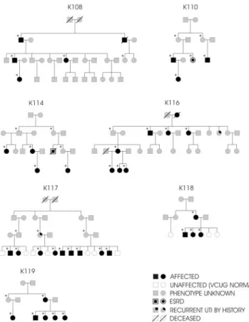
Figure 1. Pedigree structure of the seven kindreds studied.
Squares represent males, circles represent females. Black sym-bols: affected individuals; white symsym-bols: unaffected individu-als (normal voiding cystourethrography [VCUG]); gray sym-bols: affection status unknown. The symbols with double contour are patients in whom ESRD developed. A line through the symbols means the individual is deceased. *Individuals genotyped for this study.
produced by mutations in different genes (locus heterogeneity), such that the genetic cause of disease differs between families. Linkage analysis under heterogeneity is performed assuming that a proportion of families (␣) is linked to the marker tested, whereas the remaining families are unlinked. The LOD score for each family is therefore maximized over two parameters (␣ and the recombination fraction, ), yielding the HLOD. The total HLOD at a given locus is calculated by the sum of the HLOD from all of the kindreds studied. This analytic method results in minimal expenditure of degrees of freedom and has been shown to be a powerful tool for detection of linkage (25). In addition, to avoid erroneous exclusion of a locus because of mis-specification of the disease model, we performed nonparametric, allele-sharing analysis using permissive scoring functions (exponential model, with the “all” scoring function and equal weighting between families) as implemented in ALLEGRO. Published thresholds for
genome-wide significance (LOD ⱖ3.3), replication (P ⬍ 0.01), and exclusion of linkage (LODⱕ ⫺2) were used (20). Studies exploring the properties of the HLOD statistic suggested that an HLOD of approxi-mately 1.2 corresponds to P⫽ 0.01 (25).
Because these thresholds have been obtained assuming analysis of a large number of small pedigrees with an infinitely dense map, they may not be pertinent to data sets that vary in pedigree structure and information content. We therefore performed simulations to evaluate the power of our pedigrees to detect linkage and obtain the empiric thresholds for significance of our findings. We performed simulations of genotypes with ALLEGRO, using the same family structure and affection status as our VUR pedigrees, and specifying a 20-cM genetic map with marker spacing and heterozygosities equivalent to those used at our candidate intervals. To obtain the null distribution for our linkage statistics, we first performed simulations of genotypes under
Table 1. Pairwise total LOD scores at different recombination fractions (
)
Marker name Chr cM 0.0 0.01 0.05 0.1 0.2 0.3 0.4
GATA124C08N
1
129.37
⫺0.33
⫺0.28
⫺0.13
⫺0.04
0
0
⫺0.01
GATA133A08Q
1
137.59
⫺6.29
⫺5.41
⫺3.44
⫺2.18
⫺0.96
⫺0.42
⫺0.14
D1S1627
1
139.02
⫺2.61
⫺2.29
⫺1.49
⫺0.93
⫺0.37
⫺0.14
⫺0.05
D1S2809
1
144.38
⫺0.48
⫺0.26
0.21
0.44
0.47
0.31
0.11
D1S534
1
151.88
⫺0.55
⫺0.45
⫺0.19
⫺0.03
0.06
0.05
0.02
D1S498
1
155.89
⫺3.92
⫺3.55
⫺2.61
⫺1.84
⫺0.93
⫺0.43
⫺0.15
D1S2777
1
161.05
0
0.1
0.32
0.41
0.35
0.18
0.02
D1S1653
1
164.09
⫺0.73
⫺0.62
⫺0.29
⫺0.04
0.15
0.11
0.01
D1S1679
1
170.84
⫺5.3
⫺4.88
⫺3.71
⫺2.73
⫺1.49
⫺0.74
⫺0.28
AAC023
3
94.20
⫺5.29
⫺4.25
⫺2.31
⫺1.20
⫺0.31
⫺0.04
0.01
D3S3653
3
107.19
⫺4.15
⫺3.16
⫺1.61
⫺0.84
⫺0.22
⫺0.02
0.03
D3S3681
3
109.22
⫺8.22
⫺6.87
⫺4.31
⫺2.78
⫺1.30
⫺0.61
⫺0.23
D3S4529
3
112.42
⫺2.76
⫺2.25
⫺1.29
⫺0.68
⫺0.15
0.00
0.01
D6S1959
6
34.23
⫺1.91
⫺1.59
⫺0.8
⫺0.3
0.09
0.15
0.08
D6S1022
6
44.41
⫺2.59
⫺2.35
⫺1.63
⫺1.07
⫺0.46
⫺0.18
⫺0.05
D6S1051
6
50.75
⫺3.93
⫺3.5
⫺2.34
⫺1.48
⫺0.54
⫺0.12
0.04
D6S1017
6
63.28
⫺0.88
⫺0.7
⫺0.29
⫺0.06
0.06
0.02
⫺0.03
D6S2410
6
73.13
⫺1.77
⫺1.48
⫺0.82
⫺0.41
⫺0.06
0.03
0.02
D6S1053
6
80.45
⫺3.52
⫺3.1
⫺2.09
⫺1.39
⫺0.65
⫺0.29
⫺0.11
D6S1031
6
88.63
⫺1.74
⫺1.58
⫺1.12
⫺0.72
⫺0.24
⫺0.05
0.00
D10S1432
10
93.92
⫺1.54
⫺1.3
⫺0.7
⫺0.29
0.05
0.11
0.07
D10S2327
10
100.92
⫺0.89
⫺0.59
⫺0.03
0.24
0.36
0.29
0.16
D10S2470
10
112.58
⫺6.02
⫺5.39
⫺3.81
⫺2.64
⫺1.29
⫺0.55
⫺0.15
D10S677
10
117.42
⫺7.29
⫺6.25
⫺3.86
⫺2.32
⫺0.82
⫺0.2
0.02
D10S1239
10
125.0
⫺5.27
⫺4.8
⫺3.41
⫺2.26
⫺0.98
⫺0.35
⫺0.06
D10S1425
10
136.67
⫺2.08
⫺1.84
⫺1.24
⫺0.83
⫺0.41
⫺0.17
⫺0.03
D10S1230
10
142.78
⫺4.82
⫺4.37
⫺3.02
⫺2.01
⫺0.92
⫺0.39
⫺0.13
D10S1222
10
156.27
⫺4.36
⫺3.83
⫺2.74
⫺2.01
⫺1.09
⫺0.52
⫺0.17
D10S1248
10
165.27
⫺4.98
⫺4.45
⫺3.19
⫺2.25
⫺1.14
⫺0.52
⫺0.18
D19S714
19
42.0
⫺6.06
⫺5.27
⫺3.37
⫺2.06
⫺0.73
⫺0.17
0.02
D19S433
19
51.88
⫺4.36
⫺3.74
⫺2.27
⫺1.29
⫺0.38
⫺0.08
⫺0.01
D19S559
19
68.08
⫺3.77
⫺3.3
⫺2.11
⫺1.3
⫺0.52
⫺0.2
⫺0.07
D19S246
19
78.08
1.19
1.25
1.42
1.49
1.3
0.89
0.42
D19S589
19
87.66
⫺0.24
⫺0.24
⫺0.21
⫺0.17
⫺0.09
⫺0.04
⫺0.01
D19S254
19
100.61
⫺3.99
⫺3.59
⫺2.59
⫺1.84
⫺0.93
⫺0.41
⫺0.11
the hypothesis of no disease locus in the interval. We performed 1000 simulations to provide a solid estimate of the empiric LOD score threshold corresponding to Pⱕ 0.01. The simulated data were then analyzed with ALLEGRO under the same model previously delineated and HLOD were determined for each run. The HLOD calculated from these 1000 simulated runs provide the distribution of the HLOD sta-tistic under the hypothesis of no disease locus in the interval, the null hypothesis. The HLOD in the top 1% and 5% of the distribution define the point-wise empiric thresholds for type I error at Pⱕ 0.01 and P ⱕ 0.05, respectively, and therefore constitute our thresholds for replica-tion of linkage at Pⱕ 0.01 and P ⱕ 0.05 significance levels.
Inadequate study power may also hinder replication of original linkage findings. Therefore, we used simulations to assess the power of our study sample to detect linkage under various levels of locus het-erogeneity. We simulated genotypes of our pedigree sample (1000 replicates) with the assumption of linkage under each of the following conditions: (1) genetic homogeneity; (2) 50% locus heterogeneity; and (3) 28.6% locus heterogeneity (two of seven families linked). We then analyzed this second set of simulated pedigrees with ALLEGRO to obtain the maximum and average expected LOD (eLOD) scores with the models above. The fraction of LOD scores exceeding the thresholds for genome-wide significance and linkage replication provided our power to detect linkage with various levels of heterogeneity.
Results
Patients
We studied seven Caucasian pedigrees from Italy (K108, K110, K114, K116) and the United States (K117, K118, K119). All were ascertained via an index case with VUR documented by VCUG. These seven multigenerational kindreds comprised 140 individuals (Figure 1); we collected DNA from 105 individuals. In total, 41 individuals were classified as affected (36 based on a positive VCUG, two based on the presence of ESRD, one by the presence of documented recurrent urinary tract infections in childhood, and two by UPJ obstruction documented by sonogram). Eight patients were unaffected based on negative VCUG performed at age⬍6 yr; 91 individuals were considered as having unknown phenotype because they did not have a
clear phenotype (e.g., undocumented episodes of urinary tract infections or history of enuresis) or were asymptomatic and were not investigated by VCUG. There were 14 males and 27 females affected (male/female ratio ⫽ 0.52). The age at the diagnosis was 9.5⫾ 11 yr (range, 1 mo to 45 yr). Among those affected, 14 patients underwent corrective surgery, ESRD de-veloped in two patients, and one of them underwent kidney transplantation. Consistent with previous reports, some indi-viduals had associated urologic abnormalities: in K110, one patient had renal stones; in K116, one patient had a pelvic kidney; and in K117, one patient had a paraureteral diverticu-lum. All of the affected individuals in K114 also had associated UPJ obstruction documented by abdominal sonogram. Finally, K117 is noteworthy because it has a theoretical maximal LOD score⬎3 and can alone enable gene localization with genome-wide significance.
In these pedigrees, male-to-male transmission and a higher ratio of females to males argued against X-linked inheritance. The absence of consanguinity and occurrence of the disease in multiple generations made recessive inheritance unlikely. Con-sistent with the literature, the pattern of transmission was most consistent with multifactorial inheritance or an autosomal-dominant inheritance with reduced penetrance (7).
Simulation Studies
In addition to using published criteria for replication, we used simulations to establish empiric thresholds for replication of linkage. In 1000 simulations under the null hypothesis of no disease locus in the interval, we observed an HLODⱖ0.84 a total of 10 times and an HLODⱖ0.51 a total of 50 times. These data established the empiric threshold for replication of para-metric linkage at Pⱕ 0.01 and P ⱕ 0.05, respectively (20). The nonparametric LOD scores corresponding to these thresholds were 1.88 and 1.3, respectively.
To determine the power of our sample to detect linkage, we next performed simulations under the assumption of linkage
Table 2. Multipoint LOD scores in VUR families
Average
eLOD LOD1p13 LOD3p12 LOD6p21 LOD6p21 10q24LOD 10q26LOD 19q13LOD
Candidate gene
?
ROBO2
HLA
CDC5L
PAX2
?
USF2
cM location
144 to 164
108
45
66
125
165
59
K 108
0.63
⫺0.6
0.38
0.44
0.29
⫺0.74
0.11
0.43
K 110
0.95
⫺0.38
⫺1.33
⫺1.05
⫺0.03
⫺0.44
0.16
⫺0.21
K 114
1.19
⫺0.18
⫺0.48
⫺0.56
⫺0.26
⫺1.48
⫺0.52
⫺0.23
K 116
1.56
⫺0.96
⫺2.56
⫺1.6
⫺1.44
⫺1.23
⫺0.55
⫺1.25
K 117
3.35
⫺1.43
⫺1.59
⫺1.24
⫺1.86
⫺0.53
⫺1.36
⫺0.76
K 118
0.51
⫺0.59
⫺1.5
⫺0.57
⫺0.63
0.4
⫺0.33
0.07
K 119
1.3
0.96
⫺1.06
⫺0.11
0.09
⫺0.41
⫺0.64
⫺0.11
Total LOD (
␣ ⫽ 100%)
9.49
⫺3.18
⫺8.14
⫺4.69
⫺3.84
⫺4.43
⫺3.13
⫺2.06
HLOD (
␣ ⫽ 50%)
3.45
⫺0.46
⫺1.37
⫺0.96
⫺0.69
⫺1.06
⫺0.86
⫺0.45
Nonparametric LOD
8.15
0.74
0.02
0.01
0.31
0.06
0.29
0.06
Expected LOD (eLOD) for individual kindreds were obtained based on affected-only analysis with 1000 simulations, assuming homogeneity. On chr01, LOD scores shown were obtained at the location with maximized HLOD (D1S1653, 164 cM). VUR indicates vesicoureteral reflux; HLOD, heterogeneity LOD.
with various levels of genetic heterogeneity (1000 replicates each). Under genetic homogeneity, the average parametric ex-pected LOD for our kindreds was 1.4 (Table 2). For parametric analysis, simulations yielded a maximum expected LOD score of 11.14 (average 9.49⫾ 1.65) under homogeneity, 11.14 (aver-age 3.45⫾ 2.34) under 50% locus heterogeneity, and 9.07 (av-erage 1.71⫾ 1.83) under 28.6% locus heterogeneity (two fami-lies out of seven linked). For nonparametric analysis, we obtained a maximum expected LOD scores of 12.89 (average 8.15⫾ 1.52) under homogeneity, 9.96 (average 2.76 ⫾ 2.11) with 50% heterogeneity, and 8.67 (average 1.36⫾ 1.57) with 28.6% heterogeneity. The threshold for replication (at Pⱕ 0.01) was exceeded in 100%, 85%, and 53% of simulated runs with ho-mogeneity, 50% heterogeneity, and 28.6% heterogeneity, re-spectively. These data indicate that our sample had ample power not only to replicate linkage but also to achieve genome-wide significance with at least 50% locus heterogeneity.
Analysis of Linkage
Tables 1 and 2 show the pair-wise and multipoint LOD scores at the candidate intervals, respectively. Under genetic homoge-neity, multipoint LOD scores were strongly negative, excluding linkage to all candidate loci. In addition, our data do not support statistically significant linkage to these loci under a model of 50% genetic heterogeneity (negative HLOD at all intervals). Varying the percentage of linked families to maxi-mize the HLOD also did not achieve statistical significance at any of these intervals, with a maximal HLOD of 0.55 on chro-mosome 19 (D19S246) and 50% percent of pedigrees linked. This peak score was obtained approximately 10 cM (16 Mb) away from our candidate gene (USF2) and is significantly lower than our empiric threshold for replication of linkage at the 1% level. These results did not change significantly with alternative analyses. As expected, lowering the disease allele frequency or the phenocopy rate strengthened the evidence against linkage at all intervals. Similarly, nonparametric analysis did not reveal significant linkage to any of the candidate loci (maximum non-parametric LOD of 0.74 on chromosome 1), demonstrating that absence of linkage cannot be attributed to mis-specification of disease model. Finally, secondary analyses with exclusion of K114, a pedigree with associated UPJ obstruction, did not suf-ficiently increase the LOD scores at candidate intervals to achieve statistical significance.
Discussion
Replication studies are important components of genetic in-vestigations because they help determine the level of heteroge-neity of the trait and assess the potential for type 1 error (20,26). Thus, although the estimated probability of false-positive find-ings in genome scans is⬍0.05 when a threshold LOD score of 3.3 is used, Lander and Kruglyak emphasized that replication studies should be also performed (20). Replication studies may have difficulty in detecting linkage to reported loci because initial positive reports tend to overestimate the genetic effect (20,27). This bias occurs because initial reports tend to benefit from random fluctuations that push the LOD score above sta-tistical significance, whereas follow-up studies regress to the
true mean (20,27). Moreover, variation in methodology, ascer-tainment criteria, and study population may also complicate the ability to replicate initial findings (20,27). In the case of VUR, one of the major obstacles for linkage studies is the relative rarity of large pedigrees because of reduced penetrance of the trait (7). Reduced penetrance necessitates affected-only analysis and this constraint, combined with locus heterogene-ity, diminishes the ability to assemble a well-powered cohort. Our study, however, benefited from relatively large kindreds, including one of the largest families reported to date (K117); this kindred alone should be sufficiently large to support gene localization at the genome-wide level. Moreover, our pedigrees resembled Caucasian cohorts previously described, particularly the one described by Feather et al. (12), and we used similar ascertainment criteria and linkage models described in their article. Our simulations indicated that we were well powered to replicate linkage or achieve significance at the genome-wide level, with a maximum expected LOD score of 11.14 under homogeneity (for comparison we estimate the maximum ex-pected LOD in the pedigrees by Feather et al. at approximately 8.7) (12). Nevertheless, despite analyses under many alternative models, we found no evidence of linkage to loci previously implicated in humans. Our best interval barely reached the threshold for replication at Pⱕ 0.05 on chromosome 19, but this maximal score was achieved 16 Mb distal to the candidate gene (USF2) and did not hold up to correction for multiple testing. These data highlight the need to use stringent criteria to avoid false replication of linkage.
The lack of replication is most likely caused by overestima-tion of the risk attributable to previously reported loci and substantial genetic heterogeneity of the trait. This heterogeneity is demonstrated by human and animal studies indicating that VUR can be caused by defects in a panel of genes expressed in the lower urinary tract. For example, loss of the uroplakin III gene causes VUR in the mouse (28); similarly, tissue-specific ablation of calcineurin causes ureteropelvic obstruction (29). Mice lacking either ROBO2 or its ligand, SLIT2, also have urogenital developmental abnormalities that include supernu-merary ureteric buds that remain inappropriately connected to the nephric duct and failure of the ureters to connect to the bladder (30). The number of mutant mice exhibiting renal de-velopmental defects is rapidly growing, reflecting the intricate biology of kidney development (31). These animal data high-light the potential for genetic heterogeneity of human disease and simultaneously provide a substantial list of candidate genes for human VUR.
The candidate gene approach had generally met with limited success in human VUR (e.g., screening of PAX2 or uroplakin genes) (32,33). Lu et al. have, however, recently reported dis-ruption of the ROBO2 gene in one patient with a balanced translocation and a phenotype that included VUR; in addition, mutational screening of patients with nonsyndromic VUR re-vealed two nonconservative amino acid substitutions (19). The full description of these patients is not available at the time of writing this report, but these preliminary data indicate that
ROBO2 accounts for a small fraction of VUR cases (19).
to this locus was not supported in our seven kindreds. How-ever, we expected to detect linkage to the 1p13 locus because it accounted for disease in the majority (78%) of the families Feather et al. studied (12). Among our kindreds, however, only K119 showed a positive LOD score on chromosome 1p13, a proportion (14.3%) insufficient to replicate linkage. Because we had 85% power to replicate with 50% locus heterogeneity, our data suggest that the chromosome 1p13 interval may account for disease in smaller fraction of VUR kindreds, perhaps in as little as 15% and probably less than half of VUR families overall.
Together with the results of animal studies, our data raise the potential that mutations in different genes each account for disease in a small proportion of VUR families. In this scenario, the search for VUR genes may be very complicated and might have to rely on extensive screening of candidate genes in both familial and sporadic cases. Linkage studies of the trait would require a large number of average size pedigrees or would have to rely on pedigrees such as K117 that have a sufficiently large number of affected individuals to map a locus on their own. To be successful, comprehensive studies combining all approaches will likely be required. Accordingly, after testing the most promising candidate loci reported in humans, we are now proceeding with a genome-wide search to detect novel VUR loci in our kindreds.
Acknowledgments
We thank the patients and members of the kindreds studied for their generous contribution to this project. A.G.G. is supported by a Young Investigator Award from the Emerald Foundation and the Irving Clin-ical Scholar Program.
References
1. Peters PC, Johnson DE, Jackson JH Jr: The incidence of vesicoureteral reflux in the premature child. J Urol 97: 259 –260, 1967
2. Stephens FD, Lenaghan D: The anatomical basis and dy-namics of vesicoureteral reflux. J Urol 87: 669 – 680, 1962 3. Tanagho EA, Guthrie TH, Lyon RP: The intravesical ureter
in primary reflux. J Urol 101: 824 – 832, 1969
4. Scott JE, Swallow V, Coulthard MG, Lambert HJ, Lee RE: Screening of newborn babies for familial ureteric reflux.
Lancet 350: 396 – 400, 1997
5. Ardissino G, Dacco V, Testa S, Bonaudo R, Claris-Appiani A, Taioli E, Marra G, Edefonti A, Sereni F: Epidemiology of chronic renal failure in children: Data from the ItalKid project. Pediatrics 111: e382–387, 2003
6. Ichikawa I, Kuwayama F, Pope JCt, Stephens FD, Miyazaki Y: Paradigm shift from classic anatomic theories to con-temporary cell biological views of CAKUT. Kidney Int 61: 889 – 898, 2002
7. Eccles MR, Bailey RR, Abbott GD, Sullivan MJ: Unravelling the genetics of vesicoureteric reflux: a common familial disorder. Hum Mol Genet 5: 1425–1429, 1996
8. Noe HN: The long-term results of prospective sibling re-flux screening. J Urol 148: 1739 –1742, 1992
9. Noe HN, Wyatt RJ, Peeden JN Jr, Rivas ML: The transmis-sion of vesicoureteral reflux from parent to child. J Urol 148: 1869 –1871, 1992
10. Chapman CJ, Bailey RR, Janus ED, Abbott GD, Lynn KL: Vesicoureteric reflux: Segregation analysis. Am J Med Genet 20: 577–584, 1985
11. Pasch A, Hoefele J, Grimminger H, Hacker HW, Hilde-brandt F: Multiple urinary tract malformations with likely recessive inheritance in a large Somalian kindred. Nephrol
Dial Transplant 19: 3172–3175, 2004
12. Feather SA, Malcolm S, Woolf AS, Wright V, Blaydon D, Reid CJ, Flinter FA, Proesmans W, Devriendt K, Carter J, Warwicker P, Goodship TH, Goodship JA: Primary, non-syndromic vesicoureteric reflux and its nephropathy is genetically heterogeneous, with a locus on chromosome 1.
Am J Hum Genet 66: 1420 –1425, 2000
13. Mackintosh P, Almarhoos G, Heath DA: HLA linkage with familial vesicoureteral reflux and familial pelvi-ureteric junction obstruction. Tissue Antigens 34: 185–189, 1989 14. Groenen PM, Vanderlinden G, Devriendt K, Fryns JP, Van
de Ven WJ: Rearrangement of the human CDC5L gene by a t(6;19)(p21;q13.1) in a patient with multicystic renal dys-plasia. Genomics 49: 218 –229, 1998
15. Eccles MR, Schimmenti LA: Renal-coloboma syndrome: A multi-system developmental disorder caused by PAX2 mutations. Clin Genet 56: 1–9, 1999
16. Ogata T, Muroya K, Sasagawa I, Kosho T, Wakui K, Saka-zume S, Ito K, Matsuo N, Ohashi H, Nagai T: Genetic evidence for a novel gene(s) involved in urogenital devel-opment on 10q26. Kidney Int 58: 2281–2290, 2000
17. Groenen PM, Garcia E, Debeer P, Devriendt K, Fryns JP, Van de Ven WJ: Structure, sequence, and chromosome 19 localization of human USF2 and its rearrangement in a patient with multicystic renal dysplasia. Genomics 38: 141– 148, 1996
18. Izquierdo L, Porteous M, Paramo PG, Connor JM: Evi-dence for genetic heterogeneity in hereditary hydrone-phrosis caused by pelvi-ureteric junction obstruction, with one locus assigned to chromosome 6p. Hum Genet 89: 557–560, 1992
19. Lu W, Peters R, Ferguson H, et al.: Disruption of ROBO2 is associated with vesicoureteral reflux. J Am Soc Nephrol 15: 32A, 2004
20. Lander E, Kruglyak L: Genetic dissection of complex traits: Guidelines for interpreting and reporting linkage results.
Nat Genet 11: 241–247, 1995
21. Klemme L, Fish AJ, Rich S, Greenberg B, Senske B, Segall M: Familial ureteral abnormalities syndrome: Genomic mapping, clinical findings. Pediatr Nephrol 12: 349 –356, 1998
22. Lathrop GM, Lalouel JM: Easy calculations of lod scores and genetic risks on small computers. Am J Hum Genet 36: 460 – 465, 1984
23. Cottingham RW Jr, Idury RM, Schaffer AA: Faster sequen-tial genetic linkage computations. Am J Hum Genet 53: 252–263, 1993
24. Gudbjartsson DF, Jonasson K, Frigge ML, Kong A: Allegro, a new computer program for multipoint linkage analysis.
Nat Genet 25: 12–13, 2000
25. Abreu PC, Hodge SE, Greenberg DA: Quantification of type I error probabilities for heterogeneity LOD scores.
Genet Epidemiol 22: 156 –169, 2002
26. Vieland VJ: The replication requirement. Nat Genet 29: 244 –245, 2001
in estimation of locus-specific effects from genomewide scans. Am J Hum Genet 69: 1357–1369, 2001
28. Hu P, Deng FM, Liang FX, Hu CM, Auerbach AB, Shapiro E, Wu XR, Kachar B, Sun TT: Ablation of uroplakin III gene results in small urothelial plaques, urothelial leakage, and vesicoureteral reflux. J Cell Biol 151: 961–972, 2000 29. Chang CP, McDill BW, Neilson JR, Joist HE, Epstein JA,
Crabtree GR, Chen F: Calcineurin is required in urinary tract mesenchyme for the development of the py-eloureteral peristaltic machinery. J Clin Invest 113: 1051– 1058, 2004
30. Grieshammer U, Le M, Plump AS, Wang F, Tessier-Lavigne M, Martin GR: SLIT2-mediated ROBO2 signaling restricts kidney induction to a single site. Dev Cell 6: 709 – 717, 2004
31. Yu J, McMahon AP, Valerius MT: Recent genetic studies of mouse kidney development. Curr Opin Genet Dev 14: 550 – 557, 2004
32. Jiang S, Gitlin J, Deng FM, Liang FX, Lee A, Atala A, Bauer SB, Ehrlich GD, Feather SA, Goldberg JD, Goodship JA, Goodship TH, Hermanns M, Hu FZ, Jones KE, Malcolm S, Mendelsohn C, Preston RA, Retik AB, Schneck FX, Wright V, Ye XY, Woolf AS, Wu XR, Ostrer H, Shapiro E, Yu J, Sun TT: Lack of major involvement of human uroplakin genes in vesicoureteral reflux: Implications for disease heteroge-neity. Kidney Int 66: 10 –19, 2004
33. Kelly H, Ennis S, Yoneda A, Bermingham C, Shields DC, Molony C, Green AJ, Puri P, Barton DE: Uroplakin III is not a major candidate gene for primary vesicoureteral reflux.
Eur J Hum Genet 2004