䊚 2006 by the INTERNATIONALSOCIETY OFENDOVASCULARSPECIALISTS Available at www.jevt.org
⽧CLINICAL INVESTIGATION ⽧
Carotid Artery Stenting With Filter Protection in
High-Risk Patients Showing Severe Electroencephalographic
Alterations During Carotid Endarterectomy
Roberto Gandini, MD1; Alessio Spinelli, MD1; Sebastiano Fabiano, MD1;
Vittorio Colangelo, MD1; Maria Grazia Marciani, MD, PhD2; Andrea Romigi, MD2; and
Giovanni Simonetti, MD, PhD1
Departments of 1Diagnostic Imaging and Interventional Radiology and 2Neuroscience,
University of Tor Vergata, Rome, Italy.
⽧ ⽧
Purpose: To describe the results and efficacy of stent treatment in patients with carotid
stenosis who had aborted carotid endarterectomy procedures due to the appearance of severe electroencephalographic (EEG) alterations.
Methods: A retrospective study was conducted of 18 patients (11 men; mean age 72 years,
range 62–84) with symptomatic high-grade carotid artery stenoses (ⱖ70%) who experi-enced severe EEG alterations during carotid endarterectomy, causing the procedure to be aborted. Twelve patients had shown no hemodynamic alterations during preoperative transcranial Doppler evaluation after external compression of the common carotid artery; the remaining 6 could not be evaluated. The patients were referred for carotid artery stent-ing (CAS); 7 had contralateral internal carotid artery stenosis and 5 had contralateral oc-clusion. Endovascular intervention was carried out using standard techniques under filter protection. Follow-up was scheduled at 3, 6, and 12 months.
Results: All patients were successfully treated without immediate complications. The EEG
did not display any significant alterations during the endovascular procedure. Mean follow-up was 43 months. Magnetic resonance imaging at 6 months showed no signs of cerebral ischemia. Color Doppler ultrasound imaging documented normal stent patency in all pa-tients.
Conclusion: Patients with symptomatic severe carotid stenosis (ⱖ70%) who are considered
at risk due to the appearance of severe EEG alterations during surgical treatment may benefit from CAS with respect to both major and minor complications. Larger studies are needed to confirm these findings.
J Endovasc Ther 2006;13:451–456
Key words: carotid endarterectomy, carotid artery, stenosis, electroencephalography,
ce-rebral ischemia, carotid angioplasty, stent
⽧ ⽧
The authors have no commercial, proprietary, or financial interest in any products or companies described in this article.
Address for correspondence and reprints: Vittorio Colangelo, MD, Viale Oxford 81, 00133 Rome, Italy. Fax: 39-06-2090-2404; E-mail: [email protected]
Endovascular treatment of carotid stenosis is a valid alternative to carotid endarterectomy (CEA) owing to improved devices and tech-niques and a growing number of studies showing that carotid artery stenting (CAS) with cerebral protection is not inferior to CEA
in terms of major and minor postprocedural complications.1–4
The role of the endovascular technique is particularly important in high-risk patients in whom the surgical approach may be danger-ous due to inadequate cerebral
collateraliza-tion. Furthermore, concomitant contralateral stenosis or occlusion could pose a greater risk of neurological complications due to abrupt reduction in blood flow after clamping of the carotid vessels. In this respect, the North American Symptomatic Carotid Endarterec-tomy Trial (NASCET) showed an increase in perioperative risk after endarterectomy in pa-tients with contralateral internal carotid artery (ICA) occlusion5; but it is uncertain whether a contralateral occlusion could cause a peripro-cedural stroke during CEA.6,7 For CAS, how-ever, a few authors have found that endovas-cular treatment in the setting of bilateral disease is not encumbered by this peripro-cedural risk.8–10
Electroencephalography (EEG) is one of the most commonly used methods of monitoring possible cerebrovascular insufficiency during CEA.11Variations in EEG activity may be due to surgery (i.e., clamping) or nonsurgical causes, such as anesthetic level, blood pres-sure, other physiological factors.12 Specific signs of altered electrical activity during sur-gery may induce the surgeon to abort the pro-cedure, especially when such alterations ap-pear early and in patients with comorbidities. Patients afflicted with atherosclerotic disease usually have involvement in multiple vascular territories, so, apart from the possible coro-nary comorbidities, the vascular autoregula-tory system may be altered, thus affecting the redistribution of flow and cerebral perfusion during the procedure.
Our study evaluated the results and efficacy of angioplasty and carotid stenting with ce-rebral protection in patients in whom CEA had been aborted because of the appearance of serious EEG alterations during carotid clamping.
METHODS
Patient Characteristics
From December 1998 to May 2004, the en-dovascular team of our department treated 331 patients for occlusive disease in 363 ICAs. Eighteen of these patients (11 men; mean age 72 years, range 62–84) with a symptomatic de novo ICA stenosis ⬎70% were referred for CAS after surgical endarterectomy was
abort-ed due to serious EEG alterations during ca-rotid clamping. Comorbidities included dia-betes (n⫽13), hypertension (n⫽13), prior stroke (n⫽3), hypercholesterolemia (n⫽10), and obesity (n⫽5).
Twelve (67%) of the 18 patients had had a preoperative transcranial Doppler (TCD) scan performed while compressing the common carotid artery (CCA) to simulate clamping. Seven patients showed no significant hemo-dynamic alterations in the contralateral mid-dle cerebral artery. The other 5 patients, all with contralateral ICA occlusion, showed mild hemodynamic alterations without significant blood flow reduction, so the surgical team planned to use an intraoperative shunt during CEA in these patients. The remaining 6 pa-tients lacked a suitable acoustic window for TCD, but none of them had contraindications to surgery and so underwent CEA.
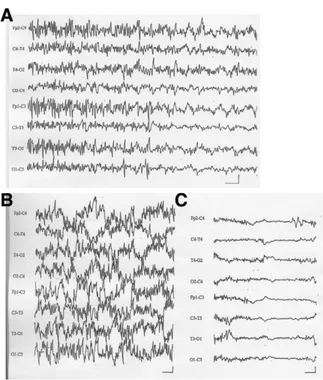
Within the first 20 seconds after clamping, each patient manifested serious EEG alter-ations (Fig. 1), either a decrease in amplitude of all activity, including delta and faster fre-quencies, or increased amplitude and dura-tion of delta waves. Our surgeons considered the precocious appearance of major EEG al-terations after clamping as a possible threat to the positioning of a carotid-carotid shunt, even though it can be performed in ⬃60 sec-onds. Considering the patients’ comorbidities, the surgeons did not feel that they could po-sition a shunt safely, so they aborted the pro-cedures.
Carotid Stent Procedure
When the patients were referred for endo-vascular treatment, color Doppler sonograph-ic analysis of plaque morphology document-ed hyperechogenicity in 9 cases, mixdocument-ed morphology in 7 cases, and hypoechogenicity in 2, one of which showed an ulcerated plaque. Computed tomography (CT) and/or magnetic resonance angiography (MRA) con-firmed the significant stenosis and provided further information on the circle of Willis and the anatomy of the supra-aortic vessels. Sev-en patiSev-ents had contralateral ICA stSev-enoses (4 ⬎70% and 3 between 50% and 60%), and 5 had contralateral occlusions (2 CCA, 3 ICA);
Figure 1⽧(A) In this patient with left carotid artery stenosis, carotid clamping induced an increase in amplitude of slow waves (1-Hz frequency) and si-multaneous reduction in faster frequencies local-ized in the left front, central, and temporal regions. (B) In another patient with right carotid stenosis and contralateral carotid occlusion, the EEG before clamping showed widespread fast frequencies with high voltage. (C) Right carotid clamping in-duced severe, bilateral reduction in amplitude of faster frequency components. EEGs were recorded with a 0.3-Hz high band-pass filter and a 50-Hz low band-pass filter. The horizontal bars indicate 1 sec-ond; the vertical bars are 20V.
the other 6 had no significant disease in the contralateral carotid circulation.
The patients were pretreated with 100 mg/d of aspirin and 75 mg/d of clopidogrel for at least 4 to 5 days prior to treatment. During CAS, continuous EEG monitoring was per-formed using an analogue 10-channel system polygraph (Vega 10; EBNeuro, Florence, Italy). The time constant was 0.3 seconds, and a 50-Hz low-pass filter was needed in the operating room’s electrical environment. Electrodes were positioned according to the 10–20 Inter-national System to monitor 8 EEG channels (Fp2, C4, T4, O2, Fp1, C3, P3, O1). The record-ing was begun before the procedure and con-tinued throughout, with careful attention to
the EEG during the following critical phases: guide catheter manipulation in the CCA, ad-vancement of the catheter or long introducer into the CCA, guidewire manipulation through the stenosis, predilation, crossing the stenosis with the protection filter, stent ad-vancement and release, post dilation, and fil-ter recovery.
During the procedure, heparin boluses (50– 100 U/kg) were administered to maintain the activated coagulation time around 300 sec-onds. All the procedures were performed with continuous heparinized saline wash to pre-vent the formation of clots or air emboli in the system. In 7 cases, the ‘‘over-the-wire’’ tech-nique was used, in which the ipsilateral ex-ternal carotid artery was catheterized from a femoral access with a 0.035-inch hydrophilic guidewire and diagnostic catheters. After ex-changing for a stiff or Amplatz guidewire, a long 6-F introducer (Cook Inc., Bloomington, IN, USA) was advanced in the CCA. In 9 cases, catheterization of the CCA was done with a 0.035-inch stiff hydrophilic guidewire and 7-F guiding catheter. Owing to pronounced tor-tuosity of the supra-aortic vessels (type III arch) in 2 patients, a coaxial telescopic system was used, in which a 5-F (125-cm) diagnostic catheter and 0.035-inch stiff guidewire were inserted inside a catheter or a long introducer and advanced simultaneously. An EPI FilterWire cerebral protection filter (Boston Scientific, Natick, MA, USA) was deployed in all patients. Predilation with a 3-mm balloon was necessary in only 1 case because of a tight stenosis. Monorail Carotid Wallstents (Boston Scientific) were used exclusively. At-ropine (0.5–1.0 mg) was administered before stent dilation, which was done with 5- to 6-mm balloons (Fig. 2) for an average of 3 to 7 seconds.
Postoperatively, clopidogrel was continued for 6 weeks and aspirin indefinitely. Follow-up consisted of clinical examination with color Doppler imaging before discharge and at 3, 6, and 12 months, with semiannual examina-tions thereafter.
RESULTS
In all patients, the filter was correctly placed distal to the stenosis with no technical
prob-Figure 2⽧(A) This DSA shows evidence of signif-icant ostial stenosis of both left and right ICAs. (B) Postprocedural DSA after stent deployment in the left ICA.
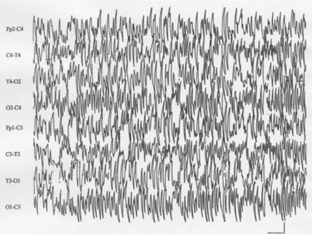
Figure 3⽧An EEG showing no significant changes in amplitude or frequency during each phase of the endovascular procedure (0.3-Hz high band-pass fil-ter, 50-Hz low band-pass filter). The horizontal bar indicates 1 second; the vertical bar is 20V. lems. Positioning the stent was successful in
all cases, which lasted an average of 23 min-utes (range 12–47) from the femoral artery puncture to sheath removal. In 4 patients, macroscopic debris was found inside the fil-ter.
Continuous intraoperative EEG monitoring (Fig. 3) did not show significant cerebral al-terations during any phase of the procedure. The final angiogram showed good stent po-sition with restoration of the physiological vessel lumen (residual stenosis⬍20%) and no changes in the circle of Willis.
Over a mean 43-month follow-up (range 12–65), no patient showed either clinical evi-dence of neurological deficits at the 3 and 6-month examinations or MR signs of cerebral ischemia at 6 months. All stents were patent at the 12-month follow-up.
DISCUSSION
With recent developments in cerebral protec-tion systems, miniaturizaprotec-tion of devices, and evolutionary stent materials, CAS has be-come a safer technique, with a low risk of complications.1–4 The Pro-CAS registry of 3267 procedures confirmed the technical suc-cess of CAS (98%), with 0.6% mortality and 1.2% major and 1.3% minor stroke rates.4The combined stroke and death rate was 2.8%. The randomized SAPPHIRE trial compared the
results of CEA (151 patients) to those of CAS under cerebral protection (156 patients) in symptomatic patients with ⱖ50% stenosis or asymptomatic patients with ⱖ80% stenosis.3 Notably, several CAS patients had been ex-cluded from the surgical protocol because of at least 1 risk factor, such as a high degree of heart failure, severe pulmonary disease, con-tralateral carotid occlusion, previous surgery with restenosis, or previous neck radiothera-py. The study clearly demonstrated a lower rate of complications (i.e., mortality, peripro-cedural neurological events, and myocardial infarction) at 30 days in the CAS patients, sup-porting the validity of protected CAS as an option in the treatment of carotid stenoses.
In our patients, the use of a cerebral pro-tection system seems to have played a fun-damental role in allowing continuous ante-grade flow in the target carotid axis, thus preserving cerebral perfusion. During CEA, on the other hand, the abrupt reduction in cere-bral blood flow due to carotid clamping in pa-tients with contralateral ICA or CCA occlusion may increase the risks of the procedure. The NASCET study reported a 14% increased risk of cerebral ischemia in patients with contra-lateral ICA occlusion, directly related to a de-crease in cerebral blood flow due to bilateral carotid pathology.5 In a systematic review of 1729 patients from the European Carotid
Sur-gery Trial, Rothwell et al.13described a 2-fold risk of perioperative stroke in patients with contralateral carotid occlusion. On the other hand, Samson et al.14did not find any signif-icant change in surgical perioperative risk for patients with significant occlusions or steno-sis of the contralateral ICA. Nevertheless, we believe that the presence of a contralateral oc-clusion or significant stenosis can be a risk factor during endarterectomy, particularly in high-risk patients with diffuse atherosclerosis and comorbidities.
In a study by Samson et al.,142 major types of clamp-related EEG variations were de-tailed: attenuation of all activity by at least 75% and/or aⱖ2-fold increase in ⱕ1-Hz delta activity, which is what occurred in our study patients. In the absence of a contralateral ca-rotid occlusion or severe stenosis, EEG changes with clamping are usually ipsilateral. Bilateral clamp-related changes typically arise when contralateral blood flow is severely compromised. The incidence of unilateral changes is more than twice that of bilateral changes.12 The incidence of the major varia-tions noted above range from 3.4% to 12.5%15 owing to several variables, such as subjective elements in assessing non-processed EEG signals, different high band-pass filter fre-quencies, varying anesthetic techniques, and different patient populations.
Since diffuse atherosclerosis in the entire arterial district can alter flow regulatory sys-tems mediated by intraparietal receptors, pa-tients without significant stenosis or occlu-sion of the contralateral carotid axis can nonetheless be considered at high surgical risk. Moreover, the absence of communicat-ing arteries may explain the intolerance to clamping of the contralateral carotid. In 2 of our 18 cases, the DSA performed before the stent procedure confirmed an anatomical var-iation characterized by the absence of the an-terior and posan-terior communicating arteries. In the remaining 16 cases, no anatomical var-iations of the intracranial vessels were de-tected.
Some authors report that TCD is an excel-lent means of predicting cerebral ischemia during carotid surgery.16 Consistent with the work by Lagneau et al.,17 our study showed that the preliminary evaluation with TCD does
not present a reliable prediction of hemody-namic compensation in the contralateral ca-rotid territory. In addition, the frequent ab-sence of an acoustic window for TCD and the difficulty of external compression to simulate clamping in patients with widespread parietal calcification reduce the potential value of TCD. In our group, 7 patients who did not show hemodynamic alterations in the prelim-inary TCD evaluation demonstrated severe EEG alterations within the first 20 seconds af-ter clamping, which suggests widespread ath-erosclerotic pathology with abnormal adap-tation of flow redistribution in the intracranial vessels. This hypothesis may explain the se-vere EEG alterations in those patients with regular patency and caliber of the contralat-eral carotid artery.
Conclusion
In our experience, stent placement with a protection system may represent the treat-ment of choice in patients with high surgical risk because of stenosis or occlusions of the contralateral carotid artery or because of se-rious, widespread atherosclerotic pathology that involves the supra-aortic vessels. Fur-thermore, the application of carotid stenting in high-risk patients suggests that endovas-cular management of carotid stenoses can be a valid alternative to CEA, achieving high technical success and a low rate of compli-cations.
REFERENCES
1. CARESS Steering Committee. Carotid Revas-cularization using Endarterectomy or Stenting Systems (CARESS): phase I clinical trial. J
En-dovasc Ther. 2003;10:1021–1030.
2. Goodney PP, Schermerhorn ML, Powell RJ. Current status of carotid artery stenting. J Vasc
Surg. 2006;43:406–411.
3. Yadav JS, Wholey MH, Kuntz RE, et al. Pro-tected carotid-artery stenting versus endarter-ectomy in high-risk patients. N Engl J Med. 2004;351:1493–1501.
4. Theiss W, Hermanek P, Mathias K, et al. Pro-CAS. A prospective registry of carotid angio-plasty and stenting. Stroke. 2004;35:2134–2139. 5. Ferguson GG, Eliasziw M, Barr HW, et al. The North American Symptomatic Carotid
Endar-terectomy Trial: surgical results in 1415 pa-tients. Stroke. 1999;30:1751–1758.
6. Mozes G. High-risk carotid endarterectomy.
Semin Vasc Surg. 2005;18:61–68.
7. Grego F, Antonello M, Lepidi S, et al. Is contra-lateral carotid artery occlusion a risk factor for carotid endarterectomy? Ann Vasc Surg. 2005; 19:882–889.
8. Mathur A, Roubin GS, Gomez CR, et al. Elective carotid artery stenting in the presence of con-tralateral occlusion. Am J Cardiol. 1998;81: 1315–1317.
9. Mericle RA, Kim SH, Lanzino G, et al. Carotid artery angioplasty and use of stents in high-risk patients with contralateral occlusions. J
Neu-rosurg. 1999;90:1031–1036.
10. Sabeti S, Schillinger M, Mlekusch W, et al. Contralateral high grade carotid artery stenosis or occlusion is not associated with an in-creased risk for poor neurological outcome af-ter carotid stenting. Radiology. 2004;230:70–76. 11. Findlay JM, Marchak BE, Pelz DM, et al. Carotid endarterectomy: a review. Can J Neurol Sci. 2004;31:22–36.
12. Blume WT, Ferguson GG, McNeill DK.
Signifi-cance of EEG changes at carotid endarterec-tomy. Stroke. 1986;17:891–897.
13. Rothwell PM, Slattery J, Warlow CP. Clinical and angiographic predictors of stroke and death from carotid endarterectomy: systematic review. BMJ. 1997;315:1571–1577.
14. Samson RH, Showalter DP, Yunis JP. Routine carotid endarterectomy without a shunt, even in the presence of a contralateral occlusion.
Cardiovasc Surg. 1998;6:475–484.
15. Sundt TM, Sharbrough FW, Piepgras DG, et al. Correlation of cerebral blood flow and electro-encephalographic changes during carotid end-arterectomy: with results of surgery and he-modynamics of cerebral ischemia. Mayo Clin
Proc. 1981;56:533–543.
16. McDowell HA, Gross GM, Halsey JH. Carotid endarterectomy monitored with transcranial Doppler. Ann Surg. 1992;215:514–518.
17. Lagneau P, Baujat B, Anidjar S, et al. Is trans-cranial Doppler a worthwhile examination for preoperative evaluation of the circle of Willis? Evaluation of 137 carotid endarterectomies performed under regional anesthesia. Int