Post-translational modifications and
molecular interactions regulating
VEGFR2 activity
PhD Thesis
Scuola Normale Superiore
Lucia Pattarini
INDEX
SYNOPSIS ...1
INTRODUCTION ...3
BLOOD VESSEL DEVELOPMENT...4
Pathological and physiological angiogenesis ...6
Vascular Endothelial Growth Factors ...8
VEGF-A ...8
PlGF...12
VEGF-B ...13
VEGF-C and VEGF-D ...14
VEGF Receptors ...15
VEGFR1...17
VEGFR2...19
VEGFR3...28
Semaphorins and plexins in vessel biology...29
Neuropilins...31
Neuropilin1 ...32
PROTEIN FUNCTION REGULATION BY ACETYLATION...37
Lysine acetyltransferases (KATs) ...39
Lysines deacetylases (KDACs) ...40
Crosstalk between acetylation and other PTMs in regulating protein function ...42
Acetylation of non-nuclear proteins ...44
Acetylation and blood vessel biology ...46
VISUALIZATION OF PROTEIN INTERACTIONS USING FLUORESCENT PROTEINS...50
GFP: Structure, folding and fluorescence properties ...51
GFP optimization and spectral variants ...53
Other fluorescent proteins...55
Fluorescent proteins as a powerful tool in biology...55
Fluorescence (Foster) Resonance Energy Transfer (FRET) ...57
Imaging FRET for cell biology applications...58
Intermolecular FRET...59
FRET applied to transmembrane receptor biology...60
MATERIALS AND METHODS ...63
PLASMIDS...64
CELL CULTURE, TRANSFECTION AND TREATMENT...65
ANTIBODIES...66
WESTERN BLOT AND IMMUNOPRECIPITATION...67
IMMUNOFLUORESCENCE...68
STRUCTURAL MODELLING...70
RESULTS...71
RESULTS PART I...73
VEGFR2 IS ACETYLATED BY P300...73
VEGFR2 is acetylated in PAE cells...73
p300 increases VEGFR2 acetylation in vivo ...74
IDENTIFICATION OF VEGFR2 ACETYLATION SITES...79
At least five VEGFR2 residues are acetylated...79
Substitution of lysine with arginine strongly reduces VEGFR2 acetylation..83
VEGFR2 ACETYLATION INFLUENCES RECEPTOR PHOSPHORYLATION AND STABILITY...84
VEGFR2 acetylation increases VEGF-induced tyrosine phosphorylation....85
Mutation of lysine 1053 to arginine impairs VEGFR2 tyrosine phosphorylation but has no effect on protein stability ...86
Substitution of five lysines with arginines reduces VEGFR2 stability ...88
Effect of VEGFR2 acetylation on VEGF-dependent desensitization ...91
RESULTS PART II...93
FLUORESCENCE RESONANCE ENERGY TRANSFER...93
VEGFR2-EGFP characterization...97
Localization and activity of fluorescent Neuropilin1 ...99
VISUALIZATION OF VEGFR2-NP1 INTERACTION IN EC USING FRET...102
FRET FOR THE VISUALIZATION OF NEUROPILIN1 COMPLEXES...105
EFFECT OF ANTI-NP1 BLOCKING ANTIBODIES ON FRET-DETECTED NP1 COMPLEXES...109
ANTI-PLGF INHIBITS NP1 COMPLEXES FORMATION...113
DISCUSSION ...117
DISCUSSION PART I...118
VEGFR2 is modified by acetylation ...118
Molecular players in VEGFR2 acetylation and deacetylation ...120
Crosstalk between VEGFR2 phosphorylation and acetylation ...122
VEGFR2 stability and acetylation ...123
VEGFR2 acetylation and endothelial cell function...124
DISCUSSION PART II...126
FRET as a powerful technique to study protein-protein interaction...126
FRET allows analysis of VEGFR2-Neuropilin1 interactions in ECs...127
NP1-NP1 complex formation ...130
FRET for anti-NP1 antibody validation...131
BIBLIOGRAPHY...135
SYNOPSIS
The work described in this thesis has been mainly focused on the study of a key molecule involved in blood vessel formation, the tyrosine kinase receptor VEGFR2.
Considering that VEGFR2 biology should be tighly regulated to allow proper blood vessel formation and maintenance, we investigated two different mechanims influencing VEGFR2 activity: post translational modification and receptor complex formation.
Since VEGFR2 biology is governed through protein modication, mainly phosphorylation, we decided to investigate the possible role of acetylation in VEGFR2 activity. Combining biochemical and proteomic studies, we showed that VEGFR2 is modified by acetylation. Starting from this observation, we further investigated the impact of VEGFR2 acetylation on protein stability and phosphorylation in response to ligand. These findings are of particular interest, since, to our knowledge, this is the first report that a tyrosine kinase receptor might be regulated by acetylation.
Additionally, we decided to elucidate the interaction of VEGFR2 with its coreceptor Neuropilin1, with particular attention to the Neuropilin1 molecule, by taking advantage of the FRET imaging technique. Collectively, our work characterizes VEGFR2-Neuropilin1 and Neuropilin1-Neuropilin1 complex formation in response to VEGFs and SEMA3A. Altough we do not provide direct evidence for Neuropilin1 direct signalling, our data suggest that Neuropilin1 oligomer formation might be a key step in Neuropilin1 biology.
Chapter 1
INTRODUCTION
INTRODUCTION
Blood vessel development
The blood vessel network supplies oxygen and metabolites, as well as immune cells, to all organs and tissues of our body. Establishing a functional vascular system is a complex event, governed by a perfect orchestration of cellular and molecular players.
Vessel development could be reduced to different processes, often overlapping in nature: formation, branching, remodelling-pruning, stabilization, and specialization (Jain, 2003).
De novo vessel formation could be explained by at least two mechanisms, vasculogenesis and angiogenesis (Figure 1.1) (Folkman, 2006). During embryonic development, haemangioblasts migrate and aggregate to give rise to blood islands (vasculogenesis), characterized by endothelial cell (EC) progenitors in the outer layer and haematopoietic progenitors in the inner layer (Choi et al., 1998; Huber et al., 2004; Risau, 1991; Vogeli et al., 2006). Cells of the primary plexus stain positive for specific markers, such as CD34, Tie2, Sca-1 and Vascular Endothelial Growth Factor Receptor 2 (VEGFR2) (Coultas et al., 2005; Faloon et al., 2000; Lancrin et al., 2009). The primitive vascular plexus undergoes remodelling through angiogenesis; this process combines sprouting, bridging and branching that convert existing primitive vessels in a mature circulation. Vascular remodelling requires proliferation, survival, migration, and differentiation of ECs (Carmeliet, 2003; Cleaver and Melton, 2003; Hanahan, 1997; Risau, 1991). Accordingly, several molecules are involved in these processes, and more than 30 genes, such as Vascular Endothelial Growth Factor (VEGF), Tie 2 and Transforming Growth Factor-β (TGFβ), are essential for vascular development (Carmeliet et al., 1996; Dickson et al., 1995; Dumont et al., 1994; Ferrara et al., 1996). A newly formed vessel network requires
stabilization, provided mainly by the recruitment of mural cells (MC) and the generation of extracellular matrix (ECM) (Cleaver and Melton, 2003; Jain, 2003). At least four different pathways contribute and control this process, essentially providing communication between endothelial and mural cells. They are activated by a ligand/receptor pair interaction: PDGF/PDGFR-β, SIP1/EGD1, Ang-Tie and TGF-β/TGF-βR (reviewed in (Armulik et al., 2005)).
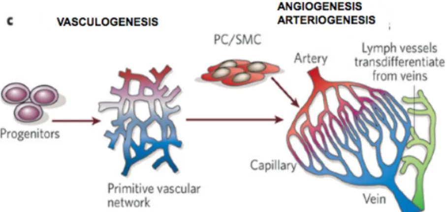
Figure 1.1. Mechanisms of vessel development. A primitive vascular
network is formed by endothelial cell progenitors in a process defined as vasculogenesis; blood vessel remodelling, stabilization and specialization during angiogenesis give rise to a mature vessel network. PC: pericytes, SMC: smooth muscle cells. Adapted from Carmeliet, Nature 2005.
An additional step in blood vessel formation is vessel specialization, a still poorly characterized tissue- and organ-specific process, which includes the formation of functional junctions between ECs and MCs, capillaries diversification as well as arterious-venous specification (Ruoslahti, 2002).
transcription factor COUP-TFII regulates differentiation towards the venous phenotype, inhibiting Neuropilin1 and Notch pathways (You et al., 2005). Interestingly, a mature blood vessel network displays a striking parallel with a nerve fiber network, suggesting the existence, even at a molecular level, of common pathways during development. Actually, compelling evidence establishes a growing role for axon guidance molecules even in vessel guidance, complementing and cooperating with canonical pathways; among them, the Semaphorin-Neuropilin1-Plexin and Netrin pathways (Carmeliet and Tessier-Lavigne, 2005).
Pathological and physiological angiogenesis
Blood vessels do not undergo major modifications in healthy adult organism, and angiogenesis is restricted to cycling ovary and placenta during pregnancy. Nonetheless, even though quiescent, ECs and therefore vessels are still angiogenesis-competent, as they can respond to hypoxic and inflammatory stimuli, as it happens during wound healing.
Imbalance of the angiogenic stimulus, both towards excessive and insufficient angiogenesis, is characteristic of several pathologies; as a consenquence, optimal regulation of angiogenesis represents a crucial issue in human physiology and pathology (Figure 1.2).
Excessive angiogenesis accompanies inflammatory and ocular disorders, in addition to tumor growth (Carmeliet, 2005a). The observation that tumor growth relies on blood supply led to the hypothesis that tumor angiogenesis inhibition could provide a successful strategy to treat cancer (Folkman, 1971). Consequently, given the key role in angiogenesis for the VEGF pathway (Ferrara et al., 2003), several VEGF-targeting strategies are exploited in cancer treatment, including neutralizing VEGF and anti-VEGFR antibodies, soluble anti-VEGFRs and tyrosine kinase inhibitors (Ferrara and Kerbel, 2005). Even though several clinical trials are currently exploiting more than 20 anti-VEGF agents, many unresolved issues, such
as development of drug resistance, still limit the efficacy of this therapy (Ellis and Hicklin, 2008; Ellis and Reardon, 2009).
Figure 1.2. The angiogenetic switch. Several molecules are involved in
fine balancing angiogenesis in human physiology and pathology, functioning both as positive and negative regulators. Adapted from
Hanahan and Folkman, Cell 1996.
To overcome these difficulties, while initial anti-angiogenesis strategies exploited mainly EC-targeting agents, more recent findings encourage the targeting of different cell types that indirectly influence tumor angiogenesis, such as cells of the immune system (Carmeliet, 2005a; Ferrara and Kerbel, 2005). In addition, the lack of success of many anti-angiogenic therapies is influenced by the complex interaction between cellular and molecular players that characterizes vessel biology.
Conversely, insufficient angiogenesis characterizes many ischemic diseases, such as limb and heart ischemia, leading to inadequate vessel formation. Therefore, induction of revascularization by delivering pro-angiogenic molecules represents an appealing therapy for patients with ischemic diseases (Baumgartner and Isner, 2001; Isner et al., 2001). Results obtained from several clinical trials, that exploited recombinant
indicate that the best therapeutical strategy is the combination of pro-angiogenic agents (Rissanen and Yla-Herttuala, 2007). Adeno Associated Virus-based vectors represent, for their tropism and low inflammatory potential, a good candidate for cardiovascular gene therapy (Giacca, 2007). Concluding, several clinical trials are currently ongoing for vessel imbalance-related diseases, and again, due to its role as a master switch in the angiogenic process, VEGF has been extensively studied.
The most interesting features of VEGF and VEGFRs, with particular attention to VEGFR2, will be discussed in the following paragraphs.
Vascular Endothelial Growth Factors
Currently, the VEGF family comprises seven members, namely PlGF (Placenta Growth Factor), VEGF-A, VEGF-B, VEGF-C, VEGF-D, VEGF-E and sv-VEGF (snake venom VEGF). They act mainly as inducers and modulators of vasculogenesis, angiogenesis and vascular remodelling in
vivo, even if their influence is not restricted to endothelial cells (Carmeliet
and Storkebaum, 2002; Carmeliet and Tessier-Lavigne, 2005; Ferrara et al., 2003; Matsumoto and Claesson-Welsh, 2001) (Figure 1.4).
Active, secreted forms of VEGF members form homo and hetero-dimers, thus increasing signalling diversity; their biological effects are mainly mediated by their receptors VEGFR1, VEGFR2 and VEGFR3, in association with coreceptors, such as those belonging to the Neuropilin, Heparan Sulfate Proteoglycan (HSPG) and integrin families (Jakobsson et al., 2006; Soker et al., 1998; Soldi et al., 1999).
VEGF-A
VEGF-A, also referred as VEGF, was discovered as an essential player in angiogenesis (Ferrara and Henzel, 1989). Remarkably, VEGF-A is required
in vivo for proper development of the cardiovascular system; even the
since VEGF-A is haploinsufficient (Carmeliet et al., 1996; Ferrara et al., 1996). Recent data pointed out the importance of VEGF also in vessel maintenance: VEGF produced by endothelial cells is crucial for vascular homeostasis, through a cell autonomous VEGF signalling (Lee et al., 2007); this observation confirms the pleiotropic role of VEGF in vessel biology. Importantly, VEGF-A has been originally described as Vascular Permeability Factor (VPF), due to its ability to increase vascular permeability, disrupting vascular barrier integrity (reviewed in (Weis and Cheresh, 2005)).
In accordance with its key role in vessel formation, VEGF availability is controlled at many levels, including transcription, mRNA stability and translation, post translational modification and binding affinity.
At the transcriptional level, many stimuli, including growth factors, p53 mutation, nitric oxide (NO), hormones, cytokines and cellular stress control VEGF expression (Takahashi and Shibuya, 2005). In particular, hypoxia, via Hypoxia Inducible Factor 1 (HIF-1) is the major positive regulator of VEGF expression. Hypoxic conditions induce accumulation of the highly instable α subunit of HIF-1, leading to the formation of an active transcriptional activator that binds to the Hypoxia Responsive Elements (HRE) in the 5ʼ flanking region of the VEGF promoter (Pages and Pouyssegur, 2005).
Hypoxia is a key factor for VEGF stabilization also at mRNA level, controlling mRNA stability by binding of regulatory proteins to the 3ʼ Untranslated Region (UTR), as well as mRNA translation via IRES sequences present in the 5ʼ UTR (Stein et al., 1998).
The observation that Dicer protein mutant mice are characterized by severely compromised blood vessel formation, displaying reduced levels of VEGF-A mRNA, suggest that also miRNA have a role in controlling VEGF
Human VEGF-A gene, located on chromosome 6, is about 25 Kb long and comprises 8 exons (Tischer et al., 1991). VEGF mRNA undergoes alternative splicing of exons 6 and 7, which encode for binding motifs to heparin and similar molecules. In humans, alternative splicing give rise to at least nine isoforms: VEGF121, VEGF145, VEGF148, VEGF162, VEGF165,
VEGF183, VEGF189 and VEGF206 (Figure 1.3). In mice all isoforms are one
aminoacid shorter than their human counterparts. Another isoform, VEGF165b, is generated by exon 8 distal splice site selection, and differs
from VEGF165 only in the carboxy-terminal six amino acids, thus resulting in
a change of the aminoacidic sequence form CDKPRR to SLTRKD (Harper and Bates, 2008).
Figure 1.3. VEGF-A splicing. Alternative splicing of VEGF-A mRNA gives
rise to almost nine VEGF isoforms, characterized by different length (small numbers) and therefore different binding properties. A different distal splice site selection in exon 8 originates VEGF165b. Adapted from Takahashi and Shibuya, Clinical Science 2005.
Remarkably, even if VEGF165b seems to bind to VEGFR2 with the same
affinity as VEGF165, it lacks angiogenic properties, and is therefore defined
as anti-angiogenic (Woolard et al., 2004). The molecular bases of VEGF165b
properties were recently clarified: VEGF165b is not able to engage
VEGFR2 phosphorylation at tyrosines 1054-1059 in the activation loop (Kawamura et al., 2008b).
The VEGF165 isoform is the one preferentially expressed, followed by
VEGF121 and VEGF189; how differential splicing of these isoforms is
regulated is however largely unknown.
VEGF165, a secreted homodimer with moderate affinity for heparin, is a
powerful inducer of endothelial cell migration, proliferation, survival and vascular permeability (Leung et al., 1989; Senger et al., 1983). The importance of VEGF165 in vessel development is highlighted by the
observation that VEGF164 mice are normal and healthy, while VEGF120
puppies exhibit serious vascular remodelling defects, including defective branching (Stalmans et al., 2002). Altogether, the use of transgenic animals expressing selectively VEGF isoforms indicates that they play distinct roles in vascular morphogenesis and arterial development, a feature depending on their diffusibility and differential interaction with VEGF coreceptors, as discussed later (Carmeliet et al., 1999; Ruhrberg et al., 2002; Stalmans et al., 2002).
In conclusion, VEGF is a key regulator of angiogenesis in health ad disease, stimulating endothelial cell migration and division, and is critical for vascular development. Therefore gene expression regulation, alternative splicing, in addition to other mechanism, contribute to the tight regulation of VEGF activity, crucial for the formation and maintenance of a proper blood vessel network.
It is noteworthy to mention that recent studies also indicate that VEGF might exert its effect on a variety of cell types, besides ECs. For instance VEGF appeared to be critical to prevent motor neuron degeneration, also exerting a direct action on neurons (Zacchigna et al., 2008a). A potent effect of VEGF in promoting survival and regeneration of skeletal muscle
observed an effect of VEGF165 on bone marrow (BM) derived CD11b+ cells,
as these cells expressed VEGF receptors. In particular we observed the induction of VEGF-dependent BM cells migration, proliferation and secretion of cytokines able to trigger smooth muscle cell recruitment (Zacchigna et al., 2008c).
VEGF has a role also in tissue homeostasis, for instance in liver biology (LeCouter et al., 2003).
PlGF
PlGF is a secreted factor, and like VEGF-A, can undergo differential splicing, giving rise to at least four different isoforms, PlGF-1, PlGF-2, PlGF-3 and PlGF-4 (Yang et al., 2003).
Several gene inactivation studies have revealed that PlGF deficient mice, differently from VEGF-A, are viable and healthy, suggesting that endogenous PlGF is not necessary for vascular development and physiological vessel maintenance in healthy adult organisms (Carmeliet et al., 2001).
At a cellular level, PlGF acts as a pleiotropic factor, stimulating angiogenesis directly targeting endothelial and mural cells, and indirectly through the recruitment of pro-angiogenic cell (Clauss et al., 1996; Hattori et al., 2002; Luttun et al., 2002). Its pleiotropic activity is reflected by the observation that PlGF stimulates EC growth, migration and survival, while is mainly a chemoattractant for BM progenitors (Cao, 2009). PlGF-activity on these cells is mediated by binding to VEGFR1, but not to VEGFR2, and probably to Neuropilin1 and Neuropilin2, two PlGF coreceptors; downstream intracellular signalling switches on a series of pro-angiogenic genes (Autiero et al., 2003b).
Interestingly, animal knock out studies have revealed that PlGF activity is fundamental for the angiogenic and inflammatory switch in various pathologies, such as tumor growth and ischemia, raising the possibility that
PlGF might be a disease-specific angiogenic factor (Carmeliet et al., 2001). Additionally, PlGF levels are undetectable in most organs in healthy conditions, while many cell types, including ECs, SMCs, and BM progenitors express PlGF in pathological conditions (Fischer et al., 2008). Taken together these results suggest that PlGF targeting might reduce pathological angiogenesis without affecting healthy blood vessels. A monoclonal blocking antibody recognizing mPlGF (αPlGF) inhibits the growth and metastasis in more than 10 tumor models in mice. Inhibition of tumor growth depends on the pleiotropic effect of this antibody: i) αPlGF not only inhibits tumor vessel growth, but also causes regression of pre-existing tumor vessels; ii) it is anti-inflammatory, inhibiting recruitment of Tumor Associated Macropharges (TAM); iii) it impairs tumor lymphangiogenesis, mainly through macrophage inhibition (Fischer et al., 2007).
Therefore genetic and pharmacological studies have identified PlGF as a possible therapeutic target for anticancer therapy, particularly in combination with VEGF-VEGFR inhibitors.
VEGF-B
The VEGF-B gene generates, through alternative splicing, two protein isoforms, a heparin-binding isoform of 167 amino acids and a diffusible isoform of 186 amino acids, differing at their C-terminus (Li et al., 2001; Olofsson et al., 1996a; Olofsson et al., 1996b). In addition to VEGFR1, and similar to the VEGFA isoforms containing exon 7 of the VEGFA gene (including VEGF-A165) and to PlGF, both VEGFB167 and VEGFB186 also
bind NP1 and NP2, expressed on endothelial as well as on other cell types (Li et al., 2001).
The VEGFB gene displays a unique expression pattern with prominent expression in the heart during embryonic development (Lagercrantz et al.,
vasculature, impaired recovery from cardiac ischemia, and, most notably, decreased heart size (Bellomo et al., 2000). In another mouse strain, the VEGFB knock out specifically generates atrial conduction abnormalities (Aase et al., 2001). In addition, recent work reassessing the role of VEGFB167 during ischemia has indicated that this factor, delivered either as
a recombinant protein, or using adenoviral vectors or through the implantation of transfected myoblasts, significantly increased revascularization of the infarcted myocardium, however failed to enhance vascular growth in the skin or ischemic limb (Li et al., 2008).
VEGF-B overexpression has been proven to induce myocardium specific angiogenesis and arteriogenesis in rabbits and pigs upon acute infarction (Lahteenvuo et al., 2009).
Heart and skeletal muscle are the tissues characterized by the highest VEGF-B expression, even if many different tissues, including ECs and brain, usually express VEGF-B (Li et al., 2001); conversely, the role of VEGF-B in tumor growth remains elusive. At the cellular level, VEGF-B receptors are VEGFR1 and NP1, and VEGF-B stimulates directly endothelial cell migration and growth (Cao, 2009).
VEGF-C and VEGF-D
VEGF-C and VEGF-D share peculiar characteristics: they present unique N and C terminal extensions different from the other VEGF members, they bind and activate VEGFR3 in addition to VEGFR2 and their affinity for this receptor is increased upon their proteolitic cleavage (Lohela et al., 2009). VEGF-C gene is composed by seven exons, and this protein is expressed in the heart, small intestine, placenta, ovary and the thyroid gland in adults (Roy et al., 2006). VEGF-C is essential for lymphangiogenesis, as lack of VEGF-C in mice leads to a complete absence of lymph vessels and embryonic lethality, while blood vasculature grows normally (Karkkainen et al., 2004).
VEGF-D is 48% identical to VEGF-C, and it is expressed in many adult tissues including the vascular endothelium, heart, skeletal muscle, lung, and bowel (Roy et al., 2006).
VEGF-D has been shown to be largely dispensable for the development of lymphatic system, and its physiological role needs to be further clarified (Baldwin et al., 2005), even if in vitro this factor is able to stimulate migration and proliferation of endothelial cells; additionally, this factor seems to have a role in tumor angiogenesis and lymphangiogenesis (Achen et al., 2001; Stacker et al., 2001).
Recently, data obtained using double KO mice for VEGF-C and VEGF-D, showed that both VEGF-C and VEGF-D are displensable for embryonic angiogenesis; additionally, it has been shown that VEGF-C/D KO does not phenocopy VEGFR3 KO (Haiko et al., 2008).
The discovery of VEGF homologues in the genome of parapoxvirus Orf (VEGF-E) and in snake venom (svVEGF) finally confirms the pleiotropic role of VEGF family members (Shibuya, 2003).
VEGF Receptors
A key element in the complex regulation of VEGF activity is represented by the VEGF-receptors (VEGFR): VEGFR1 (or Flt1), VEGFR2 (or KDR, Flk1) and VEGFR3 (Flt4), expressed by several cell types (Figure 1.4). VEGFRs belong to the Tyrosine Kinase Receptor (TKR) superfamily. These consist of an extracellular domain composed by seven Immunoglobulin (Ig) like domains (Ig), a short transmembrane and a juxtamembrane segment, and are characterized by a split intracellular tyrosine kinase domain interrupted by a 70 aa long kinase insert domain (Carmeliet, 2005b; Matsumoto and
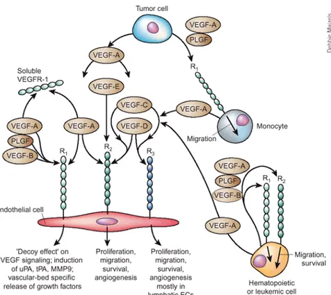
Figure 1.4. Interaction between VEGFs and VEGFRs. VEGFs can
selectively bind different VEGFRs, expressed on the cell surface of endothelial cells, monocytes and tumor cells. The three VEGF tyrosine kinase receptors can form homo and hetero-dimers as a consequence of ligand binding, eventually leading to intracellular signalling. Adapted from
Ferrara et al, Nature Medicine 2003.
VEGF binding to VEGFRs is the initial step for signal transduction, accompanied by receptor homo and hetero-dimerization. Therefore, the characterization of receptor dimer dynamics represents a crucial step in understanding receptor activation. Tyrosine kinase activation parallels
dimer formation and leads to autophosphorylation; finally, phosphorylated receptors recruit intracellular partners.
VEGFR1
VEGFR1/Flt1, together with VEGFR2, has a primary role in angiogenesis, playing a complex regulatory role (Shibuya, 2006). Even if VEGFR1 and VEGFR2 are structurally similar, VEGFR1 function is different and multifaceted, functioning both as a negative and positive regulator of angiogenesis (Olsson et al., 2006). Interestingly, VEGFR1 has ten fold higher affinity for VEGF compared to VEGFR2 (Waltenberger et al., 1994). VEGFR1 activity results from its binding to three different gene products belonging to the VEGF family, namely VEGF-A, VEGF-B and PlGF.
The concept that VEGFR1 has a negative regulatory role during early embryogenesis was suggested by the observation that VEGFR1 null mice die at embryonic day 8.5-9.0, displaying a disorganized vascular endothelium and overgrowth of endothelial cells (Fong et al., 1995). Interestingly, VEGFR1 kinase activity seems to be dispensable during embryogenesis, since mice expressing a VEGFR1 mutant that lacks the tyrosine kinase domain develop an essentially normal vasculature (Hiratsuka et al., 1998). Therefore, VEGFR1 has been proposed to act as a VEGF-A trap, thus preventing excessive VEGFR2 activation during embryonic development (Shibuya, 2001). A physiological role of this endogenous VEGF-A trap was demonstrated in adult life; a soluble VEGFR1, known as sFlt1 and expressed also by human placenta (Shibuya et al., 1990), is essential in order to preserve corneal avascularity (Ambati et al., 2006; Ambati et al., 2007).
Nonetheless, VEGFR1 has a functional role in active transduction of VEGF signalling, well clarified in cells lacking other VEGFRs. Monocytes/macrophages migration in response to VEGF-A is mediated by
Zacchigna et al., 2008c). Additionally, VEGFR1-mediated migration of haematopoietic bone marrow progenitors initiates the pre-metastatic niche in mouse models (Kaplan et al., 2005), even if this finding has been recently discussed (Dawson et al., 2009a; Dawson et al., 2009b).
VEGFR1 signalling.
Despite its ability to bind VEGF-A with more then 10-fold higher affinity than VEGFR2, VEGFR1 undergoes only a weak phosphorylation, even if all kinase motifs are conserved (Waltenberger et al., 1994). Relevant to this issue, VEGFR1 and VEGFR2 share a 43% of overall homology, lower in the extracellular domain (33%) and higher in the kinase domains (70%). Nevertheless, the mechanism responsible for VEGFR1 kinase-impaired activity is still debated. On one hand the juxtamembrane domain, probably by forming an intracellular structure inhibiting accessibility of regulatory sequences, has been implicated in kinase repression (Gille et al., 2000). On the other hand, it has been proposed that substitution of a single aminoacid from aspartic acid at position 1050 (conserved among many RTKs) to asparagine (in VEGFR1) is linked with its decoy activity, in particular by inhibiting phosphorylation in the activation loop (Meyer et al., 2006). Additionally, taking advantage of overexpression studies to overcome weak signals, several VEGFR1 tyrosine residues were identified as phosphorylated, together with their interacting partners (among others SH2, p38/PI3K, Grb2 and Nkc) (Olsson et al., 2006; Shibuya, 2006). Further observations indicate that different VEGFR1 ligands, VEGF-A and PlGF, induce different phosphorylation patterns (Autiero et al., 2003a). Nevertheless, the complexity of VEGFR1 downstream signalling is far from being clarified.
An additional regulation level is provided by the interplay between different VEGFRs, usually coexpressed by endothelial cells. In particular, VEGFR1 has the ability to form heterodimers with other VEGFRs, thus modulating
their response both in a negative and positive manner. In support of its inhibitory role it has been shown that Flt1 can suppress VEGFR2 mediated proliferation but not migration of endothelial cells (Zeng et al., 2001). Conversely, it has been demonstrated that PlGF-driven VEGFR1 activation leads to amplification of VEGFR2-mediated angiogenesis, through intermolecular trans-phosphorylation of Flk1; this observation suggests an inter- and intra-molecular crosstalk between VEGFR1 and VEGFR2 (Autiero et al., 2003b).
VEGFR1, together with Neuropilin1 and Neuropilin2, constitutes the functional receptor for PlGF in cells where it is primarily expressed, such as tumor cells. For this reason, an anti-PlGF blocking antibody, inhibiting binding to VEGFR1 and formation of VEGFR1-Neuropilin1 complexes, inhibits also the growth and metastasis of various tumors (Fischer et al., 2007). These results strengthen the functional role of VEGFR1 in pathological conditions such as tumor growth.
Thus, the role and the mechanism by which VEGFR1 supports angiogenesis are complicated and likely involve several different mechanisms, including VEGFR1 decoy activity, formation of complexes with other receptors as well as direct signalling.
In order to further clarify VEGFR1 biology, it will be particularly interesting to study in vivo interaction between VEGFR1 and VEGFR2, and how this interaction might give rise to different biological effects.
VEGFR2
The experimental evidence that VEGFR2 signalling is required for cardiovascular development (Shalaby et al., 1995) and that has a major role in neovascularization in both physiological and pathological conditions has stimulated a general interest in understanding VEGFR2 biology. Therefore, due to its key role in vessel biology, several aspects regulating
Expression
During development, VEGFR2 is detectable from E7.5 in mesodermal cells of the tail region; VEGFR2 positive cells migrate and differentiate into primitive endothelial cells (Shalaby et al., 1995). During adult life VEGFR2 is expressed mostly in vascular and lymphatic ECs. Even if lower levels of VEGFR2 are detected in haematopoietic stem cells, neurons, osteoblasts as well as megakaryocites (Matsumoto and Claesson-Welsh, 2001), VEGFR2 biology has been so far analyzed mainly in EC. The 5ʼ non-coding region and the 3ʼ region of the first intron are required to properly regulate VEGFR2 expression in ECs (Shibuya and Claesson-Welsh, 2006). Recent data show that Extra Cellular Matrix (ECM) is able to control VEGFR2 expression in vitro and in vivo, and that this action is mediated by p190RhoGAP (Mammoto et al., 2009).
In accordance to its major regulatory role in angiogenesis, VEGFR2 is upregulated during pathological angiogenesis, such as in tumors, and VEGF-A has a positive effect on VEGFR2 expression by means of a positive feedback mechanism (Shibuya and Claesson-Welsh, 2006). Ligand binding and dimerization
In addition to VEGF-A (Kd=75-760 pM), VEGFR2 is able to bind VEGF-E,
sv-VEGF as well as processed VEGF-C and VEGF-D. Thereby, VEGFR-2 represents the major mediator of VEGF-A-induced proliferation, migration and permeability in EC.
Since the observation that VEGFR2-VEGF binding requires Ig domain 2 and 3 of the receptor and is likely to occur at the ligand dimer interface, many crystallography studies have focused on the VEGF-VEGFR2 interaction in order to develop anti-angiogenic drugs (Roskoski, 2008). Ligand binding is accompanied, as in many RTK, by receptor dimerization, the first step toward receptor activation.
Figure 1.5. VEGFR2 activity regulation. VEGFs induce VEGFR2
phosphorylation, and in addition receptor activation is tightly regulated by coreceptors expressed even in trans. Additional cellular mechanisms contribute to fine tune VEGFR2 signalling.
In particular, Ruch and coworkers, based on Electro Microscopy observations of soluble molecules, propose that VEGF induces transition of VEGFR-2 extracellular domains from an highly flexible conformation to a more stable, rigid arrangement, which is stabilized by homotypic interactions of membrane-proximal and membrane-distal immunoglobulin-like domains (in particular involving the Ig domain 7). In the full-length protein, the rigid arrangement of two receptor monomers is probably
et al., 2007).
Nonetheless, the precise mechanism linking VEGF binding and receptor homo and hetero-dimerization is still far from being clarified.
Trafficking
Since protein trafficking controls the relative amount of receptor available for VEGF binding at the plasma membrane, this aspect is crucial in fine-tuning VEGF activity at the cellular level. Nonetheless, many issues regarding VEGFR2 trafficking are still debated, such as internalization through caveolin-1 vesicles or clathrin coated pits (Mukherjee et al., 2006). Surprisingly, in HUVE cells more than 40% of VEGFR2 protein was found to be localized in an internal vesicular pool positive for early endosomal compartment markers (EEA1, Rab4); VEGF stimulation not only induces VEGFR2 downregulation, as expected for a TKR, but also redistribution to a late endosomal compartment and finally recycling of the receptor (Gampel et al., 2006). Apart from intrinsic tyrosine kinase activity, required for VEGFR2 trafficking and degradation (Ewan et al., 2006), the stimuli controlling the endocytic itinerary of VEGFR2 are still undisclosed.
Moreover, it is still not clear whether VEGFR2 internalization and degradation depend on c-Cbl mediated ubiquitination or on PKC-mediated VEGFR2 C-tail serine phosphorylation (Duval et al., 2003; Singh et al., 2005).
The complexity of VEGFR2 trafficking is emphasized by the observation that internalized receptors are still able to induce intracellular signalling, and therefore molecules such as Vascular Endothellial (VE)-cadherin, controlling VEGFR2 internalization, might finally control VEGF signalling (Lampugnani et al., 2006).(Jakobsson et al., 2006)
Coreceptors
An additional mechanism that regulates VEGFR2 downstream signalling is represented by membrane associated VEGFR2 coreceptors, such as
Neuropilins (Neufeld et al., 2002), Heparan Sulfate Proteoglycans (HSPGs), and VE-cadherin (Carmeliet and Collen, 2000). Interestingly, the interaction between VEGFR2 and some coreceptors can occur in trans: it has been shown that VEGF165-VEGFR2 signalling is increased in EC, when HSPG
are expressed in trans in a co-culture system (Jakobsson et al., 2006). Moreover, VEGFR2 was found to be part of a mechano-sensory complex triggered by fluid shear stress and comprising PECAM-1 and VE-cadherin, that leads to the conformational activation of integrins (Tzima et al., 2005). VE-cadherin, a specific component of endothelial adherent junctions, has been found to limit VEGFR2 mediated mitogenic signal (Grazia Lampugnani et al., 2003). This VE-cadherin induced inactivation, mediated by decreased receptor phosphorylation, can be interrupted by receptor internalization (Lampugnani et al., 2006).
Finally, integrins have been found to associate with VEGFR2 in EC, and play a crucial role in balancing VEGFR2 activity. For instance, the αvβ3 integrin, upon binding to its ligand vitronectin, enhances VEGFR-2 phosphorylation, PI 3-kinase activity, focal adhesion dynamics as well as proliferation and migration of ECs triggered by VEGF-A (Napione et al., 2007; Soldi et al., 1999). In contrast to the αvβ3/vitronectin (or fibrin) pair, collagen I, the ligand of α1β3 and α2β1 integrins, exerts an inhibitory action reducing VEGF-A-induced VEGFR-2 autophosphorylation by recruiting the tyrosine phosphatase SHP2 (Mitola et al., 2006).
Structure and kinase activity
Relatively few tyrosine (Tyr) residues were identified as phosphorylation sites in VEGFR2 including human Tyr951 (949 in the mouse sequence) and 996 (994) in the kinase insert domain, Tyr1054 (1052) and 1059 (1057) in the kinase domain, and Tyr1175 and 1214 in the C-terminal tail. Proteomic studies identified three additional tyrosines as phosphorylated at a low
(Matsumoto et al., 2005). On the other hand, Tyr1175 and 1214 were identified as two major VEGF-A dependent autophosphorylation residues (Takahashi and Shibuya, 2005) (Figure 1.6).
Since VEGFR2 is a tyrosine kinase enzyme, its phosphorylation is linked to its structure, as VEGF-A binding leads to receptor dimerization, protein kinase activation and trans-autophosphorylation. Therefore, comprehension of VEGFR2 structure is functional in order to understand its activity.
From a structural point of view, human VEGFR2 kinase domain can be subdivided in:
• a proximal kinase domain (residues 827-931)
• a kinase insert domain (residues 932-998); this segment, although not directly involved in catalysis, is important in signal transduction as a docking site for cellular proteins
• a distal kinase domain (residues 999-1158), containing both the catalytic and the activation loop.
Two lobes, similarly to other TKRs such as EGFR, characterize the catalytic core of VEGFR2, with the active site located in the cleft between the N-terminal and the C-N-terminal lobe. The smaller N-lobe is predominantly formed by antiparallel β-sheets and contains the glycin-rich ATP-Phosphate binding loop, while the larger C-lobe is characterized by α-helices and includes the activation and catalytic loops (McTigue et al., 1999). Conformational changes at the level of these lobes are linked to inter-conversion of the kinase from inactive to active state and finally to catalysis. In particular, when VEGFR2 is an inactive state, the activation loop that comprises two tyrosines (1054 and 1059) is in a “closed” conformation that prevents substrate binding. Phosphorylation in the activation loop stabilizes it in its active, “opened” conformation (Roskoski, 2008) (see
Figure 1.6. VEGFR2 intracellular part. Human VEGFR2 intracellular
domains are highlighted, while white bars and residue numbers indicates the main phosphorylated tyrosines. In addition, position of the catalytic and activation loops is shown.
An interesting issue is how VEGFR2 dephosphorylation is regulated. To our knowledge, SHP2 and VE-PTP are the only tyrosine phosphatases that have been demonstrated to interact and dephosphorylate VEGFR2 (Mellberg et al., 2009; Mitola et al., 2006); in particular SHP2 favours VEGFR2 internalization, and might be responsible for collagen I negative regulation of VEGFR2 signalling (Mitola et al., 2006).
Downstream signalling
The multiplicity of cellular functions controlled by the VEGF-VEGFR2 axis is reflected by the diversity of signalling cascades that are activated upon ligand binding. Accordingly, a complex network of signal transduction leads to cell migration, cell survival, cell proliferation, vascular permeability, actin remodelling and focal adhesion turnover.
The characteristics of VEGFR2 downstream signalling are additionally modulated by the formation of different membrane signalling complexes, in addition to association with other VEGFRs and coreceptors such as Neuropilins, as discussed later.
PLCγ. Endothelial cell culture experiments demonstrated that phosphorylated Tyr1175 represents the single major site for PLCγ binding to VEGFR2, and has a key role in the activation of the PLCγ-PKC-MAPK
the relevance of Tyr1175 (1173 in mice) and its downstream signalling in
vivo. Knock-in mice with a single aminoacid substitution (Tyrosine 1173 to
phenylalanine) died at E8.5-9.5 as a consequence of endothelial and haematopoietic cells defects, such as blood vessel deficiency, comparable to VEGFR2 null mice. Conversely, substitution of the highly phosphorylated, but not required for PLCγ signalling, Tyr 1212 (1214 in human) with phenylalanine, gave rise to viable and fertile mice (Sakurai et al., 2005).
Even PLCγ activity is fine tuned by VEGFR2 signalling: c-Cbl, recruited and phosphorylated by VEGFR2, promotes PLCγ ubiquitination, inhibiting signal transduction (Singh et al., 2007).
Recently, VEGF induced PLCγ-PKC pathway activation was linked to VEGF -target genes regulation, through phosphorylation of HDAC7 and its nuclear export, thus providing a direct connection between VEGF stimulation and histone acetylation (Ha et al., 2008).
PI3K. Another event that strictly relies on Tyr1175 phosphorylation is the
activation of the PI3K-Akt-PKB pathway for cell survival and migration, as inferred form studies in endothelial cells (Dayanir et al., 2001; Gerber et al., 1998). Shb is one of the adaptor molecules binding to pTyr1175 and mediating PI3K-dependent endothelial cell migration (Holmqvist et al., 2004). Nonetheless, since VEGFR2 induced PI3K activation is not so strong, activation of an additional pathway to sustain VEGF induce migration is likely to occur (Takahashi and Shibuya, 2005). One single specific PI3K isoform, namely 110α, is the preferential mediator of VEGF-A dependent migration of endothelial cells in vitro, and has been linked with developmental angiogenesis in vivo (Graupera et al., 2008).
Recent studies highlighted the role of Akt and its substrate Gridin in neovascularization during adult life, as mediators of VEGF-triggered vascular remodelling (Kitamura et al., 2008).
Ras. Even if VEGF is not a powerful mitogen for EC, Ras dependent and
independent Raf-MEK-MAPK pathway activation seems to occur depending on cell types (Olsson et al., 2006).
Gab and Grb2. Grb2, Gab1 and Gab2 are scaffolding adaptors involved in
many TKR signalling such as EGFR. Gab1 and Grb2 were shown to be involved in VEGFR2 downstream signalling, and a model inferred from in
vitro data includes a direct interaction between VEGFR and Grb2, the latter
able to bind Gab1. This system has been linked to PI3K activation, endothelial cell migration and capillary formation (Laramee et al., 2007).
TSAd/VRAP. Tyr951 (mouse 949), an additional VEGFR2 phosphorylation
site, has been identified as the binding site for the T-cell specific adaptor (TSAd, or VEGFR-associated protein VRAP) (Matsumoto et al., 2005). The same group demonstrated that VEGFR2-TSAd coupling is responsible of actin reorganization and therefore endothelial cell migration, but seems not to be linked to cell proliferation; additionally, TSAd is probably involved in tumor angiogenesis.
Cdc42 p38 MAPK. EC migration downstream to VEGFR2 is also regulated
by phosphorylation of Tyr1214 (1212), which triggers sequential activation of Cdc42 and SAPK2/p38, finally driving the SAPK2/p38-mediated actin remodelling of stress fibers in endothelial cells exposed to VEGF (Lamalice et al., 2004). Additional molecules, such as IQGAP1, are able to bind VEGFR2, and induce both VEGF induced migration and proliferation (Meyer et al., 2008)
FAK-paxillin. VEGF promotes cell migration through a RhoA-ROCK
dependent mechanism, activating Focal Adesion Kinase (FAK) and finally paxillin (Le Boeuf et al., 2006).
VEGFR2 post-translational modifications
different PTMs in addition to tyrosine phosphorylation, crucial for receptor activation.
Glycosilation is important in VEGFR2 protein maturation; the VEGFR2 pool present at the plasma membrane is N-glycosilated (Takahashi and Shibuya, 1997).
Poly-ubiquitination is one of the signals involved in receptor down-regulation upon VEGF stimulation, even if the details of this modification are still unknown (Duval et al., 2003).
VEGFR3
VEGFR3/Flt4 constitutes the receptor for the VEGF-C and VEGF-D family members. The observation that, in adults, VEGFR3 expression is restricted to the lymphatic endothelium and the use of genetic models linked this receptor mainly to lymphatics development and maintenance (Kaipainen et al., 1995; Veikkola et al., 2001). In particular, VEGFR3 signalling is required for lymphatic endothelial cells sprouting as well as lymphatic vessel maintenance through the inhibition of apoptosis (Alitalo et al., 2005). Nonetheless, VEGFR3 gene targeted mice exhibit defects in arterial-venous remodelling of the primary vascular plexus, leading to embryonic lethality from day E9.5 (Dumont et al., 1998). Therefore, during embryonic development, VEGFR3 activity is not restricted to lymphatics, but has an important function in blood vessel development. Recent data extended the role of VEGFR3 in angiogenesis also during adult life; VEGFR3 was found to be highly expressed in angiogenic sprouts, while targeting of VEGFR3 signalling resulted in decreased sprouting, vascular density, vessel branching and endothelial cell proliferation in mouse angiogenesis models (Tammela et al., 2008). VEGFR3 is also found to be upregulated in tumor microvasculature, thus opening the possibility to exploit VEGFR3 targeting agents to inhibit tumor growth (Saharinen et al., 2004).
The extension of VEGFR3 role not only to lymphatic biology, but also to pathological and embryonic development, warrants further studies to define the molecular mediators of these diverse activities, still poorly explored. VEGFR3 signalling
Two conserved tyrosine residues in the kinase domain of VEGFR3 are probably responsible for its kinase activity, and additional tyrosine phosphorylation sites have been identified in the VEGFR3 C-terminal tail (Dixelius et al., 2003). Signal transduction downstream to VEGFR3 has been only partially characterized: Shc2/Grb2 interact directly with the receptor, while downstream intracellular mediators identified until now comprise ERK1/2, PI3F-Akt, STAT3 and STAT5 transcription factors (Olsson et al., 2006).
As already mentioned in the case of VEGFR1, VEGFR3 signalling can be modulated by the interaction with VEGFR2 and other co-receptors, in this case Neuropilin2. The formation of these complexes is biologically relevant and, for example, VEGFR2-VEGFR3 hetero-dimers might form in vivo both in lymphatic cells and subtypes of endothelial cells, resulting in differential phosphorylation sites and finally differential signalling (Dixelius et al., 2003). All these observation suggest that VEGFR3, besides its fundamental role in lymphatic vessel development and maintenance, also acts as a regulator of vascular network formation. In this respect VEGFR3 may constitute an additional target of anti-angiogenic therapies.
Semaphorins and plexins in vessel biology
Initially described as axon guidance molecules, semaphorins are also implicated in the regulation of neural development and organ morphogenesis, together with angiogenesis and invasive tumor growth (Larrivee et al., 2009; Serini et al., 2009). For instance, as semaphorins and
Kessler, 2008). This family of membrane-bound and secreted proteins influences cytoskeletal remodelling, integrin-dependent adhesion, cell proliferation, apoptosis and differentiation (Kruger et al., 2005). All semaphorins contain an amino terminal sema domain, required for signalling, and were subdivided into eight groups: invertebrate semaphorins in group 1 and 2, vertebrate semaphorins in groups 3 to 7, and viral semaphorins in group 8. Interestingly, class3 semaphorins are the only secreted vertebrate semaphorins and, with one exception, require Neuropilin binding to signal through class A plexins.
Plexins represent the main functional family of semaphorin receptors. In mammals, nine plexins have been identified so far, subdivided in four subfamilies based on homology: PlexinA1 to A4, Plexin B1 to B3, Plexin C1 and PlexinD1 (Franco and Tamagnone, 2008). The extracellular domains of these single pass transmembrane receptors are distinguished by the presence of sema, PSI and IPT domains. While semaphorins belonging to classes 4-7 as well as SEMA3E bind directly to specific plexins and activate plexin-mediated signal transduction, other class 3 semaphorins binds to Neuropilins, while Plexins (PlexinAs and PlexinD1) serve as signal transduction elements.
Plexins are characterized by a unique, but highly conserved, cytoplasmic region, which has been associated to multiple signal transducers (Kruger et al., 2005). For instance, plexin intracellular domain contains GTPase-activating protein (GAP)-like motifs, able to interact with G-protein R-Ras, and activates Rho-GTPases. Additionally, it has been shown that plexins become tyrosine phosphorylated, even if the regulatory role of this posttranslational modification need to be further investigated (Franco and Tamagnone, 2008).
Neuropilins
Neuropilins (Neuropilin1/NP1 and Neuropilin2/NP2), initially identified as receptors for several class 3 semaphorins, in association with plexin family receptors mediate repulsive axon guidance in the developing nervous system (Fujisawa, 2004).
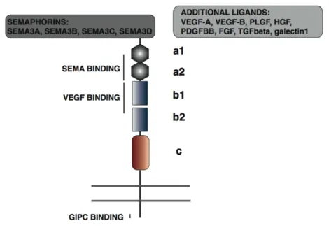
Even if Neuropilin1 and Neuropilin2 share only 44% homology, they have similar structural features: the extracellular part of these single pass transmembrane receptors has two Complement binding domains (CUB, or a1 and a2), two coagulation factor V/VII homology domains (b1 and b2) ad a single meprin domain (MAM or c). Notably, the intracellular domain of neuropilins is only 40-aminoacid long, displays poor homology with other proteins and lacks enzymatic activity. Despite structural homologies, Neuropilin1 and Neuropilin2 differ for binding and signal properties (Neufeld et al., 2002). In particular, SEMA3A binds only to NP1, SEMA3F and SEMA3G interact only with NP2, while SEMA3B, SEMA3C and SEMA3D bind both receptors. Additionally, Neuropilin1 and Neuropilin2 also display specific and mutually selective binding to factors not belonging to the SEMA family, such as VEGF-A, VEGF-B, VEGF-C, PlGF, PDGF-bb, FGF2, TGFβ, HGF and galectin (Figure 1.7).
Moreover, Neuropilins differ in VEGF-A isoform binding, as NP2 binds to VEGF165 and VEGF145, while NP1 binds VEGF165 and possibly VEGF121, as
discussed afterwards. The biological difference between NP1 and NP2 is enforced by their non-redundant role in development, as NP1 deficient mice die during mid-gestation with defects in the heart, vasculature, and nerve projection (Kawasaki et al., 1999), while NP2 KO mice are viable and display only defects in nerve projection (Chen et al., 2000; Giger et al., 2000). Interestingly, the double NP1/NP2 knockout mouse had a more severe abnormal vascular phenotype than either NP1 or NP2 single
knockouts, resembling the phenotypes of VEGF and VEGFR-2 knockouts (Takashima et al., 2002).
Neuropilin1
Neuropilin1 is a functional, transmembrane receptor, able to mediate signalling from structurally distinct ligands during nervous system, heart and vascular development, as shown in Figure 1.7. In particular Neuropilin1 expression in EC is required for cardiovascular development (Gu et al., 2003), and is important for the formation of the capillary plexus, partially independently from blood flow (Jones et al., 2008).
Figure 1.7. Interaction of Neuropilin1 with different ligands. NP1
ligands belonging to the semaphorin and other families are shown. Predicted binding sites for SEMA, VEGF and GIPC are indicated by bars. Additionally, the structural domains of Neuropilin1 CUB (a1, a2), FV/FVIII (b1/b2) and MAM (c ) are shown.
Neuropilin1 was shown to bind to VEGF-A isoforms containing exon 7 (the heparin binding domain) and therefore common usage of the NP1 receptor by factors, such as VEGF165 and SEMA3A, raised the possibility of a
competition or at least a cross regulation.
VEGF and SEMA3A bind to two distinct, even if partially overlapping, domains on NP1 extracellular part (Gu et al., 2002), and structural studies show that VEGF and SEMA3A do not compete directly for NP1 binding (Appleton et al., 2007; Vander Kooi et al., 2007). Nonetheless, several data show how SEMA3A and VEGF165 can have opposite effect on cell migration
and survival (for a review see (Kruger et al., 2005)); recent findings open the possibility that SEMA3A and VEGF can act by promoting independent signals downstream to NP1 (Acevedo et al., 2008; Guttmann-Raviv et al., 2007).
Even if Neuropilin1 was initially proposed to simply act by favouring VEGF165, but not VEGF121, binding to VEGFR2, thus enhancing
VEGF-induced chemotaxis in cultured endothelial cells (Miao et al., 1999; Soker et al., 1996; Soker et al., 2002; Soker et al., 1998; Whitaker et al., 2001), recent data clearly suggest a more complicated picture for the Neuropilin1 – VEGFR2 system activity in EC. This model implies that VEGF121, which
does not have the Neuropilin1 binding region coded by exon 7, is a weaker VEGFR2 activator as compared to VEGF165. Nonetheless, one group
showed that both Neuropilin1 and 2 are able to enhance VEGF121-induced
signal transduction by VEGFR2 in PAE and HUVE cells (Shraga-Heled et al., 2007). Another group provided evidence that VEGF121 is also able to
directly bind Neuropilin1 by means of the tail region instead of exon 7, but without inducing the formation of VEGFR2-Neuropilin1 complexes (Pan et al., 2007b). More recently, formation of stable VEGF165-NP1-VEGFR2
vivo through p38 signalling (Kawamura et al., 2008a). In conclusion,
VEGF165,and not VEGF121, induces the formation of VEGFR2-Neuropilin1
complexes, even if the dynamics of these functional angiogenic receptor clusters have never been studied in detail (Soker et al., 2002; Whitaker et al., 2001).
Genetic and biochemical studies generated strong evidence for a role of Neuropilin1 in vascular morphogenesis; therefore is not surprising that even NP1 has been exploited as a target for anti-cancer therapy. In particular, two antibodies, blocking respectively VEGF and SEMA binding to NP1, synergize with anti-VEGF in reducing tumor growth, even diminishing vascular density and EC-pericyte association. (Liang et al., 2007; Pan et al., 2007a).
Neuropilin1 signalling
Due to its short and catalytically inactive intracellular tail, for a long time Neuropilin1 has been considered only able to enhance VEGF-induced signal through VEGFR2 interaction (Figure 1.8). Recent evidence suggests that Neuropilin1 mediates HUVEC adhesion to ECM (Murga et al., 2005) as well as increases VEGF induced EC survival (Wang et al., 2007), independently from VEGFR2.
Notably, a protein called RGS-GAIP-interacting protein (GIPC or synectin), involved in vesicle trafficking, was found to interact with Neuropilin1 (Cai and Reed, 1999). The PDZ-binding domain of Neuropilin1, involved in GIPC1 interaction, seems to be required for VEGFR2-NP1 complex formation in vitro (Prahst et al., 2008), as well as to promote integrin internalization and indirectly EC adhesion (Valdembri et al., 2009). This interaction is important in vivo for proper angiogenesis in zebrafish (Wang et al., 2006) while it has possibly a role in branching morphogenesis in mice (Chittenden et al., 2006).
How Neuropilin1 might initiate an intracellular signalling cascade in response to an extracellular stimulus is still unresolved. It has been shown that NP1 is differentially glycosaminoglycan-modified in EC and SMC (Shintani et al., 2006), but no other PTMs has been characterized so far, suggesting a signalling mechanism different from post translational modifications.
Figure 1.8. Models for Neuropilin1 activity in enhancing VEGF signalling. In the first model (left), enhanced signalling is due to enhanced
VEGF binding to VEGFR2, while in the second model (right) Neuropilin1 contributes directly to signalling, independently from VEGFR2, and even trough GIPC1. The role of Neuropilin1 homo-complexes formation in signalling is still debated.
form dimers, probably not only through the MAM domain as initially supposed (Nakamura et al., 1998), but also through the transmembrane domain, and the formation complexes seems to be involved in Neuropilin1 signalling (Roth et al., 2008).
Concluding, Neuropilin1 seems to act in regulating EC functions by almost two different mechanisms: by enhancing VEGF binding to VEGFR2 and by a VEGFR2 independent signalling, but how these mechanisms are regulated is still an unresolved question.
Protein function regulation by acetylation
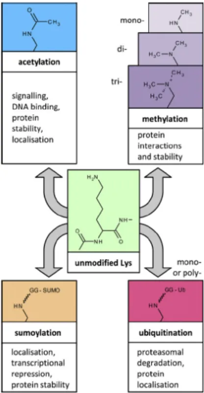
Proteins are naturally modified by several mechanisms, including phosphorylation, ubiquitination, sumoylation, neddylation, acetylation, methylation and isomerization. In addition, the combination of PTMs generates a great potential for cross-regulation. In particular lysine can be the target of multiple, mutually exclusive modifications, as shown in Figure 1.9.
Figure 1.9. Lysine modifications. The ε-amino group of lysine can be
modified by acetylation and several covalent modifications, resulting in different biological outcomes. Adapted from Spange, International Journal
Lysine acetylation is a reversible process that entails transfer of an acetyl group from Acetyl coenzyme A to the ε-amino group of a lysine residue, thus removing the positive charge from the lysine. Both protein acetylation and deacetylation requires dedicated enzymes, defined as lysine acetyltransferases (KATs) and deacetylases (KDACs), respectively (Allis et al., 2007).
Even if acetylation has been initially defined as a histone-specific modification, recent reports demonstrate that more than 100 proteins, both nuclear and cytoplasmic, are acetylated in one or more residues (Glozak et al., 2005; Kim et al., 2006; Vidali et al., 1968) (see Table 1).
Some representative examples will be provided to illustrate the complex role of acetylation in regulating protein function.
PROTEIN CATEGORY SELECTED EXAMPLES
CORE HISTONES H2A, H2B, H3, H4 NON-HISTONE CHROMATIN
PROTEINS HMGB
KAT p300/CBP, PCAF
TRANSCRIPTION FACTORS p53, STAT3, c-Myc, MyoD, E2F, Rp, NFkB, HIF
CYTOSKELETAL PROTEINS ALPHA-TUBULIN, cortactin
CHAPERONES Hsp90
MITOCHONDRIAL PROTEINS ACETYLCoA synthase, thioredoxin VIRAL PROTEINS Adenoviral E1A, HIV Tat and Integrase TRANSMEMBRANE RECEPTORS Type I INFR
Table 1. Selected acetylated proteins. Acetylated proteins are listed, and
cytoplasmic acetylated proteins are highlighted. References: Histones (Roth et al., 2001); HMGB (Sterner et al., 1979); p300, PCAF (Santos-Rosa et al., 2003; Thompson et al., 2004); transcription factors (Galbiati et al., 2005; Gu and Roeder, 1997; Jeong et al., 2002; Marzio et al., 2000; Polesskaya et al., 2000; Sakaguchi et al., 1998; Sartorelli et al., 1999); cytiskeletal proteins (Hubbert et al., 2002; Zhang et al., 2007); chaperones (Kovacs et al., 2005); mitochondrial proteins (Starai et al., 2002); viral proteins (Cereseto et al., 2005; Kiernan et al., 1999; Madison et al., 2002; Marzio et al., 1998; Ott et al., 1999; Zhang et al., 2000); INFR (Tang et al., 2007).
Lysine acetyltransferases (KATs)
Several proteins have been found to have intrinsic lysine acetyltransferase activity, while no acetylation consensus site on target proteins has been characterized until now. Nonetheless, acetylases modify very few lysines within a given protein, thus indicating that some specificity exists; moreover, proteomic studies suggest that acetylation is favoured in the context of specific secondary structural characteristics (Kim et al., 2006). According to sequence similarity, KATs can be subdivided into three major groups.
The Gcn5-related N-acetyltransferases (GNATs) family includes GCN5, PCAF, Elp3, Hat1, Hpa2 and Nut1. GCN5 and PCAF are characterized by a Histone Acetyl Transferase (HAT) domain and by a bromodomain, that possesses specific acetyl-lysine recognizing ability; these proteins are co-transcriptional activators able to acetylate histones, but they can acetylate non-histone protein as well (Yang, 2004). Interestingly, the Elp3 subunit of the elongator complex possesses acetyltransferase activity (Wittschieben et al., 1999) and has been recently identified as the enzyme responsible for α-tubulin lysine modification (Creppe et al., 2009).
The p300/CBP family has been extensively characterized for its pleiotropic role in cell biology (Goodman and Smolik, 2000). Both p300 and CBP, two proteins with a high homology, work as transcriptional co-activators, and are able to catalyze acetylation of non-histones substrates.
The third major group is constituted by the MYST family: human MOZ (monocytic leukaemia zinc finger protein), yeast Ybf2, yeast Sas2 and mammalian Tip60 (Avvakumov and Cote, 2007). This large and diverse family has been poorly characterized in comparison with GNATs and p300/CBP.
In accordance with their important role in nuclear, as well as cytoplasmic, protein functions, KAT enzymatic activity can be regulated by multiple mechanisms.
• Most KATs actually exist as part of multi-subunit complexes in vivo; and their function and the specificity of the catalytic subunit depend on the nature of the complex (Lee and Workman, 2007).
• Some KATS, such as p300 and CBP are auto-acetylated (Karanam et al., 2006), and acetylation seems to be linked to enzyme activation. • In addition to acetylation, KATs are regulated by several PTMs:
ubiquitination, phosphorylation and sumoylation.
• Enzymatic activities of PCAF and p300/CBP are regulated by their interaction with transcription factors as well as viral proteins.
• Sub cellular compartmentalization has been shown to be an important mechanism for KAT and KDAC function, for example in response to interferon (Tang et al., 2007).
Lysines deacetylases (KDACs)
Known eukaryotic deacetylases are divided in two families: the HDACs (Histone Deacetylase) and Sirtuins. These two major groups display a different cofactor requirement: HDACs, strictly related to the yeast Hda1/Rpd3 proteins, require a divalent Zinc cation for deacetylation, while Silent information regulator 2 (SIR2)-related enzymes rely on NAD+ for their catalytic activity (Imai et al., 2000). Despite structural and functional diversity, both families not only contribute to histone deacetylation, but also participate in other cellular processes by targeting non-histone substrates as well.
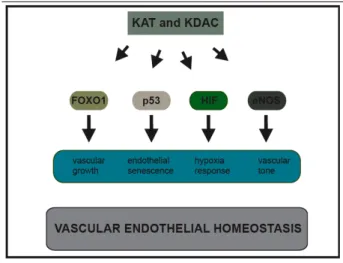
HDACs have a major role in controlling negatively gene expression, through chromatin compaction that favours transcriptional repression. Nonetheless, it is now clear how they have also more specific functions, especially in the regulation of key transcriptional factors, such as HIF1-α
(Haberland et al., 2009; Jeong et al., 2002). In addition, in vitro and in vivo studies revealed highly specific functions for the 11 mammalian HDAC isoforms, which differ in structure, enzymatic functions, sub cellular localization and expression patterns.
Class I is constituted by HDAC1, 2, 3 and 8. These Rpd3-related molecules are characterized by a high enzymatic activity towards histones, are ubiquitously expressed and are usually located into the nucleus (Taunton et al., 1996).
HDAC4, 5, 7 and 9 belong to Class IIa family. These proteins have restricted expression patterns: HDAC7 for example is enriched in ECs (Chang et al., 2006), while HDAC 5 and 9 are highly expressed in heart tissue (Chang et al., 2004). Interestingly, phosphorylation of these HDACs has been linked to nuclear-cytoplasm shuttling (McKinsey et al., 2000). Finally, Class II HDACs repress transcription by a not completely elucidated mechanism, since they are characterized by a low, if any, enzymatic activity (Haberland et al., 2009).
Class IIb has two members: HDAC6 and HDAC10. Interestingly, HDAC6 represents the main cytoplasmic mammalian deacetylase (Zhang et al., 2008). Additionally, it has been demonstrated that this enzyme can directly deacetylate cytoplasmic proteins, such as α-tubulin, cortactin, as well as IFNRα (Hubbert et al., 2002; Tang et al., 2007; Zhang et al., 2008).
HDAC 11 is the only Class IV member; this enzyme is enriched in some tissues, such as brain and heart, but has been poorly characterized from a functional point of view (Gao et al., 2002).
Intriguingly, many HDAC inhibitors (HDACi) are actually under investigation, even in human clinical trials, for their effectiveness in the treatment of a variety of disorders (Buchwald et al., 2009).