SYSTEMATIC REVIEW ARTICLE
1389-5575/19 $58.00+.00 © 2019 Bentham Science Publishers
Clinical Management of Oral Lichen Planus: A Systematic Review
Luca Oberti
1, Lucchese Alberta
2, Petruzzi Massimo
3, Carinci Francesco
4and Lauritano Dorina
1* 1Department of Medicine and Surgery, Centre of Neuroscience of Milan, University of Milano-Bicocca, 20126 Milan, Italy; 2Multidisciplinary Department of Medical and Dental Specialties, University of Campania- Luigi Vanvitelli, 80138 Naples, Italy; 3Interdisciplinary Department of Medicine, University of Bari, 70121 Bari, Italy; 4Department of Morphology, Surgery and Experimental Medicine, University of Ferrara, 44121 Ferrara, ItalyA R T I C L E H I S T O R Y Received: April 17, 2018 Revised: February 12, 2019 Accepted: February 22, 2019 DOI: 10.2174/1389557519666190301144157
Abstract: Aim: The clinical management of OLP represents a considerable challenge for the oral phy-sician. The aim of this review is to assess the main intervention used in the management of OLP and the efficacy of every type of treatment.
Materials and Methods: We searched and analyzed PubMed database for articles on OLP
manage-ment. Only randomized controlled trials, comparing an active treatment with placebo, or between dif-ferent active treatments, were considered in this systematic review. Only patients with symptomatic OLP were included and interventions of all types were considered (topical treatment, systemic drugs, non pharmacological intervention).
Results: A total of 25 randomized controlled trials were examined and included in this review. Steroids
are the most frequently employed drug in the treatment of OLP and their efficacy and safety are dem-onstrated. Also calcineurin inhibitors and photo-dynamic therapy are used in different studies for OLP management, with positive results.
Conclusion: Topical steroids remain the first-line treatment for symptomatic OLP, however, many
dif-ferent pharmacological and non-pharmacological therapies would represent a valid alternative for its management, but, nowadays they require further investigations.
Keywords: Calcineurin inhibitors, Cytochrome P450, OLP randomized controlled trials, OLP treatment, Oral lichen planus, Photo-dynamic therapy, Steroids, cytokine, gene regulation, inflammation, interleukin-6.
1. INTRODUCTION
Oral lichen planus (OLP) is a chronic inflammatory immuno-mediated disorder that affects the epithelium of oral mucous membranes [1]. OLP has an overall prevalence of 0.2-2% [2], with a higher incidence in the female population (M:F = 1:2) [3]. The pathogenesis of OLP relates to an abnormal activation of CD8+ T-lymphocytes, which causes the destruction of basal cells of the epithelium [3].
OLP has different clinical presentations: the reticular form, which has characteristic white striae (Wickham striae); the atrophic-erosive type, with erythematous and ulcerated areas, often surrounded by white striae; the plaque-like form; the papular type and the bullous type. The most affected areas are the buccal mucosa and the dorsal surface of the tongue [4].
The main symptoms associated with OLP are pain and a localized burning sensation; lesions are typical of the *Address correspondence to this author at the Department of Medicine and Surgery, Centre of Neuroscience of Milan, University of Milano-Bicocca, 20126 Milan, Italy; Tel: 00393356790163;
E-mail: [email protected]
atrophic-erosive type, but are rarely present in reticular forms [3].
It is important to distinguish OLP from oral lichenoid lesions, which can be similar in appearance, but the latter represent a reaction to specific causal factors (e.g. dental materials, traumas, drugs) [5].
Managing OLP is aimed at reducing symptoms and the manifestation of lesions, characterized by periods of exacer-bation and quiescence. The first-line treatment is represented by the use of topical corticosteroids, although other effective therapies, like photodynamic therapy and the use of cal-cineurin inhibitors have recently been made available. 2. MATERIALS AND METHODS
We examined the main publications related to OLP man-agement, identified by searching the PubMed electronic da-tabase. We used the following search items: oral lichen planus, oral lichen planus management, oral lichen planus treatment, oral lichen planus therapy, randomized trial oral lichen planus, oral lichen planus placebo, oral lichen planus
steroids, oral lichen planus laser, oral lichen planus photo-dynamic therapy, oral lichen planus calcineurin inhibitors, and oral lichen planus curcumin.
The search was run in September 2017 and a limited up-date literature search was performed in December 2017.
The results were automatically filtered in order to include only studies published in English, in the last 5 years (2013-2017). Literature reviews and case-reports were discarded, and only randomized clinical trials (RCTs) conducted in hu-mans were considered.
Finally, we used the following eligibility criteria: • Types of studies: randomized controlled trials; • Publishing date: from 2013 to 2017;
• Publishing language: English language;
• Types of participants: patients over 18 years old with symptomatic, clinically or histologically diag-nosed OLP, with no signs of dysplasia of lesions and without severe or uncontrolled systemic dis-eases;
• Types of intervention: comparison between differ-ent active treatmdiffer-ents or between an active treatmdiffer-ent and placebo;
• Types of outcome measures: symptoms and/or clinical aspects of lesions.
A total of 25 studies were included in our review. Ex-cluding the literature reviews, 216 citations remained from the first search. Of these, 158 studies were selected, which were related to OLP treatment. After then excluding all the case-reports, cohort studies and non-randomized trials, 44 studies remained; 7 additional studies were discarded be-cause the full texts were not available and 2 trials were ex-cluded because they were not conducted in humans. Finally, 35 studies were examined, and 10 of these were not included in this review because they did not meet the eligibility crite-ria. Detailed procedures of the literature search and article screening are shown in Fig. (1).
Data on the characteristics of the population sample (number of patients, sex, age, clinical form of OLP), the therapeutic interventions (drugs used in the trial, route of administration, side effects, duration of follow-up) and on the results of each treatment were extracted from all the stud-ies.
A total of 1060 patient samples were considered: the number of patients in each study was quite homogeneous, with an average of about 40-50 patients per study. The smallest sample was that examined by Kapoor et al. (2014), limited to 15 patients, while the largest trial was that con-ducted by Kazancioglu et al. (2015), consisting of 120 sub-jects.
Regarding the gender and age of patients, there was a similar distribution in all the studies: the sample predomi-nantly contained females (the average percentage of female patients was 60-70%), with the average age of patients be-tween 40 and 60 years old. This confirms the typical
distri-bution of the disease, with a higher prevalence in women after 40-50 years [7].
Concerning the clinical forms of OLP examined in the trials, it was observed that the majority of patients recruited for receiving different treatment were affected by atrophic-erosive lichen, which is the main symptomatic form of OLP. This is, moreover, in accordance with the main literature reviews, with our previous research articles and with the consensus conferences on the subject, which establish that OLP treatment is only recommended in cases of sympto-matic lesions [6, 8, 9].
In detail, among all the trials considered, 13 exclusively concerned atrophic-erosive forms of OLP; 11 examined any form of symptomatic OLP, and only one study concerned exclusively symptomatic reticular forms.
2.1. Inclusion Criteria
The main inclusive criteria were: • Clinical diagnosis of OLP;
• Histological diagnosis of OLP (according to WHO criteria, the characteristic histological features in-clude infiltration of T-cells in a band-like pattern in the basal and subepithelial layer; thickening of the epithelium with superficial hyperkeratosis and parakeratosis; and vacuolar degeneration of the ba-sal cells with the presence of numerous Civatte bod-ies);
• Presence of symptomatic lesions.
Apart from two studies, for which the diagnosis of OLP was only clinical [10, 11], all the trials examined also in-cluded histological confirmation of OLP in addition to the clinical diagnosis.
Furthermore, most of the studies conducted without the use of topical corticosteroids (first-choice therapy), but with a second-line therapy, recruited only patients who were pre-viously subjected – unsuccessfully - to conventional treat-ment for OLP.
Finally, three studies also introduced a temporal inclu-sion criterion: the persistence of leinclu-sions (at least 2 months for Liu et al. and Amirchaghmaghi et al., at least 1 year for Bacci et al.).
2.2. Exclusion Criteria
The exclusion criteria were much more varied; some studies were more restrictive and excluded patients with sys-temic diseases or who were undergoing any pharmacological therapy, while others selected larger samples of patients. In general, the main exclusion criteria were:
• Age < 18 years; • Pregnancy or lactation;
• Uncontrolled systemic diseases (hepatopathies, co-agulopathies, immune or haematological diseases, diabetes);
Fig. (1). Flow chart showing selection of studies for the review.
• History of allergic reactions to the active ingredient used in the clinical trial;
• Presence of histological signs of dysplasia in the bi-opsy specimen;
• Evidence of lichenoid lesions due to specific causes (dental materials, drugs inducing lichenoid reac-tions, etc.);
• Previous therapy for OLP in the period preceding experimentation (from 2 weeks to 3 months before
the trial; on average 2 weeks for topical treatments and 4 weeks for systemic therapy, as reported in the studies of Lee et al., Lopez Jornet and Aznar-Cayuela, and Amirchaghmaghi et al.);
• Incompletion of the trial.
It should be noted that some studies also excluded smok-ers [11, 12, 13] or heavy drinksmok-ers of alcohol [11] and pa-tients with genital or skin lichenoid lesions [14, 15, 16, 17, 18], or patients affected by other types of oral lesions, differ-ent from those caused by OLP [19, 20, 21].
Finally, all trials using curcumin excluded patients with a history of gastric or duodenal ulcers, due to the irritative effect of turmeric on gastro-intestinal mucosa, and patients undergoing orthodontic treatment [22, 23].
3. RESULTS
All 25 examined studies were randomized controlled trials; 22 of these were parallel group studies, 1 was a cross-sectional study [21] and 2 were split-mouth design studies [11, 12]. Furthermore, 4 were single-blind studies [14, 19, 24, 25] and 9 were double-blind studies [10, 16, 20, 21, 22, 23, 26, 27, 28].
Table 1 and Fig. (1) show the main characteristics of the included studies.
3.1. Study Design
Among all the studies, 7 compared active treatment with a placebo [10, 20, 21, 23, 28, 29], and one of these studies compared placebos with multiple treatments [30]. A total of 14 clinical trials compared two different active treatments [11, 12, 13, 15, 16, 17, 18, 22, 26, 27, 31, 32, 33, 34]; 3 stud-ies compared two different drugs belonging to the same class [19, 26, 27]; finally, one study compared two different routes of administration of the same drug [14].
In these clinical trials, different therapeutic approaches to OLP were considered and compared, ranging from the more conventional treatments to the less consolidated ones.
In the analysed literature, the most frequently recurrent therapy was based on the use of topical corticosteroids [12-19, 22, 24, 25, 30-34], followed by photo-dynamic therapy [13, 17, 30, 32-34], then by treatment with calcineurin in-hibitors, such as tacrolimus and pimecrolimus [15, 16, 26, 27, 31], and finally by topical therapy with curcumin gel [18, 22, 23]. We also observed the use of various other types of intervention, namely cryotherapy [12], ozone therapy [30], excisional surgery [11], inhibitors of neo-angiogenesis [24], tocopherol [21], hydroxychloroquine [10], chamomile [28], hyaluronic acid [29], and Gluscosides of Paeony Capsule (GPC) [25].
3.2. Outcomes
To evaluate the treatment efficacy, almost all the studies used the criteria of Carrozzo and Gandolfo, who divided treatment success according to: complete remission, partial resolution and non-success.
The outcomes evaluated regarding these therapeutic ap-proaches were different; in relation to the symptomatology - generally characterized by a burning sensation and pain - the majority of studies evaluated symptoms by using the Visual Analog Scale (VAS), and 4 studies used the Numerical Rat-ing Scale (NRS) [10, 11, 18, 19]. Only two studies did not consider symptomatology as an outcome [15, 27], while the results of one study were based exclusively on evaluating the improvement of symptoms, using the VAS [13].
The improvement of the clinical aspect of the lesions was assessed using the Thongprasom scale (5 = white striae with erosive area >1 cm2, 4 = white striae with erosive area <1
cm2, 3 = white striae with atrophic area >1 cm2, 2 = white striae with atrophic area <1 cm2, 1 = only white striae, 0 = no
lesions); the principal exceptions are represented by some studies that used alternative scales, but were still comparable to the classification system of Thongprasom [15, 18, 20, 27], and three studies only recorded the dimensional change of the lesion without using any classification system [10, 19, 29].
In all the studies, measurements were made using a periodontal probe, a calibre, or a calibrated tongue depressor. Alternative outcomes were the evaluation of histological changes [24]; the analysis of plasma IL-6 and IL-8 levels [27]; and the observation of an improvement in the quality of life after treatment using the Oral Health Impact Profile (OHIP-14, a scale that evaluates the functional alteration caused by the disease) [14, 28], the functional alteration scale of Lilleby [32] and the Hospital Anxiety-Depression Scale [28].
4. DISCUSSION 4.1. Steroids
Topical application of corticosteroids is considered the first treatment for symptomatic OLP, due to their effective-ness and safety. Steroids have an important local anti-inflammatory action and they are able to suppress the im-mune activity of T lymphocytes [14].
There are many different corticosteroids that can be used in managing OLP, however, nowadays, the most widely rec-ommended are triamcinolone acetonide 0.1%, which has the mildest action, clobetasol propionate 0.05% and dexametha-sone 0.05%, which instead have greater anti-inflammatory activity [6].
In the examined trials, the most frequently used formula-tions were oral paste and oral gel for both triamcinolone and clobetasol while, dexamethasone was used as an oral solu-tion for rinses, in the totality of these studies.
Topical application of the drug is always performed after oral hygiene, and in the 30 minutes following application the patient should not eat, drink or smoke [15, 26, 31].
In relation to treatment duration, studies have set different protocols; some of these, such as that conducted by Bakthiari et al. (2017) scheduled 2 weeks of treatment, others, such as that by Lee et al. (2013), instead continued treatment for 6 weeks, and others still for 2 months (Mostafa et al., 2017). In general, most of the trials examined proposed an average dura-tion of therapy for 4 weeks, with 2-4 applicadura-tions per day. The majority of literature reviews recommend treatment of 1 month, as confirmed by Lodi et al. and Scully et al. [6, 8].
According to the study by Sivaraman et al. (2016), where the efficacy of topical triamcinolone and clobetasol was compared, the latter was observed to have a significantly greater efficacy and was able to improve symptoms and re-duce the clinical appearance of lesions after treatment of just 1 week. This observation is in line with the literature on the subject; Gupta et al. described that clobetasol is the first-choice drug in the treatment of OLP, because of its major efficacy compared to triamcinolone [8, 35].
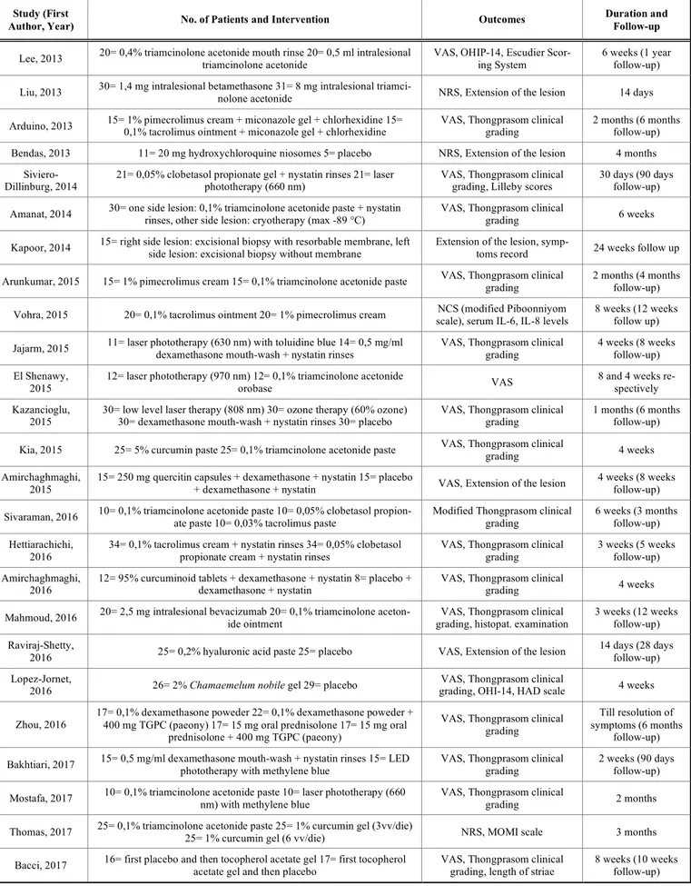
Table 1. Characteristics of included studies. Study (First
Author, Year) No. of Patients and Intervention Outcomes
Duration and Follow-up
Lee, 2013 20= 0,4% triamcinolone acetonide mouth rinse 20= 0,5 ml intralesional triamcinolone acetonide VAS, OHIP-14, Escudier Scor-ing System 6 weeks (1 year follow-up)
Liu, 2013 30= 1,4 mg intralesional betamethasone 31= 8 mg intralesional triamci-nolone acetonide NRS, Extension of the lesion 14 days
Arduino, 2013 15= 1% pimecrolimus cream + miconazole gel + chlorhexidine 15= 0,1% tacrolimus ointment + miconazole gel + chlorhexidine VAS, Thongprasom clinical grading 2 months (6 months follow-up) Bendas, 2013 11= 20 mg hydroxychloroquine niosomes 5= placebo NRS, Extension of the lesion 4 months
Siviero-Dillinburg, 2014 21= 0,05% clobetasol propionate gel + nystatin rinses 21= laser phototherapy (660 nm) VAS, Thongprasom clinical grading, Lilleby scores 30 days (90 days follow-up) Amanat, 2014 30= one side lesion: 0,1% triamcinolone acetonide paste + nystatin rinses, other side lesion: cryotherapy (max -89 °C) VAS, Thongprasom clinical grading 6 weeks
Kapoor, 2014 15= right side lesion: excisional biopsy with resorbable membrane, left side lesion: excisional biopsy without membrane Extension of the lesion, symp-toms record 24 weeks follow up
Arunkumar, 2015 15= 1% pimecrolimus cream 15= 0,1% triamcinolone acetonide paste VAS, Thongprasom clinical grading 2 months (4 months follow-up)
Vohra, 2015 20= 0,1% tacrolimus ointment 20= 1% pimecrolimus cream scale), serum IL-6, IL-8 levels NCS (modified Piboonniyom 8 weeks (12 weeks follow up)
Jajarm, 2015 11= laser phototherapy (630 nm) with toluidine blue 14= 0,5 mg/ml dexamethasone mouth-wash + nystatin rinses VAS, Thongprasom clinical grading 4 weeks (8 weeks follow-up) El Shenawy,
2015
12= laser phototherapy (970 nm) 12= 0,1% triamcinolone acetonide
orobase VAS
8 and 4 weeks re-spectively Kazancioglu,
2015 30= low level laser therapy (808 nm) 30= ozone therapy (60% ozone) 30= dexamethasone mouth-wash + nystatin rinses 30= placebo VAS, Thongprasom clinical grading 1 months (6 months follow-up) Kia, 2015 25= 5% curcumin paste 25= 0,1% triamcinolone acetonide paste VAS, Thongprasom clinical grading 4 weeks Amirchaghmaghi,
2015
15= 250 mg quercitin capsules + dexamethasone + nystatin 15= placebo
+ dexamethasone + nystatin VAS, Extension of the lesion
4 weeks (8 weeks follow-up) Sivaraman, 2016 10= 0,1% triamcinolone acetonide paste 10= 0,05% clobetasol propion-ate paste 10= 0,03% tacrolimus paste Modified Thongprasom clinical grading 6 weeks (3 months follow-up)
Hettiarachichi, 2016
34= 0,1% tacrolimus cream + nystatin rinses 34= 0,05% clobetasol propionate cream + nystatin rinses
VAS, Thongprasom clinical grading
3 weeks (5 weeks follow-up) Amirchaghmaghi,
2016
12= 95% curcuminoid tablets + dexamethasone + nystatin 8= placebo + dexamethasone + nystatin
VAS, Thongprasom clinical
grading 4 weeks Mahmoud, 2016 20= 2,5 mg intralesional bevacizumab 20= 0,1% triamcinolone aceton-ide ointment grading, histopat. examination VAS, Thongprasom clinical 3 weeks (12 weeks follow-up)
Raviraj-Shetty,
2016 25= 0,2% hyaluronic acid paste 25= placebo VAS, Extension of the lesion
14 days (28 days follow-up) Lopez-Jornet,
2016 26= 2% Chamaemelum nobile gel 29= placebo
VAS, Thongprasom clinical
grading, OHI-14, HAD scale 4 weeks
Zhou, 2016
17= 0,1% dexamethasone poweder 22= 0,1% dexamethasone poweder + 400 mg TGPC (paeony) 17= 15 mg oral prednisolone 17= 15 mg oral
prednisolone + 400 mg TGPC (paeony)
VAS, Thongprasom clinical grading
Till resolution of symptoms (6 months
follow-up) Bakhtiari, 2017 15= 0,5 mg/ml dexamethasone mouth-wash + nystatin rinses 15= LED phototherapy with methylene blue VAS, Thongprasom clinical grading 2 weeks (90 days follow-up)
Mostafa, 2017 10= 0,1% triamcinolone acetonide paste 10= laser phototherapy (660 nm) with methylene blue VAS, Thongprasom clinical grading 2 months
Thomas, 2017 25= 0,1% triamcinolone acetonide paste 25= 1% curcumin gel (3vv/die) 25= 1% curcumin gel (6 vv/die) NRS, MOMI scale 3 months
Intralesional injections of corticosteroids represent an effective treatment for OLP, which can guarantee a signifi-cant improvement of symptoms, and lead to the healing of lesions, but it is only reserved for the most serious cases that do not respond to topical therapy [6]. The analysed articles show a protocol for this treatment characterized by one injec-tion per week, which should be performed in the sub-epithelial connective tissue, for about 2-4 weeks [14, 19]; in particular Liu et al. (2013) compared injections with triam-cinolone and injections with betamethasone and they high-lighted that betamethasone is significantly more effective [19]. Moreover, Lee et al. (2013) observed that there were no statistically significant differences between the topical appli-cation and the intralesional injections of triamcinolone. Lodi et al. reported the efficacy of steroid injections, and con-firmed that triamcinolone is the most used drug [6].
Thongprasom and Dhanuthai found that intralesional injection of steroids gave variable results; in addition, they can be painful and have localized side effects such as mu-cosal atrophy [36]. It should be noted that the use of topical steroids is associated with various side effects, probably more so than those induced by many other second-line drugs, but not too serious to prevent their use [8]; the analysed stud-ies demonstrated two main adverse effects, namely a burning sensation and irritation of the mucous membranes, and oral candidiasis [14], the latter is easily resolvable by adding anti-fungal drugs to the therapy, such as rinsing with nystatin (100000 U/ml, 3 times a day) or application of miconazole gel (2%, once a day) [16, 26].
Gonzalez-Moles reported that the only adverse effect with long-term use of topical steroids is oral candidiasis [37]. Alrashdan et al. reported that systemic absorption of topical steroids has been described in different studies but, in almost all cases, it has no clinical relevance [3]. Ramadas et al. con-ducted a clinical trial on the systemic absorption of 0.1% triamcinolone acetonide and they found that, at the indicated doses, the drug is completely safe and does not cause any systemic absorption [38].
The side effects of corticosteroids in injection solutions are, instead, less frequent [14], but much more serious, as when they are systemically absorbed, they can possibly cause transient hyperglycaemia and cushingoid features [19]. Topical corticosteroid therapy thus remains the first-line treatment for OLP [3, 6, 8, 35]. The percentage of complete remission of lesions after topical steroid therapy varies be-tween 47 and 75%, with a high number of cases of partial remission [30, 32].
Lodi et al. reported that there is insufficient evidence to establish that a specific corticosteroid is more effective than others in the treatment of OLP [39]. Furthermore, Farhi e Dupin described that there is no reliable evidence to support a greater efficacy of certain topical steroid formulations compared to others [40].
4.2. Calcineurin Inhibitors
Calcineurin inhibitors are immunomodulatory drugs with inhibitory activity towards calcineurin, a calcium-dependent
protein with phosphatase activity, responsible for the im-mune response [16, 31].
This group of drugs includes tacrolimus, pimecrolimus and cyclosporine.
Tacrolimus (0.1%) and pimecrolimus (1%) represent alternatives for patients who are refractory to steroid therapy. Conrotto et al. and Yoke et al. found that cyclosporine did not provide any additional benefit compared to steroids, and responses were comparable to those of steroids. For this rea-son, cyclosporine cannot be used as a first-line treatment for OLP [41, 42].
Tacrolimus and pimecrolimus are well-tolerated drugs, especially pimecrolimus [31]; the most common side effects associated with this therapy are hyperpigmentation of the mucous membranes, a transient burning sensation immedi-ately after application [16], xerostomia, and gastro-oesophageal reflux [26]. Guo et al. reported no adverse ef-fects, while Resende et al. described mild and transient side effects [43, 44]. Ribero et al. also found that patients re-ported a transitory burning sensation and altered taste sensa-tion [45].
Our review of randomized controlled trials shows discor-dant results; according to Sivaraman et al. (2016), corticos-teroids are significantly more effective than tacrolimus, while Arunkumar et al. (2015) showed that there were no statistically significant differences; Hettiarachchi et al. (2016) instead reported that tacrolimus was more effective in the treatment of OLP [15, 16, 31].
Radfar et al. and Guo et al. both reported that there was no evidence to support that topical tacrolimus was more ef-fective and safer than topical corticosteroids for OLP man-agement [43, 46], while Resende et al. reported an improve-ment in all 15 patients treated with tacrolimus, with a com-plete remission in 86% of cases [44]. Sonthalia et al. de-scribed a 95% of either complete or partial remission after a 2-month treatment with tacrolimus [47]. After the same ther-apy period Malik et al. found a complete remission in 55% cases of patients treated with tacrolimus [48].
Conversely, in the retrospective analysis conducted by Ribero et al., only 20% of patients achieved complete remis-sion at 2 months, and 33% achieved remisremis-sion after a 6-month treatment with tacrolimus [45]. The average duration of tacrolimus treatment is 2 months, with four applications per day.
It is evident, both from the study by Arduino et al. (2013) and from the trial conducted by Vohra et al. (2015), that both tacrolimus and pimecrolimus have been shown to be efficient in improving symptoms and in reducing the clinical appearance of lichenoid lesions, but that there are no statisti-cally significant differences between these two drugs in rela-tion to their efficacy [26, 27]. However, it seems that treat-ment with pimecrolimus tends to guarantee more stable re-sults over time, with a lower risk of relapse, according to that described by Arduino et al. [26]. In contrast with the analysed trials, Lodi et al. reported that there is no reliable evidence to show that pimecrolimus is more effective than a placebo [39].
These drugs must be investigated further to solve two main problems related to their use, namely the possible sys-temic absorption observed in some studies [26], and the po-tential malignant transformation that the drug may possibly induce [26, 31].
For these reasons, the majority of the studies published on this issue do not consider calcineurin inhibitors as a suitable first-line therapy. In contrast, Resende et al. and Shipley and Spivakovsky concluded that tacrolimus 0.1% could be used as a first-line therapy because it is a safe and effective medication that improves the clinical appearance of the lesion and reduces pain, as well as the histopathological features of OLP [44, 49].
4.3. Comparison between Corticosteroids and Photody-namic Therapy
Photodynamic therapy for treating OLP exploits two dif-ferent actions, namely biostimulation induced by a laser, generally a diode laser with variable wavelength (630-980 nm), and the release of reactive oxygen species (ROS) by the irradiated photosensitive substance [6, 34, 50, 51].
Thus, photodynamic therapy functions by enhancing phagocytic activity, increasing the number of lymphatic ves-sels and their diameter, through the normalization of capil-lary permeability and by rebalancing the microcirculation [30].
This is a safe procedure that does not expose the patient to bleeding or to scar formation [17, 32]. A burning sensa-tion and local oedema are the only known side effects [17]. The treatment protocol is very similar in all the studies car-ried out, and is characterized by one-three sessions of 2-2.5 minutes per week for 2 months [13, 17, 30, 32, 34]. Jajarm et al. (2015) advised performing two sessions of photodynamic therapy per day instead of just one [34].
Among the six studies using this treatment method, four of them use only a laser and two combine the action of the laser with that of the photosensitive agent; Jajarm et al. used toluidine blue, while Mostafa et al. (2017) used methylene blue [17, 34].
Kvaal et al. found that topical methyl 5-aminolevulinate (MAL) photodynamic therapy showed lasting improvement after a single treatment [50], while Sobaniec et al. obtained excellent results using a gel containing 20% chlorin e6-polyvinylpyrrolidone and 10% dimethyl sulfoxide, which was applied directly onto the lesion [52].
Comparing the effectiveness between photodynamic treatment and application of topical steroids, some authors, including Dillenburg et al. (2014), Bakhtiari et al. (2017), and Jajarm et al. reported that there were no statistically sig-nificant differences [32, 33, 34], but rather, according to Ja-jarm et al. dexamethasone would be significantly more ef-fective than laser therapy in improving the symptoms due to OLP [34]. According to El Shenawy and Eldin (2015) and Kazancioglu and Erisen (2015), photodynamic therapy is significantly less efficient than topical application of corti-costeroids for treating OLP [13, 30]. Conversely, the study
by Mostafa et al. reported that the effectiveness of photody-namic therapy was significantly greater than that of steroid therapy for OLP [17].
Al-Maweri et al. confirmed that low-level laser therapy is effective in managing symptomatic OLP, and can be used as an alternative to corticosteroids. However, due to various methods and substantial variations in laser parameters, more RCTs with larger sample sizes are highly warranted [53]. Spanemberg et al. concluded that the use of laser treatment for OLP lesions has already been consolidated and is safe; nevertheless, further research is needed, especially random-ized, controlled clinical trials with long-term follow-up in order to create care protocols for managing oral disorders [54].
This therapeutic approach, therefore, does not represent a first-line treatment, but may play an important role in OLP patients who do not tolerate topical corticosteroid therapy, or those who are unresponsive to this treatment [6].
4.4. Curcumin
Curcumin is one of the three curcuminoids contained in Curcuma Longa, a plant often used in oriental medicine due to its well-known anti-inflammatory, antioxidant, biostimu-lant and anti-cancer properties [22, 35]. Its anti-inflammatory activity is expressed by inhibiting arachidonic acid metabo-lism that reduces the synthesis of prostaglandins and leukot-rienes; moreover, it can modulate the immune response by stimulating the activation of macrophages, T lymphocytes and natural killer lymphocytes [18]. The analysed studies show that there are no statistically significant differences between the use of a 5% curcumin oral paste and the use of topical triamcinolone [22]. Singh et al. described curcumin as a new safe option for treating OLP, but they found that its effectiveness is lower than that of steroids [55].
Chainani-Wu et al. conducted two studies using two dif-ferent doses of curcuminoid; with a low-dose of curcumin, no significant effects were observed in patients, so they could not conclude whether curcumin is effective for manag-ing OLP. The second trial, with an increased dose of curcu-min, reported a greater reduction in clinical signs and symp-toms compared to the placebo. For this reason, they con-cluded that curcumin - at a dose of 6 g per day - is effica-cious in managing OLP, with no side effects [56, 57].
According to Thomas et al. (2017), the use of lower per-centages of curcumin (for example 1%) is significantly less efficient compared to topical corticosteroids [18]. Finally, the study conducted by Amirchaghmaghi et al. (2016) re-ported no significant differences between the use of soluble tablets of curcumin and placebo [23].
4.5. Other Pharmacological Treatments
In recent years, many experimental treatments have been proposed for managing OLP, however, only a few of them have been shown to be really effective and, even then, it is still necessary to carefully study their effects, evaluating their efficacy through more clinical trials on larger samples.
Fig. (2). List of therapies according to effectiveness for OLP treatment.
The most used drugs are:
• Cyclosporine (topical application or rinses), which are able to reduce inflammation by inhibiting syn-thesis of cytokines; however, they have a high cost, a bad taste and they cause a transient burning sensa-tion immediately after applicasensa-tion [6, 35];
• Topical retinoids or, rarely, systemic retinoids, that have antioxidant action [51, 58]. Scardina et al. re-ported that, with isotretinoin, none of the cases of reticular OLP showed improvement, while 35% pa-tients with erosive OLP responded favourably to a high concentration of isotretinoin. Dalirsani et al. compared vitamin A as a mouthwash with triamci-nolone for managing OLP, and it was seen that combining triamcinolone and vitamin A mouthwash was more effective in the clinical resolution of le-sions compared to triamcinolone mouthwash alone. However, topical retinoids should be considered as a second-line therapy for treating OLP [59,60];
• Tocopherol, an important antioxidant, is able to contribute to the integrity of the cell membrane. According to a cross-sectional study conducted by Bacci et al. (2017), tocopherol could significantly reduce the extent of the lesions in OLP, without, however, leading to an improvement in symptoms [21];
• Antimalarial drugs can induce improvements in pa-tients with OLP [61], although they may be in-volved in the genesis of lichenoid reactions [6]. The most used antimalarial drug is hydroxychloroquine, which seems to be significantly effective in treating OLP, as shown by a study by Bendas et al. (2013), where, after a 4-month treatment, an evident im-provement of the lesions was obtained without col-lateral effects and without relapse [10]. Rivas-Tolosa et al. confirmed with their trial that antima-larial drugs could be useful for treating oral erosive lichen planus, because they are easily administered and affordable, with few adverse effects [61].
In the examined clinical trials, the use of different drugs was tested; Raviraj-Shetty et al. (2016) obtained significant results using a 0.2% hyaluronic acid paste [29]; Lopez Jornet and Aznar-Cayuela (2016) conducted a study on patients with OLP treated with chamomile gel (Chamaemelum no-bile), and observed a complete remission in 20% of patients, obtaining (in relation to the improvement of symptoms) sta-tistically significant results [28].
Zhou et al. (2016) demonstrated that the use of topical dexamethasone associated with total GPC, a substance typi-cal of Chinese medicine with anti-inflammatory, im-munoregulatory and antioxidant properties, allows better results to be obtained compared to the exclusive use of corti-costeroids. These findings only emerge when the total GPCs are taken for a long time, at least 4 months [25].
Mahmoud and Afifi (2016) instead proposed the use of intralesional injections of bevacizumab, an anti-angiogenetic agent, in patients suffering from atrophic-erosive lichen planus, and they obtained histological and clinical improve-ments, which were significantly greater than those treated with topical triamcinolone [24].
Finally, a study conducted by Amirchaghmaghi et al. (2015) on quercetin, a flavonoid with anti-inflammatory, antioxidant and anti-cancer properties, showed that it had no statistically significant efficacy [20].
4.6. Other Non-pharmacological Treatments
Kazancioglu and Erisen (2015) proposed ozone therapy as a treatment for OLP; this method was effective in both reducing the symptoms and improving the clinical appear-ance of the lesions, with significantly better results than those treated with placebo or with laser therapy, and compa-rable to those obtained with topical application of corticos-teroids [30].
Moreover, Amanat et al. (2014) showed that cryotherapy (-89°C under local anaesthesia of the affected mucosa), which could be a proposed method for treating OLP, does not show better results than patients treated with topical ster-oids [12]. Evidence-based data on the use of these techniques for managing OLP are lacking as there are insufficient RCTs in the literature, but these modalities may serve as potential therapies for treating refractory OLP [35].
Nowadays, therefore, the preferential non-pharmacological treatment, in addition to photodynamic therapy, is surgical excision [6]. In this case, application of resorbable membranes after excision does not seem to achieve better results, as demonstrated by Kapoor et al. (2014).
Axell and Henriksen conducted a study in which, after surgical excision of the lesion, the surgical site received a soft tissue graft; the healthy graft remained free of lesions and appeared clinically healthy at follow-up visits [62]. CONCLUSION
The examined studies confirm that the most efficient and effective therapy in patients with symptomatic OLP is topi-cal treatment with corticosteroids, which, nowadays, remains
the first-line therapy for this type of lesion [35]. The use of intralesional steroids, although fairly uncommon, can repre-sent a valid alternative in cases that only partially respond to topical steroid therapy [6], while the use of systemic corti-costeroids is only indicated in patients whose condition is unresponsive to topical first- and second-line therapies, or with a relevant involvement of oral mucosa [8].
From our review of RCTs of OLP, which can also be seen from other literature reviews, it can be concluded that, in the case of relapses or refractory lesions, second-line ther-apy consists of the use of calcineurin inhibitors, and in the use of topical preparations of curcumin and retinoids [51]. Photodynamic therapy is another valid choice in all these cases [50] and, when the lesion is circumscribed, excisional surgery also represents an important alternative in OLP [6].
Fig. (2) shows the main treatment modalities for manag-ing OLP. Numerous nutraceutical treatments need to be in-vestigated further with other randomized trials; they can now only be applied in association with the common first-line therapies.
None of the other therapies have been found to be better than topical steroids for managing OLP; for this reason, the use of other therapies should only be reserved for extensive lesions and refractory cases, and should only be prescribed by a specialist physician.
CONSENT FOR PUBLICATION Not applicable.
FUNDING None.
CONFLICT OF INTEREST
The authors declare no conflict of interest, financial or otherwise.
ACKNOWLEDGEMENTS Declared none.
REFERENCES
[1] Mustafa MB, Porter SR, Smoller BR, Sitaru C. Oral mucosal
mani-festations of autoimmune skin diseases. Autoimmunity Reviews,
2015, 14: 930-951.
[2] Bascones-Martinez A, Garcia-Garcia V, Meurman JH, Requena-Caballero L. Immune-mediated diseases: what can be found in the
oral cavity? International Journal of Dermatology, 2015, 54:
258-270.
[3] Alrashdan MS, Cirillo N, McCullough M. Oral lichen planus: a
literature review and update. Arch Dermatol Res, 2016, 308:
539-551.
[4] Andreasen JO. Oral lichen planus: a clinical evaluation of 115
cases. Oral Surg Oral Med Oral Pathol. 1968; 25:31-41.
[5] Gissi DB, Venturi M, Gabusi A, De Martino A, Montebugnoli L.
Le patologie autoimmunitarie del cavo orale. Reviews in Health
Care, 2011, 2(2): 33-55.
[6] Lodi G, Scully C, Carrozzo M, Griffiths M, Sugerman PB, Thong-prasom K. Current controversies in oral lichen planus: report of an
international consensus meeting. Part 2. Clinical management and malignant transformation. Oral Surg Oral Med Oral Pathol Oral
[7] Lodi G, Scully C, Carrozzo M, Griffiths M, Sugerman PB, Thong-prasom K. Current controversies in oral lichen planus: report of an
international consensus meeting. Part 1. Viral infections and etiopathogenesis. Oral Surg Oral Med Oral Pathol Oral Radiol
En-dod, 2005, 100(1): 40-51.
[8] Scully C, Beyli M, Ferreiro MC, Ficarra G, Gill Y, Griffiths M, Holmstrup P, Mutlu S, Porter S, Wray D. Update on oral lichen
planus: etiopathogenesis and management. Crit Rev Oral Biol
Med, 1998, 9(1):86-122.
[9] Al-Hashimi I, Schifter M, Lockhart PB, Wray D, Brennan M, Migliorati CA, Axéll T, Bruce AJ, Carpenter W, Eisenberg E, Ep-stein JB, Holmstrup P, Jontell M, Lozada-Nur F, Nair R, Silverman B, Thongprasom K, Thornhill M, Warnakulasuriya S, Van der Waal I. Oral lichen planus and oral lichenoid lesions: diagnostic
and therapeutic considerations. Oral Surg Oral Med Oral Pathol
Oral Radiol Endod, 2007, 103(1):25,1-25,12.
[10] Bendas ER, Abdullah H, El-Komy MHM, Kassem MAA.
Hy-droxychloroquine niosomes: a new trend in topical management of oral lichen planus. International journal of Pharmaceutics, 2013,
458: 287-295.
[11] Kapoor A, Sikri P, Grover V, Malhotra R, Sachdeva S. Evaluation
of efficacy of a bioresorbable membrane in the treatment of oral li-chen planus. Dent Res J, 2014, 11(3): 386-394.
[12] Amanat D, Ebrahimi H, Zahedani MZ, Zeini N, Pourshahidi S, Ranjbar Z. Comparing the effects of cryotherapy with nitrous oxide
gas versus topical corticosteroids in the treatment of oral lichen planus. Indian J Dent Res, 2014, 25(6): 711-716.
[13] El Shenawy HM e Eldin AM. A comparative evaluation of
low-level laser and topical steroid therapies for the treatment of ero-sive-atrophic lichen planus. Macedonian Journal of Medical
Sci-ences, 2015, 3(3): 462-466.
[14] Lee YC, Shin SY, Kim SW, Eun YG. Intralesional injection versus
mouth rinse of triamcinolone acetonide in oral lichen planus: a randomized controlled study. Otolaryngology–Head and Neck
Sur-gery, 2013, 148(3): 443-449.
[15] Sivaraman S, Santham K, Nelson A, Laliytha B, Azhalvel P, Dee-pak JH. A randomized triple-blind clinical trial to compare the
ef-fectiveness of topical triamcinolone acetonate (0.1%), clobetasol propionate (0.05%), and tacrolimus orabase (0.03%) in the man-agement of oral lichen planus. J Pharm Bioall Sci, 2015, 8(5):
86-89.
[16] Hettiarachchi PVKS, Hettiarachchi RM, Jayasinghe RD, Sitheeque M. Comparison of topical tacrolimus and clobetasol in the
man-agement of symptomatic oral lichen planus: a double-blinded, ran-domized clinical trial in Sri Lanka. J Invest Clin Dent, 2016, 1–6
[17] Mostafa D, Moussa E, Alnouaem M. Evaluation of photodynamic
therapy in treatment of oral erosive lichen planus in comparison with topically applied corticosteroids. Photodiagnosis and
Pho-todynamic Therapy, 2017, 19: 56-66.
[18] Thomas AE, Varma B, Kurup S, Jose R, Chandy ML, Kumar SP, Aravind MS, Ramadas AA. Evaluation of efficacy of 1%
curcumi-noids as local application in management of oral lichen planus: in-terventional study. Journal of Clinical and Diagnostic Research,
2017, 11(4): ZC89-ZC93.
[19] Liu C, Xie B, Yang Y, Lin D, Wang C, Lin M, Ge L, Zhou H.
Efficacy of intralesional betamethasone for erosive oral lichen planus and evaluation of recurrence: a randomized, controlled trial. Oral Surg Oral Med Oral Pathol Oral Radiol, 2013, 116:
584-590.
[20] Amirchaghmaghi M, Delavarian Z, Iranshahi M, Taghi Shakeri M, Mozafari PM, Mohammadpour AH, Farazi F, Iranshahy M. A
ran-domized placebo-controlled double blind clinical trial of quercetin for treatment of oral lichen planus. J Dent Res Dent Clin Dent
Prospect, 2015, 9(1): 23-28.
[21] Bacci C, Vanzo V, Frigo AC, Stellini E, Sbricoli L, Valenti M. Topical tocopherol for treatment of reticular oral lichen planus: a randomized, double-blind, crossover study. Oral Diseases, 2017, 23: 62-68.
[22] Kia SJ, Shirazian S, Mansourian A, Fard LK, Ashnagar S.
Com-parative efficacy of topical curcumin and triamcinolone for oral li-chen planus: a randomized, controlled clinical trial. JDTUMS,
2015, 12:11.
[23] Amirchaghmaghi M, Pakfetrat A, Delavarian Z, Ghalavani H, Ghazi A. Evaluation of the efficacy of curcumin in the treatment of
oral lichen planus: A randomized controlled trial. Journal of
Clini-cal and Diagnostic Research, 2016, 10(5): ZC134-ZC137. [24] Mahmoud MM e Afifi MM. Anti-angiogenic therapy
(bevacizu-mab) in the management of oral lichen planus. Eur J Oral Sci,
2016, 124: 119-126.
[25] Zhou L, Cao T, Wang Y, Yao H, Du G, Tian Z, Tang G. Clinical
observation on the treatment of oral lichen planus with total gluco-sides of paeony capsule combined with corticosteroids.
Interna-tional Immunopharmacology, 2016, 36: 106-110.
[26] Arduino PG, Carbone M, Della Ferrera F, Elia A, Conrotto D, Gambino A, Comba A, Calogiuri PL, Broccoletti R. Pimecrolimus
vs. tacrolimus for the topical treatment of unresponsive oral ero-sive lichen planus: an 8 week randomized double-blind controlled study. JEADV, 2014, 28: 475-482.
[27] Vohra S, Singal A, Sharma SB. Clinical and serological efficacy of
topical calcineurin inhibitors in oral lichen planus: a prospective randomized controlled trial. International Journal of Dermatology,
2016, 55: 101-105.
[28] Lopez Jornet P e Aznar-Cayuela C. Efficacy of topical chamomile
management vs placebo in patients with oral lichen planus: a ran-domized double-blind study. JEADV, 2016, 30: 1783-1786.
[29] Raviraj-Shetty R, Burde KN, Guttal KS. The efficacy of topical
hyaluronic acid 0.2% in the management of symptomatic oral li-chen planus. Journal of Clinical and Diagnostic Research, 2016,
10(1): ZC46-ZC50.
[30] Kazancioglu HO e Erisen M. Comparison of low-level laser
ther-apy versus ozone therther-apy in the treatment of oral lichen planus.
Ann Dermatol, 2015, 27(5): 485-491
[31] Arunkumar S, Kalappanavar A, Annigeri RG, Kalappa SG.
Rela-tive efficacy of pimecrolimus cream and triamcinolone acetonide paste in the treatment of symptomatic oral lichen planus. Indian J
of Dent, 2015, 6(1): 14-19.
[32] Dillenburg CS, Martins MAT, Munerato MC, Marques MM, Car-rard VC, Filho MSA, Castilho RM, Martins MD. Efficacy of laser
phototherapy in comparison to topical clobetasol for the treatment of oral lichen planus: a randomized controlled trial. Journal of
Biomedical Optics, 2014, 19(6): 068002-1:1-9
[33] Bakhtiari S, Mojahedi SM, Azari-Marhabi S, Namdari M, Rankohi ZE. Comparing clinical effects of photodynamic therapy as a novel
method with topical corticosteroid for treatment of oral lichen planus. Photodiagnosis and Photodynamic Therapy, 2017.
[34] Jajarm HH, Falaki F, Sanatkhani M, Ahmadzadeh M, Ahrari F, Shafaee H. A comparative study of toluidine blue-mediated
pho-todynamic therapy versus topical corticosteroids in the treatment of erosive-atrophic oral lichen planus: a randomized clinical con-trolled trial. Lasers Med Sci, 2015, 30:1475-1480.
[35] Gupta S, Ghosh S, Gupta S. Interventions for the management of
oral lichen planus: a review of the conventional and novel thera-pies. Oral Diseases, 2017, 23(8): 1029-1042.
[36] Thongprasom K, Dhanuthai K. Steroid in the treatment of lichen
planus: a review. J Oral Sci, 2008, 50: 377-385.
[37] Gonzalez-Moles MA. The use of topical steroids in oral pathology. Med Oral Patol Oral Cir Bucal, 2010, 16: 827-831.
[38] Ramadas AA, Jose R, Arathy SL, Kurup S, Chandy ML, Kumar SP. Systemic absorption of 0,1% triamcinolone acetonide as
topi-cal application in management of oral lichen planus. Indian J Dent
Res, 2016, 27(3): 230-235.
[39] Lodi G, Carrozzo M, Furness S, Thongprasom K. Interventions for
treating oral lichen planus. Cochrane Database Syst Rev, 2011,
7:CD001168.
[40] Farhi D, Dupin N. Pathophysiology, etiologic factors, and clinical
management of oral lichen planus, part I: facts and controversies.
Clinics in Dermatology, 2010, 28: 100-108.
[41] Conrotto D, Carbone M, Carrozzo M, Arduino P, Broccoletti R, Pentenero M, Gandolfo S. Ciclosporin vs. clobetasol in the topical
management of atrophic and erosive oral lichen planus: a double-blind, randomized controlled trial. Br J Dermatol, 2006, 154:
139-145.
[42] Yoke PC, Tin GB, Kim MJ, Rajaseharan A, Ahmed S, Thongpra-som K, Chaimusik M, Suresh S, Machin D, Bee WH, Seldrup J. A
randomized controlled trial to compare steroid with cyclosporine for the topical treatment of oral lichen planus. Oral Surg Oral Med
[43] Guo CL, Zhao JZ, Zhang J, Dong HT. Efficacy of topical
tac-rolimus for erosive oral lichen planus: a meta-anlaysis. Chin Med
Sci J, 2015, 30(4):210-217.
[44] Resende JPM, Chaves MDGAM, Aarestrup FM, Aarestrup BV, Olate S, Netto HD. Oral lichen planus treated with tacrolimus
0,1%. Int J Clin Exp Med, 2013, 6(10): 917-921.
[45] Ribero S, Stieger M, Quaglino P, Hongang T, Bornstein MM, Naldi L, Borradori L. Efficacy of topical tacrolimus for oral lichen
planus: real-life experience in a retrospective cohort of patients with a review of the literature. J Eur Acad Dermatol Venereol,
2015, 29: 1107-1113.
[46] Radfar L, Wild RC, Suresh L. A comparative treatment study of
topical tacrolimus and clobetasol in oral lichen planus. Oral Surg
Oral Med Oral Pathol Oral Radiol, 2008, 105(2): 187-193. [47] Sonthalia S, Singal A. Comparative efficacy of tacrolimus 0.1%
ointment and clobetasol propionate 0.05% ointment in oral lichen planus: a randomized double-blind trial. Int J Dermatol, 2012; 51:
1371-1378.
[48] Malik U, Gupta S, Malik SD, Vashishth S, Zaheeruddin, Raju MS.
Treatment of symptomatic oral lichen planus (OLP) with 0.1% tac-rolimus powder in Oraguard-B - A pilot prospective study. Saudi
Dent J, 2012, 24: 143-148.
[49] Shipley CA, Spivakovsky S. Tacrolimus or clobetasol for
treat-ment of oral lichen planus. Evidence-based Dentistry, 2016, 17(1):
16.
[50] Kvaal SI, Angell-Petersen E, Warloe T. Photodynamic treatment of
oral lichen planus. Oral Surg Oral Med Oral Pathol Oral Radiol,
2013, 115: 62-70.
[51] Yang H, Wu Y, Ma H, Jiang L, Zeng X, Dan H, Zhou L, Chen Q.
Possible alternative therapies for oral lichen planus cases refrac-tory to steroid therapies. Oral Surg Oral Med Oral Pathol Oral
Ra-diol, 2016, 121 (5): 496-509.
[52] Sobaniec S, Bernaczyk P, Pietruski J, Cholewa M, Skurska A, Dolinska E, Duraj E, Takajuk G, Paniczko A, Olszewska E, Pie-truska M. Clinical assessment of the efficacy of photodynamic
ther-apy in the treatment of oral lichen planus. Lasers Med Sci, 2013,
28(1): 311-316.
[53] Al-Maweri SA, Kalakonda B, Al-Soneidar WA, Al-Shamiri HM, Alakhali MS, Alaizari N. Efficacy of low-level laser therapy in
management of symptomatic oral lichen planus: a systematic re-view. Lasers Med Sci, 2017, 32: 1429-1437.
[54] Spanemberg JC, de Figueiredo MAZ, Cherubini K, Salum FG.
Low-level laser therapy: a review of its apllications in the man-agement of oral mucosal disorders. Altern Ther Health Med, 2016,
22(6): 24-31.
[55] Singh V, Pal M, Gupta S, Tiwari SK, Malkunje L, Das S. Turmeric
– a new treatment option for lichen planus: a pilot study. Natl J
Maxillofac Surg, 2013, 4(2): 198-201.
[56] Chainani-Wu N, Silverman S Jr, Reingold A, Bostrom A, McCul-loch C, Lozada-Nur F. A randomized, placebocontrolled,
double-blind clinical trial of curcuminoids in oral lichen planus.
Phy-tomedicine, 2007, 14: 437-446.
[57] Chainani-Wu N, Madden E, Lozada-Nur F, Silverman S.
High-dose curcuminoids are efficacious in the reduction in symptoms and signs of oral lichen planus. J Am Acad Dermatol, 2012, 66:
752-760.
[58] Petruzzi M, Lucchese A, Lajolo C, Lauritano D, Serpico R. Topical
Retinoids in Oral Lichen Planus Treatment: An Overview.
Derma-tology, 2013, 226 (1): 61-67.
[59] Scardina GA, Messina P, Carini F, Maresi E. A randomized trial
assessing the effectiveness of different concentrations of isotreti-noin in the management of lichen planus. Int J Oral Maxillofac
Surg, 2006, 35: 67-71.
[60] Dalirsani Z, Taghavi Zenouz A, Mehdipour M, Alavi F, Java-dzadeh Y. Comparison of the effect of combination of
triamci-nolone acetonide and vitamin a mouthwash with triamcitriamci-nolone mouthwash alone on oral lichen planus. J Dent Res Dent Clin Dent
Prospects, 2010, 4: 21-24.
[61] Rivas-Tolosa N, Requena C, Llombart B, Alcalá R, Serra-Guillén C, Calomarde L, Nagore E, Guillén C, Sanmartín O. Antimalarial
drugs for the treatment of oral erosive lichen planus. Dermatology,
2016, 232(1): 86-90.
[62] Axell T, Henriksen BM. Treatment of gingival lichen with free
palatal grafts. J Oral Pathol Med, 2007, 36: 105-109.
DISCLAIMER: The above article has been published in Epub (ahead of print) on the basis of the materials provided by the author. The Edito-rial Department reserves the right to make minor modifications for further improvement of the manuscript.