R E S E A R C H A R T I C L E
Open Access
The World Society of Emergency Surgery
(WSES) spleen trauma classification: a
useful tool in the management of splenic
trauma
Federico Coccolini
1,2*, Paola Fugazzola
1,2, Lucia Morganti
3, Marco Ceresoli
1,4, Stefano Magnone
2, Giulia Montori
2,
Matteo Tomasoni
1,2, Stefano Maccatrozzo
2, Niccolò Allievi
2, Savino Occhionorelli
3, Yoram Kluger
4, Massimo Sartelli
5,
Gian Luca Baiocchi
6, Luca Ansaloni
1,2and Fausto Catena
7Abstract
Background: The World Society of Emergency Surgery (WSES) spleen trauma classification meets the need of an evolution of the current anatomical spleen injury scale considering both the anatomical lesions and their physiologic effect. The aim of the present study is to evaluate the efficacy and trustfulness of the WSES classification as a tool in the decision-making process during spleen trauma management.
Methods: Multicenter prospective observational study on adult patients with blunt splenic trauma managed between 2014 and 2016 in two Italian trauma centers (ASST Papa Giovanni XXIII in Bergamo and Sant’Anna University Hospital in Ferrara). Risk factors for operative management at the arrival of the patient and as a definitive treatment were analyzed. Moreover, the association between the different WSES grades of injury and the definitive management was analyzed.
Results: One hundred twenty-four patients were included. At multivariate analysis, a WSES splenic injury grade IV is a risk factor for the operative management both at the arrival of the patients and as a definitive treatment. WSES splenic injury grade III is a risk factor for angioembolization.
Conclusions: The WSES classification is a good and reliable tool in the decision-making process in splenic trauma management.
Keywords: Spleen trauma, Classification, Validation, Practice, Surgery, Outcome, Non-operative management, Quality
Introduction
The most commonly used classification of splenic trauma is the American Association for the Surgery of Trauma (AAST)-Organ Injury Severity Score (OIS). It was initially ideated to allow the comparison between different series of patients; then, it has been used as a classification system to drive treatment
strategies. It is based on spleen lesion anatomy [1].
This scale was validated by several studies with large
sample sizes [2–4] showing as both the management
at the patient arrival (operative management (OM) vs non-operative management (NOM)), and the NOM failure rate was associated with the ASST lesion grade in patients with blunt splenic trauma. In fact, the anatomy of the lesions plays a fundamental role in determining the conditions of the patients. In some situations, however, patient conditions lead to an emergent transfer to the operating room (OR) with-out the opportunity to define the grade of splenic lesions before the surgical exploration. In these cases, © The Author(s). 2019 Open Access This article is distributed under the terms of the Creative Commons Attribution 4.0
International License (http://creativecommons.org/licenses/by/4.0/), which permits unrestricted use, distribution, and reproduction in any medium, provided you give appropriate credit to the original author(s) and the source, provide a link to the Creative Commons license, and indicate if changes were made. The Creative Commons Public Domain Dedication waiver (http://creativecommons.org/publicdomain/zero/1.0/) applies to the data made available in this article, unless otherwise stated.
* Correspondence:[email protected]
1
General, Emergency and Trauma Surgery Department, Bufalini Hospital, Viale Ghirotti 268, 47521 Cesena, Italy
2General, Emergency and Trauma Surgery Department, Papa Giovanni XXIII
Hospital, Bergamo, Italy
the physiopathologic status of the patients leads the therapeutic decision, more than the anatomy of the splenic lesions. Moreover, there are patients with high-grade splenic lesions without hemodynamic re-percussions that can be managed with NOM thanks to the modern tools in bleeding management. As a counterpart, there exists a cohort of patients with
hemodynamic instability requiring urgent surgical
intervention due to low-grade splenic injuries. In May 2017, during the World Society of Emergency Surgery (WSES) World Congress in Campinas, Brazil, the final version of the WSES guidelines on spleen
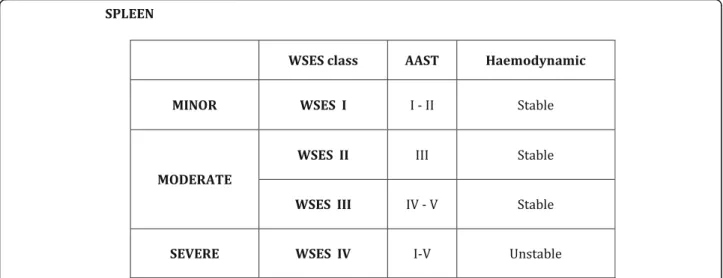
trauma was approved (Fig. 1) [5]. The WSES grading
system takes into account both the patient’s condition and the anatomy of lesions.
The aim of the present study is to evaluate the effi-cacy and trustfulness of the WSES classification as a tool in decision-making process during spleen trauma management.
Methods
This is an analysis of two prospectively enrolled adult patient cohorts with blunt splenic trauma managed between 2014 and 2016 in two Italian trauma centers (TC) (ASST Papa Giovanni XXIII in Bergamo and Sant’Anna University Hospital in Ferrara) stratified ac-cording to the WSES classification. Ethical committee and patients’ consent to participate were waived be-cause no personal or sensible data were recorded and no specific intervention was adopted other than the usual clinical practice. Patients’ characteristics were collected (age, sex, comorbidity, ASA (American Soci-ety of Anesthesiologists) score, antiplatelet or anti-coagulant therapy). Trauma mechanism of injury, patient conditions at the arrival in the emergency
department (ED) (systolic blood pressure (SBP), heart rate (HR), shock index (SI), need of red blood cell (RBC) transfusion), blood gas test (pH, base excess (BE), lactates (Lac)), blood exams (CBC, platelet count, INR, fibrinogen), and eco-fast results were
reported. We defined a patient “hemodynamically
unstable” if, after resuscitation in the ED and without vasoactive drugs, he/she had a SBP lower than 90 mmHg, a shock index higher than 1, or a BE lower than − 5.
For patients who underwent CT at the arrival, the AAST classification for the splenic injury, the number of abdominal quadrants with hemoperitoneum, and the presence of vascular lesions (contrast blush (CB), pseudoaneurysm (PSA), arterovenous fistula (AVF)) were reported. For patients who underwent urgent surgical intervention, intraoperative (for splenecto-mized patients) or postoperative CT findings were registered. The Injury Severity Score (ISS) and the presence of associated abdominal, pelvic, or cerebral lesions were reported. Patients were classified accord-ing to the 2017 WSES classification. The management at the arrival (observation, distal angioembolization (AE), proximal AE, splenectomy, intraperitoneal pack-ing, hemostasis of the splenic injury, surgical inter-vention for other organ lesions), the time between the arrival in the ED and the first urgent intervention, and the need of further intervention during hospital stay (AE or splenectomy) have been recorded.
It was defined OM if the patient underwent urgent surgical intervention at the arrival at the ED and if during the surgical procedure, a splenectomy or a hemostatic splenic technique (e.g., splenic packing or splenorrhaphy) was performed. The NOM could include AE or not. Failure of NOM (fNOM) was defined as the
need of performing a splenectomy after starting NOM. To validate the 2017 WSES classification, the risk factors for OM at the arrival of the patient and for OM as a de-finitive treatment (including both patients treated with OM at the arrival and patients operated for fNOM) have been analyzed. It was verified if the WSES grade was a risk factor for OM at the arrival and as a definitive treatment for adult patients with blunt splenic trauma.
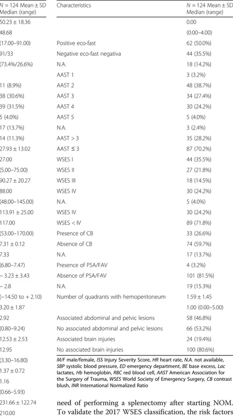
Table 1 Patient characteristics
Characteristics N = 124 Mean ± SD Median (range) Age (years) 50.23 ± 18.36 48.68 (17.00–91.00) M/F 91/33 (73.4%/26.6%) Trauma mechanism of injury
-Invested pedestrian 11 (8.9%) -Car 38 (30.6%) -Motorbike 39 (31.5%) -Bike 5 (4.0%) -Precipitation 17 (13.7%) -Others 14 (11.3%) ISS 27.93 ± 13.02 27.00 (5.00–75.00) HR at arrival in ED (bpm) 90.27 ± 20.27 88.00 (48.00–145.00) SBP at arrival in ED (mmHg) 113.91 ± 25.00 117.00 (53.00–170.00) pH 7.31 ± 0.12 7.33 (6.80–7.47) BE (mmol/L) − 3.23 ± 3.43 − 2.8 (−14.50 to + 2.10) Lac 3.20 ± 1.87 2.92 (0.80–9.24) Hb (g/dL) 12.53 ± 2.53 12.95 (3.30–16.80) INR (s) 1.37 ± 0.72 1.16 (0.66–5.93) Fibrinogen (mg/dL) 231.66 ± 122.74 210.00 (26.00–1120.00) Platelets (× 103/mL) 218.92 ± 72.27 220.00 (55.00–460.00) Number of RBC units transfused in ED 0.48 ± 0.96
Table 1 Patient characteristics (Continued)
Characteristics N = 124 Mean ± SD
Median (range) 0.00
(0.00–4.00)
Positive eco-fast 62 (50.0%)
Negative eco-fast negativa 44 (35.5%)
N.A. 18 (14.2%) AAST 1 3 (3.2%) AAST 2 48 (38.7%) AAST 3 34 (27.4%) AAST 4 30 (24.2%) AAST 5 5 (4.0%) N.A. 3 (2.4%) AAST > 3 35 (28.2%) AAST≤ 3 87 (70.2%) WSES I 44 (35.5%) WSES II 27 (21.8%) WSES III 18 (14.5%) WSES IV 30 (24.2%) N.A. 5 (4.0%) WSES IV 30 (24.2%) WSES < IV 89 (71.8%) Presence of CB 33 (26.6%) Absence of CB 74 (59.7%) N.A. 17 (13.7%) Presence of PSA/FAV 4 (3.2%) Absence of PSA/FAV 101 (81.5%) N.A. 19 (15.3%)
Number of quadrants with hemoperitoneum 1.59 ± 1.45 1.00 (0.00–5.00) Associated abdominal and pelvic lesions 58 (46.8%) No associated abdominal and pelvic lesions 66 (53.2%) Associated brain injuries 24 (19.4%) No associated brain injuries 100 (80.6%) M/F male/female, ISS Injury Severity Score, HR heart rate, N.A. not available, SBP systolic blood pressure, ED emergency department, BE base excess, Lac lactates, Hb hemoglobin, RBC red blood cell, AAST American Association for the Surgery of Trauma, WSES World Society of Emergency Surgery, CB contrast blush, INR International Normalized Ratio
Statistical analysis
Continuous variables were expressed as mean and standard deviation; categorical data were expressed as
proportions and percentages. t test was used for
con-tinuous variables with normal distribution and the Mann-Whitney test for non-normal distribution vari-ables. Parametric variables were compared with chi-square test. Multivariate models were calculated with the linear logistic regression method including all the variables resulted significantly associated (p < 0.05) with the selected outcome at univariate analysis. All the statistical analysis was performed with IBM SPSS 20 (IBM Corp. released 2011; IBM SPSS Statistics for Windows, Version 20.0; Armonk, NY: IBM Corp.).
Results
The study includes 124 patients older than 17 years with blunt splenic lesion, of whom 66 managed in ASST Papa Giovanni in Bergamo and 58 in Sant’Anna University Hospital in Ferrara. The two groups of
pa-tients were similar in terms of epidemiological
features, trauma mechanism of injury, ISS, and
splenic injury grade. Patient characteristics are re-ported in Table 1.
NOM rate was 53.2% (66 patients) and OM rate 46.0% (58 patients). Among OM patients, we had 84.5% (49 patients) of patients treated with splenec-tomy and 15.5% (9 patients) with hepatic and splenic packing (in patients with hepatic lesion associated) and/or splenic hemostasis (Table 2).
Among NOM patients, 22 underwent AE (17.8% of total patients and 33.3% of NOM patients) at the arrival or during the hospital stay (Table2).
Risk factors for OM at the arrival of patient in the ED, including the WSES splenic injury grade, were
analyzed with univariate (Table 3) and multivariate
(Table 4) analysis.
At the multivariate analysis, the WSES IV splenic injury grade was found as the only one risk factor for
OM at the arrival of patients (OR 5.44, p = 0,049)
(Table 4).
The risk factors for OM as a definitive treatment were analyzed, including both patients treated with OM at the arrival in the ED and patients operated for fNOM. The OM was applied on 53.2% of patients as a definitive treatment.
Risk factors emerging from univariate and multivariate analyses are shown in Tables5and6.
The WSES grade IV (OR 7.22, p = 0,029) and ISS
value higher than 25 (OR 5.75, p = 0,013) were found
as the only significant risk factors at the multivariate analysis (Table 6).
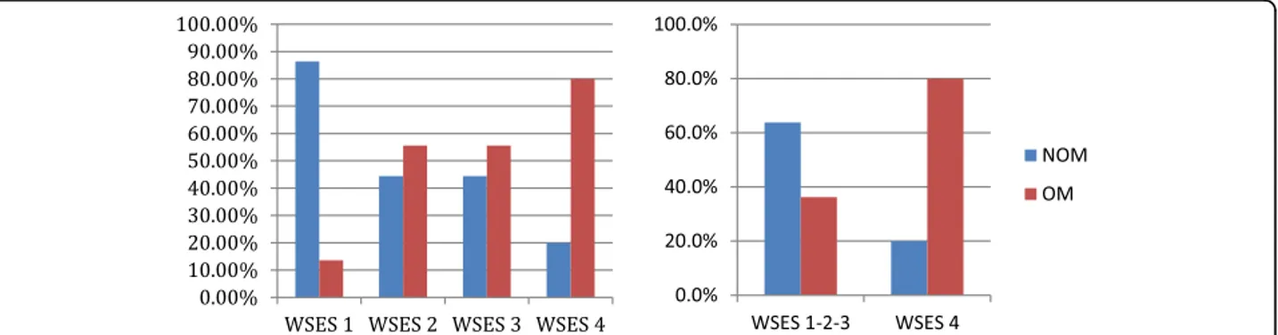
The previous analysis showed as OM rate, both at the arrival of patient and as a definitive treatment, increased with the increasing of the WSES splenic injury grade, in particular for the WSES grade IV
compared with lower grade (Figs. 2 and 3).
The present study verified also if the AAST and WSES classifications were predictive for AE at the ar-rival of patient with splenic injury or during hospital stay. While an AAST grade higher than 3 was not a significant risk factor for AE (AAST > 3 (20.0%) vs
AAST ≤ 3 (17.2%), n.s.), a WSES splenic injury grade
of III was found as a significant risk factor (WSES 3
(38.9%) vs WSES 1-2-4 (13.9%), p = 0.010).
Discussion
After the introduction of AE and the modern tools in bleeding management, the NOM failure rate
de-creased from 23–67% to 4–42% [6–10] and it was no
longer associated with the AAST injury grade (i.e.,
anatomical degree of lesion) [11]. So it has been
ac-cepted that the physiopathologic status of the pa-tients, more than the anatomy of the splenic lesions,
should lead the therapeutic decision in splenic
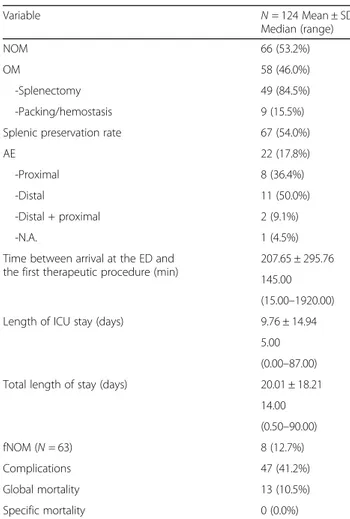
Table 2 Patient outcomes
Variable N = 124 Mean ± SD Median (range) NOM 66 (53.2%) OM 58 (46.0%) -Splenectomy 49 (84.5%) -Packing/hemostasis 9 (15.5%)
Splenic preservation rate 67 (54.0%)
AE 22 (17.8%)
-Proximal 8 (36.4%)
-Distal 11 (50.0%)
-Distal + proximal 2 (9.1%)
-N.A. 1 (4.5%)
Time between arrival at the ED and the first therapeutic procedure (min)
207.65 ± 295.76 145.00 (15.00–1920.00) Length of ICU stay (days) 9.76 ± 14.94
5.00 (0.00–87.00) Total length of stay (days) 20.01 ± 18.21
14.00 (0.50–90.00) fNOM (N = 63) 8 (12.7%) Complications 47 (41.2%) Global mortality 13 (10.5%) Specific mortality 0 (0.0%)
NOM non-operative management, OM operative management, N.A. not available, fNOM failure of non-operative management, ICU intensive care unit
trauma. Furthermore, many studies [8, 12–16] showed that the vascular lesions (CB, PSA, AVF), which have significant incidence also in low-grade in-juries [12, 16], were predictive factors for NOM fail-ure and that they should be considered indications to Table 3 Univariate analysis of risk factors for OM at the arrival
of patient at the ED
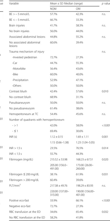
Variable Mean ± SD Median (range) p value
NOM OM Age < 55 years 42.3% 57.7% n.s. Age > 55 years 50.0% 50.0% Age (years) 50.54 ± 18.17 49.87 ± 18.73 n.s. 49.35 (18.00–91.00) 48.00 (17.00–85.60) No anticoagulant/antiplatelet drugs 48.8% 51.2% n.s. Anticoagulant/antiplatelet drugs 40.0% 60.0% HR (mean ± SD) 85.95 ± 18.66 95.24 ± 21.07 0.009 Median (range) (bpm) 80.00 (48.00– 133.00) 95.00 (55.00– 145.00) HR < 120 bpm 58.7% 41.3% n.s. HR > 120 bpm 46.8% 53.2% SBP(mmHg) 120.40 ± 21.35 106.51 ± 26.92 0.002 120.00 (70.00– 170.00) 105.00 (53.00– 167.00) SBP > 90 mmHg 60.4% 39.6% 0.001 SBP < 90 mmHg 21.7% 78.3% Shock index < 1 60.2% 39.8% 0.002 Shock index > 1 26.9% 73.1% AAST 1 100.0% 0.0% < 0.001 AAST 2 81.3% 18.7% AAST 3 44.1% 55.9% AAST 4 26.7% 73.3% AAST 5 0.0% 100.0% AAST≤ 3 66.7% 33.3% < 0.001 AAST > 3 22.9% 77.1% WSES I 86.4% 13.6% < 0.001 WSES II 44.4% 55.6% WSES III 44.4% 55.6% WSES IV 20.0% 80.0% WSES I-II-III 63.8% 36.2% < 0.001 WSES IV 20.0% 80.0% ISS 24.38 ± 12.68 32.05 ± 12.27 < 0.001 22.00 (5.00–75.00) 29.00 (9.00–66.00) ISS < 25 72.0% 28.0% 0.001 ISS > 25 40.9% 59.1% Lac 3.01 ± 1.90 3.51 ± 1.85 n.s. 2.66 (0.80–9.24) 3.08 (1.30–8.00) BE (mmol/L) − 3.34 ± 3.82 − 3.06 ± 2.88 n.s. − 2.80 (− 14.50– 2.10) − 2.90 (− 9.50–1.80) pH 7.32 ± 0.07 7.28 ± 0.16 n.s. 7.34 (7.13–7.43) 7.29 (6.80–7.47) Hb (g/dL) 13.31 ± 2.33 11.39 ± 2.63 < 0.001 13.60 (5.60–16.80) 11.70 (3.30–16.40) Hb > 12 g/dL 66.7% 33.3% 0.001 Hb≤ 12 g/dL 37.9% 62.1%
Table 3 Univariate analysis of risk factors for OM at the arrival of patient at the ED (Continued)
Variable Mean ± SD Median (range) p value
NOM OM
BE >− 5 mmol/L 57.7% 42.3% n.s. BE <− 5 mmol/L 66.7% 33.3%
Brain injuries 41.7% 58.3% n.s. No brain injuries 56.0% 44.0%
Associated abdominal lesions 44.8% 55.2% n.s. No associated abdominal
lesions
60.6% 39.4%
Trauma mechanism of injury n.s. -Invested pedestrian 72.7% 27.3% -Car 44.7% 55.3% -Motorbike 56.4% 43.6% -Bike 60.0% 40.0% -Precipitation 52.9% 47.1% -Others 50.0% 50.0% Contrast blush 42.4% 57.6% 0.010 No contrast blush 68.9% 31.1% Pseudoaneurysm 50.0% 50.0% n.s. No pseudoaneurysm 61.4% 38.6% Hemoperitoneum at TC 54.4% 45.6% n.s. Number of quadrants with hemoperitoneum
- > 1 42.0% 58.0% < 0.001 -≤ 1 69.4% 30.6% INR (s) 1.12 ± 0.15 1.69 ± 1.11 0.001 1.15 (0.66–1.38) 1.23 (1.04–5.05) INR > 1.5 s 23.5% 76.5% 0.014 INR < 1.5 s 55.7% 44.3% Fibrinogen (mg/dL) 215.52 ± 53.98 168.23 ± 67.51 0.020 205.00 (156.0– 491.00) 173.00 (26.00– 260.00) Fibrinogen≤ 200 mg/dL 38.1% 61.9% 0.031 Fibrinogen > 200 mg/dL 60.4% 39.6% PLT/mm3 217.38 ± 49.76 198.29 ± 83.93 n.s. 220.00 (137.00– 315.00) 190.00 (156.00– 401.00) Positive eco-fast 33.9% 66.1% < 0.001 Negative eco-fast 72.7% 27.3% RBC transfusion at the ED 34.6% 65.4% 0.032 No RBC transfusion at the ED 58.2% 41.8%
ISS Injury Severity Score, HR heart rate, SBP systolic blood pressure, ED emergency department, BE base excess, Lac lactates, Hb hemoglobin, RBC red blood cell, AAST American Association for the Surgery of Trauma, WSES World Society of Emergency Surgery, PLT platelet, INR International Normalized Ratio
AE. Vascular lesions are not considered in the AAST classification. The WSES spleen trauma classification considers both the anatomical injury grade and the clinical conditions of the patients, so it can be con-sidered as a complete tool to lead splenic trauma management, especially if associated to dedicated guidelines. From the analysis emerged, all the factors related to OM and fNOM are those linked to the physiology of the patients and more than the anat-omy. AAST classes related to the OM + fNOM mainly for the anatomical basis that represents a proxy even of the physiological conditions. WSES classes consider even the physiology from the begin-ning, and in fact, the patient stratification is slightly different (Table 5).
Actually, in fact, the possibility to not operate spleen trauma and to manage them with NOM is be-coming mandatory in right patients and in all those systems where enough facilities are present. The NOM percentage can furthermore be considered as a proxy of the preparedness of the system to manage with severe trauma with advanced strategies, allowing preserving as many patients as possible from opera-tive procedures. To obtain this result is necessary to set a system where classification and management of traumatized patients are driven by updated patient stratification tool and guidelines. Present classifica-tion associated to the last released guidelines might definitively allow for an improvement in spleen in-jured patient management. As showed in the analysis, in fact, it more strictly adheres to the necessities of the common clinical practice. As a counterpart, how-ever, the variability within the different members even from a single department accounts for the real life data.
Population of the present study represents the typ-ical case mix of two Italian trauma centers. The
cases presented in Italy are the most part victim of blunt trauma. In general, few penetrating traumas are treated in Italian hospitals. The NOM rate
re-ported in literature ranged from 60 to 95% [17–20]
and includes both studies conducted in structures with local protocols for splenic trauma management and study conducted in structures in which trauma management was based on the single surgeon experi-ence and common sense. Present study renders the actual situation in management of splenic injury in trauma centers without the application of a shared guideline, and so it gives a good representation of the real situation. The NOM rate is 53.2%, and it can be considered a not-high rate. In fact, even pa-tients with low injury grade were splenectomized. Present data showed, even in this context, as the WSES spleen injury grade IV is a significant risk fac-tor for OM, both at the arrival of the patients and as a definitive treatment. Furthermore, a WSES spleen injury grade III is a risk factor for AE (WSES 3
(38.9%) vs WSES 1-2-4 (13.9%), p = 0.010). WSES
grade IV represents the only factor related to the OM as management at the patient admission. In fact, the hemodynamic status is the only determinant of the necessity to proceed to operating room. The ana-tomical grade of damage is not influent on the emer-gency management in presence of hemodynamic instability at admission. However, the relative high OM rate, also in lower injury grade (OM rate is 36.2% in WSES I,I, and III injury grade), reflects the need for standardized and widely shared guideline in order to increase conservative management. Even if in presence of such a big variability in patient man-agement, the WSES classification showed to be ef-fective in driving the management. Therefore, the benefits deriving from the use the WSES trauma spleen classification could have their greatest expres-sion if associated with the application of the widely approved WSES spleen trauma guidelines. Their combined large-scale application could realistically increase successful NOM rate and improve the spleen trauma management.
The limitations of this study are that this is an observational study, even if prospective, and that patients did not have isolated spleen injury and so the associated lesions could have partially influenced results; however, as said, it reports the reality in the trauma centers’ daily practice. As a counterpart, however, this study stresses the necessity to diffuse and apply a common way to proceed. This will allow to reduce the number of operated patients and to improve the management quality by reducing even the short- and long-term morbi-mortality of unneces-sary laparotomies and splenectomies.
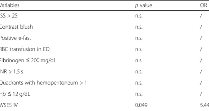
Table 4 Multivariate analysis of risk factors for OM at the arrival of patient at the ED Variables p value OR ISS > 25 n.s. / Contrast blush n.s. / Positive e-fast n.s. / RBC transfusion in ED n.s. / Fibrinogen≤ 200 mg/dL n.s. / INR > 1.5 s n.s. /
Quadrants with hemoperitoneum > 1 n.s. /
Hb≤ 12 g/dL n.s. /
WSES IV 0.049 5.44
ISS Injury Severity Score, CB contrast blush, ED emergency department, RBC red blood cell, SI shock index, AAST American Association for the Surgery of Trauma, Hb hemoglobin, WSES World Society of Emergency Surgery, INR International Normalized Ratio
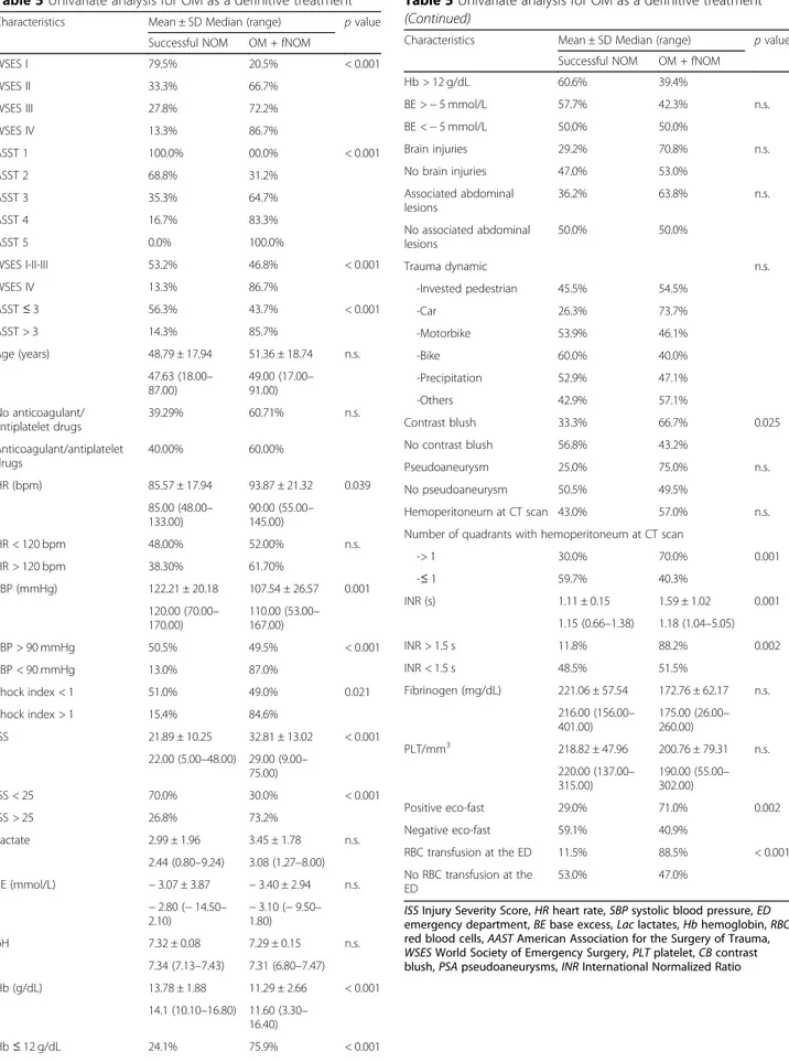
Table 5 Univariate analysis for OM as a definitive treatment
Characteristics Mean ± SD Median (range) p value Successful NOM OM + fNOM
WSES I 79.5% 20.5% < 0.001 WSES II 33.3% 66.7% WSES III 27.8% 72.2% WSES IV 13.3% 86.7% ASST 1 100.0% 00.0% < 0.001 ASST 2 68.8% 31.2% ASST 3 35.3% 64.7% ASST 4 16.7% 83.3% ASST 5 0.0% 100.0% WSES I-II-III 53.2% 46.8% < 0.001 WSES IV 13.3% 86.7% ASST≤ 3 56.3% 43.7% < 0.001 ASST > 3 14.3% 85.7% Age (years) 48.79 ± 17.94 51.36 ± 18.74 n.s. 47.63 (18.00– 87.00) 49.00 (17.00– 91.00) No anticoagulant/ antiplatelet drugs 39.29% 60.71% n.s. Anticoagulant/antiplatelet drugs 40.00% 60.00% HR (bpm) 85.57 ± 17.94 93.87 ± 21.32 0.039 85.00 (48.00– 133.00) 90.00 (55.00– 145.00) HR < 120 bpm 48.00% 52.00% n.s. HR > 120 bpm 38.30% 61.70% SBP (mmHg) 122.21 ± 20.18 107.54 ± 26.57 0.001 120.00 (70.00– 170.00) 110.00 (53.00– 167.00) SBP > 90 mmHg 50.5% 49.5% < 0.001 SBP < 90 mmHg 13.0% 87.0% Shock index < 1 51.0% 49.0% 0.021 Shock index > 1 15.4% 84.6% ISS 21.89 ± 10.25 32.81 ± 13.02 < 0.001 22.00 (5.00–48.00) 29.00 (9.00– 75.00) ISS < 25 70.0% 30.0% < 0.001 ISS > 25 26.8% 73.2% Lactate 2.99 ± 1.96 3.45 ± 1.78 n.s. 2.44 (0.80–9.24) 3.08 (1.27–8.00) BE (mmol/L) − 3.07 ± 3.87 − 3.40 ± 2.94 n.s. − 2.80 (− 14.50– 2.10) − 3.10 (− 9.50–1.80) pH 7.32 ± 0.08 7.29 ± 0.15 n.s. 7.34 (7.13–7.43) 7.31 (6.80–7.47) Hb (g/dL) 13.78 ± 1.88 11.29 ± 2.66 < 0.001 14.1 (10.10–16.80) 11.60 (3.30– 16.40) Hb≤ 12 g/dL 24.1% 75.9% < 0.001
Table 5 Univariate analysis for OM as a definitive treatment (Continued)
Characteristics Mean ± SD Median (range) p value Successful NOM OM + fNOM
Hb > 12 g/dL 60.6% 39.4% BE >− 5 mmol/L 57.7% 42.3% n.s. BE <− 5 mmol/L 50.0% 50.0% Brain injuries 29.2% 70.8% n.s. No brain injuries 47.0% 53.0% Associated abdominal lesions 36.2% 63.8% n.s. No associated abdominal lesions 50.0% 50.0% Trauma dynamic n.s. -Invested pedestrian 45.5% 54.5% -Car 26.3% 73.7% -Motorbike 53.9% 46.1% -Bike 60.0% 40.0% -Precipitation 52.9% 47.1% -Others 42.9% 57.1% Contrast blush 33.3% 66.7% 0.025 No contrast blush 56.8% 43.2% Pseudoaneurysm 25.0% 75.0% n.s. No pseudoaneurysm 50.5% 49.5% Hemoperitoneum at CT scan 43.0% 57.0% n.s. Number of quadrants with hemoperitoneum at CT scan
-> 1 30.0% 70.0% 0.001 -≤ 1 59.7% 40.3% INR (s) 1.11 ± 0.15 1.59 ± 1.02 0.001 1.15 (0.66–1.38) 1.18 (1.04–5.05) INR > 1.5 s 11.8% 88.2% 0.002 INR < 1.5 s 48.5% 51.5% Fibrinogen (mg/dL) 221.06 ± 57.54 172.76 ± 62.17 n.s. 216.00 (156.00– 401.00) 175.00 (26.00– 260.00) PLT/mm3 218.82 ± 47.96 200.76 ± 79.31 n.s. 220.00 (137.00– 315.00) 190.00 (55.00– 302.00) Positive eco-fast 29.0% 71.0% 0.002 Negative eco-fast 59.1% 40.9% RBC transfusion at the ED 11.5% 88.5% < 0.001 No RBC transfusion at the ED 53.0% 47.0%
ISS Injury Severity Score, HR heart rate, SBP systolic blood pressure, ED emergency department, BE base excess, Lac lactates, Hb hemoglobin, RBC red blood cells, AAST American Association for the Surgery of Trauma, WSES World Society of Emergency Surgery, PLT platelet, CB contrast blush, PSA pseudoaneurysms, INR International Normalized Ratio
Conclusions
The WSES classification is a good and reliable tool in
the decision-making process in splenic trauma
management. Abbreviations
AAST:American Association for the Surgery of Trauma;
AE: Angioembolization; AG: Angiography; ASA: American Society of Anesthesiologists; AVF: Arterovenous fistula; BE: Base excess; CB: Contrast blush; ED: Emergency department; fNOM: Failure of non-operative manage-ment; HR: Heart rate; INR: International normalized ration; LAC: Lactates;
NOM: Non-operative management; OIS: Organ Injury Severity Score; OM: Operative management; OR: Odds ratio; PSA: Pseudoaneurysm; RBC: Red blood cell; RR: Risk ratio; SBP: Systolic blood pressure; SI: Shock index; SNOM: Successful non-operative management; TC: Trauma center; WSES: World Society of Emergency Surgery
Acknowledgements Not applicable Authors’ contributions
FeCo, PF, LM, and MC contributed to the manuscript conception, literature revision, and analysis. LA, FaCa, SM, YK, and GLB helped with the analysis. Table 6 Multivariate analysis of risk factors for OM as a
definitive treatment Variables p value OR INR > 1.5 s n.s. / RBC transfusion in ED n.s. / Hb≤ 12 g/dL n.s. / ISS > 25 0.013 5.75 Contrast blush n.s. / Positive e-fast n.s. /
Quadrants with hemoperitoneum > 1 n.s. /
WSES IV 0.029 7.22
ISS Injury Severity Score, ED emergency department, RBC red blood cell, SI shock index, AAST American Association for the Surgery of Trauma, Hb hemoglobin, WSES World Society of Emergency Surgery, CB contrast blush, INR International Normalized Ratio
Fig. 2 OM and NOM rate at the arrival of patient according to WSES splenic injury grade (NOM, Non Operative Management; OM, Operative Management)
Fig. 3 OM and NOM rate as a definitive treatment according to the WSES splenic injury grade (SNOM, Successful Non Operative Management; OM, Operative Management; FNOM, Failure of Non Operative Management)
FeCo and PF drafted the paper that was critically reviewed by MS, SO, MT, NA, and SM. All the authors read and approved the final version of the manuscript.
Funding None
Availability of data and materials Not applicable
Ethics approval and consent to participate Not applicable
Consent for publication Not applicable Competing interests
The authors declare that they have no competing interests. Author details
1
General, Emergency and Trauma Surgery Department, Bufalini Hospital, Viale Ghirotti 268, 47521 Cesena, Italy.2General, Emergency and Trauma Surgery
Department, Papa Giovanni XXIII Hospital, Bergamo, Italy.3General and Emergency Surgery Department, Sant’Anna University Hospital, Ferrara, Italy.
4
Emergency and Trauma Surgery, Rambam Medical Centra, Haifa, Israel.
5General Surgery, Macerata Hospital, Macerata, Italy.6Department of Clinical
and Experimental Sciences, University of Brescia, Brescia, Italy.7Emergency Surgery Department, Parma University Hospital, Parma, Italy.
Received: 30 January 2019 Accepted: 22 May 2019
References
1. Moore EE, Cogbill TH, Jurkovich GJ, Shackford SR, Malangoni MA, Champion HR. Organ injury scaling: spleen and liver (1994 revision). J Trauma. 1995; 38(3):323–4.
2. Peitzman AB, Heil B, Rivera L, Federle MB, Harbrecht BG, Clancy KD, Croce M, Enderson BL, Morris JA, Shatz D, Meredith JW, Ochoa JB, Fakhry SM, Cushman JG, Minei JP, McCarthy M, Luchette FA, Townsend R, Tinkoff G, Block EF, Ross S, Frykberg ER, Bell RM. Blunt splenic injury in adults: multi-institutional study of the Eastern Association for the Surgery of Trauma. J Trauma. 2000;49(2):177–87.
3. Tinkoff G, Esposito TJ, Reed J, Kilgo P, Fildes J, Pasquale M, Meredith JW. American Association for the Surgery of Trauma Organ Injury Scale I: spleen, liver, and kidney, validation based on the National Trauma Data Bank. J Am Coll Surg. 2008;207(5):646–55.
4. Bhangu A, Nepogodiev D, Lal N, Bowley DM. Meta-analysis of predictive factors and outcomes for failure of non-operative management of blunt splenic trauma. Injury. 2012;43(9):1337–46.
5. Coccolini F, Montori G, Catena F, Kluger Y, Biffl W, Moore EE, et al. Splenic trauma: WSES classification and guidelines for adult and pediatric patients. World J Emerg Surg. 2017;12:40.
6. Haan JM, Bochicchio GV, Kramer N, et al. Nonoperative management of blunt splenic injury: a 5-year experience. J Trauma. 2005;58(3):492–8. 7. Smith HE, Biffl WL, Majercik SD, et al. Splenic artery embolization: have we
gone too far? J Trauma. 2006;61(3):541–4.
8. Gavant ML, Schurr M, Flick PA, Croce MA, Fabian TC, Gold RE. Predicting clinical outcome of nonsurgical management of blunt splenic injury: using CT to reveal abnormalities of splenic vasculature. AJR Am J Roentgenol. 1997;168(1):207–12.
9. Bhullar IS, Frykberg ER, Tepas JJ 3rd, et al. At first blush: absence of computed tomography contrast extravasation in grade IV or V adult blunt splenic trauma should not preclude angioembolization. J Trauma Acute Care Surg. 2013;74(1):105–11.
10. Skattum J, Naess PA, Eken T, et al. Refining the role of splenic angiographic embolization in high-grade splenic injuries. J Trauma Acute Care Surg. 2013; 74(1):100–3.
11. Requarth JA, D’Agostino RB Jr, Miller PR. Nonoperative management of adult blunt splenic injury with and without splenic artery embolotherapy: a meta-analysis. J Trauma. 2011;71(4):898–903.
12. Marmery H, Shanmuganathan K, Mirvis SE, Richard H, Sliker C, Miller LA, et al. Correlation of multidetector CT findings with splenic arteriography and surgery: prospective study in 392 patients. J Am Coll Surg. 2008;206:685–93. 13. Boscak AR, Shanmuganathan K, Mirvis SE, et al. Optimizing trauma
multidetector CT protocol for blunt splenic injury: need for arterial and portal venous phase scans. Radiology. 2013;268(1):79–88.
14. Schurr MJ, Fabian TC, Gavant M, Croce MA, Kudsk KA, Minard G, Woodman G, Pritchard FE. Management of blunt splenic trauma: computed tomographic contrast blush predicts failure of nonoperative management. J Trauma. 1995;39(3):507–12 discussion 512–3.
15. Shanmuganathan K, Mirvis SE, Boyd-Kranis R, Takada T, Scalea TM. Nonsurgical management of blunt splenic injury: use of CT criteria to select patients for splenic arteriography and potential endovascular therapy. Radiology. 2000;217(1):75–82.
16. Marmery H, Shanmuganathan K, Alexander MT, et al. Optimization of selection for nonoperative management of blunt splenic injury: comparison of MDCT grading systems. AJR Am J Roentgenol. 2007;189(6):1421–7. 17. Scarborough JE, Ingraham AM, Liepert AE, Jung HS, O’Rourke AP, Agarwal SK.
Nonoperative management is as effective as immediate splenectomy for adult patients with high-grade blunt splenic injury. J Am Coll Surg. 2016;223(2):249– 58.https://doi.org/10.1016/j.jamcollsurg.2016.03.043. Epub 2016 Apr 23. 18. Olthof DC, van der Vlies CH, Goslings JC. Evidence-based management and
controversies in blunt splenic trauma. Curr Trauma Rep. 2017;3:32–7.https:// doi.org/10.1007/s40719-017-0074-2.
19. Cadeddu M, Garnett A, Al-Aneni K, et al. Management of spleen injuries in the adult trauma population: a ten-year experience. Can J Surg. 2006;49:386–90. 20. Skattum J, Loekke RJV, Titze TL, Bechensteen AG, Aaberge IS, Osnes LT, et al.
Preserved function after angioembolisation of splenic injury in children and adolescents: a case control study. Injury. 2014;45:156–9.
Publisher’s Note
Springer Nature remains neutral with regard to jurisdictional claims in published maps and institutional affiliations.