GLAUCOMA
Histological findings of failed gold micro shunts in primary
open-angle glaucoma
Luca Agnifili&Ciro Costagliola&Michele Figus&Giovanna Iezzi&Adriano Piattelli&
Paolo Carpineto&Rodolfo Mastropasqua&Marco Nardi&Leonardo Mastropasqua
Received: 6 March 2011 / Revised: 5 July 2011 / Accepted: 28 July 2011 / Published online: 23 August 2011 # Springer-Verlag 2011
Abstract
Background To describe the histological features of failed gold micro shunts (GMS) in unsuccessful implantations for refractory primary open-angle glaucoma (POAG).
Methods This was an interventional case series study. Five eyes of five glaucomatous patients with unsuccessful GMS
implantation underwent shunt removal. Each device was sectioned into three portions: proximal or anterior chamber (AC) portion, middle or scleral portion and distal or suprachoroidal (SC) portion. The histological analysis was performed throughout the whole extent of the shunt, describing both the inner spaces and the outer surface. Results At the moment of removal all devices were correctly located into the SC space and in AC, with the exception of a case presenting corneal endothelial contact. The mean intra-ocular pressure before GMS removal was 30.4±5.3 mmHg, and the mean time of GMS removal after implantation was 6.8±2.5 months. No significant histolog-ical differences were documented among the five analyzed devices. The main feature was the presence of a thick connective capsule-like reaction surrounding both the proximal and distal ends and invading the posterior and anterior grid holes, whereas a more loosely arranged connective tissue was observed within the inner channels. Signs of surface fibrosis of the middle-scleral portion and inflammatory cell infiltration of the device were not documented in any of the cases.
Conclusions Failed GMS implantations presented connec-tive tissue filling all the inner spaces and creating a thick fibrotic capsule surrounding the ends of the device. This modification isolated the GMS from the AC and SC space, impeding aqueous flows throughout the shunt.
Keywords Gold micro shunt . Suprachoroidal space . Refractory glaucoma . Bleb-less surgery . Histology of failed GMS
Introduction
Currently, the most diffuse and reliable procedures for lowering intraocular pressure (IOP) in medically uncon-trolled glaucoma are filtration surgery and drainage device
None of the authors have a proprietary interest in the development or marketing of any of the products mentioned in this article. No funding or support has been received for the study.
There was no role of any sponsors or funders in the design and conduct of the study, in the collection, analysis, and interpretation of the data, and in the preparation, review, or approval of the manuscript. The authors have full control of all primary data, and they agree to
allow Graefe’s Archive for Clinical and Experimental Ophthalmology
to review their data if requested.
L. Agnifili (*)
Department of Medicine and Ageing Science, Ophthalmic Clinic, University of Chieti-Pescara,
Via dei Frentani, 114, 66100 Chieti, Italy e-mail: [email protected] C. Costagliola
Department for Health Sciences, Ophthalmic Clinic, University of Molise,
Campobasso, Italy
M. Figus
:
M. NardiDepartment of Neuroscience, Ophthalmology, University of Pisa, Pisa, Italy
G. Iezzi
:
A. PiattelliDepartment of Odontostomalogy Sciences, University of Chieti-Pescara,
Chieti, Italy
P. Carpineto
:
R. Mastropasqua:
L. MastropasquaDepartment of Medicine and Ageing Science, Ophthalmic Clinic, University G. d’Annunzio of Chieti-Pescara,
Via dei Vestini snc, 66100 Chieti, Italy
implantation [1–4]. Both work by creating a new drainage route allowing aqueous humor (AH) to drain from the anterior chamber (AC), thereby circumventing pathological outflow obstruction. The aqueous then flows into sub-conjunctiva leading to the formation of a filtering bleb [5,6].
However, both procedures can lead to significant early post-operative complications and long-term bleb encapsulation, the latter being widely recognized as the main cause of surgical failure and exhausting post-operative management [7,8].
In recent years, different surgical approaches which exploit alternative aqueous outflow pathways have been evaluated, with the aim of avoiding sub-conjunctival aqueous filtration and formation of filtering blebs.
One such procedure involves the use of gold micro shunt (GMS) devices, which are ultrathin 24-karat gold, biocom-patible flat plates containing micro-tubular channels that create a bridge between the AC and suprachoroidal (SC) space. As a result, the device forces the aqueous flow between the two compartments, taking advantage of the natural gradient of hydrostatic pressure between the AC and SC spaces [9]. Hence, AH is removed via the choroid vascular system or by gradually flowing through the scleral layers and then the conjunctiva. The final purpose of GMS implantation is to enhances uveo-scleral outflow without direct aqueous sub-conjunctival filtration and creation of a bleb.
To date, only two studies have evaluated the efficacy and safety of the GMS implantation and the possible mechanisms of action of the device. Melamed et al. [10] reported a 79% surgical success rate with a 32.6% reduction of post-operative IOP, without relevant post-operative complications and with-out the formation of filtering blebs in any of the patients. Mastropasqua et al. [11] reported a 57% surgical success rate, with a 22.6% reduction of IOP, also without relevant post-operative complications and the formation of filtering blebs. In the latter study, the authors also evaluated possible aqueous outflow pathways exploited by the shunt. The reported results suggested that trans-scleral aqueous perco-lation, in addition to the choriocapillaris resorption, may represent one of the main outflow routes lowering IOP. However, to date, no previous published reports have specifically investigated the mechanisms which can lead to the device failure. The aim of this study was to analyze histological features of failed GMS implantations.
Material and methods
This was an interventional case series study. The research adhered to the tenets of the Declaration of Helsinki, and informed consent was obtained from all patients prior to enrolment.
Our institutional review board approved the project. The study was carried out on five eyes of five Caucasian
patients with a previous diagnosis of refractory primary open-angle glaucoma (POAG) who were referred to the Ophthalmic Clinics of the Universities of Pisa and Chieti-Pescara, Italy (four females and one male, with an age range between 50 and 68 years, mean 61.2 ± 7.1 years). All patients had a history of two failed incisional procedures (MMC augmented trabeculectomy), followed by GMS implantation (SOLX, Boston, MA, USA) at least 1 year after the last trabeculectomy. In all eyes, the GMS was originally inserted in the superior-temporal region, with no significant intra- or early post-operative complications reported. A detailed description of the shunt and surgical implantation technique were previously reported [10].
At enrolment, all included eyes presented an uncontrolled glaucoma with IOP ranging from 22 to 36 mmHg under maximal tolerated medical therapy and were scheduled to undergo a new drainage device implantation after the GMS removal.
In all patients the gold shunt was correctly positioned, as verified by gonioscopy and ultrasound biomicroscopy (UBM, Humphrey 840 System, San Leandro, CA, USA) for AC and suprachoroidal portions respectively. In one case, a contact of the shunt with the corneal endothelium, with associated signs of moderate focal corneal oedema, was observed (UBM and slit-lamp features).
Even though the failure was not considered to be an indication for the device removal, the GMS was removed to avoid any intra- and/or post-operative mechanical interact between the gold shunt and the AC portion of the new drainage device; its equatorial plate was planned to be inserted in the superior-temporal region.
Microshunts were removed under local anaesthesia using a peribulbar injection. A bridle suture was placed around the superior rectus muscle, or alternatively a corneal traction suture was placed at 12 o’clock. A fornix-based conjunctival flap was fashioned by sub-conjunctival irriga-tion with lidocaine and forceps. The previous scleral incision used to insert the GMS was localized (2.5-3 mm posterior to the limbus) and splayed by a 45-degree knife, until the device appeared. The implant was first pushed towards the AC by using an insulin needle (or a bevel up in case of tight intra-tissutal shunt adherence) in order to expose the supra-choroidal part, and then gently removed by anchoring the posterior rounded edges. Finally, the sclera and the conjunctiva were tightly closed with 10–0 nylon and 7–0 Vicryl sutures respectively. The eyes were patched at the end of the procedure, and all patients were treated with dexamethasone–tobramycine fixed combina-tion eye drops (five times daily) and ciprofloxacin tablets 500 mg (twice daily); the topical therapy was tapered in 2 weeks, while the systemic therapy was stopped after 7 days.
Each removed GMS was fixed and stored in 10% neutral buffered formalin for 1 to 3 days before the histological analysis.
Histological processing and evaluation
The ground sections were obtained as described by Piattelli et al. [12] The specimens were dehydrated in an ascending series of alcohol rinses, and embedded in glycol methacry-late resin (Technovit 7200 VLC, Kulzer, Wehrheim, Germany). After polymerization, each shunt was sectioned widthwise (perpendicularly to the main axis of the shunt) using a high-precision diamond disk to about 150μm, and was ground down to about 30μm. Two slides stained with basic fuchsin and toluidine blue to study the presence and the features of the connective tissue, were obtained for each specimen.
For the histological analysis, devices were divided into three portions: a proximal portion represented by the anterior grid (AC end), a middle or scleral portion containing the channels, and a distal portion represented by the posterior grid with round edges (SC end). The histological evaluation was made throughout the whole length of the devices, both on the outer surface and into the channels and the grid of holes of both ends. The evaluation was performed using a light microscope (Laborlux S, Leitz, Wetzlar, Germany) connected to a high-resolution video camera (3CCD, JVC KY-F55B, Milan, Italy), and inter-faced to a monitor and personal computer. This optical system was associated with a digitizing pad (Matrix Vision GmbH, Milan, Italy) and a histometry software package with image-capture functionalities (Image-Pro Plus 4.5, Media Cybernetics Inc, Immagini & Computer Snc, Milan, Italy). The specimen sections and the subsequent
histolog-ical analysis were made by two different operators (GI and AP respectively).
Results
The mean time of GMS removal after implantation was 6.8± 2.5 months, with a mean IOP of 30.4±5.3 mmHg. The demographics and clinical characteristics of enrolled patients before the GMS removal are summarized in Table 1. The histological findings of the five removed GMS did not present significant differences, even though the case with corneal contact showed AC portion features enhanced, but similar, with respect to the other devices (Table2).
All analyzed shunts showed a marked connective colonization of the outer surface, inner channels and end grid holes. The distribution and the characteristics of fibrotic tissue throughout the shunt surface were not uniform in thickness, density, surface adherence and localization, whereas the inner fibrosis showed different density features between the middle portion and the AC and SC ends.
The shunt fibrosis was arbitrarily graded on the basis of the thickness and density of connective tissue, from mild to very intense.
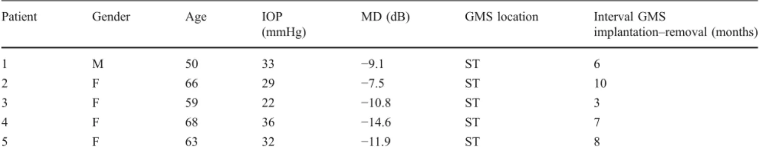
When analyzing the AC end, we did find a very thick, fibrous, and capsule-like tissue composed of several connective layers surrounding both faces of the grid (often asymmetrically) and the external edges (Fig.1a). The tissue tightly adhered to the outer surface and filled all grid holes (Fig. 1b, c); spindle cells, probably corresponding to fibroblasts, were documented between the connective layers of both the outer and inner portions of the fibrous tissue (Fig. 1b, c).
Table 1 Patients characteristics and surgical data
Patient Gender Age IOP
(mmHg)
MD (dB) GMS location Interval GMS
implantation–removal (months)
1 M 50 33 −9.1 ST 6 2 F 66 29 −7.5 ST 10 3 F 59 22 −10.8 ST 3 4 F 68 36 −14.6 ST 7 5 F 63 32 −11.9 ST 8 M = male F = female
IOP: intra-ocular pressure MD: mean defect dB: decibel
GMS: gold micro shunt ST: superior-temporal
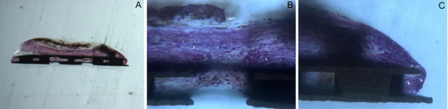
At the outer surface of the middle-scleral portion, the fibrotic tissue was absent or poorly represented, occasionally showing small areas of interrupted and weakly adherent thin connective layers; a capsule-like reaction was not documented in any case (Fig.2a). Conversely, the inner channels were filled with loosely arranged connective tissue partially or completely obstructing the lumen, occasionally showing spindle cells (Fig.2b).
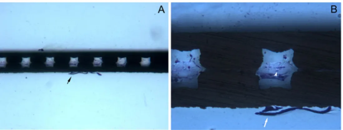
At the SC end of the devices, a thin and thickly arranged, poorly cellulated, and tightly adherent connective tissue was observed on both the outer surfaces of the posterior grid and the round edges (Fig.3a). Each hole of the grid was partially or completely filled with dense connective tissue, directly coming from the outer faces of the shunt (Fig. 3b), occasionally showing spindle cells (Fig. 3c). No signs of inflammatory cell infiltration were observed in any portion of the shunts.
Discussion
The interest for suprachoroidal devices in refractory glaucoma has increased after the initial results of Ozdamar, [13] who introduced the first successful suprachoroidal drainage technique to divert aqueous humor from the anterior chamber to suprachoroidal space. GMS represents one of the latest suprachoroidal drainage devices for refractory uncontrolled glaucoma, developed to reduce hypotony induced early post-operative complications and avoid bleb formation.
In the present study, we analyzed the histological findings of unsuccessful GMS,
Recent studies [10, 11] reported encouraging results concerning the safety and efficacy of GMS implantation, but with the exact mechanisms of IOP reduction only partially determined. Morphological evidence at the site of implantation provided by in vivo confocal microscopy, anterior segment
Table 2 Histological findings of different GMS portions
GMS: gold micro shunt AC: anterior chamber SC: suprachoroidal NA: not applicable
Patient Sector fibrosis AC portion Middle portion SC portion
1 Surface +++ ± ++ Grid holes ++ NA ++ Channels NA +++ NA Surface ++ + ++ 2 Grid holes +++ NA +++ Channels NA ++ NA Surface ++ ± +++ 3 Grid holes + NA ++ Channels NA +++ NA Surface +++ – ++ 4 Grid holes + NA +++ Channels NA ++ NA Surface +++ ± +++ 5 Grid holes ++ NA ++ Channels NA ++ NA
Fig. 1 Histological section of the proximal portion (anterior chamber) of a failed GMS. a Frontal section of the anterior GMS grid showing the presence of a very thick and tightly adherent incomplete connective capsule, evident only on the iris face and on the edges of the device. Arrow indicates a partial detachment of the capsule, which had most probably occurred during the staining process. A marked dense connective colonization was also evident within the holes of the
grid, directly in contact with the outer capsule. Acid fuchsin–toluidine
blue 40 ×. b, c Aspects at higher magnification of the outer surface, of the inner holes and of the edge of the device. Some scattered spindle cells appear between the connective layers (arrowheads). The capsule-like connective tissue presents partially detached at the superficial part
optical coherence tomography and ultrasound biomicroscopy supported the theory that AH, once reaching the supra-choroidal space, drains via the supra-choroidal vascular system or gradually permeates through the scleral layers, without the creation of a bleb. On the other hand, to date, the mechanisms which lead to long-term GMS failure have not been investigated, even if the results reported by Mastropasqua et al. [11] supposed the formation of a fibrotic capsule around the device or the occlusion of the inner channels by connective tissue as the cause in unsuccessful cases.
Consistently, our study did show the presence of connec-tive tissue either into the inner channels and both end grid holes or around the outer surface of the failed shunts.
The main features of our case series were connective tissue thickly arranged on the external surfaces and within the end grid holes, and more loosely structured into the inner spaces of the middle-scleral portion of the devices. Interestingly, the distribution and the characteristics of the fibrotic tissue
throughout the shunt surface were not uniform in thickness, density, surface adherence and localization.
The connective tissue was remarkably thick and densely arranged, forming a partial capsule-like structure at the AC end, while presenting thinner (but with a similar density) at the SC end forming a completely surrounding capsule. These features were not different among the analyzed devices, with the exception of the case presenting corneal contact, where we did find a very intense connective reaction of the AC portion, probably enhanced also by a local inflammatory stimulation. The surface adherence of the connective tissue at the AC and SC ends was tight and did not appear to differ, while the middle-scleral portion did not show evident signs of external fibrosis. One hypothesis to explain this feature could be the presence of a narrow space surrounding the middle portion (which lies and is tightly compressed between the deep scleral layers (70–80% of scleral depth)) which could prevent the external fibrotic reaction.
More-Fig. 2 Histological section of the middle portion. a Frontal section showing some of the inner channels partially or completely filled with a loosely arranged connective tissue. The outer surfaces did not show evidence of organized fibrotic reaction, except for the presence in small random areas of short traits of thin, loose, acellulated and poorly
adherent connective tissue (black arrow). Acid fuchsin–toluidine blue
40 ×. b. At higher magnification, each channel appear filled by a very loose connective tissue, containing some spindle cells (white arrow-head). White arrow indicates a short trait of thin, loosely arranged and
poorly adherent connective tissue. Acid fuchsin–toluidine blue 200 ×
Fig. 3 Histological section of the distal portion (suprachoroid). a Frontal section of the posterior GMS grid, showing the presence of a very tightly adherent capsule-like connective tissue on both faces and on the edges of the device. A partial yet dense connective colonization was also evident within the holes of the grid, directly in contact with
the superficial capsule. Acid fuchsin–toluidine blue 40 ×. b At higher
magnification, channels appear incompletely filled by connective tissue. c. Scattered spindle cells are evident within the capsule-like
over, the connective fibers probably adhered very poorly on the smooth and regular surface of the middle portion while presenting tightly anchored at the holed and irregular surfaces of the opposite end. Thus, we cannot rule out inadvertent tissue detachment on the middle portion during shunt removal.
With regard to the looser nature and the partiality of the inner channel fibrosis, one may suppose some modification induced by the fixation process (mainly for the partial aspect of the filling connective tissue), or a less intense fibroblast concentration and activity. This may be hypothet-ically due to a progressively reduced fibroblast migration into the channels when the end grid fibrosis became intense and obstructed the holes.
Two synergistic mechanisms may be considered as the potential causes leading to the fibrosis of failed shunts. First, a direct activation and proliferation of SC fibroblasts induced by the intra-operative suprachoroid manipulation or/and a possible migration of Tenon’s fibroblasts through the scleral incision. Moreover, the scleral incision probably permits a direct sub-conjunctival filtration in the early post-operative period with AH humor by-passing, at least partially, the shunt. This may facilitate the fibroblast colonization of the inner spaces of the device because of the absence of AH wash-out effect.
Portney et al. [14] reported fibroblastic reaction in the SC space after implantation of a cyclodialysis silicone implant. In a rabbit model of the suprachoroidal shunt [15], massive formation of scar tissue was observed as early as 2 weeks after surgery, with the histologic examination revealing fibrosis with similarities to proliferative vitreoretinopathy specimens. The authors suggested that scar tissue derives from fibroblasts transformed from retinal pigment epithelial cells of suprachoroid, as a consequence of the surgical trauma. Alternatively, fibroblasts reach the SC space via the lumen of the tube from the AC or enter the SC space via scleral pores and venous channels from epibulbar Tenon.
The second mechanism, which may be considered as synergistic to the first, is most probably linked to the molecular composition of the glaucomatous aqueous humor. AH in glaucoma is known to contain several cytokines which affect fibroblast activity, especially TGF-β2, whose AC levels
are very significantly increased with respect to healthy eyes [16,17]. It has been widely demonstrated that TGF-β2is one
of the most potent stimulator of human fibroblast and its activity is responsible for the post-surgery conjunctival bleb scarring [17]. Moreover, Cvenkel et al. [18] reported higher pre-operative AH levels of TNF-α and IL-6 in patients with unsuccessful trabeculectomy, and proposed that they may activate the conjunctival fibroblasts. Further studies [19,20] documented high AH levels of others pro-fibrogenic cytokines such as IL-1 or vascular endothelial growth factors (VEGF) in POAG patients. Also, the creation of a by-pass
from the AC to the SC space allows AH to be forcedly directed toward the shunt, conveying and storing at the site of implantation a great amount of pro-fibrotic agents with an hypothetical gradient from AC to SC. Even though we did not analyze the AH of patients before GMS removal, high levels of pro-fibrogenic cytokines were possible in our cases. On the basis of all these considerations, the thinner aspect of SC capsule with respect to AC capsule could been due to a fibroblast colonization and a connective reaction which began at the SC portion and continued through the whole shunt towards the AC. At this level, where the shunt has a wider free surrounding space, and where AH pro-fibrotic molecules were probably more concentrated, the fibrosis was more intense and the capsule appeared thicker. The relative thickness of the AC fibrosis could account for the partiality of the AC capsule described in all failed devices, since it could be a consequence of inadvertent tissue detachment during the removal and/or fixation processes.
Finally, also the properties and the modality of interaction of the gold alloy with living tissues cannot be ruled out as trigger stimuli for shunt fibrosis, even though no previous histological data were available for ocular tissue reaction to gold implantation, and though opposing evidence was reported for gold and extra-ocular tissue integration [21–23]. All these histological modifications isolated the shunt from the choroidal vascular bed and the sclera, impeding the aqueous outflow.
Because of the significant risk of GMS failure, especially in refractory glaucoma [43%, with a mean follow-up of 15.4 (5.4) months] [11], new drainage devices and bleb-less surgeries are required in clinical practice.
The iStent (Glaukos Corp., Laguna Hills, CA, USA) improved aqueous outflow by means of a patent channel created through the trabecular meshwork into Schlemm’s canal via ab-interno placement of the device. As recently reported [24,25], implantation of the iStent concomitant with cataract extraction significantly increased trabecular outflow facility, reduced IOP and the number of medications at 1 year.
New suprachoroidal drainage devices, such as the Transcend CyPass glaucoma implant, are under clinical evaluation with, to date, no results yet available for safety and efficacy.
Canaloplasty, a new non-penetrating bleb-less glaucoma surgery that combines the principles of viscocanalostomy with a circumferential distension of SC showed significant reduction in IOP, fewer post-operative glaucoma medications and limited surgical complications compared to trabeculectomy in glaucomatous patients with up to 3 years of follow-up. [26] The principal limitation of the present study is the small number of analyzed cases. The second limitation is that the cutting–grinding technique used to treat resin-embedded specimens provides a low resolution of the cells and blood vessels. Nevertheless, this is the only way to obtain in the
same slide both tissues and metallic implant and to analyze the interactions between the device and surrounding tissues. Moreover, with this technique it is possible to obtain only a small amount of histochemistry (e.g., alkaline and acid phosphatases) whereas it is not possible to perform immuno-histochemistry. Therefore, further investigations based on different ways of histological analysis such as electron microscopy are needed to verify our findings.
It would also be interesting to evaluate the effect of post-surgical treatment with topical non-steroidal anti-inflammatory drugs such as ibuprofen (other than steroids), which has the property to reduce aqueous humor level of cytokine and fibroblast proliferation, on the surgical success. [27]
In conclusion, the present study showed that the main cause of GMS failure in refractory uncontrolled glaucoma was an intense fibrosis of the inner spaces and outer surfaces of the shunt. Even though GMS implantation overcomes the prob-lems of the incisional surgery by avoiding bleb formation, it probably does not improve the final success of the glaucoma surgery. Further studies evaluating the long-term GMS efficacy are mandatory.
References
1. Jones E, Clarke J, Khaw PT (2005) Recent advances in
trabeculectomy technique. Curr Opin Ophthalmol 16:107–113
2. Hodkin MJ, Goldblatt WS, Burgoyne CF, Ball SF, Insler MS (1995) Early clinical experience with the Baerveldt implant in
complicated glaucomas. Am J Ophthalmol 120:32–40
3. Mermoud A, Salmon JF, Alexander P, Straker C, Murray AD (1993) Molteno tube implantation for neovascular glaucoma. Long-term results and factors influencing the outcome. Ophthalmology
100:897–902
4. Coleman AL, Hill R, Wilson MR, Choplin N, Kotas-Neumann R, Tam M, Bacharach J, Panek WC (1995) Initial clinical experience
with the Ahmed Glaucoma Valve implant. Am J Ophthalmol 120:23–31
5. Benedikt O (1977) The effects of filtering operations. Klin Monatsbl Augenheilkd 170:10
6. Teng CC, Chi HH, Katzin HM (1959) Histology and mechanism of filtering operations. Am J Ophthalmol 47:16–33
7. Kee C (2001) Prevention of early postoperative hypotony by partial ligation of silicone tube in Ahmed glaucoma valve
implantation. J Glaucoma 110:466–469
8. Feldman RM, Tabet RR (2008) Needle revision of filtering blebs.
J Glaucoma 17:594–600
9. Emi K, Pederson JE, Toris CB (1989) Hydrostatic pressure of the
suprachoroidal space. Invest Ophthalmol Vis Sci 30:233–238
10. Melamed S, Ben Simon GJ, Goldenfeld M, Simon G (2009) Efficacy and safety of gold micro shunt implantation to supra-ciliary space in patients with glaucoma: a pilot study. Arch
Ophthalmol 127:264–269
11. Mastropasqua L, Agnifili L, Ciancaglini M, Nubile M, Carpineto P, Fasanella V, Figus M, Lazzeri S, Nardi M (2010) In vivo analysis of conjunctiva in gold micro shunt implantation for
glaucoma. Br J Ophthalmol 94(12):1592–1596, Epub 2010 Aug 30
12. Piattelli A, Scarano A, Quaranta M (1997) High-precision, cost-effective system for producing thin sections of oral tissues containing dental implants. Biomaterials 18:577–579
13. Ozdamar A, Aras C, Karacorlu M (2003) Suprachoroidal seton implantation in refractory glaucoma: a novel surgical technique. J
Glaucoma 12:354–359
14. Portney GL (1973) Silicone elastomer implantation cyclodialysis:
a negative report. Arch Ophthalmol 89:10–12
15. Jordan JF, Engels BF, Dinslage S, Dietlein TS, Ayertey HD, Roters S, Esser P, Konen W, Krieglstein GK (2006) A novel approach to suprachoroidal drainage for the surgical treatment of intractable
glaucoma. J Glaucoma 15:200–205
16. Tripathi RC, Li J, Chan WA, Tripathi BJ (1994) Aqueous humor in
glaucomatous eyes contains an increased level of TGF-β2. Exp
Eye Res 59:723–728
17. Picht G, Welge-Luessen U, Grehn F, Lütjen-Drecoll E (2001) Transforming growth factor beta 2 levels in the aqueous humor in different types of glaucoma and the relation to filtering bleb development. Graefes Arch Clin Exp Ophthalmol 239(3):199–207 18. Cvenkel B, Kopitar AN, Ihan A (2010) Inflammatory molecules in aqueous humour and on ocular surface and glaucoma surgery outcome. Mediators Inflamm 2010:939602, Epub 2010 May 5 19. Cunliffe IA, Richardson PS, Rees RC, Rennie IG (1995) Effect of
TNF, IL-1, and IL-6 on the proliferation of human Tenon’s capsule
fibroblasts in tissue culture. Br J Ophthalmol 79(6):590–595
20. Hu DN, Ritch R, Liebmann J, Liu Y, Cheng B, Hu MS (2002) Vascular endothelial growth factor is increased in aqueous humor
of glaucomatous eyes. J Glaucoma 11(5):406–410
21. Welander M, Abrahamsson I, Berglundh T (2008) The mucosal barrier at implant abutment of different materials. Clin Oral Impl
Res 19:635–641
22. Abrahamsson I, Berglundh T, Glantz PO, Lindhe J (1998) The mucosa attachment at different abutments. An experimental study
indogs. J Clin Periodontol 25:721–727
23. Demann ET, Stein PS, Haubenreich JE (2005) Gold as an implant in medicine and dentistry. J Long Term Eff Med Implants 15 (6):687–698
24. Samuelson TW, Katz LJ, Wells JM, Duh YJ, Giamporcaro JE, US iStent Study Group (2011) Randomized evaluation of the trabecular micro-bypass stent with phacoemulsification in patients
with glaucoma and cataract. Ophthalmology 118(3):459–467,
Epub 2010 Sep 15
25. Fernández-Barrientos Y, García-Feijoó J, Martínez-de-la-Casa JM, Pablo LE, Fernández-Pérez C, García Sánchez J (2010) Fluoropho-tometric study of the effect of the glaukos trabecular microbypass stent on aqueous humor dynamics. Invest Ophthalmol Vis Sci 51
(7):3327–3332, Epub 2010 Mar 5
26. Grieshaber MC, Pienaar A, Olivier J, Stegmann R (2010) Canaloplasty for primary open-angle glaucoma: long-term outcome.
Br J Ophthalmol 94:1478–1482
27. Tilden ME, Boney RS, Goldenberg MM, Rosenbaum JT (1990) The effects of topical S(+)-ibuprofen on interleukin-1 induced
ocular inflammation in a rabbit model. J Ocul Pharmacol 6(2):131–