INTERNATIONAL JOURNAL OF IMMUNOPATHOLOGY AND PHARMACOLOGY Vol. 18, no. 3, 445-455 (2005)
CONSTITUTIVE AND STIMULATED PRODUCTION OF VEGF BY HUMAN
MEGAKARYOBLASTIC CELL LINES: EFFECT ON PROLIFERATION AND
SIGNALING PATHWAY
L. BONSI, L. PIERDOMENICO\ M. BISCARDP, C. MARCHIONNI, S. GAVAZZP,
V.
FOSSATI , B.GHINASSI ,F. ALVIANO , D. RONDELLP, M. FRANCHINA
4,
G.P. BAGNARA
1and A. GROSSI
DepartmentofHistology, Embryology and AppliedBiology, University ofBologna, Italy;
'Interdepartmental Centerfor CancerResearch
"G.Prodi" University ofBologna, Italy;
'Division ofHematology, University ofFlorence, Italy; 'DepartmentofHematology and Oncology,
University ofIllinois, Chicago.Il; USA; 'Dept. ofObstetrics and Gynecology, University ofBologna, Italy
Received November
11,
2004 - Accepted May
31,
2005
Release of vascular endothelial growth factor (VEGF) and other candidate angiogenic factors such as
basic fibroblast growth factor and transforming growth factor
B,
may playa role in sustaining neoplastic
cell proliferation and tumor growth. We evaluated VEGF expression and synthesis in the two
erythro-megakaryocytic cell lines B1647, HEL and one erythro-megakaryocytic cell line M07 expressing erythroid
markers. In this study RT-PCR was performed to evaluate VEGF expression and that of its receptor
KDR; VEGF production was assayed by Elisa test and western blot analysis; sensitivity to VEGF was
tested by thymidine incorporation. VEGF and its receptor KDR were expressed in B1647 and HEL, both
as mRNAs and as proteins, while only KDR transcript was found in M07 cells. Only B1647 and HEL
cells showed a strong spontaneous proliferating activity. In fact, measurable amounts of VEGF were
present in the unstimulated cell medium, thus suggesting an autocrine production ofVEGF by B1647 and
HEL cells, but not by M07, which was inhibited in mRNA-silencing conditions. This production could
not be further boosted by other growth factors, whereas it was inhibited by
TGF-~l.Finally, analysis of
She signal transduction proteins following stimulation with VEGF indicated that only p46 was tyrosine
phosphorylated. These data indicate that leukemic cells may be capable of autocrine production ofVEGF
which, in turn, maintains cell proliferation, possibly mediated by She p46 phosphorylation.
Vascular endothelial growth factor (VEGF), a potent inducer of angiogenesis and a stimulator of endothelial cell proliferation, differentiation and survival, is involved in the development of blood vessels during embryogenesis and promotes vascular permeability and migration of monocytes through endothelial tissue. Itis a homo dime ric glycoprotein with a structural similarity to Platelet-Derived Growth Factor (PDGF), and can appear in five isoforms, two bound to the cell surface or to the extracellular matrix (189 and 206) and three soluble (121, 145 and 165)
forms that induce proliferation of endothelial cells and
in vivo
angiogenesis (1). Production of VEGF isregulated by growth factors such as Fibroblast Growth Factor 4, PDGF, Tumor Necrosis Factor-a ( (TNF-a),
Transforming Growth Factor-B (TGF-~»,
Keratinocyte Growth Factor, Insuline Growth Factor, Interleukin I~,Interleukin 6, Thrombopoietin (TPO) and Oncostatin M (2). VEGF binds to tyrosine-kinase receptors identified as VEGFR-I (fms-like tyrosine kinase l-Fltl ), VEGFR-2 (Fetal liver kinaseI-Flkl/ Kinase Domain Insert Receptor-KDR), predominantly
Keywords: VEGF, human cell lines, VEGF-R2, cytokine production, She protein
Mailing address: Laura Bonsi, PhD
Department of Histology, Embryology and Applied Biology University of Bologna
Via Belmeloro, 8 - 40126 Bologna - Italy Ph: +39-051-2094100 - Fax: +39-051-2094110
E-mail: [email protected]
445
0394-6320 (2005)
Copyright© by BIOLIFE, s.a.s. This publication and/or article is for individual use only and may not be further reproduced without written permission from the copyright holder. Unauthorized reproduction may resulis in financial and other penalties
446
L. BONS) ET AL.expressed in endothelial cells, and VEGFR-3. Angiogenesis may also be implicated in the pathogenesis of solid (3) and hematologic malignancies (4), since VEGF mRNA has been found to be over-expressed in solid tumors (5), and a role, albeit indirect, has been proposed for it in sustaining the proliferation of leukemic cells (6-7).Ithas been shown that megakaryocytic cell lines and human megakaryocytes express VEGF transcripts, and that constitutive secretion of VEGF by CD41
+
cells is further increased by IL-3 and TPO (8-9).In this study, we tested the role of VEGF in sustaining leukemic cell growth, by evaluating the constitutive and stimulated synthesis of VEGF, as well as its role in modulating cell proliferation, in the Bl647 (10), HEL (11) and M07 (12) erythro-megakaryocytic cell lines. Lastly, the tyrosine phosphorylation of the signaling cascade She protein (a member of the c-Src family) was evaluated following stimulation with VEGF.
MATERIALS AND METHODS
eel/lines
The human erythro-megakaryocytic cell lines B1647
and HEL were maintained in Iscove's Modified
Dulbecco's Medium (lMDM) supplemented with 5%
human serum and 10% Fetal Calf Serum (FCS)
respectively (all medium constituents were purchased from BioWhittaker, Walkersville, MD, USA). The human megakaryocytic factor-dependent cell line M07 was maintained in IMDM supplemented with 5% FCS and 10 ng/mL IL3 (BioSurce, Camarillo, CA, USA). For starvation, cells were washed twice in PBS, resuspended in serum free medium (lMDM without serum or factors) and incubated for 24 hours.
To induce VEGF release, the starved cells were cultured in 24-well plates at a concentration of 1x10⁶ per
mL of serum free medium in the presence or absence of the following growth factors: Erythropoietin (Epo, Roche, Monza, Italy) 2U/mL, TPO (R&D systems, Minneapolis, MN, USA) 20 ng/mL, fms-like tyrosine kinase 3 (Flt3, ICN Biomedica1s, CA, USA) 100 ng/mL (alone and in combination with TPO), Hepatocyte Growth Factor (HGF,
ICN Biomedica1s, CA,USA) 20 ng/mL, TGF-~1 (R&D
systems, Minneapolis, MN, USA) 5 ng/mL, and incubated for 48 hours at 37°C, 5% C02 and 95% humidified air.
At the end of incubation supernatants were recovered by centrifugation at 1200 rpm for 10 min and frozen for Elisa analysis. Supernatants from non stimulated cells
were concentrated five times by Amicon concentrator filter
(Millipore Corporation, Bedford, MA, USA) and
immunoprecipitated with an antiserum recognizing VEGF (Sigma, Saint Louis, MO, USA) so that the protein released could also be detected by western blot analysis.
Immunoprecipitation and western blot analysis
Starved cells (10 x106
) were incubated with or without
VEGF (Sigma) 150 ng/mL at 37°C for 5, 10, 15,30 min, extracted by cold lysis buffer (50mM TrisHCI pH 7.5, 1mM EDTApH 8 and 150mM NaCI) plus1% Triton X"100 and a
mixture of protease inhibitors (1 mmol/liter
phenylmethylsulfonyl fluoride, 10 ug/ml, leupeptin, 0.15 units/mL aprotinin, 1 ug/ml, pepstatin A) for 20minat 4°C, and centrifuged at 13,000 g for 20 min ( all electrophoresis reagents were purchased from Sigma). The supernatant protein concentration was determined by Bradford assay (13). The clarified supernatant (protein concentration 500 ug) was pre-cleaned for 1 h with 50 ul. ofSepharose protein A (3 mg/sample). The pre-cleaned celllysates were then adsorbed by antisera (anti-KDR- Sigma, anti-VEGF Sigma, and anti-She Santa Cruz, California, USA) coupled to protein A-Sepharose (Sigma). Bound proteins were washed several times in lysis buffer and eluted in boiling Laemmli buffer. Eluted proteins were subjected to 6% (for KDR detection), 10% (for She detection) and 12% (for VEGF
detection) SDS-polyacrylamide gel electrophoresis
according to the protein molecular weight. Proteins were then transferred electrophoretically to nitrocellulose (HYBOND, Amersham, Buckinghamshire, England). In order to block non-specific binding, the membranes were incubated in blocking solution (5% low fat milk in 20 mM Tris-HCI,pH 7.4, and 150 mM NaCl-TBS and 0.1% Tween 20) for 1h. Antisera (anti-KDR- Chemicon- Temecula, Ca 92590 USA, anti-VEGF, anti pTyr and anti-She Santa Cruz, California, USA) were then added at the same solution, and the incubation was carried out overnight at 4°C. For detection, the filters were washed four times (15 min each wash) with TBS 0.1% Tween 20 and allowed to react for 1h at room temperature with peroxidase-conjugated secondary antibody. The enzyme was removed by washing as above.
The filters were reacted for 5 min with a
chemiluminescence reagent (SuperSignal Substrate, Pierce, Rockford, Illinois, USA) and exposed to an autoradiography film for 1-15 min (BIOMAX, Eastman Kodak, Rochester, NY, USA). In order to re-probe, nitrocellulose filters were first stripped of antibody by 62 mM Tris-HCI, pH 6.7, 2%
SDS, 100 mM~-mercaptoethanol,washed and probed with
another antibody (14).
ELISA Test
Enzyme linked immuno-sorbent assay (ELISA;
Int. J. Immunopathol. Pharmacol.
447
for VEGF concentration in M07, B1647 and HEL cell supernatants were performed following the manufacturer's instructions. The lower limit of detection was less than 5 pg/mL. Samples were analysed in duplicate and the results are a mean of three experiments.
Apoptosis
Apoptosis was evaluated from necrotic HEL, B1647 and M07 cells with an Annexin-V-FLUOS Staining Kit (Roche Diagnostics, Germany). Cells were treated with
TGF-~1 (5 ng/mL) for 48 hours and then collected
washed and incubated for 15 min with Annexin-
V~
fluorescein. Propidium iodide was added to differentiate apoptotic from necrotic cells. Samples were analyzed on a flow cytometer using a 488 nm excitation.
RT-PCR
Total cellular RNA was prepared by the TriPure
Isolation reagent method. First strand cDNA was
synthesized with 1 ug of total RNA in a reverse buffer containing 100 pmol of oligo dT, dNTPs 0.25 mM, DTT 10 mM, 20 U ofRnase Inhibitor and 50 U ofM-MuLV RT. The mixture was incubated for 1 hour at 42°C, heated for 5 minutes at 94 °C and stored at -20°C until used. PCR amplifications were carried out in a thermal cycler with cDNA derived from 200 ng of total RNA, reaction buffer lOx, MgCI2 2mM, dNTP O.2mM, primers 0.2 j.!M and 1.25 U of Taq DNA polymerase. Primers were designed and
synthesized for VEGF (sense 5'
-CGAAGTGGTGAAGTTCATGGATG-3'; antisense 5'-TTCTGTATCAGTCTTTCCTGGTGAG-3') and KDR (sense 5'-AAAACCTTTTGTTGCTTTTTGA-3'; antisense 5'-GAAATGGGATTGGTAAGGATGA-3'). Each cycle included a denaturation step (95°C for 1 min), an annealing step (55°C for 1 min) and an extension step (72 °C for 1 min). The PCR products were visualized on a 2% agarose gel stained by ethidium bromide.
Sensitivity to VEGF
Cells were washed twice in PBS, re-suspended in serum free medium and incubated for 24 hours. Cell cultures were performed in serum free medium (lMDM) in triplicate, in 96-well microplates at a concentration of 50 x 103 per 200 ul, per well in the presence or absence ofVEGF at different concentrations (10, 50, 100, 150 ng/mL). Antibody against VEGF (Biodesign, Saco, ME) (2 ug/ml.) was added to each VEGF concentration in the B1647 and HEL experiments alone.
After 24 and 48 hours' culture the cells were labeled with [methyl-3H] thymidine (lCN Biomedicals, CA,USA) at ImCi/well for 4 hours, and were assayed for incorporated radioactivity using a multiple cell harvester (Skatron, Oslo, Norway) and a Beta-counter (LKB, Uppsala, Sweden). In
order to confirm the possible autocrine VEGF production, an RNA silencing interference mechanism was used.
Preparation ofsiRNA duplexes
The targeted region was selected from a given cDNA sequence beginning 50 to 100 nucleotides downstream of the start codon. To design target-specific siRNA duplexes, we selected sequences of type AA(N 19)TT changing the last two nucleotides of the siRNA sense to dTdT (sense siRNA:5' -ccaugaacuuucugcugucdTdT-3', antisense siRNA:
5' -gacagcagaaaguucauggdTdT-3 '). A selected siRNA
sequence also underwent a BLAST search against the human genome sequence to ensure that only one gene ofthe human genome was targeted. The siRNA duplex was chemically synthesized by Proligo - Genset Oligos (Hamburg, Germany) (15).
Silencing ofthe expression of VEGF gene by siRNA
Human cell lines HEL and B1647 were grown at 37°C in IMDM supplementedwith 10% FCS for HEL and 5% human serum for B1647. The day before transfection cells were diluted with fresh medium at a concentration of lxl 06/Inl.
On the day of transfection the cells were transferred to a 24-well plate at a concentration of2.5 x lO'zmlin 200 ul of medium per well, without antibiotics (16). Exogenous delivery of siRNA duplexes to B 1647 and HEL cell lines
was carried out with the transfection reagent
Oligofectamine'" Reagent (Invitrogen, life technologies). For each well 2.5 III (20 IlM) siRNA duplex was mixed with 40 III of Opti-MEM I. In a separate tube 1 III of Oligofectamine" Reagent was added to 6.5 ul of OptiMEM I, for each well, gently mixed and incubated for 7 -10 minutes at room temperature. The final solutions were prepared, mixing them gently by inversion and incubating for 20 - 25 minutes at room temperature to allow for formation of liposome complexes. Cells were overlaid with 50 III of complexes for each well, incubated for 4 hours at 37°C in a C02 incubator, and 125 III growth medium was finally added. After 24, 48, and 72 hours the cells were stained with trypan blue and counted in the light microscope.
RESULTS
VEGF R-2 (KDR) and VEGF mRNA expression
and protein
Our first approach to analysing KDR and VEGF
in the cell lines was by RT-PCR. We observed that
Bl647 and REL cells expressed mRNAs of both
KDR and VEGF, while the M07 cell line expressed
KDR- but not VEGF mRNA (Fig. I). For evaluation
448
L.HOlliSIET AL.Fig. 1.Expressionof KDR and VEGF mRNA expression in M07, HEL and B1647 cells by RT-PCR. A faint band corre spondingto KDRexpressionisvisible in M07whilethe same cells do not expressVEGF mRNA.KDR and its ligand
VEGF areexpressedin B1647 and HELcell lines.
Lane1:molecular weightmarker MWM XlV;Lane2:M07 cell line;Lane3:HELcell line; Lane4:B1647cellline,Lane 5: Positive controlfor KDR: bonemarrowmononuclear cel/s. Positivecontrolfor VEGF: HepG2 (Human hepatocyt e cell line),Lane6:negative control: no mRNA;Lane7:molecular weightmarker MWMIX.
Fig. 2. KDR protein exp ress ion in the three cell lines studied.M07 cellsdo not express KDR protein despitethe weak exp ression ofKDR transcript. The evidenceof KDR in B1647and HELcellsisconfirmedby KDRexpression in Human Umbilical Vein EndothelialCells (H UVE Cs) usedas a positive control. An ep ithe lial cell line (5637)
and a lymphoblastic cell line (Ju rkat) }vere us ed as negative controls.
Lane I :Positive control (HUVECelilysate); Lanes2-3: Negativecontrols -lane2 (5637cell lysate) lane3(Jurkat cell lysate), Lane4:B1647 cell lysate;Lane5:HEL cell lysate;Lane6:M07 celllysate
Sampleswere subjectto immunoprecipitationand blotting with thesame antibody.
6
5
4
3
2
1
KDR
-KDR -239 bp VEGF -607 / -535 bp -403 bp 7--
----
.-. 2 3 4 5of KDR protein, cell Iysates were immunoprecipitated with an anti-KDR antibody and immunoblotted with the same antibody. It emerged that B 1647 and HEL cells expressed a 210 kDa protein consistent with the presence of KOR. By contrast, the immunoprecipitation ofM071ysates did not contain the protein, despite expressing KOR transcript (Fig.2).
VEGF protein analysis was carried out on supernatants from starved (48 hours)B1647 and HEL cells by Western blot analysis.A 21 kOa band was evident on the nitrocellulose filters, corresponding to VEGF (Fig.3). The identity of the band was confirmed by recombinant VEGF 165, as a positive control. Another band at 18 kDa was observed, possibly corresponding to non-glycosylated VEGF 165.The corresponding medium ofM07 cells was not tested for the presence of protein, as it did not expressVEGF transcript.
VEGF production
VEGF production was tested by Elisa assay in cell supernatants both in basal condition and following stimulation (for 48 hours) with Epo, TPO, Flt-3 (alone and in combination with TPO), HGF and
TGF-~1 (Fig. 4).B 1647 and HEL cells produced 900 pg/mLand 500 pg/mL, respectively. No significant changes were seen in the presenceof Epo, TPO, Flt-3, TPO and Flt-3 or HGF, while TGF-~1 caused a significant decrease in VEGF concentration in both
cell lines (p=0.03). This result could not be ascribed to a reduction in cell number, which remained unmodified over the time of incubation, while the analysis of apoptotic cell death by annexin V and propidium iodide staining revealed no difference between TGF ~1 treated and untreated cells (HEL untreated cells: 3.3% and TGF ~1 treated: 3.4%,
B 1647 untreated cells: 1.43% and TGF ~1 treated: 1.91%).
Sensitivityto VEGFs
To evaluate the proliferation effect of VEGF,
B1647, HEL, and M07 cells were maintained in serum free conditions in the presence of various concentrations of VEGF (l0, 50, 100, 150 ng/mL)
for 24 and 48 hours' culture, after which a thymidine incorporation assay was performed.
No response was detected in M07 proliferation after stimulation with different concentrations of VEGF (Fig.5A).A dose-dependent proliferation of B 1647 cells was evident as early as 24 hours after incubation with VEGF (Figs 5B and 5C). In HEL cell experiments no significant differences were detected between treated and untreated cells after 24 hours of culture (Fig. 50), while a plateau was reached at 50 ng/mL after 48 hours (Fig. 5E). In both cell lines antibody to VEGF decreased the proliferation to control values at 24 and 48 hours of incubation (Figs 5B, 5C and 5E). In B1647 cell proliferation induced by VEGF at 150 ng/mLcould
lot. J. Immuoopathol. Pharmaco l.
449
+42 kDa
+21
. 18
2 3
Fig. 3. Western blot analysisofconditioned medium (CM) from the erythromegakaryocytic cell lines. Twenty-four hours before incubation, thecells were starved ofserum to
exclude exogenousprotein.Supernatantswereconcentrated five timesin order to betterdetectthe protein by western blot technique.Lane1:B1647 CM Lane2:HEL CM Lane 3
:rh VEGF165(100ng).
Sampleswere subject to immunoprecipitation and blotting with thesame antibody.
not be neutralized completely by anti-VEGF
antibody
.
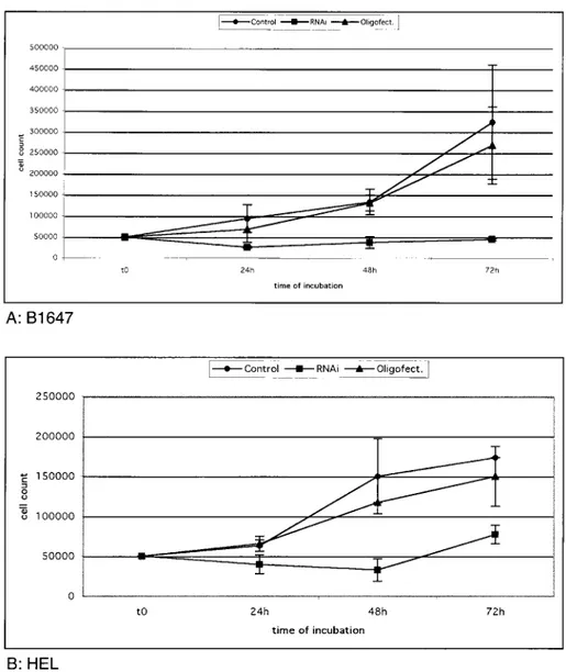
Small interfering RNAs
Specific VEGF gene silencing was documented
by counting the cells stained with try
pan
bl
ue
after
24
,
48 and 72 hours of incubation (Figs 6A and 6B)
.
VEGF RNA
i
s able to silence the gene
t
ranscription
and b
lock B
1647 and BEL cell growth
.
As
oligofectamine is a potent transfection vector, in
order
to
introduce
siRNA
into
the
cells
oligofectamine contro
l
was necessary, in case it
should
h
ave a toxic effect. In both ce
ll
lines the serum
free and the oligofectamine contro
l
curves were
simila
r
(Figs 6A-6B)
.
D
espite silencing conditions
an increase in the BEL cell count was observed after
48 hours, very likely due to the presence of
untransfected cells in the culture
.
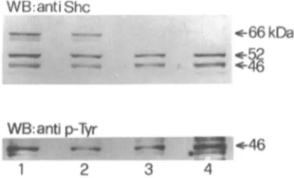
Shephosphorila tion
We
also
evaluated
She
a
daptor
protein
phosphory
lation
upon KD
R
activat
ion
w
ith
VEGF in
BEL and
B1
647. In mamm
alian
cells S
he
is present
in three isoforms P66
,
P52 and P46
,
produced by
alternative translation
.
B
y immunoprecipitation of
B1647 and BEL cells with an antibody reactive to
She p66, p52
,
p46 we found that the
B 1
647 cell line
expressed only p52shc and p46shc proteins (Fig
.
7
,
lanes 3 and 4), whereas BEL cells expressed all three
isoform proteins (Fig
.
7
,
lanes 1 an
d
2).
B
y
r
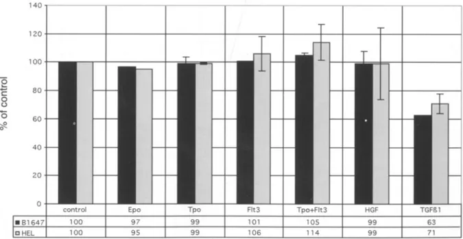
e-VEGFstim ul at ed prod uc ti on 14 0 120 100 (5 "-80 C 0 o
'0
60 ;,g 0 40 20 0control Epo Tpo Flt3 Tpo+Flt3 HGF
. 81647 100 97 99 101 105 99
[]HEL 100 95 99 10 6 114 99 71
Fig. 4. VEGF production insupe rnatants from stimulated and unstimulatedB1647and HELcells(ELISA test)after48
hoursofincubation .Data are exp ressed as a percentageofthecontrol(sup ernatantfrom unstimulatedcells)and represent
the mean ofthreeexperim ents.No factors were ableto induce VEGF production in the twocell lines. TGF~1caused a significantdecreasein VEGFsynthes is in both celllines (p=0.03).
450
L. BONSI ET AL.j _ _Control ____ RNAi~-Oli9;f~ct] 500000 450000 400000 350000 300000 . / 250000
/
...
200000.c-:
1SOOOO T~ 100000 T-1
---=
"1 _ _ 50000-
= 0 to 24h 48h 72h time of incubation A: M07 VEGF 24 e 48h~ntrol _RNAi - . - oligofect.] 250000 200000 ~ 150000
----<::~
~
::J a c "ii 100000 u.>:
.---I
50000-'-
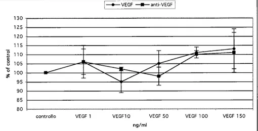
---I I 0 to 24h 48h 72h time of incubation B: B1647 VEGF 24h !_VEGF _anti-VEGFI 450 400 350 300g
..----t: 250. /
a c '0 200 V/ '
~ 150 ~ -r---
T ~-±_______Jo
F=- V
100 = l. SO 0controllo VEGF 1 VEGFlO VEGF 50 VEGF 100 VEGF 150
ng/ml
Int. J. ImmunopathoI. PharmacoI.
E
VE.GF _anti-VEGf] 130 125 120 115]
110 T---~
c: a 105 (J----
"'-...~~. /
/
'0 100 -.f!.<,
. /
...,
95 90 85 80cantralla VEGF 1 VEGF10 VEGF 50 VEGF 100 VEGF 150
ng/ml D: HEL VEGF 24h FVEGF=--anti-VEGFI
451
180 160 140]
120 c: 100 a (J '0 80 -.f!. 60 40 20 0-
---
.---
---cantralla E: HEL VEGF 48h VEGF 1 VEGF10 ng/ml
VEGF 50 VEGF 100 VEGF 150
Fig. 5. Effect ofdifferent VEGF concentrations on M07 (panel A), B1647 (panels B-C), and HEL (panels D-E) cells. After24and48hours 'incubation the proliferation was evaluated as 3H-TdR incorporation and measured as counts per min (cpm) expressed as a % of the control. Compared to M07 cells, 50 ng/mL (p=O. 02) and 10 ng/mL (p=0.006) was sufficient to increase B1647 cell proliferation after24and48hours respectively; 50 ng/mL was needed to stimulate HEL cells significantly after48hours (p=0.003).
Data represent a mean ofthree experiments
+
SEM.probing the same nitrocellulose membrane with an
antibody reactive to phosphotyrosine-containing
proteins, in both cell lines only p46shc proved to be
phosphorylated following stimulation with VEGF at
a concentration of 100 ng/mL for 5 min. However no
difference was detected between stimulated and
unstimulated cells, a finding that is most likely due to
autocrine VEGF production in these cell lines,
despite 24 hour cell starvation.
DISCUSSION
In normal hematopoiesis VEGF is produced by
human megakaryocytes and released by these
precursor cells and platelets (8, 17). Our study
indicates that leukemic cell lines of the
erythro-megakarycocytic phenotype, such as HEL and B1647,
also constitutively produce VEGF and express KDR.
This is not the case in all instances, a fact shown by the
lack ofVEGF mRNA and KDR protein in M07 cells.
452
L. BONSI ET AL.The same VEGF-R isofonn was first demonstrated in HEL cells (18), but no data was previously available in other cell lines.
In a previous study we demonstrated that proliferation of B 1647 cells was not modulated by hemopoietic cytokines, while in the presence of TPO the transcripts of specific erythroid proteins, such as~
andyglobins increased (l0). In that work we observed that only B 1647 and HEL proliferated in the presence ofVEGF in the culture medium and while a maximum stimulating dose was not found in B1647, a plateau in the proliferation curve of HEL cells was observed at
the concentration of 50 nglmL VEGF. These results are in agreement with the observation that only these two cell lines, but notM07,actually produce KDR protein. The suggestion that VEGF may have a role in sustaining leukemic cell growth is further reinforced by the observation that proliferation was inhibited by the antibody to VEGF in thymidine incorporation and the number of cells was reduced in the RNA interference experiments because of the block on autocrine production. In our setting this last method (19-20) stressed the role ofVEGF gene in regulating B 1647and HEL cell growth.
/ '
/ . /
»:-:
,~ T-r
_ _ I . -= 1---50000 100000 150000 §300000 8250000 500000 ] 450000 350000 400000 0; o 200000 to 24h 48h 72h time of incubation A:B1647 250000 200000 ~ 150000 c:"
0 o 0:; 100000 o 50000 0 to B:HEL 24h 48h time of incubation 72hFig.6. Silencing of VEGF expression in B1647 (panel A) and in HEL (panel B) cell lines. After 24, 48, 72 hours from
transfection with2.5f!l (20 f!M) VEGF siRNA duplexes, trypan blue stained cells were counted.
The growth relative to the cells treated with the transfection agent (Oligofectamine) isreported. Bars represent the means ofthree experiments+SEM.
lot.J. ImmuoopathoI. PharmacoI.
453
Fig. 7. Involvement of She proteins in the KDR signal
pathway in HEL (1-2) and B1647 (3-4) cell lines. Lysates were immunoprecipitated with anti She protein and immunoblotted with the same antibody (upper panel) while She phophorylation was detected after stripping (lower panel). Lanes 1 and 3: immunoprecipitates from
unstimulated cells; Lanes 2 and 4: immunoprecipitates
from cells stimulated with 100 ng/mL VEGFfor 5 min. WB: western blotting.
The possible
role
of VEGF
in
leukemic
proliferation
is
consistent
with
three
more
observations: a) myeloblast and immature myeloid
cells, but not erythroid precursors and lymphoid cells,
co-express VEGF and its receptor (21); b) antiVEGF
antibodies promote the formation of CFU-GEMM
and BFU-E colonies in methy1cellulose cultures of
bone
marrow
cells
from
patients
with
myelodysplastic syndromes, by inhibiting TNFa and
IL1P production from BM mononuclear and stromal
cells (21); c) anti leukemic activity by agents such as
AS203 (7) inhibits VEGF production by leukemic
cells and induces endothelial cell apoptosis, thereby
reducing the release of leukemic cell stimulating
cytokines, including VEGF.
In
our
experimental
conditions
cytokine
stimulation failed to increase VEGF protein
secretion by B1647 and HEL cells, while M07
cells did not produce VEGF, which is consistent
with the lack of the transcript. This is in contrast
with the observation of Bobik et al. that TPO
caused a time-and dose-dependent increase in the
levels of VEGF released by the megakaryocytic
cell line CMK, and the c-mpl expressing UT7 cells.
On the other hand, our data indicate that TGF-P, a
multifunctional polypeptide that regulates the
proliferation and differentiation of various types of
cells, has a strong and direct inhibitory effect on the
synthesis ofVEGF. In fact the evident inhibition of
WB
:antiShc
-
-::~
+66kDa+46
thymidine incorporation in the presence of TGF-p
observed in B 1647 and HEL cells did not
correspond to a decrease in the number of cells in
culture at the time of supernatant collection (data
not shown). This is in contrast with the suggestion
by Pertovaara et al. that the angiogenic effect of
TGF-p 1 on endothelial cells is partly mediated by
a paracrine induction ofVEGF in other surrounding
cell types (22) as well as with the alleged TGF-p
driven increase in VEGF expression by osteoblasts
and osteoblast-like cells (23-24). Western blot
analysis of VEGF production in B1647 and HEL
supernatants revealed bands at different molecular
weight besides the 21 kDa band corresponding to
rhVEGF 165 : 42 kDa, 37 kDa, 30 kDa and 18 kDa.
As also reported by Mohle et al, 18 kDa band
corresponds to non-glycolysated VEGFl65 whereas
30-37 kDa bands were found by other authors in rat
pituitary tumor cell line GH3 (25). VEGF mediates
its pleiotropic action by binding to KDR with an
intrinsic protein tyrosine kinase activity. The first
step in signal activation is dimerization of receptor
KDR after binding with VEGF and this process is
followed by tyrosine autophosphorylation of kinase
domain and cytoplasmic protein "target". We
studied She, one of these target proteins called
"adaptors" because of their capacity for interacting
with proteins of a multimolecular complex (26).
Three different isoforms of She, 46 kDa, 52 kDa
and 66 kDa, are known: the 46 and 52 kDa isoforms
are expressed ubiquitously, whereas the 66 kDa
isoform is predominantly found in cells of epithelial
origin (27-28). In our experiments only p46 was
phosphorylated after the binding ofVEGF with its
receptor KDR although B1647 cells also expressed
p52 and HEL cells expressed both p52 and p66.
Itcould be of interest to investigate the role of p52
and p66 in the VEGF induced proliferation of the
erythro-megakaryoblastic cell lines, because p52
and p66, but not p46, have been found to be
associated with activation EGFR-Src complex in the
human carcinoma A431 cell line (29).
In
conclusion,
our
findings
support
the
relationship between angiogenesis and leukemic
growth: a) VEGF is produced by some of the
leukemic cell lines, and TGF-p
is the only
modulator ofVEGF synthesis; b) VEGF regulates
cell proliferation that is inhibited by antibodies to
454
L. BONSI ET AL.VEGF; c) VEGF
binding to KDR transduces a
proliferative signal through the phosphorylation of
p46
shC•
ACKNOWLEDGMENTS
This work was supported by grants from the
Italian Ministry for University and Research:
MURST ex 60% and 40%. We thank Dr Ralph
Nisbet for checking the manuscript.
REFERENCES
1. Neufeld G., T. Cohen, S. Gengrinovitch and Z.
Poltorak.1999. Vascular endothelial growth factor (VEGF) and its receptors. FASEB J. 13:9.
2. Hermanns H.M., S. Radtke, F. Schaper, P.C.
Heinrich andI. Behrmann. 2000. Non-redundant
signal transduction of interleukin-6-type cytokines.J. BioI. Chern. 275:40742.
3. Kerbel R. 2000. Tumor angiogenesis: past, present
and the near future. Carcinogenesis 21:505.
4. KiniA.R., L.A. Peterson, M.S. Tallman and M.W.
Lingen. 2001. Angiogenesis in acute promyelocytic leukemia: induction by vascular endothelial growth factor and inhibition by all-trans retinoic acid.Blood
97:3919.
5. Paley P.J., K.A. Staskus, K. Gebhard, D.
Mohanraj, L.B. Twiggs, L.F. Carson and S. Ramakrishnan. 1997. Vascular endothelial growth factor expression in early stage ovarian carcinoma.
Cancer 80:98.
6. Dias S., Z. Hattori, K. Zhu, B. Heissig, M. Choy,
W. Lane, Y. Wu, A. Chadburn, E. Hyjek, M. Gill, D.J. Hicklin, L. Witte, M.A. Moore and S. Rafii. 2000. Autocrine stimulation of VEGFR-2 activates human leukemic cell growth and migration. J. Clin. Invest. 106:511.
7. Roboz G.J., S. Dias, G. Lam, W.J. Lane, S.L.
Soignet, R.P. jr Warrell and S. Rafii. 2000. Arsenic trioxide induces dose- and time-dependent apoptosis of endothelium and may exert an antileukemic effect via inhibition of angiogenesis. Blood 96:1525.
8. MohleR,D. Green, M.A.S. Moore, R.L. Nachman
and S. Rafii. 1997. Constitutive production and thrombin-induced release of vascular endothelial
growth factor by human megakaryocytes and
platelets.PNAS 94:663.
9. BobikR,Y. Hong, G. Breier, J.F. Martin and J.D.
Erusalimsky. 1998. Thrombopoietin stimulates
VEGF release from c-Mpl-expressing cell lines and hemopoietic progenitors. FEBS Lett. 423:10.
10. Bonsi L., A. Grossi, P. Strippoli, F. Tumietto,R
Tonelli, A.M. Vannucchi, A. Ronchi, S. Ottolenghi, G. Visconti, G.c. Avanzi, L. Pegoraro and G.P. Bagnara. 1997. An erythroid and megakaryocytic common precursor cell line (B1647) expressing both
c-mpl and erythropoietin receptor (Epo-R)
proliferates and modifies globin chain synthesis in response to megakaryocyte growth and development factor (MGDF) but not to erythropoietin (Epo).Br.J. Haematol. 98:549.
11. Martin P. and T. Papayannopoulou. 1982. HEL cells: a new human erythroleukemia cell line with spontaneous and induced globin expression.Science
216:1233.
12. Avanzi G.C., M.F. Brizzi, J. Giannotti, A.
Ciarletta, Y.c. Yang, L. Pegoraro and S.c. Clark. 1980. M-07e human leukemic factor-dependent cell line provides a rapid and sensitive bioassay for the human cytokines GM-CSF and IL3. J. Cell. Physiol.
145:458.
13. Bradford M.M.. 1986, A rapid and sensitive method for the quantitation of microgram quantities of protein utilizing the principle of protein-dye binding.Anal. Biochem. 72:248.
14. Marchisio M., P.M. Grimley,A. Di Baldassarre, E. Santavenere and S. Miscia. 2004. Novel shift of Jak/Stat signalling characterizes the protective effect of aurintricarboxylic acid (ATA) from tumor necrosis factor-a toxicity in human B lymphocytes. Int. J. Immunopathol. Pharmacol. 17:5.
15. Elbashir S.M., J. Harborth, W. Lendeckel, A. YaIcin, K. Weber and T. Tuschl. 2001. Duplexes of 21-nucleotide RNAs mediate RNA interference in cultured mammalian cells.Nature 411:494.
16. Elbashir S.M., J. Harborth, K. Weber and T. Tuschl. 2002. Analysis of gene function in somatic mammalian cells using small interfering RNAs.
Methods 26:199.
17. Weltermann A., M. Wolzt, K. Petersmann, C. Czerni, U. GraselIi, K. Lechner and P.A. Kyrle.
Int. J. Immnnopathol. Pharmacol.
455
1999. Large amounts of vascular endothelial growth factor at the site ofhemostatic plug formation in vivo.
Arterioscler. Thromb. Vase. BioI. 19:1757.
18. Ziegler B.L., M. Valtieri, G. Almeida Porada, R. De Maria, R. Muller, B. Masella, M. Gabbianelli,
I.Casella, E. Pelosi, T. Bock, E.D. Zanjani and C. Peschle. 1999. KDR receptor: a key marker defining hemopoietic stem cell. Science 285: 1553.
19. Tuschl T., P.D. Zamore, R. Lehmann, D.P. Bartel and P.A. Sharp. 1999. Targeted mRNA degradation by double-stranded RNA in vitro. Gene. Dev. 13:3191.
20. Harborth J. , S.M. Elbashir, K. Bechert, T. Tuschl and K. Weber. 2001. Identification of essential genes in cultured mammalian cells using small interfering RNAs.J.Cell Sci. 114:4557.
21. Bellamy W.T, L. Richter, D. Sirjani, C. Roxas, B. Glinsmann-Gibson, Y. Frutiger, T.M. Grogan and A.F. List. 2001. Vascularendothelialcell growth factor is an autocrine promoter of abnormal localized immature myeloid precursors and leukemia progenitor formation in myelodysplastic syndromes. Blood 97:1427.
22. Pertovaara L., A. Kaipanen, T. Mustonen, A. Orpana, N. Ferrara, O. Saksela and K. Alitalo. 1994. Vascular Endothelial Growth Factor is induced in response to transforming growth factor-beta in
fibroblastic and epithelial cells. J. Biol. Chem.
269:6271.
23. Saadeh P.B., B.J. Mehrara, D.S. Steinbrech, M.E. Dudziak, J.A. Grzenwald, J.S. Luchs, J.A. Spector,
H. Ueno, G.K. Gittes and M.T. Longaker. 1999. Transforming growth factor-beta 1 modulates the expression of vascular endothelial growth factor by osteoblasts. Am.J.Physiol. 277:628.
24. Sodin-Semrl S., A. Spagnolo, B. Barbaro, J. Varga and S.Fiore. 2004. LipoxinA4 counteracts synergistic activation of human fibroblast-like synoviocytes. Int.
J.Immunopathol. Pharmacol. 17: 15
25. Vidal S., M.e. Oliveira,K.Kovacs, B.W. Scheithauer and R. Lloyd. 2000. Immunolocalization of vascular endothelial growth factor in the GH3 cell line. Cell
Tissue Res. 30:83.
26. Pelicci G., L. Lanfrancone, F. Grignani, J. McGlade, F. Cavallo, G. Forni, I. Nicoletti, F. Grignani, T. Pawson and P.G. Pelicci. 1992.A novel transforming protein (she) with an SH2 domain is implicated in mitogenic signal transduction. Cell 70:93.
27. Bonfini L., E. Migliaccio, G. Pelicci, L.
Lanfrancone and P.G. Pelicci. 1996. Not all She roads lead to Ras. Trends Biochem. Sci. 21:257. 28. Migliaccio E., M. Giorgio, S. Mele, G. Pelicci, P.
Reboldi, P.P. Pandolfi, L. Lanfrancone and P.G. Pelicci. 1999. The p66 she adaptor protein controls oxidative stress response and life span in mammals.
Nature 402:309.
29. Sato K., M. Kimoto, M. Kakumoto, D.
Horiuchi, T. Iwasaki, A.A. Tokmakov and Y. Fukami. 2000. Adaptor protein She undergoes translocation and mediates up-regulation of the tyrosine kinase c-Src in EGF -stimulated A431 cells. Genes Cells 5:74.