SCUOLA DI MEDICINA
Dipartimento di Medicina Traslazionale
PhD Program in Sciences and Medical Biotechnology
XXXI CYCLE
DIFFUSE LARGE B-CELL LYMPHOMA
GENOTYPING ON THE LIQUID BIOPSY
Tutor: Coordinator:
Chiar.mo Prof. Gianluca Gaidano Prof.ssa Marisa Gariglio
Candidate: Dr Fary Diop
Matricula: 20005154
2
INDEX
SUMMARY ... 3
SOMMARIO ... 5
1.
INTRODUCTION ... 6
1.1. LIQUID BIOPSY AND CELL-FREE DNA ... 6
1.2. DIFFUSE LARGE B-CELL LYMPHOMA (DLBCL) ... 7
1.3. GENETIC LESIONS COMMON TO GCB AND ABCDLBCL ... 8
1.4. GENETIC LESIONS IN GCBDLBCL ... 11
1.5. GENETIC LESIONS IN ABCDLBCL ... 12
1.6. DLBCL TREATMENT ... 14
2.
AIM OF THE STUDY ... 16
3.
METHODS ... 17
3.1. PATIENTS ... 17
3.2. GENOMIC DNA(GDNA) EXTRACTION... 18
3.3. PLASMA CFDNA EXTRACTION ... 19
3.4. LIBRARY DESIGN FOR HYBRID SELECTION ... 19
3.5. CAPP-SEQ LIBRARY PREPARATION AND ULTRA-DEEP NEXT GENERATION SEQUENCING ... 20
3.6. BIOINFORMATIC PIPELINE FOR VARIANT CALLING ... 21
3.7. SANGER SEQUENCING... 22
3.8. STATISTICAL ANALYSIS ... 22
4.
RESULTS ... 23
4.1. CHARACTERISTICS OF THE STUDY COHORTS ... 23
4.2. PLASMA CFDNA GENOTYPING DISCLOSES SOMATIC MUTATIONS IN DLBCL-ASSOCIATED GENES ... 23
4.3. VALIDATION OF BIOPSY-FREE DLBCL GENOTYPING... 25
4.4. LONGITUDINAL MONITORING OF DLBCL GENOTYPE BY USING PLASMA CFDNA ... 28
5.
DISCUSSION ... 30
FIGURES LEGENDS ... 58
SUPPLEMENTARY FIGURE LEGENDS ... 69
3
SUMMARY
Accessible and real-time genotyping for diagnostic, prognostic, or treatment purposes is
increasingly impelling in diffuse large B-cell lymphoma (DLBCL). Cell-free DNA (cfDNA) is shed into the
blood by tumor cells undergoing apoptosis and can be used as source of tumor DNA for the
identification of DLBCL mutations, clonal evolution, and genetic mechanisms of resistance. In this
study, we aimed at tracking the basal DLBCL genetic profile and its modification upon treatment using
plasma cfDNA. Ultra-deep targeted next generation sequencing of pretreatment plasma cfDNA from
DLBCL patients discovered DLBCL-associated mutations that were represented in >20% of the alleles
of the tumor biopsy with >90% sensitivity and∼100% specificity. Plasma cfDNA genotyping also
allowed for the recovery of mutations that were undetectable in the tissue biopsy, conceivably
because, they were restricted to clones that were anatomically distant from the biopsy site.
Longitudinal analysis of plasma samples collected under (R-CHOP) chemotherapy showed a
rapid clearance of DLBCL mutations from cfDNA among responding patients. Conversely, among
patients who were resistant to R-CHOP, basal DLBCL mutations did not disappear from cfDNA and,
moreover, among treatment-resistant patients, new mutations were acquired in cfDNA that marked
resistant clones selected during the clonal evolution. These results demonstrate that cfDNA
4
represented somatic tumor mutations and is a real-time and noninvasive approach to tracking clonal
5
SOMMARIO
Il DNA libero circolante (cell-free DNA, cfDNA) viene rilasciato nel sangue dalle cellule tumorali che
vanno incontro a meccanismi di apoptosi o necrosi, e può essere utilizzato come fonte di DNA tumorale
per l'identificazione delle mutazioni nel linfoma diffuso a grandi cellule B (DLBCL), dell'evoluzione
clonale e dei meccanismi genetici di resistenza. In questo studio, abbiamo mirato a rintracciare il
profilo genetico alla diagnosi del DLBCL e la sua modifica dopo trattamento, utilizzando il cfDNA
estratto da plasma. Il cfDNA e il DNA genomico ottenuto dalla biopsia tessutale di 50 pazienti affetti
da DLBCL sono stati sottoposti a ultra-deep next generation sequencing, rilevando mutazioni
tipicamente associate al DLBCL e rappresentate in> 20% degli alleli della biopsia tumorale, con una
sensibilità> 90% e una specificità del 100%. La genotipizzazione del cfDNA al plasma ha permesso di
identificare mutazioni che non erano rilevabili nella biopsia tissutale, presumibilmente perché, a causa
dell'eterogeneità spaziale del tumore, erano limitate ai cloni che erano anatomicamente distanti dal
sito della biopsia. L'analisi longitudinale dei campioni di plasma raccolti durante la chemioterapia con
Rituximab-ciclofosfamide-doxorubicina-vincristina-prednisone (R-CHOP) ha mostrato una rapida
scomparsa delle mutazioni dal cfDNA tra i pazienti rispondenti al trattamento. Al contrario, nel cfDNA
dei pazienti resistenti a R-CHOP, non solo le mutazioni identificate alla diagnosi non scomparivano dal
cfDNA, ma emergevano nuove mutazioni acquisite, indicando la presenza di cloni resistenti selezionati
6
1. INTRODUCTION
1.1.
Liquid biopsy and cell-free DNA
Rarity of neoplastic cells in the biopsy imposes major technical hurdles that prevented large
genomic studies in aggressive lymphomas, such as diffuse large B-cell lymphoma (DLBCL). Limitations
in accessing fresh tumor material from DLBCL tissue biopsies has prevented the rapid translation of
DLBCL gene mutations into prognostic or predictive tools for the clinical practice. Also, serial sampling
of tumors to track the acquisition of drug-resistance mutations requires a re-biopsy, which may not be
routinely feasible in the clinical practice. Therefore, alternative accessible sources of tumor DNA may
help to complement the molecular diagnostic analyses that are routinely carried out on formalin-fixed
paraffin-embedded (FFPE) tissue biopsies.1 Liquid biopsy-based genotyping from serum/plasma or
other body fluids coupled with advanced molecular technologies can provide non-invasive method for
genetic analyses. These samples contain cell-free nucleic acids (cfNAs) which are valuable markers in
different diagnostic protocols.2 Cell-free fragments of DNA (cfDNA) are shed into the bloodstream by
tumor cells undergoing apoptosis.3 Accessing tumor cfDNA through the bloodstream has clear
sampling advantages and allows serial monitoring of disease genetics in real time. cfDNA is also
7
imposed by tissue biopsies in the reconstruction of the entire cancer clonal architecture, and to identify
resistant clones that are dormant in non-accessible tumor sites.3
In DLBCL, cfDNA has been quantified or used to track the tumor clonotypic immunoglobulin gene
rearrangement for minimal residual disease monitoring.4-7 Conversely, the evidence that cfDNA
mirrors the underlying tumor genetics in DLBCL and the proof that cfDNA can be used to track in real
time clonal evolution-driven resistance upon treatment are currently limited to retrospective series.8,9
1.2. Diffuse large B-cell lymphoma (DLBCL)
DLBCL is the most common B cell non-Hodgkin lymphoma (B-NHL) in the adult, is an
aggressive disease that remains incurable in approximately 30% of patients.10 DLBCL arises from the
clonal expansion of B cells in the germinal center (GC), a specialized microenvironment that forms in
secondary lymphoid organs upon encounter of a naïve B cell with its cognate antigen, in the context
of T-cell dependent co-stimulation.10
Using gene expression profiling (GEP), two molecularly distinct forms of DLBCL were
identified, resembling either germinal center B-cells (GCB) or activated B-cells (ABC), while additional
15-30% of cases remain unclassified.11 These two forms were morphologically overlapping but had
8
Consistent with their putative “cell-of-origin” (COO), GCB DLBCLs display high-level
expression of the master regulator BCL6 and harbor hypermutated immunoglobulin genes with
ongoing somatic hypermutation, whereas ABC DLBCLs show activation of NF-
B and BCR signalingpathways, and upregulation of genes required for plasmacytic differentiation.12The COO classification
has been shown to identify distinct DLBCL prognostic subgroups, with GCB DLBCLs being associated
with a significantly better outcome as compared with the ABC subgroup.13,14
1.3. Genetic lesions common to GCB and ABC DLBCL
A large number of genetic lesion are involved in DLBCL pathogenesis, and some are
commonly shared among GCB and ABC DLBCL. In particular, one commonly disrupted program in
DLBCL is represented by epigenetic remodeling. The genetic lesions include i) alterations of histone
modification genes; ii) alterations deregulating BCL6; iii) loss of immune surveillance mechanisms, and
iv) other lesions.
i) Alterations of histone modification genes: up to 30% of cases, with some preference for
GCB-DLBCL, harbor mutations and/or deletions inactivating CREB- Binding Protein (CREBBP) and, more
rarely, E1A Binding Protein P300 (EP300), two ubiquitously expressed acetyltransferases that modify
lysine residues on both histone and non-histone nuclear proteins, modulating the activity of a large
9
remove the C-terminal HAT domain and amino acid changes that impair its affinity for Acetyl-CoA,
severely reducing its enzymatic activity.
At least one third of DLBCLs feature mutations in the Lysine Methyltransferase 2D (KMT2D)
gene. KMT2D encodes for a methyltrasferase that controls epigenetic transcriptional regulation by
mono-, di- and trimethylating the lysine 4 position of histone 3 (H3K4). While the consequences of
KMT2D mutations in DLBCL have not been elucidated yet, most events are predicted to generate
severely truncated proteins lacking the catalytic SET domain, which is required for its
methyltransferase activity.10
ii) Alterations deregulating BCL6: chromosomal rearrangements of the BCL6 (B-cell
Lymphoma 6 protein) locus characterize as many as 35% of DLBCL patients, although with two- to
three-fold higher frequencies in ABC-DLBCLs. These balanced, reciprocal recombination events
juxtapose the coding domain of BCL6 downstream to heterologous promoters derived from alternative
chromosomal partners, leading to deregulated expression of an intact protein, in part by preventing
its downregulation during post-GC differentiation.10,16
In addition to genetic lesions directly affecting the BCL6 gene, DLBCL have devised a number
of ways to deregulate the BCL6 function indirectly. Indeed, about 10-15% of DLBCL patients harbor
gain-of-function somatic mutations in the Myocyte Enhancer Binding Factor 2B (MEF2B) transcription
10
Moreover, in 5% of DLBCL cases, loss-of-function mutations/deletions of F-Box Protein 11 (FBXO11)
impair proteasomal-mediated degradation of the BCL6 protein, which is controlled by this E3 ubiquitin
ligase.19
iii) Loss of immune surveillance mechanisms: in 29% of cases, the beta-2-microglobulin (B2M)
gene is lost because of structurally disruptive mutations and/or deletions, and another 30% of cases
lack B2M expression in the absence of genetic lesions, suggesting the existence of additional genetic
or epigenetic mechanisms of inactivation.20 B2M encodes for an invariant subunit of the HLA class I
(HLA-I) complex, which is expressed on the surface of all nucleated cells and is required for recognition
by cytotoxic T lymphocytes. As a result, over 60% of DLBCL lack surface HLA-I expression, which in turn
may favor lymphomagenesis by allowing evasion from immune surveillance.21
iv) Other lesions: Mutations and deletions of TP53 (Tumor Protein 53) remain an important
pathogenic lesion in ~20% of all DLBCL.22,23 In particular, TP53 mutations affecting its DNA binding
domain are most important from a prognostic standpoint.24 Also shared across both DLBCL subtypes
are mutations of the Forkhead box protein O1 (FOXO1) transcription factor. These events cluster
around a phosphorylation site required for AKT-mediated nuclear-cytoplasmic translocation and
inactivation of FOXO1, and were suggested to enhance its activity by preventing its nuclear export
11
1.4. Genetic lesions in GCB DLBCL
Few lesions had been found preferentially associated with GCB-DLBCL, including i)
chromosomal translocations of BCL2 and MYC; ii) PTEN alterations; iii) mutations of EZH2 and, iv)
mutations in the Gα13 pathway.
i) Chromosomal translocations of BCL2 and MYC: BCL2 (B-cell lymphoma 2) is deregulated in
diffuse large B-cell lymphoma. The t(14;18) translocation causes constitutive overexpression of BCL2
by juxtaposing it to immunoglobulin heavy chain gene enhancer elements. This translocation is found
in about 35-45% of GCB cases.26 MYC rearrangements have been detected in approximately 5% to 14%
of DLBCL and these alterations are frequently associated with BCL2 or BCL6 rearrangements.10,27 Both
BCL2 and MYC translocations lead to ectopic expression of the involved protein, in part by allowing
escape from BCL6-mediated transcriptional repression.10
ii) PTEN alterations: Phosphatase and tensin homolog (PTEN) is a tumor suppressor gene
that can resist the function of PI3K and negatively regulate AKT activity.28 PTEN deletions (mostly
heterozygous) are detected in 11.3% of DLBCL, and showed opposite prognostic effects in patients
with AKT hyperactivation and in MYC rearranged DLBCL patients.29 PTEN mutations, detected in 10.6%
of patients, are associated with upregulation of genes involved in central nervous system function,
12
play important roles in PTEN expression and loss, and that loss of PTEN tumor-suppressor function
contributes to the poor survival of DLBCL patients with AKT hyperactivation.29
iii) Mutations of EZH2: Histone methyltransferase enhancer of Zeste homolog 2 (EZH2)
encodes a subunit of polycomb-repressive complex 2 (PRC2) and is responsible for the trimethylation
of lysine 27 of histone H3 (H3K27me3).30 The EZH2 gene is mutated in about 23% of GCB-DLBCLs. The
most frequent mutation is a missense affecting the amino acid tyrosine at position 641 (p.Y641), which
encodes the catalytic site of the SET domain, resulting in a mutant protein that acts synergistically with
the wild-type enzyme to increase histone H3 trimethylation.31,32 Increased EZH2 expression of tumor
cells is associated with better prognosis in GCB DLBCL.33
iv) Mutations in the Gα13 pathway: Deep sequencing studies of GCB-DLBCL have revealed
mutations in G Protein Subunit Alpha 13 (GNA13),34 that encodes for Gα13. Approximately 20% of
GCB-DLBCLs are characterized by structurally damaging mutations in various components of a G-protein
coupled inhibitory circuit that regulates the growth and local confinement of GCB cells.10 Mutations in
the Gα13 signaling pathway in DLBCL result in loss of function, and that restoration and/or activation
of this signaling pathway help reduce tumor growth and progression.35
13
The genomic landscape of ABC-DLBCL is associated with genetic lesions leading to
constitutive activation of the NF-
B transcription factor,36 and it involves i) Mutations activating theBCR signaling pathway; ii) Mutations activating the Toll-Like Receptor (TLR) pathway and, iii) Mutations
inactivating negative regulators of NF-
B.i) Mutations activating the BCR signaling pathway: ABC-DLBCL cells were found to display a
chronic, active form of BCR (B-cell receptor) signaling.37 Over 20% of patients harbor somatic mutations
in the Ig superfamily members CD79B (Cluster of Differentiation 79 B) and, at lower frequencies,
CD79A.10,37,38 In most cases, the mutations replace the first tyrosine residue (p.Y196) in the cytoplasmic
immunoreceptor tyrosine-based activation motifs (ITAMs).
These events are thought to circumvent negative feedback circuits that attenuate BCR
signaling, thus maintaining it chronically active. In ~9% of ABC-DLBCL, activation of BCR and NF-
B issustained by oncogenic mutations of the Caspase Recruitment Domain Family Member 11 (CARD11)
gene. CARD11 is a major component of the “signalosome” complex, the coordinated recruitment of
which is required for proper transduction of BCR signaling.39 These events cluster in the exons encoding
for the protein coiled-coil domain and enhance the ability of CARD11 to transactivate NF-
B targetgenes.37,39
ii) Mutations activating the Toll-Like Receptor (TLR) pathway: MYD88 (Myeloid differentiation
14
residue within the TIR (Toll/IL1 receptor) domain, leading to a L265P substitution.38,40 This mutation
induces IRAK4 (Interleukin-1 receptor-associated kinase 4) kinase activity and phosphorylation through
the spontaneous assembly of a protein complex containing IRAK1 and IRAK4, which in turn can activate
NF-
B and JAK/STAT3 transcriptional responses.41iii) Mutations inactivating negative regulators of NF-
B: Almost one third of ABC-DLBCLharbor biallelic TNFAIP3 (TNF Alpha Induced Protein 3) truncating mutations and/or deletions.42
TNFAIP3 encodes for a dual function ubiquitin-modification enzyme involved in the termination of
NF-
B responses triggered by TLR and BCR stimulation. TNFAIP3 mutations are thought to induce inappropriately prolonged NF-
B responses.42,431.6. DLBCL treatment
At present, the combination of rituximab, cyclophosphamide, doxorubicin, vincristine and
prednisone (R-CHOP) is the gold standard treatment for DLBCL.44 Rituximab is an antibody directed
against the CD20 protein, which is primarily found on the surface of B cells and is present on many
lymphoma cells,45 while cyclophosphamide, doxorubicin, vincristine and prednisone are chemotherapy
agents.46 About 50% to 70% of patients may be cured by R-CHOP chemotherapy. Nevertheless, R-CHOP
is found to be inadequate in 30% to 40% of patients.47 For these patients, different processes may
15
event in young patients, may be observed in 5% of patients older than age 70 years. R-CHOP failures
are principally due to either primary refractoriness or relapse after reaching a complete response
(CR).48
Mutations of clinical importance in DLBCL affect the TP53 gene, whose variants are
consistently associated with poor prognosis among patients treated with R-CHOP,49 and the CARD11,
CD79A, CD79B, and MYD88 genes, whose variants predict the benefit or no benefit from ibrutinib, the
16
2. AIM OF THE STUDY
In this study we aim to demonstrate that cfDNA genotyping of DLBCL:
i) Is as accurate as genotyping of the diagnostic biopsy to detect somatic mutations of allelic
abundance >20%;
17
3. METHODS
3.1. Patients
The study had a prospective, observational, non-intervention, uni-centered design and
consisted in the longitudinal collection of peripheral blood (PB) samples and clinical data from DLBCL
patients treated with R-CHOP at the University of Eastern Piedmont.
Inclusion criteria were: i) male or female adults >18 years; ii) diagnosis of untreated DLBCL
after pathological revision; iii) treatment with R-CHOP; iv) evidence of a signed informed consent. A
total of 50 previously untreated DLBCL patients fulfilled the inclusion criteria were recruited in the
study from November 2013 to August 2015 as training series (n=30) and from September 2016 to
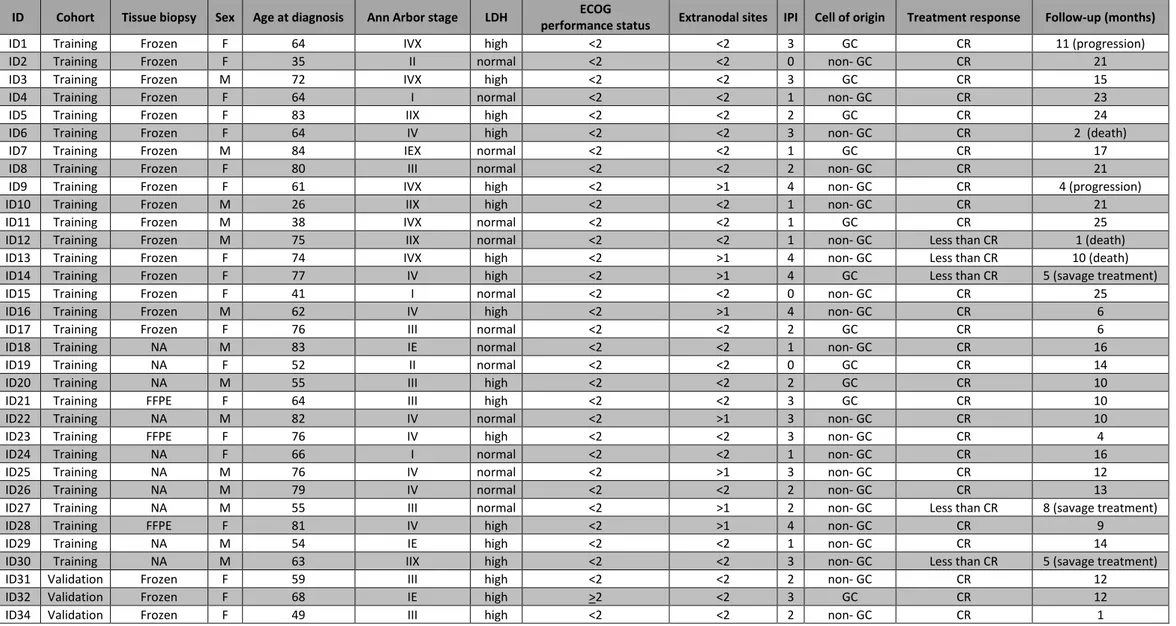
October 2016 as validation cohort (n=20) (Table 1). All patients received R-CHOP treatment.
The following biological material was collected: i) cfDNA isolated from plasma at diagnosis
before treatment start, during R-CHOP courses (on day 1 of each course before treatment infusion), at
the end of treatment and at progression, and ii) normal germline genomic DNA (gDNA) extracted from
PB granulocytes after Ficoll separation.
For comparative purposes, tumor gDNA from the paired DLBCL diagnostic tissue biopsy was
available for 36 patients (extracted from fresh specimens in 25 cases and from FFPE specimens in 11
cases). In the remaining 14 cases, the leftover of the FFPE diagnostic biopsy was not available or gave
18
temporal proximity of the tumor tissue biopsy (7-14 days after diagnostic tissue biopsy) and before
starting treatment.
Clinical information were prospectively maintained in the University of Eastern Piedmont
lymphoma database. Disease response was assessed by PET/CT.51 Patients provided informed consent
in accordance with local IRB requirements and Declaration of Helsinki.
Paired plasma cfDNA and normal gDNA from granulocytes collected from 6 healthy donors
were used to set the experimental and biological background of the ultra-deep next generation
sequencing (NGS) approach. The study was approved by the Ethical Committee of the Ospedale
Maggiore della Carità di Novara affiliated with the University of Eastern Piedmont (Protocol Code CE
112/15).
3.2. Genomic DNA (gDNA) extraction
PB granulocytes were separated by Ficoll gradient density centrifugation as source of normal
germline gDNA. Tumor gDNA was isolated from the fresh or FFPE diagnostic tissue biopsies containing
>70% of tumor cells as estimated by morphology and immunohistochemistry.
Tumor and normal gDNA were extracted by using the “salting out” protocol.52 PB was diluted
1:2 with physiological solution (NaCl 0.9%) and then centrifuged in a gradient differentiation Sigma
19
to obtain mononuclear white blood cells and granulocytes. Cells were lysed with Lysis Buffer (Tris-HCl
1M, pH 8.2, NaCl 5M, EDTA 0.5M), SDS 20% and digested with pronase E (20 mg/mL).
Samples were incubated at 37°C overnight in a shaking incubator. Proteins were precipitated
with 6M NaCl, and subsequently discarded after centrifugation at 3200 rpm for 20 minutes. DNA was
isolated by precipitation with pure ethanol and washed three times with 75% ethanol. The excess
ethanol was evaporated, and the DNA was dissolved with TE buffer (Tris-HCl 1M, pH 8.2 and EDTA
0.5M).
3.3. Plasma cfDNA extraction
30 ml of PB samples were collected in EDTA tubes and centrifuged at 820 g for 10 min to
separate plasma from cells within one hour from collection. Plasma was then further centrifuged at
13000 g for 10 min to pellet and remove any remaining cells and stored at -80°C until DNA extraction.
cfDNA was extracted from 1 ml aliquots of plasma immediately after thawing by using the QIAamp
circulating nucleic acid kit (Qiagen, Hilden, Germany) and quantified using a SYBR green-based
real-time qPCR assay for the β-globin gene (180-bp amplicon) carried out on StepOnePlus Real-Time PCR
System (Step One software 2.0; Applied Biosystems, Foster City, CA, USA).
20
A targeted resequencing gene panel including coding exons and splice sites of 59 genes
(target region: 207299bp) that are recurrently mutated in DLBCL and other mature B-cell tumors has
been specifically designed for this project. The gene panel allowed a priori the recovery of at least one
clonal mutation in 92.6% (95% CI: 83.6-97.5%) of DLBCL patients, as documented by in silico validation
against public genomic datasets of DLBCL.53-56
3.5. CAPP-seq library preparation and ultra-deep next generation sequencing
In the training cohort (n=30), the gene panel was analyzed in plasma cfDNA collected at
diagnosis, during R-CHOP courses, at the end of treatment, and at progression (total cfDNA
samples=127), and, for comparative purposes to filter out polymorphisms, in germline gDNA from the
paired granulocytes. The tumor gDNA from the paired tissue biopsy was also investigated to test the
accuracy of cfDNA genotyping.
In the validation cohort (n=20), the gene panel was investigated in plasma cfDNA collected
at diagnosis, and in normal and tumor gDNA from paired granulocytes and tissue biopsy, respectively.
Tumor and germline gDNA from tissues (median=318 ng) were sheared through sonication (Covaris
M220 focused-ultrasonicator, Woburn, MA, USA) before library construction to obtain 200-bp
fragments. For plasma cfDNA, which is naturally fragmented, 2-717 ng (median=17 ng) of DNA were
21
The next generation sequencing (NGS) libraries were constructed using the KAPA Library
Preparation Kit (Kapa Biosystems, Wilmington, MA, USA), and hybrid selection was performed with
the custom SeqCap EZ Choice Library (Roche NimbleGen, Madison, WI, USA). Multiplexed libraries (n=6
per run) were sequenced using 300-bp paired-end runs on a MiSeq sequencer (Illumina, San Diego,
CA, USA).
3.6. Bioinformatic pipeline for variant calling
Non-synonymous somatic mutation calling in plasma cfDNA was performed separately and
in blind from mutation calling in tumor gDNA. After CAPP-seq, FASTQ sequencing reads were initially
deduped through FastUniq v1.1. Then, the deduped FASTQ sequencing reads were locally aligned to
the hg19 version of the human genome assembly using BWA v.0.6.2 and assembled into a mpileup file
using SAMtools v.1. Single nucleotide variations and indels were called in plasma cfDNA vs germline
gDNA, and tumor gDNA vs germline gDNA, respectively, with the somatic function of VarScan2.
Variants annotated as SNPs according to dbSNP 138 (with the exception of TP53 variants that
were manually curated and scored as SNPs according to the IARC TP53 database), intronic variants
mapping >2 bp before the start or after the end of coding exons, and synonymous variants were
filtered out. Two independent statistical approaches (Fisher's exact test and Z-test) were then used to
22
region. Only variants that had a significant call in both test were retained (Bonferroni adjusted test
p<6x10⁻⁸).
3.7. Sanger sequencing
Sanger sequencing was also used to validate the most abundant plasma cfDNA mutations
detected by CAPP-seq. PCR primers were designed in the Primer 3 program
(http://frodo.wi.mit.edu/primer3/). Purified amplicons were subjected to Sanger sequencing and
compared to the corresponding germline sequences using the Mutation Surveyor Version 4.0.8
software package (SoftGenetics, State College, PA, USA; http://www.softgenetics.com) after
automated and/or manual curation. cfDNA from plasma were subjected to Sanger sequencing and
compared to the corresponding germline. Mutations were then confirmed from both strands on
independent PCR products.
3.8. Statistical analysis
Sensitivity and specificity of plasma cfDNA genotyping were calculated in comparison with
23
4. RESULTS
4.1. Characteristics of the study cohorts
The study was based on a prospectively collected consecutive series of 30 newly diagnosed
DLBCL patients (training cohort) whose characteristics were consistent with an unselected cohort of
DLBCL (Table 1). Upon R-CHOP treatment, 83.3% (95% CI: 65.9-93.1%; n=25/30) of patients achieved
a PET/CT negative complete remission, while 16.7% (95% CI: 6.8-34.0%; n=5/30) failed to achieve a
complete remission. Among patients that achieved complete remission (median follow-up 6 months),
two relapsed. The median number of cfDNA molecules per ml of plasma at disease presentation was
771.7 (range: 137.2-18742.5). An independent validation series of 20 consecutive DLBCL patients was
also assessed to confirm the accuracy of plasma cfDNA genotyping (Table 1).
4.2. Plasma cfDNA genotyping discloses somatic mutations in DLBCL-associated genes
To provide the proof of principle that plasma could function as a liquid biopsy for tracking
recurrently mutated genes in DLBCL, plasma cfDNA collected at presentation from the training cohort
was genotyped by using CAPP-seq, a targeted ultra-deep NGS approach for plasma cfDNA genotyping
already validated in solid tumors. In our cohort, 80% or more of the target region covered >1000x in
all DLBCL and 80% or more of the target region covered >2000x in 17/30 DLBCL.57 Paired normal gDNA
24
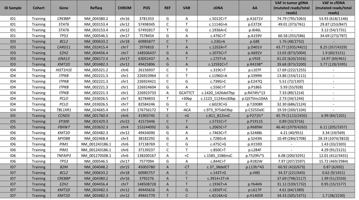
66.6% (95% CI: 47.1-82.7%; n=20/30) of patients harbored somatic mutations (total number 129;
range: 2-13 mutations per patient) that were detectable in plasma cfDNA (Table 2).
In order to validate the NGS results, cfDNA was extracted from a second aliquot of plasma,
and then subjected to a second CAPP-seq and ultra-deep-NGS sequencing. Robustness of the plasma
cfDNA CAPP-seq approach and of the bioinformatics analysis was documented by the high
concordance (R2 0.916) of variant calling from the independent duplicate experiments (Figure S1),
which consistently confirmed all the variants initially discovered in plasma cfDNA, including those of
low allele frequency, thus excluding their origin from a batch-specific experimental noise.
Sanger sequencing consistently detected all plasma cfDNA mutations showing a
representation within the sensitivity range of this approach (allele frequency >10%) (Figure S2), thus
validating the CAPP-seq results on a different experimental platform.
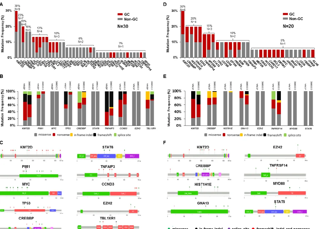
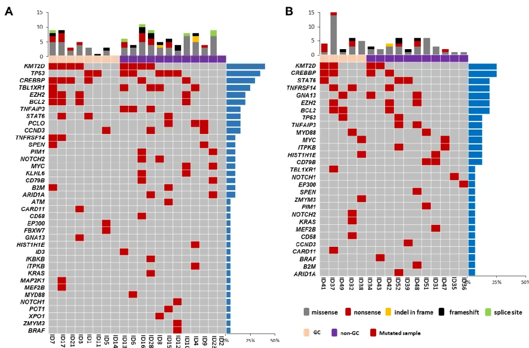
Consistent with the typical spectrum of mutated genes in DLBCL, plasma cfDNA genotyping
discovered somatic variants of KMT2D in 30.0% (95% CI: 14.7-49.4%; n=9/30) of cases, TP53 in 23.3%
(95% CI: 9.9-42.2%; n=7/30), CREBBP in 20.0% (95% CI: 7.7-38.5%; n=6/30), PIM1 and TNFAIP3 in 16.6%
(95% CI: 5.6-34.7%; n=5/30), EZH2, STAT6, and TBL1XR1 in 13.3% (95% CI: 3.7-30.7%; n=4/30), B2M,
BCL2, CARD11, CCND3 and FBXW7 in 10.0% (95% CI: 2.1-26.5%; n=3/30), CD58, CD79B and MYC in
6.6% (95% CI: 0.8-22.0%; n=2/30), EP300, GNA13, MEF2B, MYD88 and TNFRSF14 in 3.3% (95% CI:
25
of mutated genes within cell of origin categories of DLBCL, EZH2 and BCL2 mutations were as expected
more frequent in germinal-center (GC) DLBCL, while TNFAIP3 and PIM1 mutations were more frequent
in non-GC DLBCL (Figure 1A).10
Among recurrently affected genes, the molecular spectrum of mutations identified in plasma
cfDNA was highly consistent with that of variants that have been detected in tumor gDNA of published
DLBCL series and reported in the COSMIC database (Figure 1B and 1C). PIM1 and MYC were affected
by multiple hotspot mutations that were suggestive of AID (Activation-induced cytidine
deaminase)-related events (Figure 1C; Table 2).58 Notably, patient ID23, who had the t(8;14) MYC translocation,
also harbored multiple MYC mutations in cfDNA, consistent with the known accumulation of mutations
in the translocated MYC gene due to the IGH enhancer-driven misfire of somatic hypermutation (Table
2).59
Analysis of 6 healthy donors by CAPP-seq did not disclose any somatic mutations in plasma
cfDNA (Figure S3), suggesting that the ultra-deep NGS and variant calling approaches used in this study
did not pick up biological or analytical background noises in cfDNA.
4.3. Validation of biopsy-free DLBCL genotyping
The fresh or FFPE tissue biopsy of 20 DLBCL patients from the training cohort was genotyped
26
sequencing approach of FFPE samples, gDNA from paired fresh/FFPE samples processed from the same
DLBCL biopsy (n=4) was subjected to CAPP-seq. Pairwise analysis of data showed high (96%)
concordance in variant recovery from FFPE vs fresh samples (Figure S4).
In order to systematically derive the accuracy of cfDNA genotyping, the results of plasma
cfDNA genotyping and tumor gDNA genotyping (gold standard; mutation spectrum shown in Figure
2A) were then compared (Figures 3A-C) in 18 DLBCL training cases provided with the paired
plasma/tissue samples and informative because mutated within the target region. Sequencing
coverage of the tumor gDNA was comparable to that of plasma cfDNA (Figure S5).
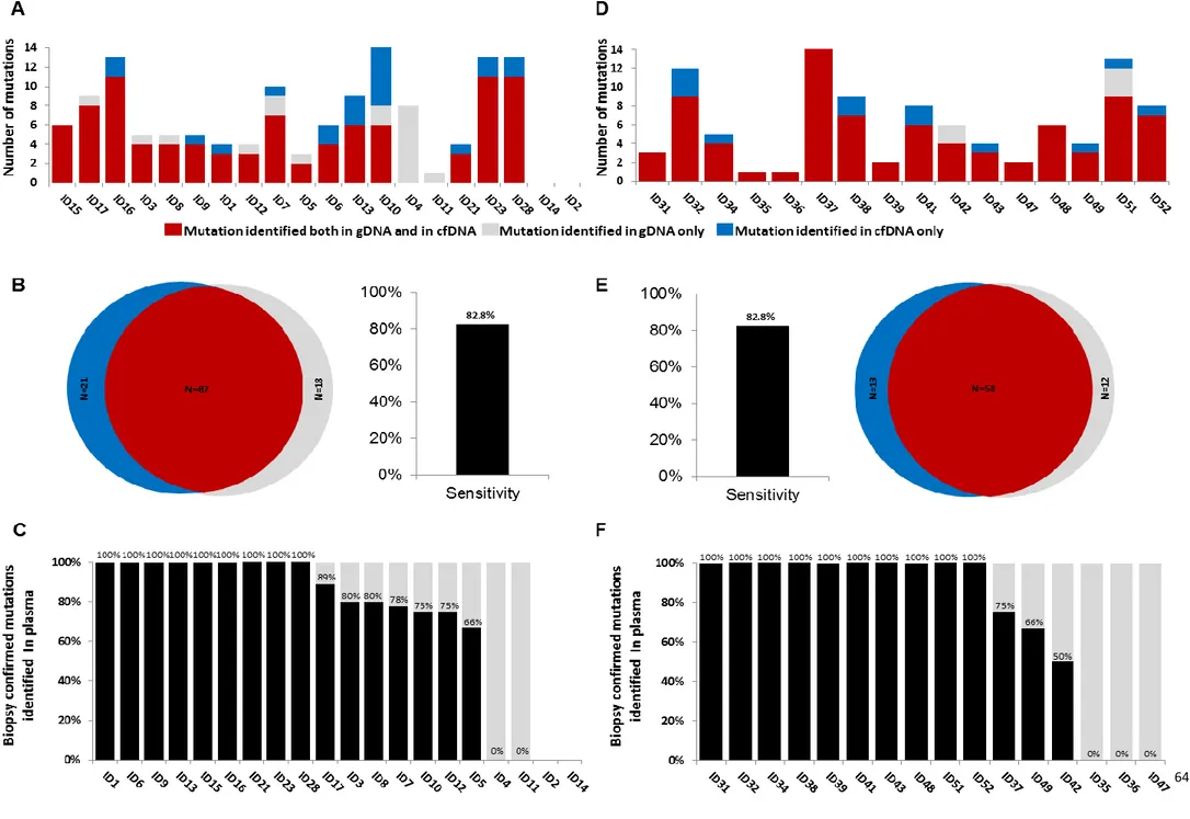
Genotyping of plasma cfDNA collected at diagnosis identified a total of 108 somatic
mutations, while genotyping of the gDNA from the diagnostic tissue biopsy identified 105 somatic
mutations. Biopsy-confirmed tumor mutations were detectable with 82.8% (n=87/105, 95% CI:
74.4-88.9%) sensitivity in pretreatment plasma cfDNA samples (Figure 3B). Biopsy-confirmed tumor
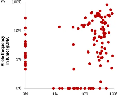
mutations not discovered in the cfDNA (n=18/105) generally had a low representation in the tissue
diagnostic biopsy (median allele frequency in the tumor biopsy=5.7%) (Figure 4A and 4B). Consistently,
by ROC analysis, cfDNA genotyping showed the highest sensitivity (97.1%; 95% CI: 89.5-99.8%;
n=68/70) in discovering mutations that were represented in >20% of the alleles of the tumor biopsy
(Figure 4B), thus demonstrating that plasma cfDNA can accurately mirror the profiles of the most
27
status in 10% (95% CI: 1.5-31.3%; n=2/20) of DLBCL cases (Figure 3C). The representation of the
variants in plasma correlated with the LDH levels at DLBCL diagnosis, but not with disease stage (Figure
S6).
Plasma cfDNA genotyping disclosed additional 21 somatic mutations that were not detectable
in the tissue biopsy, including 7 variants affecting PIM1, a known target of somatic hypermutation
(Figure 2A and 3; Table S4).58 Repeated ultra-deep NGS consistently confirmed these variants, thus
excluding their origin from a batch-specific experimental noise (Figure S2). Because the target region
that has been sequenced in the compiled 20 DLBCL patients might support a total of 18,011,440
potential non-synonymous variants if they were randomly distributed, and considering that only 21
non-synonymous mutations were detected in plasma cfDNA but not in the tumor biopsy, the false
positive rate of plasma cfDNA genotyping was 1.16x10⁻⁶ corresponding to a specificity >99.99%
compared to tumor gDNA genotyping.
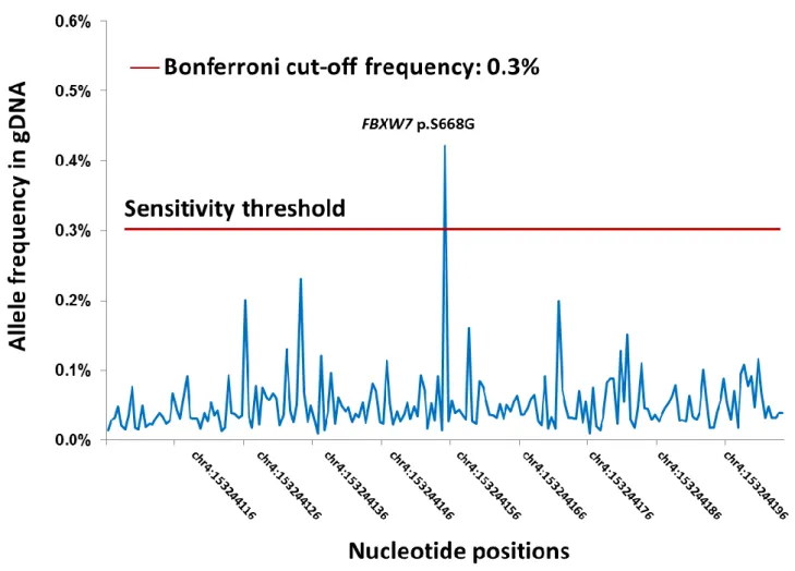
As previously reported in other cancer types,60 non-synonymous mutations occurring only in
plasma cfDNA conceivably represented tumor mutations restricted to clones that were anatomically
distant from the biopsy site, rather than false positive calls. Though the study lacked a systematic
multiregional sequencing of tumor samples from multiple anatomical sites, in support of the above
interpretation are the observations that: i) the plasma cfDNA FBXW7 p.S668G mutation of patient ID9,
28
the cerebrospinal fluid collected at the time of isolated meningeal relapse (Figures 5 and 7); and ii) all
mutations lacking in the diagnostic tissue biopsy disappeared from cfDNA upon achieving complete
remission of DLBCL.
Plasma cfDNA was genotyped by CAPP-seq in an independent cohort of 20 consecutive DLBCL.
Overall, within the interrogated genes, 85.0% (95% CI: 63.1-95.6%; n=17/20) of validation patients
harbored somatic mutations (total number 83; range: 1-12 mutations per patient) that were
detectable in plasma cfDNA (Figure 1D; Table S4). The molecular spectrum of mutations identified in
plasma cfDNA of the validation series was highly consistent with that observed in the training series
and more in general with that of published DLBCL cohorts (Figure 1E and F). CAPP-seq of paired tumor
and plasma samples from 16 DLBCL of the validation series confirmed the sensitivity of plasma cfDNA
genotyping in recovering biopsy-confirmed mutations (Figure 3 D-E). Biopsy-confirmed tumor
mutations were detectable with 82.8% (n=58/70, 95% CI: 72.2-90.0%) sensitivity in pretreatment
plasma cfDNA samples. Most of the biopsy-confirmed tumor mutations not discovered in the cfDNA
had a low representation in the tissue diagnostic biopsy <20% (Figure 4C). Consistently, by ROC
analysis, cfDNA genotyping showed the highest sensitivity (91.3%; 95% CI: 79.1-97.1%; n=42/46) in
discovering mutations that were represented in >20% of the alleles of the tumor biopsy (Figure 4D).
29
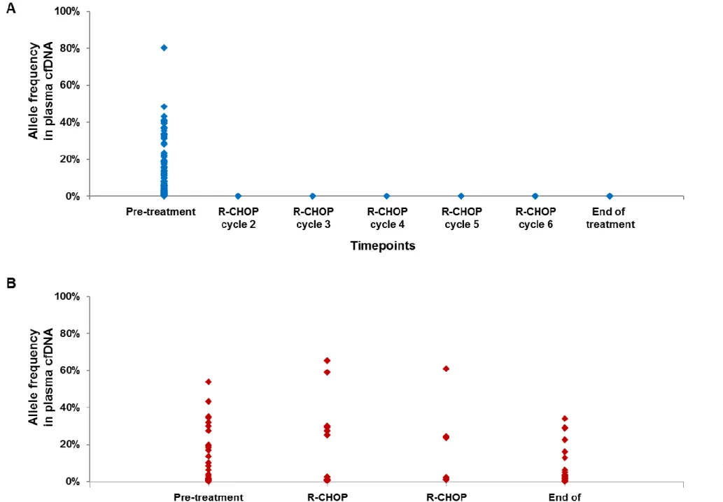
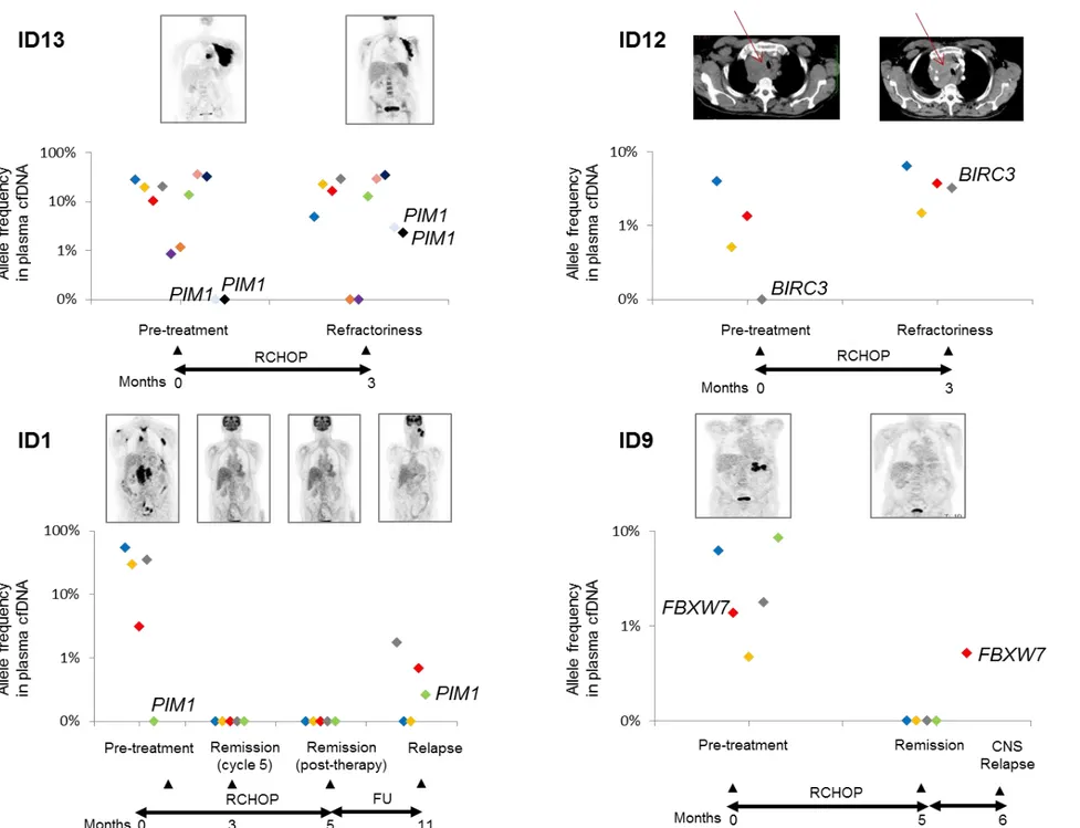
A total of 127 plasma cfDNA samples were sequentially evaluated to assess the dynamics of
mutations in plasma upon treatment with R-CHOP. Longitudinal analysis showed a rapid clearance of
DLBCL mutations in the cfDNA among responding patients (Figure 6A). Among patients who were
primary resistant to R-CHOP (ID12, ID13), DLBCL mutations did not disappear from cfDNA (Figure 6B).
In one of the two patients who responded to R-CHOP but ultimately had an early relapse in the central
nervous system (ID9), mutations were still detectable in the plasma cfDNA sample collected while the
patient was in complete remission (1-month prior relapse). In addition, among patients that were
primary refractory to R-CHOP or relapsed after treatment, new mutations appeared in the cfDNA that
conceivably marked resistant clones that were selected during the clonal evolution process taking
30
5. DISCUSSION
By applying a training-validation approach, this study shows that plasma cfDNA genotyping: i)
is as accurate as genotyping of the diagnostic biopsy to detect somatic mutations of allelic abundance
>20% in DLBCL; ii) allows the identification of mutations that are undetectable in the tissue biopsy
conceivably because restricted to clones that are anatomically distant from the biopsy site; and iii) is a
real-time and non-invasive tool to track clonal evolution and emergence of treatment resistant clones.
The identification of genomic alterations with clinical relevance in hematologic malignancies is
increasing the need for assays that can routinely identify tumor mutational profile. Among hematologic
malignancies with a leukemic component, accessibility of tumor cells in the PB has allowed the fast
incorporation of gene mutations into genetic prognostic and predictive algorithms.61-63 At variance
with other hematologic malignancies, DLBCL typically lacks a leukemic involvement, and bone marrow
dissemination is infrequent.64 Limited access to the tumor material has therefore hampered the
development and validation of molecular prognostic models in DLBCL, whose molecular stratification
represents an unmet medical need.65 On these bases, among hematologic malignancies, DLBCL is an
ideal model in which the liquid biopsy may allow a step forward in the translation of disease genetics
into clinically useful markers, especially in the era of novel agents that are active in molecular subgroup
31
Because of its >90% sensitivity and ~100% specificity, plasma cfDNA is an easily accessible
source of tumor DNA that allows to accurately profile DLBCL patients for cancer gene mutations
represented in >20% of the tumor alleles, which is the sensitivity threshold of conventional Sanger
sequencing methods that are broadly used to characterize tumor tissue specimens. On these bases,
plasma cfDNA is an effective surrogate of direct tumor genotyping by conventional sequencing for the
detection of clonally abundant mutations.
Plasma cfDNA genotyping also informs on variants that are subclonally represented in the
tumor biopsy, though a proportion (50%) of low abundance mutations (i.e. allele frequency <20% in
the tumor biopsy) has been missed by our ultra-deep NGS approach conceivably because of its
chemistry-dependent sensitivity limit of (~10-3). Indeed, the allelic fractions of mutations in tumor
biopsies and plasma samples are generally correlated, indicating that low abundant mutations in the
tumor are scantly represented in plasma. The increasing evidence that small subclones have a clinical
impact on treatment resistance and outcome in B-cell tumors prompts the development of sensitive
approaches for cfDNA that are capable of exactly mirroring both the clonal and subclonal composition
of the tumor.66 For example, incorporation of molecular barcoding in the library preparation chemistry
along with the application of in silico bioinformatics algorithms to suppress background NGS artifacts
32
Plasma cfDNA represents a complementary source of tumor DNA for DLBCL genotyping
compared to the tissue biopsy. On the one hand, the complete molecular heterogeneity of a tumor
cannot be adequately assessed by single or even multiple tissue biopsies, whereas cfDNA genotyping
captures genetic information shed from all sites of the disease. Also, plasma is an accessible source of
tumor DNA when DNA cannot be retrieved from the diagnostic tissue biopsy (i.e. because of the limited
size or poor conservation of the specimen). On the other hand, plasma cfDNA genotyping misses a
proportion of small subclonal mutations. Also, plasma cfDNA genotyping cannot accurately
differentiate of de novo DLBCL vs transformed tumors, precisely define the DLBCL cell of origin, and
fully detect tumor chromosomal translocations 9 which can instead be all routinely scored by analyzing
the tissue biopsy. These notions suggest that the liquid biopsy is not a substitute of the tumor biopsy,
but instead provides complementary information in DLBCL.
Treatment for DLBCL is currently undergoing a shift from chemotherapy towards regimens
incorporating targeted agents.44,50 Along with the clinical development of novel targeted agents in
DLBCL, mutation-driven mechanisms of resistance to these drugs are increasingly emerging.50 On these
bases, cfDNA genotyping may be used as a strategy of molecular monitoring to inform on the
acquisition of targeted drug-resistance in DLBCL. cfDNA analysis can be easily repeated at multiple
33
changes in tumor genetics, including dynamic changes in the mutation profile that occur during
therapy.59
Beside clonal evolution, longitudinal cfDNA genotyping can also inform on residual disease in
cancer.49 Our CAPP-seq approach does not reach the sensitivity of typical minimal residual disease
assays, and the number of informative cases (i.e. cases that achieved a radiological remission but
ultimately relapsed) is small in the present study cohort. Despite such limitations, the observation that,
among early relapsing patients, low level somatic mutations persisted in plasma while the disease was
apparently in remission support the development of plasma cfDNA mutational profiling as a tool for
residual disease surveillance in DLBCL. A CAPP-seq incorporating molecular barcoding coupled with
bioinformatics that allows the simultaneously tracking of multiple somatic mutations in cfDNA have
been developed to outperform immunoglobulin sequencing and radiographic imaging for the
detection of minimal residual disease in DLBCL.9
There are relatively few studies investigating the importance of the liquid biopsy in DLBCL that
have shown very promising results with different approaches. In two studies the IGHV-D-J gene
segment of the rearranged immunoglobulin was used as plasma cfDNA biomarker to identify patients
at high risk of treatment failure, while in two additional studies cfDNA was used to detect somatic
mutations.5,6,8,9 The retrospective nature of the previous studies that relied on archival material
34
pre-analytic factors that have a substantial effect on plasma cfDNA analysis, including the amount of
“contaminating” DNA coming from circulating cells. The prospective design and the standardized
protocols for sample processing used in our study allowed to stringently control pre-analytic factors
that might influence the results of plasma cfDNA genotyping. In less controlled situations such as
multicentered studies, the use of Cell-Free DNA BCT tubes, that avoid storage-related pre-analytic
biases, can provide a broadly validated way to obtain stable cfDNA samples.68
An immediate clinical application of cfDNA genotyping in DLBCL patients is the incorporation
of this assay within clinical trials to support post-hoc patient stratification according to baseline disease
genetics, to develop treatment-specific prognostic, predictive or actionable genomic biomarkers, and
35
REFERENCES
1. Rossi D, Diop F, Spaccarotella E, Monti S, Zanni M, Rasi S, Deambrogi C, Spina V, Bruscaggin
A, Favini C, Serra R, Ramponi A, Boldorini R, Foà R, Gaidano G. Diffuse large B-cell lymphoma
genotyping on the liquid biopsy. Blood. 2017;129(14):1947-1957.
2. Pös O, Biró O, Szemes T, Nagy B. Circulating cell-free nucleic acids: characteristics and
applications. Eur J Hum Genet. 2018;26(7):937-945.
3. Diaz LA Jr, Bardelli A. Liquid biopsies: genotyping circulating tumor DNA. J Clin Oncol.
2014;32(6):579-586.
4. Hohaus S, Giachelia M, Massini G, Mansueto G, Vannata B, Bozzoli V, Criscuolo M, D'Alò F,
Martini M, Larocca LM, Voso MT, Leone G. Cell-free circulating DNA in Hodgkin's and
non-Hodgkin's lymphomas. Ann Oncol. 2009;20(8):1408-1413.
5. Kurtz DM, Green MR, Bratman SV, Scherer F, Liu CL, Kunder CA, Takahashi K, Glover C, Keane
C, Kihira S, Visser B, Callahan J, Kong KA, Faham M, Corbelli KS, Miklos D, Advani RH, Levy R,
Hicks RJ, Hertzberg M, Ohgami RS, Gandhi MK, Diehn M, Alizadeh AA. Noninvasive
monitoring of diffuse large B-cell lymphoma by immunoglobulin high-throughput
sequencing. Blood. 2015;125(24):3679-3687.
6. Roschewski M, Dunleavy K, Pittaluga S, Moorhead M, Pepin F, Kong K, Shovlin M, Jaffe ES,
Staudt LM, Lai C, Steinberg SM, Chen CC, Zheng J, Willis TD, Faham M, Wilson WH. Circulating
36
tumour DNA and CT monitoring in patients with untreated diffuse large B-cell lymphoma: a
correlative biomarker study. Lancet Oncol. 2015;16(5):541-549.
7. Roschewski M, Staudt LM, Wilson WH. Dynamic monitoring of circulating tumor DNA in
non-Hodgkin lymphoma. Blood. 2016;127(25):3127-3132.
8. Bohers E, Viailly PJ, Dubois S, Bertrand P, Maingonnat C, Mareschal S, Ruminy P, Picquenot
JM, Bastard C, Desmots F, Fest T, Leroy K, Tilly H, Jardin F. Somatic mutations of cell-free
circulating DNA detected by next-generation sequencing reflect the genetic changes in both
germinal center B-cell-like and activated B-cell-like diffuse large B-cell lymphomas at the
time of diagnosis. Haematologica. 2015;100(7):e280-e284.
9. Scherer F, Kurtz DM, Newman AM, Stehr H, Craig AF, Esfahani MS, Lovejoy AF, Chabon JJ,
Klass DM, Liu CL, Zhou L, Glover C, Visser BC, Poultsides GA, Advani RH, Maeda LS, Gupta NK,
Levy R, Ohgami RS, Kunder CA, Diehn M, Alizadeh AA. Distinct biological subtypes and
patterns of genome evolution in lymphoma revealed by circulating tumor DNA. Sci Transl
Med. 2016;8(364):64ra155.
10. Pasqualucci L, Dalla-Favera R. The Genetic Landscape of Diffuse Large B Cell Lymphoma.
Semin Hematol.2015;52(2): 67–76.
11. Alizadeh AA, Eisen MB, Davis RE, Ma C, Lossos IS, Rosenwald A, Boldrick JC, Sabet H, Tran T,
Yu X, Powell JI, Yang L, Marti GE, Moore T, Hudson J Jr, Lu L, Lewis DB, Tibshirani R, Sherlock
G, Chan WC, Greiner TC, Weisenburger DD, Armitage JO, Warnke R, Levy R, Wilson W, Grever
37
MR, Byrd JC, Botstein D, Brown PO, Staudt LM. Distinct types of diffuse large B-cell
lymphoma identified by gene expression profiling. Nature. 2000;403(6769):503-511.
12. Shaffer AL 3rd, Young RM, Staudt LM. Pathogenesis of human B cell lymphomas. Annu Rev
Immunol. 2012; 30:565-610.
13. Rosenwald A, Wright G, Chan WC, Connors JM, Campo E, Fisher RI, Gascoyne RD,
Muller-Hermelink HK, Smeland EB, Giltnane JM, Hurt EM, Zhao H, Averett L, Yang L, Wilson WH,
Jaffe ES, Simon R, Klausner RD, Powell J, Duffey PL, Longo DL, Greiner TC, Weisenburger DD,
Sanger WG, Dave BJ, Lynch JC, Vose J, Armitage JO, Montserrat E, López-Guillermo A, Grogan
TM, Miller TP, LeBlanc M, Ott G, Kvaloy S, Delabie J, Holte H, Krajci P, Stokke T, Staudt LM.
Lymphoma/Leukemia Molecular Profiling Project. The use of molecular profiling to predict
survival after chemotherapy for diffuse large-B-cell lymphoma. N Engl J Med.
2002;346(25):1937-1947.
14. Wright G, Tan B, Rosenwald A, Hurt EH, Wiestner A, Staudt LM. A gene expression-based
method to diagnose clinically distinct subgroups of diffuse large B cell lymphoma. Proc Natl
Acad Sci U S A. 2003;100(17):9991-9996.
15. Goodman RH, Smolik S. CBP/p300 in cell growth, transformation, and development. Genes
& development. 2000;14(13):1553–1577.
16. Iqbal J, Greiner TC, Patel K, Dave BJ, Smith L, Ji J, Wright G, Sanger WG, Pickering DL, Jain S,
Horsman DE, Shen Y, Fu K, Weisenburger DD, Hans CP, Campo E, Gascoyne RD, Rosenwald
A, Jaffe ES, Delabie J, Rimsza L, Ott G, Müller-Hermelink HK, Connors JM, Vose JM, McKeithan
38
T, Staudt LM, Chan WC; Leukemia/Lymphoma Molecular Profiling Project. Distinctive
patterns of BCL6 molecular alterations and their functional consequences in different
subgroups of diffuse large B-cell lymphoma. Leukemia.2007;21(11):2332–2343.
17. Ying CY, Dominguez-Sola D, Fabi M, Lorenz IC, Hussein S, Bansal M, Califano A, Pasqualucci
L, Basso K, Dalla-Favera R. MEF2B mutations lead to deregulated expression of the oncogene
BCL6 in diffuse large B cell lymphoma. Nature immunology.2013;14(10):1084–1092.
18. Morin RD, Mendez-Lago M, Mungall AJ, Goya R, Mungall KL, Corbett RD, Johnson NA,
Severson TM, Chiu R, Field M, Jackman S, Krzywinski M, Scott DW, Trinh DL, Tamura-Wells J,
Li S, Firme MR, Rogic S, Griffith M, Chan S, Yakovenko O, Meyer IM, Zhao EY, Smailus D,
Moksa M, Chittaranjan S, Rimsza L, Brooks-Wilson A, Spinelli JJ, Ben-Neriah S, Meissner B,
Woolcock B, Boyle M, McDonald H, Tam A, Zhao Y, Delaney A, Zeng T, Tse K, Butterfield Y,
Birol I, Holt R, Schein J, Horsman DE, Moore R, Jones SJ, Connors JM, Hirst M, Gascoyne RD,
Marra MA. Frequent mutation of histone-modifying genes in non-Hodgkin lymphoma.
Nature. 2011;476(7360):298–303.
19. Duan S, Cermak L, Pagan JK, Rossi M, Martinengo C, di Celle PF, Chapuy B, Shipp M, Chiarle
R, Pagano M. FBXO11 targets BCL6 for degradation and is inactivated in diffuse large B-cell
lymphomas. Nature.2012;481(7379):90–93.
20. Challa-Malladi M, Lieu YK, Califano O, Holmes AB, Bhagat G, Murty VV, Dominguez-Sola D,
Pasqualucci L, Dalla-Favera R. Combined genetic inactivation of beta2-Microglobulin and
CD58 reveals frequent escape from immune recognition in diffuse large B cell lymphoma.
39
21. Miyashita K, Tomita N, Taguri M, Suzuki T, Ishiyama Y, Ishii Y, Nakajima Y, Numata A, Hattori
Y, Yamamoto W, Miyazaki T, Tachibana T, Takasaki H, Matsumoto K, Hashimoto C, Takemura
S, Yamazaki E, Fujimaki K, Sakai R, Motomura S, Ishigatsubo Y. Beta-2 microglobulin is a
strong prognostic factor in patients with DLBCL receiving R-CHOP therapy. Leuk
Res.2015;S0145-2126(15)30368-30374.
22. Pasqualucci L, Trifonov V, Fabbri G, Ma J, Rossi D, Chiarenza A, Wells VA, Grunn A, Messina
M, Elliot O, Chan J, Bhagat G, Chadburn A, Gaidano G, Mullighan CG, Rabadan R, Dalla-Favera
R. Analysis of the coding genome of diffuse large B-cell lymphoma. Nature
genetics.2011;43(9):830–837.
23. Monti S, Chapuy B, Takeyama K, Rodig SJ, Hao Y, Yeda KT, Inguilizian H, Mermel C, Currie T,
Dogan A, Kutok JL, Beroukhim R, Neuberg D, Habermann TM, Getz G, Kung AL, Golub TR,
Shipp MA. Integrative analysis reveals an outcome-associated and targetable pattern of p53
and cell cycle deregulation in diffuse large B cell lymphoma. Cancer cell.2012;22(3):359–372.
24. Lu TX, Young KH, Xu W, Li JY. TP53 dysfunction in diffuse large B-cell lymphoma. Crit Rev
Oncol Hematol.2016;97:47-55.
25. Trinh DL, Scott DW, Morin RD, Mendez-Lago M, An J, Jones SJ, Mungall AJ, Zhao Y, Schein J,
Steidl C, Connors JM, Gascoyne RD, Marra MA. Analysis of FOXO1 mutations in diffuse large
B-cell lymphoma. Blood.2013;121(18):3666–3674.
40
26. Schuetz JM, Johnson NA, Morin RD, Scott DW, Tan K, Ben-Nierah S, Boyle M, Slack GW, Marra
MA, Connors JM, Brooks-Wilson AR, Gascoyne RD. BCL2 mutations in diffuse large B-cell
lymphoma. Leukemia.2012;26(6):1383-1390.
27. Karube K, Campo E. MYC alterations in diffuse large B-cell lymphomas. Semin Hematol.
2015;52(2):97-106.
28. Ma Y, Zhang P, Gao Y, Fan H, Zhang M, Wu J. Evaluation of AKT phosphorylation and PTEN
loss and their correlation with the resistance of rituximab in DLBCL. Int J Clin Exp Pathol.
2015;8(11):14875-14884.
29. Wang X, Cao X, Sun R, Tang C, Tzankov A, Zhang J, Manyam GC, Xiao M, Miao Y, Jabbar K,
Tan X, Pang Y, Visco C, Xie Y, Dybkaer K, Chiu A, Orazi A, Zu Y, Bhagat G, Richards KL, Hsi ED,
Choi WWL, van Krieken JH, Huh J, Ponzoni M, Ferreri AJM, Møller MB, Parsons BM, Winter
JN, Piris MA, Li S, Miranda RN, Medeiros LJ, Li Y, Xu-Monette ZY, Young KH. Clinical
Significance of PTEN Deletion, Mutation, and Loss of PTEN Expression in De Novo Diffuse
Large B-Cell Lymphoma. Neoplasia. 2018;20(6):574-593.
30. Morin RD, Johnson NA, Severson TM, Mungall AJ, An J, Goya R, Paul JE, Boyle M, Woolcock
BW, Kuchenbauer F, Yap D, Humphries RK, Griffith OL, Shah S, Zhu H, Kimbara M, Shashkin
P, Charlot JF, Tcherpakov M, Corbett R, Tam A, Varhol R, Smailus D, Moksa M, Zhao Y,
Delaney A, Qian H, Birol I, Schein J, Moore R, Holt R, Horsman DE, Connors JM, Jones S,
Aparicio S, Hirst M, Gascoyne RD, Marra MA. Somatic mutations altering EZH2 (Tyr641) in
follicular and diff use large B-cell lymphomas of germinal-center origin. Nat Genet. 2010; 42:
181 – 185.
41
31. Elodie Bohers, Sylvain Mareschal, Philippe Bertrand, Pierre Julien Viailly, Sydney Dubois,
Catherine Maingonnat, Philippe Ruminy, Hervé Tilly & Fabrice Jardin. Activating somatic
mutations in diffuse large B-cell lymphomas: lessons from next generation sequencing and
key elements in the precision medicine era. Leukemia & Lymphoma.2015;56(5):1213-1222.
32. Wigle TJ, Knutson SK, Jin L, Kuntz KW, Pollock RM, Richon VM, Copeland RA, Scott MP. The
Y641C mutation of EZH2 alters substrate specificity for histone H3 lysine 27 methylation
states. FEBS Lett. 2011;585(19):3011-3014.
33. Lee HJ, Shin DH, Kim KB, Shin N, Park WY, Lee JH, Choi KU, Kim JY, Lee CH, Sol MY. Polycomb
protein EZH2 expression in diff use large B-cell lymphoma is associated with better prognosis
in patients treated with rituximab, cyclophosphamide, doxorubicin, vincristine and
prednisone. Leukemia & Lymphoma. 2014;55(9): 2056–2063.
34. Muppidi JR, Schmitz R, Green JA, Xiao W, Larsen AB, Braun SE, An J, Xu Y, Rosenwald A, Ott
G, Gascoyne RD, Rimsza LM, Campo E, Jaffe ES, Delabie J, Smeland EB, Braziel RM, Tubbs RR,
Cook JR, Weisenburger DD, Chan WC, Vaidehi N, Staudt LM, Cyster JG. Loss of signalling via
Ga13 in germinal center B-cell-derived lymphoma. Nature. 2014; 516(7530):254-258.
35. O'Hayre M, Inoue A, Kufareva I, Wang Z, Mikelis CM, Drummond RA, Avino S, Finkel K, Kalim
KW, DiPasquale G, Guo F, Aoki J, Zheng Y, Lionakis MS, Molinolo AA, Gutkind JS. Inactivating
Mutations in GNA13 and RHOA in Burkitt’s Lymphoma and Diffuse Large B-cell Lymphoma:
A Tumor Suppressor Function for the Gα13/RhoA Axis in B Cell. Oncogene. 2016; 35(29):
3771–3780.
42
36. Lenz G, Wright GW, Emre NC, Kohlhammer H, Dave SS, Davis RE, Carty S, Lam LT, Shaffer AL,
Xiao W, Powell J, Rosenwald A, Ott G, Muller-Hermelink HK, Gascoyne RD, Connors JM,
Campo E, Jaffe ES, Delabie J, Smeland EB, Rimsza LM, Fisher RI, Weisenburger DD, Chan WC,
Staudt LM. Molecular subtypes of diffuse large B-cell lymphoma arise by distinct genetic
pathways. Proc Natl Acad Sci U S A. 2008;105(36):13520–13525.
37. Davis RE, Ngo VN, Lenz G, Tolar P, Young RM, Romesser PB, Kohlhammer H, Lamy L, Zhao H,
Yang Y, Xu W, Shaffer AL, Wright G, Xiao W, Powell J, Jiang JK, Thomas CJ, Rosenwald A, Ott
G, Muller-Hermelink HK, Gascoyne RD, Connors JM, Johnson NA, Rimsza LM, Campo E, Jaffe
ES, Wilson WH, Delabie J, Smeland EB, Fisher RI, Braziel RM, Tubbs RR, Cook JR,
Weisenburger DD, Chan WC, Pierce SK, Staudt LM. Chronic active B-cell-receptor signalling
in diffuse large B-cell lymphoma. Nature. 2010;463(7277):88–92.
38. Kim Y, Ju H, Kim DH, Yoo HY, Kim SJ, Kim WS, Ko YH. CD79B and MYD88 mutations in diffuse
large B-cell lymphoma. Hum Pathol. 2014;45(3):556-64.
39. Knies N, Alankus B, Weilemann A, Tzankov A, Brunner K, Ruff T, Kremer M, Keller UB, Lenz
G, Ruland J. Lymphomagenic CARD11/BCL10/MALT1 signaling drives malignant B-cell
proliferation via cooperative NF-B and JNK activation. Proc Natl Acad Sci U S A.
2015;112(52):E7230-7238.
40. Ngo VN, Young RM, Schmitz R, Jhavar S, Xiao W, Lim KH, Kohlhammer H, Xu W, Yang Y, Zhao
H, Shaffer AL, Romesser P, Wright G, Powell J, Rosenwald A, Muller-Hermelink HK, Ott G,
Gascoyne RD, Connors JM, Rimsza LM, Campo E, Jaffe ES, Delabie J, Smeland EB, Fisher RI,
43
Braziel RM, Tubbs RR, Cook JR, Weisenburger DD, Chan WC, Staudt LM. Oncogenically active
MYD88 mutations in human lymphoma. Nature.2011;470(7332):115–119.
41. Rhyasen GW, Starczynowski DT. IRAK signalling in cancer. British Journal of Cancer.
2015;112(2):232-237.
42. Zhang J, Grubor V, Love CL, Banerjee A, Richards KL, Mieczkowski PA, Dunphy C, Choi W, Au
WY, Srivastava G, Lugar PL, Rizzieri DA, Lagoo AS, Bernal-Mizrachi L, Mann KP, Flowers C,
Naresh K, Evens A, Gordon LI, Czader M, Gill JI, Hsi ED, Liu Q, Fan A, Walsh K, Jima D, Smith
LL, Johnson AJ, Byrd JC, Luftig MA, Ni T, Zhu J, Chadburn A, Levy S, Dunson D, Dave SS. Genetic
heterogeneity of diffuse large B-cell lymphoma. Proc Natl Acad Sci U S A.
2013;110(4):1398-1403.
43. Compagno M, Lim WK, Grunn A, Nandula SV, Brahmachary M, Shen Q, Bertoni F, Ponzoni M,
Scandurra M, Califano A, Bhagat G, Chadburn A, Dalla-Favera R, Pasqualucci L. Mutations of
multiple genes cause deregulation of NF-kappaB in diffuse large B-cell lymphoma. Nature.
2009;459(7247):717-721.
44. Younes A, Thieblemont C, Morschhauser F, Flinn I, Friedberg JW, Amorim S, Hivert B, Westin
J, Vermeulen J, Bandyopadhyay N, de Vries R, Balasubramanian S, Hellemans P, Smit JW,
Fourneau N, Oki Y. Combination of ibrutinib with rituximab, cyclophosphamide, doxorubicin,
vincristine, and prednisone (R-CHOP) for treatment-naive patients with CD20-positive B-cell
non-Hodgkin lymphoma: a non-randomised, phase 1b study. Lancet Oncol.
2014;15(9):1019-1026.
44
45. Kwak J-Y. Treatment of Diffuse Large B Cell Lymphoma. The Korean Journal of Internal
Medicine. 2012;27(4):369-377.
46. Chiappella A, Tucci A, Castellino A, Pavone V, Baldi I, Carella AM, Orsucci L, Zanni M, Salvi F,
Liberati AM, Gaidano G, Bottelli C, Rossini B, Perticone S, De Masi P, Ladetto M, Ciccone G,
Palumbo A, Rossi G, Vitolo U. Fondazione Italiana Linfomi. Lenalidomide plus
cyclophosphamide, doxorubicin, vincristine, prednisone and rituximab is safe and effective
in untreated, elderly patients with diffuse large B-cell lymphoma: a phase I study by the
Fondazione Italiana Linfomi. Haematologica. 2013;98(11):1732-1738.
47. Tomita N, Takasaki H, Fujisawa S, Miyashita K, Ogusa E, Kishimoto K, Matsuura S, Sakai R,
Koharazawa H, Yamamoto W, Fujimaki K, Fujita H, Ishii Y, Taguchi J, Kuwabara H, Motomura
S, Ishigatsubo Y. Standard R-CHOP therapy in follicular lymphoma and diffuse large B-cell
lymphoma. J Clin Exp Hematop. 2013;53(2):121-125.
48. Coiffier B, Sarkozy C. Diffuse large B-cell lymphoma: R-CHOP failure-what to do?.
Hematology Am Soc Hematol Educ Program. 2016;2016(1):366-378.
49. Xu-Monette ZY, Wu L, Visco C, Tai YC, Tzankov A, Liu WM, Montes-Moreno S, Dybkaer K,
Chiu A, Orazi A, Zu Y, Bhagat G, Richards KL, Hsi ED, Zhao XF, Choi WW, Zhao X, van Krieken
JH, Huang Q, Huh J, Ai W, Ponzoni M, Ferreri AJ, Zhou F, Kahl BS, Winter JN, Xu W, Li J, Go
RS, Li Y, Piris MA, Møller MB, Miranda RN, Abruzzo LV, Medeiros LJ, Young KH. Mutational
profile and prognostic significance of TP53 in diffuse large B-cell lymphoma patients treated
with R-CHOP: report from an International DLBCL Rituximab-CHOP Consortium Program
Study. Blood. 2012;120(19):3986-3996.
45
50. Wilson WH, Young RM, Schmitz R, Yang Y, Pittaluga S, Wright G, Lih CJ, Williams PM, Shaffer
AL, Gerecitano J, de Vos S, Goy A, Kenkre VP, Barr PM, Blum KA, Shustov A, Advani R, Fowler
NH, Vose JM, Elstrom RL, Habermann TM, Barrientos JC, McGreivy J, Fardis M, Chang BY,
Clow F, Munneke B, Moussa D, Beaupre DM, Staudt LM. Targeting B cell receptor signaling
with ibrutinib in diffuse large B cell lymphoma. Nat Med. 2015;21(8):922-926.
51. Cheson BD, Fisher RI, Barrington SF, Cavalli F, Schwartz LH, Zucca E, Lister TA;Alliance,
Australasian Leukaemia and Lymphoma Group; Eastern Cooperative Oncology Group;
European Mantle Cell Lymphoma Consortium; Italian Lymphoma Foundation; European
Organisation for Research; Treatment of Cancer/ Dutch Hemato-Oncology Group; Grupo
Espanol de Me´dula O´ sea; German High-Grade Lymphoma Study Group; German Hodgkin’s
Study Group; Japanese Lymphoma Study Group; Lymphoma Study Association; NCIC Clinical
Trials Group; Nordic Lymphoma Study Group; Southwest Oncology Group; United Kingdom
National Cancer Research Institute. Recommendations for initial evaluation, staging, and
response assessment of Hodgkin and non-Hodgkin lymphoma: the Lugano classification. J
Clin Oncol. 2014;32(27):3059-3067.
52. Miller SA, Dykes DD, Polesky HF. A simple salting out procedure for extracting DNA from
human nucleated cells. Nucleic acids Res. 1988; 16(3):1215.
53. Pasqualucci L, Trifonov V, Fabbri G, Ma J, Rossi D, Chiarenza A, Wells VA, Grunn A, Messina
M, Elliot O, Chan J, Bhagat G, Chadburn A, Gaidano G, Mullighan CG, Rabadan R, Dalla-Favera
R. Analysis of the coding genome of diffuse large B-cell lymphoma. Nat Genet.
2011;43(9):830-837.
46
54. Lohr JG, Stojanov P, Lawrence MS, Auclair D, Chapuy B, Sougnez C, Cruz-Gordillo P, Knoechel
B, Asmann YW, Slager SL, Novak AJ, Dogan A, Ansell SM, Link BK, Zou L, Gould J, Saksena G,
Stransky N, Rangel-Escareño C, Fernandez-Lopez JC, Hidalgo-Miranda A, Melendez-Zajgla J,
Hernández-Lemus E, Schwarz-Cruz y Celis A, Imaz-Rosshandler I, Ojesina AI, Jung J,
Pedamallu CS, Lander ES, Habermann TM, Cerhan JR, Shipp MA, Getz G, Golub TR. Discovery
and prioritization of somatic mutations in diffuse large B-cell lymphoma (DLBCL) by
whole-exome sequencing. Proc Natl Acad Sci USA. 2012;109(10):3879-3884.
55. Zhang J, Grubor V, Love CL, Banerjee A, Richards KL, Mieczkowski PA, Dunphy C, Choi W, Au
WY, Srivastava G, Lugar PL, Rizzieri DA, Lagoo AS, Bernal-Mizrachi L, Mann KP, Flowers C,
Naresh K, Evens A, Gordon LI, Czader M, Gill JI, Hsi ED, Liu Q, Fan A, Walsh K, Jima D, Smith
LL, Johnson AJ, Byrd JC, Luftig MA, Ni T, Zhu J, Chadburn A, Levy S, Dunson D, Dave SS. Genetic
heterogeneity of diffuse large B-cell lymphoma. Proc Natl Acad Sci USA.
2013;110(4):1398-1403.
56. Morin RD, Mungall K, Pleasance E, Mungall AJ, Goya R, Huff RD, Scott DW, Ding J, Roth A,
Chiu R, Corbett RD, Chan FC, Mendez-Lago M, Trinh DL, Bolger-Munro M, Taylor G, Hadj
Khodabakhshi A, Ben-Neriah S, Pon J, Meissner B, Woolcock B, Farnoud N, Rogic S, Lim EL,
Johnson NA, Shah S, Jones S, Steidl C, Holt R, Birol I, Moore R, Connors JM, Gascoyne RD,
Marra MA. Mutational and structural analysis of diffuse large B-cell lymphoma using
whole-genome sequencing. Blood. 2013;122(7):1256-1265.
57. Newman AM, Bratman SV, To J, Wynne JF, Eclov NC, Modlin LA, Liu CL, Neal JW, Wakelee
HA, Merritt RE, Shrager JB, Loo BW Jr, Alizadeh AA, Diehn M. An ultrasensitive method for
quantitating circulating tumor DNA with broad patient coverage. Nat Med.
2014;20(5):548-554.
47