Contents lists available atScienceDirect
International Journal of Paleopathology
journal homepage:www.elsevier.com/locate/ijppScheuermann
’s disease in a juvenile male from the late Roman necropolis of
Torrenueva (3rd
–4th century CE, Granada, Spain)
☆
Joan Viciano
a,⁎, Sandra López-Lázaro
b, Ángela Pérez-Fernández
c, Anabel Amores-Ampuero
c,
Ruggero D
’Anastasio
a, José Miguel Jiménez-Triguero
daOperative Unit of Anthropology, Department of Medicine and Ageing Sciences,‘G. d’Annunzio’, University of Chieti–Pescara, Via dei Vestini 29, 66100 Chieti, Italy bLaboratory of Forensic Dentistry, Centre for Applied Morphology Research, Faculty of Dentistry, University of La Frontera, Avenida Francisco Salazar 01145, 4780000,
Temuco, Chile
cLaboratory of Anthropology, Department of Legal Medicine, Toxicology and Physical Anthropology, School of Medicine, University of Granada, Granada, Spain dGESPAD Al-Andalus S.L. Archaeology, 18010 Granada, Spain
A R T I C L E I N F O
Keywords:
Juvenile osteochondrosis Kyphotic spine Pelvic asymmetry Leg length discrepancy Ambulatory problems Palaeopathology
A B S T R A C T
This study details a severe case of Scheuermann’s disease (SD) in a well-preserved skeleton of a juvenile male (designated TOR302), dated to 3rd–4th century CE, from the late Roman necropolis of Torrenueva (Granada, Spain). Individual TOR302 shows an evident kyphotic curve in the thoraco-lumbar spine, which is characterised by: (i) vertebral bodies of thoracic vertebra T2, thoracic segment T4–T9, and thoraco-lumbar segment T12–L2 wedged at > 5°; (ii) slight anterior extensions of the epiphyseal ring; (iii) Schmorl’s nodes on the superior and/or inferior plates; and (iv) a Cobb angle of 75°, derived from thoracic segments T4–T9. In addition, TOR302 shows other skeletal malformations as the secondary results of abnormal growth, due to altered biomechanical forces imposed by the spinal deformity, including: (i) lateral distortion of the spine that causes a slight secondary scoliotic curve; (ii) pelvic obliquity; and (iii) discrepancy in the length of the limbs. We argue that the secondary skeletal abnormalities allowed the individual to adapt to his spinal deformity meaning he was able to walk without the aid of a stick. Despite SD being a common modern clinicalfinding, few cases have been reported in ancient skeletal remains. This case therefore represents an important contribution to the palaeopathological literature.
1. Introduction
The evaluation of pathological conditions in archaeological contexts is often a challenge, as there are far fewer resources to call upon compared to the modern clinical setting. Diseases that leave clear pathognomic signs on a skeleton are rare. However, the basis of palaeopathological diagnosis must be as close as possible to that of modern clinical medicine. For correct palaeopathological diagnosis, a broad differential diagnosis must be defined, including the full spec-trum of potential causes (Miller et al., 1996), with the use of radiology mandatory, as radiography represents an important link between modern clinical medicine and palaeopathology as a diagnostic tool that can be applied for both disciplines (De Luca et al., 2013).
Osteochondrosis is the term that is used to describe a group of
disorders that affect the growing skeleton that result from abnormal development, injury, or overuse of the growth plate and the surround-ing ossification centres (Atanda et al., 2011). Scheuermann’s disease (SD) is a juvenile osteochondrosis of the vertebral column that is characterised by a kyphotic deformity of the spine that develops in early adolescence (Ali et al., 1999; Lowe and Line, 2007). While SD is well known clinically, there have been few reports for archaeologically retrieved skeletal remains, as it is difficult to recognize certain pathological conditions for a disarticulated skeleton. Wells (1961) described a case of SD of the lumbar spine for the skeletal remains of a juvenile female from Dorset (UK), which were dated to about 1600 BCE, in the Bronze Age.Kunter (1976)published a possible case from Kamid-el-Loz (Lebanon, 4th century BCE).Cook et al. (1983)described the vertebral elements of the partial hominid skeleton known as AL-288
http://dx.doi.org/10.1016/j.ijpp.2017.04.003
Received 20 October 2016; Received in revised form 10 April 2017; Accepted 26 April 2017
☆The results of this study were presented in part at the XIX Meeting of the Italian Anthropological Association in Turin, Italy, 2011. Reference: López-Lázaro, S., Viciano, J., Amores,
A., Jiménez-Triguero, J., 2012. A probable case of Scheuermann’s disease in a juvenile male from the late Roman necropolis of Torrenueva (III–IV AD, Granada, Spain). J. Biol. Res. 85, 239–240.
⁎Corresponding author at: Operative Unit of Anthropology, Department of Medicine and Ageing Sciences,‘G. d'Annunzio’ University of Chieti–Pescara, Via dei Vestini 29, 66100
Chieti, Italy.
E-mail address:[email protected](J. Viciano).
International Journal of Paleopathology xxx (xxxx) xxx–xxx
1879-9817/ © 2017 Elsevier Inc. All rights reserved.
(‘Lucy’) from the Pliocene Hadar formation in Ethiopia. In examining 179 sailors who drowned with the British ship Mary Rose that sank in 1545 CE, Stirland (1991) noted young male skeletons that were probably affected by SD. Anderson and Carter (1994) provided a detailed description of a specimen of the Iron Age period in Kent (UK). Coughlan and Holst (2000) noted a skeleton from the Towton Hall mass grave (UK) that showed evidence of SD. Capasso (2001) reported two cases of SD from victims who died while trying toflee by sea from the ancient beach of Herculaneum (Naples, Italy), during the eruption of Vesuvius Volcano on 24–25 August, 79 CE. During examination of four skeletons that were attributed to people killed during the Battles for Zürich in 1799 CE, Meyer (2003) briefly described a possible case of SD for the skeletal remains of an adult male (skeleton no. 5595). Üstündağ and Deveci (2011) described a human skeleton of a young adult male from an Islamic cemetery in Birecik (Turkey) that was dated to the 13th–14th century CE.
The SD cases published in the palaeopathological literature tend to describe the spine in isolation. The aim of the present study was to define a case of severe SD in a well-preserved skeleton of a juvenile male from the late Roman necropolis of Torrenueva (3rd–4th century CE, Granada, Spain), along with an assortment of other abnormalities that reflect disturbances during development and growth of the musculoskeletal system. We also consider the consequences here in terms of ambulatory problems.
2. Material and methods
2.1. Background
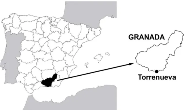
In 2008, during excavations at the foot of the Vargas’ glen and Maraute “pagus” of Torrenueva (Granada, Spain) (Fig. 1), a Roman quay was discovered next to the sea (1st–4th century CE), which was abandoned due to the economic crisis at the end of 3rd century CE, and reused as a late Roman necropolis (3th–4th century CE) and as a supply-line in Islamic times (11th–13th century CE). It was discovered by chance when houses were being built in that area. Two single tombs were discovered in the late Roman necropolis. In stratigraphic unit 312, there were few skeletal remains. However, in stratigraphic unit 315 the skeletal remains of an individual (designated TOR302) were discovered
inhumed in a tomb composed of large square tiles (tegulae bipedales) and covered by largeflat tiles (tegulae). The skeleton was in an extended supine position, with the head orientation to the west. Grave goods, including a metallic clasp andfibula, were associated with the burial (Casado-Millán et al., 2010). Individual TOR302 is stored in the Laboratory of Anthropology of the University of Granada (Spain).
These remains are the only evidence of the human presence in Torrenueva before the decline and fall of the Roman Empire. Archaeology demonstrates a predominantly rural society in late Roman Baetica, but do not explain the social relationships and economic structure in this area (Casado-Millán et al., 2010).
2.2. Preservation and completeness
Bones present from the upper body were well preserved despite fragmentation of the sternum, scapulae and ribs. The cranium, mand-ible, teeth, thoracic vertebra T1 and lumbar vertebra L3, coccyx, and the majority of the right and left carpals, metacarpals and phalanges were absent. The lower body elements were also well preserved, but the patellae and the majority of the right and left tarsals, metatarsals and phalanges were absent. The pubic and ischial bones were fragmentary. In addition, the cortical surfaces of the available bones showed no signs of taphonomic damage.
2.3. Examination methods
The sex of this individual was estimated by applying standard osteological methods based on descriptive (Bruzek, 2002; Ferembach et al., 1980) and metric (Murail et al., 2005) criteria from the pelvic features. Due to the only partial preservation of the pelvis, some observations were difficult or impossible. The age at death was estimated using morphological (Cardoso, 2008a,b; Cardoso et al., 2014) and metric (Scheuer and Black, 2000) methods.
The skeletal remains were inspected macroscopically and radio-graphically for any evidence of congenital anomalies, nutritional disorders, infectious diseases, fractures, traumatic conditions, and enthesopathies. Any signs of pathological conditions were recorded and described, and differential diagnosis was offered.
2.4. Anthropometric study of the spine
As shown inFig. 2, the following measurements were taken for the thoracic and lumbar vertebrae using digital calipers, to calculate the wedging and anterior extension of the vertebral bodies according to Digiovanni et al. (1989)andScoles et al. (1991): posterior (Fig. 2A, measure A) and anterior (Fig. 2B, measure B) heights of vertebral body, anteroposterior diameter of the superior surface of the vertebral body measured to the external margin of the ring apophysis (Fig. 2B, measure C) (not including the anterior extension), and the maximum antero-posterior diameter of the vertebral body in the midsagittal plane (Fig. 2, measure X).
The amount of wedging at each vertebral body was calculated using Equation(1): ⎛ ⎝ ⎜ ⎞ ⎠ ⎟ α arctan Y X = 2 × , o (1) where, Y= A−B 2 . (2)
The percentage of elongation of the anterior extension was calcu-lated using Equation(3):
Anterior extension X C X
(%) = − × 100.
(3)
2.5. Three-dimensional model of the spine
In clinical medicine, measurement of spinal curvature is important to assess the status of the spine shape, to monitor the progression of spinal deformities (such as kyphosis and scoliosis), and to evaluate the severity of these deformities. To evaluate the spinal curvature of this person, a three-dimensional (3D) model of the spine was constructed through acquisition of the surface geometry of each thoracic and
lumbar vertebra and the sacrum, using a structured light 3D scanner (SLS-2; David Vision Systems GmbH, Germany) and the integrated software (David LaserScanner v.3.10.4), to a resolution of 0.15 mm. The resulting surface mesh of each vertebra was articulated anatomi-cally to the adjacent vertebra mesh using professional free open-source 3D computer graphics software (Blender v.2.68, Blender Foundation). The linear dimensions of the intervertebral disc spaces (i.e., anterior and posterior disc heights) proposed byAbuzayed et al. (2010)were used to articulate the vertebrae in the 3D space. The structure of the missing L3 vertebra was constructed virtually as a mean size of the adjacent L2 and L4 vertebrae. Once the 3D model of the vertebral column was virtually reconstructed, the angle of the thoracic spine was measured.
There are several methods for measuring sagittal plane angles in the spine (e.g.,Briggs et al., 2007; Goh et al., 2000; Harrison et al., 2001). The Cobb angle (Cobb, 1948) remains one of the most commonly used techniques to describe the sagittal and coronal angular relationships along the spine (Bono et al., 2006), and it is recognized as the gold standard (Harrison et al., 2001); this was the method used here. The Cobb angle is formed by the intersection of a line parallel to the superior end-plate of the most cephalic vertebra in a particular curve, with the line parallel to the inferior end-plate of the most caudal vertebra of the curve. By convention, perpendiculars to the parallels are drawn, and the angle between their intersections is measured. The most cephalic vertebra is the vertebra that has the greatest tilt from horizontal of its superior end-plate. The most caudal vertebra is the vertebra that has the greatest tilt from horizontal of its inferior end-plate.
3. Results
3.1. Biological profile
The morphological and metric abnormalities of these skeletal remains highlight the difficulty of estimating sex and age of the
Fig. 2. Lateral and superior views of a vertebra, showing the measurements taken to estimate the vertebral wedging and to calculate the percentage increase in the anterior extension. The posterior (A, measure A) and anterior (A, measure B) heights of the vertebral body, the anteroposterior diameter of the superior surface of the vertebral body measured to the external margin of the ring apophysis (B, measure C) (not including the anterior extension), and the maximum anteroposterior diameter of the vertebral body in the midsagittal plane (measure X) were all defined and measured. The formulae used for estimation of the vertebral wedging and for computation of the percentage increase of the anterior extension are shown in the text. J. Viciano et al. International Journal of Paleopathology xxx (xxxx) xxx–xxx
individual with accuracy. According toBruzek (2002)andFerembach et al. (1980), the pelvis showed typically male morphological features for the four traits analysed: preauricular surface, greater sciatic notch, composite arch, and morphology of the iliac crest. The measurements for the pubic breadth, depth of the great sciatic notch, cotylo-sciatic breadth, and vertical acetabular diameter (Table 1) were also indicative of a male individual (Murail et al., 2005). According to these descriptive and metric criteria, this individual appears to have been a male. However, the estimated sex should be taken with caution, because the distorted and asymmetric morphology of the pelvis appears to correspond to a pattern of a disease process (see below). In addition, male features observed in adolescent coxae, as in this case, should be inconclusive for sex estimation because the coxae may represent a female whose pelvic dimensions have not yet assumed adult propor-tions. According toBuikstra and Ubelaker (1994)the growth changes during adolescence lead to distinctive differences in the male and the female pelvis, but adolescent females may be late in showing diagnostic female features, which are formed on a basic male pattern which is common in both male and female pelves until adolescence. Despite these issues, an attempt at sex assessment was made because the difference between the sexes for certain diseases/abnormalities is relevant in any palaeopathological differential diagnosis and palaeo-epidemiological studies; moreover, the sex estimation might be useful for future palaeo-epiemiological analyses (Viciano et al., 2015). Although few cases of SD have been reported for archaeologically retrieved skeletal remains, this does not mean that further cases like this will not arise in the future.
For the age of this individual, the epiphyseal union at the innominate and lower limbs (Cardoso, 2008a), the upper limbs and scapular girdle (Cardoso, 2008b) and the sacrum (Cardoso et al., 2004) was incomplete, which indicated an age of 14–18 years (innominate: ≥15 years; lower limbs: 15–18 years; upper limbs: 14–18 years; scapular girdle: 15–18 years; sacrum: ≤18 years). The diaphyseal lengths of the clavicle, tibia andfibula (Table 1) defined a younger age, of 10–16 years (clavicle: 15–16 years; tibia: ca. 12 years; fibula: 10–11 years) (Scheuer and Black, 2000). Due to the alterations and asymme-tries in the length dimensions of the long bones, the epiphyseal union of the bones was considered for the age estimation of this individual. Thus, the age at the death of individual TOR302 was estimated at 14–18 years. However, this estimated age should also be taken with caution.
Several studies have suggested that skeletal maturation is more affected by environmental insults than skeletal growth, such disease or mal-nutrition (Cardoso, 2007; Conceição and Cardoso, 2011). These studies have also highlighted that dental development is less affected by environmental insults than skeletal maturation. Therefore, a compar-ison between dental age and skeletal age might provide valuable information to determine whether there was any delay in the skeletal maturation of this individual. However, the absence of dentition in this individual means that we cannot confirm a delay in the age of the epiphyseal union.
3.2. Axial skeleton
Asymmetries were noted for the spine, for the vertebral bodies anteriorly and the arches posteriorly, as well as for its overall aspect. The cervical vertebrae demonstrated a normal morphology, whereas an evident kyphotic curve characterised the thoraco-lumbar spine.Fig. 3 illustrates the anterior, posterior and right and left lateral views of the 3D model of the spine. In frontal view, the thoracic vertebral bodies showed subtle/moderate asymmetry where the superior and inferior end-plates were not quite parallel (Fig. 4). Posteriorly, the superior and inferior thoracic articular processes were longer and the facet joint surface area was larger on the right side. The thoracic spinous processes were slightly curved towards the right, and the transverse processes were generally longer and thicker on the right side. In the lumbar region, all of these changes were more subtle; however, vertebra L5 showed failure of osseous fusion of the neural arch, which left a bony cleft (Fig. 5A). Except T3, T6 and L1, the rest of the thoracic and lumbar vertebrae had Schmorl’s nodes on the superior and/or inferior plates.
The different measurements of each thoracic and lumbar vertebra, the amount of wedging, and the presence/absence of anterior exten-sions are given inTable 2. Vertebral bodies of thoracic vertebra T2, thoracic segment T4–T9, and thoraco-lumbar segment T12–L2 were wedged at > 5° and had slight anterior extensions of the epiphyseal ring of < 16%. The global and regional Cobb angles derived from measurements taken at T4–T9 and T2–T12 were 75° and 111.5°, respectively, for the kyphotic curve (Fig. 6).
In addition to the evident kyphotic curve between T4–T9, a lateral distortion was present in the spine due to the wedging of the left hemivertebral bodies, which caused a slight secondary scoliotic curve
Table 1
Anthropometrics of individual TOR302.
Bone Relevant measure Datum (mm)
Left Right
Sacrum Maximum auricular surface height 55.36 60.63
Lateral breadth of the alae 40.43 44.03 Posterior breadth of the alae 32.85 30.98
Clavicle Maximum length 133 136
Width of the acromial epiphysis 22.50 25.20 Humerus (without proximal epiphysis; distal epiphysis fused with the diaphysis) Maximum length 268 279
Epicondylar width 53.89 52.71
Ulna (proximal epiphysis fused with the diaphysis; without distal epiphysis) Maximum length 230 236 Sub-sigmoid transverse diameter 18.94 18.04 Sub-sigmoid antero-posterior diameter 24.18 21.46 Radius (proximal epiphysis fused with the diaphysis; without distal epiphysis) Maximum length 210 220
Coxal Cotylo-pubic breadth 24.48 25.08
Depth of the great sciatic notch – 22.57 Cotylo-sciatic breadth 23.37 24.08 Vertical acetabular diameter – 56.72 Femur (proximal epiphysis fused with the diaphysis; without distal epiphysis) Maximum length 371 370
Horizontal head diameter 39.23 39.69 Vertical head diameter 38.80 39.68 Transverse subtrochanteric diameter 29.30 29.18 Antero-posterior subtrochanteric diameter – 20.52 Tibia (without proximal and distal diaphysis) Maximum length 310 – Fibula (without proximal and distal diaphysis) Maximum length 291 281
to the right. Therefore, the spine had a kyphoscoliotic curve. The Cobb angle derived from this measurement was 8.15° for the scoliotic curve. When scoliotic deformities occur in the thoracic region, this situation can lead to distortion and asymmetry of the rib cage. In this individual the right ribs showed a slight abnormally sharp dorsal angle, whereas the left ribs showed a wide dorsal angle (Fig. 7), with the presence of porous surface in most preserved fragments. The asymmetry of the rib cage was likely to have occurred due to the pressure applied to the rib bodies during the gradual progression of the scoliotic curve.
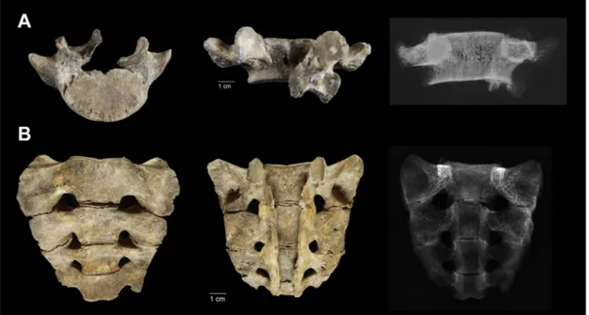
Partial agenesis of the sacrum was observed, with the lower sacral segment missing (vertebra S5) (Fig. 5B). There was also incomplete closure of the posterior sacral canal between the two halves of the neural arch. This cleft extended the entire length of the sacral neural arch, and it affected all four sacral vertebrae (from S1 to S4). The
general morphology of the sacrum was slightly asymmetric when observed in anterior view. Taking three measurements on each side of it following the definitions ofPlochocki (2002), the lateral breadth of the right alae was greater than the left one. This difference was also observable in the maximum auricular surface height; however, this condition is inverse in the posterior breadth of the alae, where the dimension is slightly lower in the right alae than the left (Table 1).
3.3. Pectoral and pelvic girdles, and appendicular skeleton
On visual examination of the pectoral girdle, the scapulae showed a moderate expression of the insertion of the triceps brachii muscle, where the infraglenoid tubercle emerges as a distinct formation of the axillary border, which assumes the form of a crest. In addition, the lateral edges
Fig. 3. Illustration of the anterior, posterior, and right and left lateral views of the three-dimensional model of the thoraco-lumbar spine of individual TOR302 (3rd–4th century CE, necropolis of Torrenueva, Granada, Spain). The blacked out vertebra corresponds to the reconstructed L3.
Fig. 4. Macroscopic and radiographic images of thoracic segments T4–T9 of the spine of individual TOR302 (3rd–4th century CE, necropolis of Torrenueva, Granada, Spain). J. Viciano et al. International Journal of Paleopathology xxx (xxxx) xxx–xxx
of both of the scapulae (although mostly the right one) were particu-larly pronounced where the teres major muscle was inserted. The right clavicle showed a well-defined imprint at the attachment site of the costoclavicular ligament, with a slightly irregular surface and the presence of small areas of osteolytic activity of limited depth (< 1 mm). The humeri, ulnae and radii showed slight shortening on the left side. The shortening of these bones was confirmed by the metric data, which showed that the maximum length of the left humerus was almost 11 mm shorter than the right one (both humeri were measured without the proximal epiphysis; the distal epiphysis was fused with the diaphysis). The left ulna and radius had maximum lengths of 6 mm and 10 mm shorter than the right ones (both ulnae and radii were measured without the distal epiphysis; the proximal epiphysis was fused with the diaphysis) (Table 1). The humeri showed pronounced
exostosis of sub-periosteal origin, which surrounded an extensive and deep osteolytic area at the attachment sites for the pectoralis major muscle. Ulnae and radii showed marked hypertrophy of the pronator quadratus and pronator teres insertions. In addition, both radii showed evident bowing of the diaphysis (Fig. 8). Finally, marked lines of attachment for flexor ligaments on palmar surfaces of the medial phalangeal row were observed.
The general appearance of the pelvis was asymmetric, which was even more evident when the two coxal bones were articulated with the sacrum, with the left coxal elevated higher than the right (Fig. 9). The superior dorsal margin of the right acetabulum showed a flattened flange that appears to form a flattening extension of the articular facet (Fig. 10), and a groove was observed along the margin of the foramen obturator on the medial lower portion. The femora showed a severe flattening of the diaphyseal superior third (hyperplatimery; 70.32 for the right femur, while that for the left femur could not be calculated) and a shortening of the neck (Fig. 11A). In addition, the postero-proximal surface of the shafts showed an extensive and deep osteolytic area at the gluteus maximus insertion site, and a marked linea aspera (Fig. 11B). Both tibiae showed a depressed surface with crateriform architecture between the proximal epiphysis and the tibial tuberosity, for the insertion of the patellar ligament and the quadriceps femoris muscle (Fig. 12A). In addition, the articular surface of the left distal epiphysis showed an osteochondritic lesion (Fig. 12B). For the dimen-sions, the femora andfibulae showed slight shortening on the right side. The discrepancy in the maximum length between the femora was 11 mm (these bones were measured without the distal epiphysis; the proximal epiphysis was partially fused with the diaphysis); the di ffer-ence in the maximum length of the fibulae (only the diaphysis was measured) was 10 mm (Table 1). Finally, the left talus showed an extension of the medial trochlear articular surface that encroached on the superior surface of the neck of the talus.
4. Discussion
There is a complex of environmental and genetic mechanisms that combine to regulate the processes of development and growth of the human body. These processes are vulnerable and can be subjected to disturbances that affect normal development of the various anatomical structures (Stevenson and Hall, 2006).
Fig. 5. (A) Fifth lumbar vertebra, showing failure of osseous fusion of the neural arch, leaving a bony cleft. (B) Partial agenesis of the sacrum (lower sacral segment S5 missing), and incomplete closure of the posterior sacral canal between the two halves of the neural arch. Individual TOR302 (3rd–4th century CE, necropolis of Torrenueva, Granada, Spain). Table 2
Vertebral measurements, body wedging angle, anterior extension, and presence/absence of Schmorl’s nodes.
Vertebra Relevant measure
Posterior height (mm) Anterior height (mm) Maximum antero-posterior diameter (mm) Vertebral wedging (°) Anterior extension (%) Schmorl nodes T1 – – – – – – T2 18.30 16.35 15.66 7.13 0.64 Present T3 17.47 16.10 17.70 4.43 0.11 Absent T4 15.55 12.58 20.04 8.48 7.93 Present T5 15.42 8.53 21.15 18.50 10.50 Present T6 16.31 11.08 23.36 12.77 15.15 Absent T7 17,05 12.93 23.46 10.04 7.97 Present T8 18.62 14.64 23.65 9.62 1.65 Present T9 19.38 17.19 23.91 5.24 2.05 Present T10 20.12 18.67 23.13 3.59 0.35 Present T11 21.61 19.64 24.36 4.63 3.37 Present T12 24.26 20.81 24.35 8.10 3.82 Present L1 25.43 21.77 23.64 8.85 0.63 Absent L2 24.67 22.41 24.09 5.37 0.00 Present L3 – – – – – – L4 23.12 23.71 24.45 -1.38 1.88 Present L5 21.11 23.94 24.24 -6.68 0.58 Present
The skeletal individual TOR302 showed an assortment of abnorm-alities that reflected disturbances during the development and growth of the musculoskeletal system (i.e., prenatal and postnatal alterations). Evidence from the skeletal remains indicates that two alterations occurred during the prenatal period: (i) partial agenesis of the sacrum, missing the last segment (vertebra S5); and (ii) incomplete closure of the posterior canal between the two halves of the neural arches of the sacrum (segments S1–S4).
The ossification pattern of the sacrum is complex, as it develops from approximately 21 separate primary centres of ossification. In the third month of prenatal life, ossification centres appear in the first and second sacral centra. Around the fourth month, ossification is evident in the centra of the third and fourth sacral segments, and in the neural arches of S1–S3. Around the fifth month, ossification occurs in the centrum of S5, and in the neural arches of S4–S5. Thus, all primary centres of ossification are present by birth. Around age 12, the sacral elements begin to unite, starting with S5 and S4, and progressing superiorly (Scheuer and Black, 2000). In individual TOR302, the centrum and lateral elements of S4 did not show signs of initial fusion with S5.
Alternatively, L5 and the sacrum showed incomplete closure of the posterior canal between the two halves of the neural arches, and two aspects can explain this skeletal anomaly: (i) the neural tube defect of spina bifida; and (ii) a cleft neural arch. According toBarnes (2012), these two aspects differ in the alignment and width of the cleft arches. The major differences are that for the neural tube defect of spina bifida, the arches are thin and pushed outwards with the widened vertebral canal and distorted pedicles, whereas for cleft neural arches, these remain within their designated alignment with a normal vertebral canal space, as in the present case study. According toChen et al. (2006), there are six types of posterior neural arch defects: (i) neurocentral synchondrosis; (ii) paraspinous cleft; (iii) spinous cleft; (iv) retro-somatic cleft; (v) spondylolysis; and (vi) retroisthmic cleft. The embryology of the vertebral column explains these threefirst types of neural arch defects. The remaining three defects either have unknown causes or are believed to be caused by overuse. In TOR302, the posterior arch defect of the sacral vertebrae was classified as ‘para-spinous cleft’ due to the failure of neural tube closure during
develop-Fig. 6. Thoracic kyphosis measured from the right lateral view using the global Cobb angle for T2–T12 (A), and the regional Cobb angle for T4–T9 (B). Individual TOR302 (3rd–4th century CE, necropolis of Torrenueva, Granada, Spain).
Fig. 7. Asymmetric ribs that developed in the presence of the scoliosis. Note the slight abnormally sharp dorsal angle in the right ribs, and the wide dorsal angle in the left ribs. Individual TOR302 (3rd–4th century CE, necropolis of Torrenueva, Granada, Spain).
ment, which prevented muscle and bone from growing around the gap. For vertebra L5, the cleft passed through the left pars interarticularis, which connects the superior and inferior articular facets. Thus, this defect was classified as unilateral ‘spondylolysis’. Although this defect can reflect defects of congenital or acquired origin, it is widely believed that most often it is acquired due to repeated microtrauma and overuse, which can eventually cause stress fractures of the pars interarticularis (Chen et al., 2006).
Individual TOR302 showed other musculoskeletal abnormalities that reflect disturbances during the growth of this individual. One of these disturbances was osteochondrosis or traction apophysitis of both tibial tuberosities. During growth, the tubercle is separated from the proximal tibial metaphysis by afibrocartilaginous plate (Scheuer and Black, 2000). When there is repetitive strain on the patellar ligament from the powerful pull of the quadriceps femoris muscles, part of the tubercle can become avulsed (Ortner, 2003). The relative mechanical weakness of the cartilaginous attachment of the patellar ligament to the developing tibial tuberosity, paired with the strong tensile forces produced by the quadriceps femoris musculature, appear to have
Fig. 8. Bowing of the diaphysis of both of the radii, and shortening of the left radius. The radii arranged according to the position of the radial tuberosity, which demonstrates the length asymmetry. Individual TOR302 (3rd–4th century CE, necropolis of Torrenueva, Granada, Spain).
Fig. 9. Asymmetric pelvis, with the left coxal elevated higher than the right, when they are articulated with the sacrum. Individual TOR302 (3rd–4th century CE, necropolis of Torrenueva, Granada, Spain).
Fig. 10. Slight bevelling (arrowhead) of the superior dorsal margin of the right acetabulum. Individual TOR302 (3rd–4th century CE, necropolis of Torrenueva, Granada, Spain).
contributed to the aetiology of this inflammatory condition, which is known as Osgood-Schlatter’s disease (Atanda et al., 2011; DiGangi et al., 2010).
The most obvious manifestation of disturbances during growth of this individual is the hyperkyphotic curve in the thoracic region, which is consistent with SD. This is an osteochondrosis that affects the
growing spine and causes severe deformity (Ali et al., 1999; Lowe and Line, 2007). Holger Welfer Scheuermann was thefirst to describe this in 1920, as different from mobile postural kyphosis (Scheuermann, 1920, 1921). He recognized from radiographs that the wedge vertebrae formation in the thoracic spine was the underlying reason for the deformity, and established that for the diagnosis of SD, at least three wedged vertebrae at the apex of the curve and end-plate irregularities of the vertebral bodies should be present. Scheuermann believed that the vertebral anomalies were caused by avascular necrosis of the vertebral ring apophysis, which results in growth disturbance of the vertebral bodies. However, more recent studies have failed to show evidence of avascular necrosis (Bradford and Moe, 1975; Ippolito and Ponseti, 1981). In 1930, Schmorl proposed another theory, in which he suggested that the kyphosis occurred as a result of intervertebral disc herniation through the growth plate of the vertebral bodies (Ali et al., 1999; Schmorl, 1930; Wenger and Frick, 1999). He believed that weakened areas along the growth plate predisposed the spine to such herniation (Schmorl’s nodes), to decrease the disc space height and alter the growth of the vertebral bodies, thus leading to progressive kyphosis. However, Schmorl’s theory is currently disputed by the finding of Schmorl’s nodes in vertebrae outside the area of kyphosis in patients with SD, as also in many healthy patients with no evidence of disease (Ali et al., 1999; Lowe, 1990; Moquin et al., 2003).Ferguson (1956)suggested that wedging of the vertebral bodies was related to the persistence of the anterior vascular groove in the adolescent, which causes inherent weakening that leads to subsequent collapse of the vertebral body. Nevertheless, there have been no signs of altered bone structure in the area of the anterior vascular groove in several studies (Lowe, 1990).Bradford et al. (1976)andLopez et al. (1988)suggested that osteoporosis might be responsible for the development of SD, although recent studies have shown that osteoporosis is not an aetiological factor of SD (Ashton et al., 2001; Gilsanz et al., 1989;
Fig. 11. (A) Severeflattening of the diaphyseal superior third (hyperplatimery) in the right femur and a shortening of the neck on both femora. (B) Extensive and deep osteolytic area at the gluteus maximus insertion site on the postero-proximal surface of the shaft of the right femur. Individual TOR302 (3rd–4th century CE, necropolis of Torrenueva, Granada, Spain).
Fig. 12. Depressed surface with a crateriform architecture between the proximal epiphysis and the tibial tuberosity (arrowhead) on the right tibia, for the macroscopic (A) and radiological (B) aspects. (C) Osteochondritic lesion on the articular surface of the distal epiphysis of the left tibia (arrowhead). Individual TOR302 (3rd–4th century CE, necropolis of Torrenueva, Granada, Spain).
Masharawi et al., 2009; Scoles et al., 1991). Other studies have suggested that the main causes of vertebral wedging in SD might be physical-activity-induced stress, trauma, inflammatory diseases, vita-min-A deficiency, poliomyelitis, disorganised intracartilaginous ossifi-cation, increased height, and increased release of growth hormone (Ali et al., 1999; Greene et al., 1985; Lowe, 1990; Murray et al., 1993; Skogland et al., 1985). Genetic transmission has also been explored as a cause of SD (Findlay et al., 1989; Halal et al., 1978; Zaidman et al., 2013), which suggested an autosomal-dominant mode of inheritance, with incomplete penetrance. Thus, although there are many proposed causes of SD, there remains a lack of evidence to exclusively support any single cause. The incidence of SD has been estimated to be 0.4% to 8.0% (Lowe, 1990; Sørensen, 1964). The sex prevalence is difficult to determine because the definition of the disease process varies. It is believed that SD typically affects males and females equally (Lowe, 1990); however, the reported ratios vary widely (Murray et al., 1993). Several studies have proposed diagnostic criteria as indicative of SD (e.g.,Bradford, 1980; Butler, 1955; Sørensen, 1964). A widely accepted definition is based on theSørensen (1964)andBradford (1980)criteria: (i) at least three consecutive adjacent vertebrae at the apex of the kyphosis with anterior wedging of 5° or more; (ii) irregular upper and lower vertebral end-plates, combined withflattening and wedging; (iii) narrowing of the intervertebral disc spaces; (iv) variable presence of Schmorl’s nodes; and (v) presence of kyphosis > 40°.
For the differential diagnosis, SD must not be confused with other syndromes or diseases, such as postural kyphosis, neoplasms, infections (e.g., brucellosis, tuberculosis), and osteoporotic or traumatic compres-sion fractures. Postural kyphosis is readily differentiated from SD because of the presence of a less acutely angled kyphosis, absence of wedging of the vertebral bodies, and disc degeneration (Lowe, 1990; Lowe and Line, 2007). Neoplasms, and multiple myeloma and bone metastases in particular, can result in vertebra destruction; however, these conditions predominate in old adults (Aebi, 2005; Greenspan and Remagen, 1998). Moreover, a pattern of non-consecutive multiple lytic lesions scattered through the vertebral column, visualisation of osteo-lytic destruction to signal the focal tumour activity that heralds decreased cortical and/or trabecular density, and multifocal skeletal involvement are more common in bone metastasis or multiple myeloma (Greenspan and Remagen, 1998). Infectious conditions, such as bru-cellosis, can result in destructive lesions of the vertebral bodies; however, the lumbar vertebrae are the site of preference (D’Anastasio et al., 2011). Widespread involvement typically results in multiple foci, and vertebral collapse is rare, while considerable repair usually occurs (Aufderheide and Rodríguez-Martín, 1998). Other infectious diseases, such as tuberculosis, can produce thoracic vertebral collapse with kyphosis (e.g., Pott’s disease), although the lower thoracic and upper lumbar vertebrae are the spinal regions that are most often affected (Aufderheide and Rodríguez-Martín, 1998; Ortner, 2003; Roberts and Buikstra, 2003). Compression fractures of the vertebral bodies due to a traumatic event can cause an angular deformity of the spine, although usually a single vertebra is involved, and with less pronounced vertebral body destruction (Ortner, 2003). In addition, there was no evidence of traumatic events from the skeleton of individual TOR302. Vertebral compression fractures due to osteoporosis can also be excluded. Osteoporosis is a metabolic disorder that is characterised by decreased bone mass and quality, and consequently an increased risk of fractures (NIH Consensus Development Panel, 2001). Another metabolic disorder that can contribute to kyphosis is rickets (a disease of infancy and childhood caused by deficiency of vitamin D) or osteomalacia (vitamin D deficiency in adults). The skeletal effects of rickets include porosity of cortical bone and deformity of the inade-quately mineralised bone under mechanical forces (Mays et al., 2006). In individual TOR302, the presence of both radii with evident bowing of the diaphysis might suggest that this was a consequence of rickets. Where bending deformity is marked as consequence of rickets, thicken-ing of cortical bone is observed on the concave side of the deformity,
which represents adaptation to the altered mechanical forces on the bones that have this bending deformity. In addition, changes in the metaphyses in the long bones are frequently observed, such as coarsening and diffuse osteopenia of the trabecular structure (Mays et al., 2006). For the vertebrae, the pattern of changes of the kyphotic spine occurs when the bone in the vertebrae becomes sufficiently weak and softened for it to be unable to resist the effects of weight bearing. Another feature is wedging of the vertebral bodies due to the expansion and pressure of the intervertebral discs against the adjacent bone, which produces a concavity in the superior and inferior surfaces of the body because of the softened state of the bone. This vertebral deformity is known as a‘biconcave codfish appearance’ (Brickley et al., 2005). Individual TOR302 did not show biconcave codfish vertebrae, and although there were bending deformities of both radii, the general osteopenia and the thinning and coarsening of the bone trabeculae of long bones were not observed, which excludes a diagnosis of rickets. Instead, the lateral curvature of both radii can be explained by the role of the pronator muscles as a mechanical stimulus involved in radial bowing. According toGaltés et al. (2009), during forearm pronation, the loading exerted on the radius by the two main pronator muscles (i.e., pronator teres, pronator quadratus), and in particular the pronator teres, can have a positive effect on radial curvature. In individual TOR302, both radii showed marked muscular insertion of both of the pronator muscles, and no radiographic signs of osteopenia were observed. Therefore, this situation might explain the curvature of these bones. Thus, the hypothesis that this is a probable case of SD is initially supported by the combination of several important pathognomonic signs and other evidence of osteochondrosis in the lower limbs observed in individual TOR302 (i.e., Osgood-Schlatter’s disease on both tibiae, osteochondritic lesion on the articular surface of the distal epiphysis of the left tibia). This is further supported by the absence of signs of infectious diseases, metabolic disorders, or fractures in the remaining bones of individual TOR302.
Individuals with spinal deformities, such as Scheuermann’s kyphosis or scoliosis, show imbalance in the sagittal or axial anatomical plane, which might indeed lead to some functional limitations. Due to the excellent preservation of the TOR302 skeleton, it was possible to see the effects of the spinal abnormalities on the pelvic girdle and appendicular skeleton, which were to accommodate the drastically altered spinal biomechanical relationships. One function of the spine is to serve as a pillar to support the weight of the body, and to maintain the head and centre of gravity above the pelvis (Dubousset, 1986). Sagittal balance is achieved by a chain of interdependent correlations between the centre of gravity of the body segment supported by the femoral heads and a variety of pelvic and spinal parameters (Husson et al., 2010). Thereby, alterations in the spinal-pelvic-femoral complex can lead to sagittal plane malalignments and can have significant consequences for the individual, not only in terms of pain and deformity, but also as ambulatory problems (Roussouly and Nnadi, 2010; Stępień, 2012).
Pelvic obliquity is a very common result of many spinal abnormal-ities (Harrison et al., 2002), such as hyperkyphosis or scoliosis. For TOR302, the left coxal was positioned higher than the right, which contributed to the obliquity of the pelvis. According to Shook and Lubicky (1997), this situation can lead to subluxation or complete dislocation of the pelvis on the elevated coxal. For TOR302, the left acetabulum did not show signs that the left coxal had been subluxed. Instead, the superior dorsal margin of the right acetabulum showed a flattened flange, which suggests that the right coxal might have been subluxed, although there was no evidence of complete dislocation. Indeed, both of the femora have a short neck, and this situation might have contributed to the subluxation on the right coxal.
Clinical medicine reports that in patients with pelvic obliquity, the leg corresponding to the elevated coxal is lengthened, while the leg on the lowered coxal is shortened (Kilgore and Van Gerven, 2010; Malanga and DeLisa, 1998; McCaw and Bates, 1991). In this way, in an individual with pelvic obliquity, the shortened leg on the lowered
coxal supports most of the weight of the upper body during periods of rest. In addition, depending on the degree of lateral pelvic tilt, the opposite leg is often flexed at the knee. However, when such an individual walks, more weight is distributed to the longer leg, and some individuals walk on tiptoe where the coxal is lowered. Never-theless, assuming these locomotor patterns or something quite similar for individual TOR302, one important question to resolve is the following: was the ambulation of the individual achieved with the aid of a walking stick? In this case, an asymmetric distribution of enthesopathies/sindesmopathies would be expected, with insertions underdeveloped for the shortened leg, which would correspond to the side for the use of a walking stick. However, both femora showed a marked muscular insertion of the gluteus maximus and a marked linea aspera, and both tibia showed a marked insertion of the patellar ligament and the quadriceps femoris muscle. Thus, the symmetric distribution of enthesopathies/sindesmopathies in the lower limbs leads us to believe that TOR302 was able to walk without the aid of a walking stick.
5. Conclusions
Individual TOR302 shows musculoskeletal abnormalities that re-flect disturbances during the growth of the individual. The most evident manifestation is the hyperkyphotic curve in the thoracic region of the spine. The combination of several important pathognomonic signs (e.g., wedging of vertebral bodies, narrowing of intervertebral disc spaces, irregular upper and lower vertebral end-plates, presence of Schmorl’s nodes), and other evidence of osteochondrosis in the lower limbs, confirm the hypothesis that individual TOR302 represents a probable case of SD.
These data suggest that although individuals who have spinal deformities can indeed have some functional limitations due to sagittal plane malalignment (e.g., ambulatory problems), the skeletal abnorm-alities did not appear to interfere with the ambulation of individual TOR302.
Conflict of interest
All authors declare no conflict of interest. Funding
This study did not receive any specific grant from funding agencies in the public, commercial, or not-for-profit sectors.
Acknowledgements
The authors would to like to thank the Villa Serena Hospital (Pescara, Italy), and especially Dr. Vincenzo Urbani, for providing X-ray images.
References
Üstündağ, H., Deveci, A., 2011. A possible case of scheuermann’s disease from Akarçay Höyük, Birecik (Şaliurfa, Turkey). Int. J. Osteoarchaeol. 21.http://dx.doi.org/10. 1002/oa.1120.
Abuzayed, B., Tutunculer, B., Kucukyuruk, B., Tuzgen, S., 2010. Anatomic basis of anterior and posterior instrumentation of the spine: morphometric study. Surg. Radiol. Anat. 32, 75–85.http://dx.doi.org/10.1007/s00276-009-0545-4.
Aebi, M., 2005. Spinal metastasis in the elderly. In: Aebi, M., Gunzburg, R., Szpalski, M. (Eds.), The Aging Spine. Springer, Berlin, pp. 120–131.
Ali, R.M., Green, D.W., Patel, T.C., 1999. Scheuermann’s kyphosis. Curr. Opin. Orthop. 11, 131–136.
Anderson, T., Carter, A.R., 1994. A possible example of Scheuermann’s disease from the iron age, deal. Kent. J. Paleopathol. 6, 57–62.
Ashton, L., Stephen, J., Nabavi-Tabrizi, A., Bleasel, J., Briody, J., 2001. Osteoporosis: a possible aetiological factor in the development of Scheuermann’s disease. J. Orthop. Surg. (Hong Kong) 9, 15–17.
Atanda Jr., A., Shah, S.A., O’Brien, K., 2011. Osteochondrosis: common causes of pain in
growing bones. Am. Fam. Phys. 8, 285–291.
Aufderheide, A., Rodríguez-Martín, C., 1998. The Cambridge Encyclopaedia of Human Paleopathology. Cambridge University Press, Cambridge.
Barnes, E., 2012. Atlas of Developmental Field Anomalies of the Human Skeleton: a Paleopathology Perspective. Wiley-Blackwell, New Jersey.
Bono, C.M., Vaccaro, A.R., Fehlings, M., Fischer, C., Dvorak, M., Ludwig, S., Harrop, J., 2006. Measurement techniques for lower cervical spine injuries: consensus statement of. Spine 3, 603–609.http://dx.doi.org/10.1097/01.brs.0000201273.39058.dd.
Bradford, D.S., Moe, J.H., 1975. Scheuermann’s juvenile kyphosis: a histologic study. Clin. Orthop. 110, 45.
Bradford, D.S., Brown, D.M., Moe, J.H., Winter, R.B., Jowsey, J., 1976. Scheuermann’s kyphosis a form of osteoporosis? Clin. Orthop. 118, 10–15.
Bradford, D., 1980. Vertebral osteochondrosis (Scheuermann’s kyphosis). Clin. Orhtop. 122, 83–90.
Brickley, M., Mays, S., Ives, R., 2005. Skeletal manifestations of vitamin D deficiency osteomalacia in documented historical collections. Int. J. Osteoarchaeol. 15, 389–403.http://dx.doi.org/10.1002/oa.794.
Briggs, A.M., Wrigley, T.V., Tully, E.A., Adams, P.E., Greig, A.M., Bennell, K.L., 2007. Radiographic measures of thoracic kyphosis in osteoporosis: Cobb and vertebral centroid angles. Skeletal Radiol. 36, 761–767. http://dx.doi.org/10.1007/s00256-007-0284-8.
Bruzek, J., 2002. A method for visual determination of sex, using the human hip bone. Am. J. Phys. Anthropol. 117, 157–168.http://dx.doi.org/10.1002/ajpa.10012.
Buikstra, J.E., Ubelaker, D.H., 1994. Standards for data collection from human skeletal remains. Arkansas Archaeological Survey Report Number 44. Arkansas.
Butler, R.W., 1955. The nature and significance of vertebral osteochondritis. Proc. R. Soc. Med. 48, 895–902.
Capasso, L., 2001. I Fuggiaschi di Ercolano: paleobiologia delle vittime dell’eruzione Vesuviana del 79 d.C. L’Erma di Bretschneider, Roma.
Cardoso, H.F.V., Pereira, V., Rios, L., 2014. Chronology of fusion of the primary and secondary ossification centers in the human sacrum and age estimation in child and adolescent skeletons. Am. J. Phys. Anthropol. 153, 214–225.http://dx.doi.org/10. 1002/ajpa.22422.
Cardoso, H.F.V., 2007. Environmental effects on skeletal versus dental development: using a documented subadult skeletal sample to test a basic assumption in human osteological research. Am. J. Phys. Anthropol. 132, 223–233.http://dx.doi.org/10. 1002/ajpa.20482.
Cardoso, H.F.V., 2008a. Epiphyseal union at the innominate and lower limb in a modern Portuguese skeletal sample, and age estimation in adolescent and young adult male and female skeletons. Am. J. Phys. Anthropol. 135, 161–170.http://dx.doi.org/10. 1002/ajpa.20717.
Cardoso, H.F.V., 2008b. Age estimation of adolescent and young adult male and female skeletons II, epiphyseal union at the upper limb and scapular girdle in a modern Portuguese skeletal sample. Am. J. Phys. Anthropol. 137, 97–105.http://dx.doi.org/ 10.1002/ajpa.20850.
Casado-Millán, P.J., Ruiz-Montes, P., Rodríguez-Aguilera, A., Rodríguez-Aguilera, J., Morcillo-Atillas, F.J., García-Consuegra Flores, J.M., Martín-Sevilla, A., Jiménez-Triguero, J.M., Serrano-Arnáez, B., 2010. Memoria científica de la actividad arqueológica preventiva mediante sondeos arqueológicos en la parcela 4.1 de la U.E. TOR-4, en el Pago de El Maraute, Torrenueva (Motril, Granada). Anuario Arqueológico de Andalucía, Consejería de Cultura, Junta de Andalucía (in press). Chen, J.J., Branstetter IV, B.F., Welch, W.C., 2006. Multiple posterior vertebral fusion abnormalities: a case report and review of the literature. Am. J. Roentgenol. 186, 1256–1259.http://dx.doi.org/10.2214/AJR.04.1874.
Cobb, J.R., 1948. Outline for the Study of Scoliosis. Instructional Course Lectures. American Academy of Orthopaedic Surgeons, Ann Arbor, pp. 261–275.
Conceição, E.L.N., Cardoso, H.F.V., 2011. Environmental effects on skeletal versus dental development II: further testing of a basic assumption in human osteological research. Am. J. Phys. Anthropol. 144, 463–470.http://dx.doi.org/10.1002/ajpa.21433. Cook, D.C., Buikstra, J.E., DeRousseau, C.J., Johanson, D.C., 1983. Vertebral pathology
on the Afar Australopithecines. Am. J. Phys. Anthropol. 60, 83–101.http://dx.doi. org/10.1002/ajpa.1330600113.
Coughlan, J., Holst, M., 2000. Health status. In: Fiorato, V., Boylston, A., Knüsel, C. (Eds.), Blood Red Roses. The Archaeology of a Mass Grave from the Battle of Towton AD 1461. Oxbow, Oxford, pp. 60–76.
D’Anastasio, R., Staniscia, T., Milia, M.L., Manzoli, L., Capasso, L., 2011. Origin, evolution and paleoepidemiology of brucellosis. Epidemiol. Infect. 139, 149–156.http://dx. doi.org/10.1017/S095026881000097X.
De Luca, S., Viciano, J., Irurita, J., López-Lázaro, S., Cameriere, R., Botella, D., 2013. Mandibular fracture and dislocation in a case study from the Jewish cemetery of Lucena (Córdoba), in south Iberian peninsula (8th–12th AD). Int. J. Osteoarchaeol. 2, 485–504.http://dx.doi.org/10.1002/oa.1267.
DiGangi, E.A., Bethard, J.D., Sullivan, L.P., 2010. Differential diagnosis of cartilaginous dysplasia and probable Osgood-Schlatter’s disease in a Mississippian individual from East Tennessee. Int. J. Osteoarchaeol. 20, 424–442.http://dx.doi.org/10.1002/oa. 1062.
Digiovanni, B.F., Scoles, P.V., Latimer, B.M., 1989. Anterior extension of the thoracic vertebral bodies in Scheuermann’s kyphosis. An anatomic study. Spine 14, 712–716.
Dubousset, J., 1986. Torticollis in children caused by congenital anomalies of the atlas. J. Bone Joint Surg. Am. 68, 178–188.
Ferembach, D., Schwidetzky, L., Stloukal, M., 1980. Recommendations for age and sex diagnoses of skeletons. J. Hum. Evol. 9, 517–549.http://dx.doi.org/10.1016/j.jchb. 2005.07.002.
Ferguson, A.B.Jr., 1956. The etiology of pre-adolescent kyphosis. J. Bone Joint Surg. 38-A, 149–157.
Findlay, A., Conner, A.N., Connor, J.M., 1989. Dominant inheritance of Scheuermann’s
juvenile kyphosis. J. Med. Genet. 26, 400–403.
Galtés, I., Jordana, X., Manyosa, J., Malgosa, A., 2009. Functional implications of radial diaphyseal curvature. Am. J. Phys. Anthropol. 138, 286–292.http://dx.doi.org/10. 1002/ajpa.20926.
Gilsanz, V., Gibbens, D.T., Carlson, M., King, J., 1989. Vertebral bone density in Scheuermann disease. J. Bone Joint Surg. Am. 71, 894–897.
Goh, S., Price, R.I., Leedman, P.J., Singer, K.P., 2000. A comparison of three methods for measuring thoracic kyphosis: implications for clinical studies. Rheumatology 39, 310–315.http://dx.doi.org/10.1093/rheumatology/39.3.310.
Greene, T.L., Hensinger, R.N., Hunter, L.Y., 1985. Back pain and vertebral changes simulating Scheuermann’s disease. J. Pediatr. Orthop. 5, 1–7.
Greenspan, A., Remagen, W., 1998. Differential Diagnosis of Tumors and Tumor-Like Lesions of Bone and Joints. Lippincott-Raven, Philadelphia, New York.
Halal, F., Gledhill, R.B., Fraser, C., 1978. Dominant inheritance of Scheuermann’s juvenile kyphosis. Am. J. Dis. Child 132, 1105–1107.
Harrison, D.E., Harrison, D.D., Cailliet, R., Janik, T.J., Holland, B., 2001. Radiographic analysis of lumbar lordosis: centroid, Cobb, TRALL, and Harrison posterior tangent methods. Spine 26, E235–E242.
Harrison, D.E., Cailliet, R., Harrison, D.D., Janik, T.J., 2002. How do anterior/posterior translations of the thoracic cage affect the sagittal lumbar spine, pelvic tilt, and thoracic kyphosis? Eur. Spine J. 11, 287–293. http://dx.doi.org/10.1007/s00586-001-0350-1.
Husson, J.L., Mallet, J.F., Parent, H., Cavagna, R., Vital, J.M., Blamoutier, A., Violas, P., 2010. The lumbar-pelvic-femoral complex: application in spinal imbalance. 96S. Orthop. Traumatol. Surg. Res. S1–S9.http://dx.doi.org/10.1016/j.otsr. 2010.03.006.
Ippolito, E., Ponseti, I.V., 1981. Juvenile kyphosis: histological and histochemical studies. J. Bone Joint Surg. Am. 63, 175–182.
Kilgore, L., Van Gerven, D., 2010. Congenital scoliosis: possible causes and consequences in a skeleton from Nubia. Int. J. Osteoarchaeol. 20, 630–644.http://dx.doi.org/10. 1002/oa.1085.
Kunter, M., 1976. Wirbelsäulenbefunde bei einer vorderasiatischen Skelettpopulation aus dem 5. J. Chr. Homo 27, 118–127.
Lopez, R.A., Burke, S.W., Levine, D.B., Schneider, R., 1988. Osteoporosis in Scheuermann’s disease. Spine 13, 1099–1103.
Lowe, T.G., Line, B.G., 2007. Evidence based medicine: analysis of Scheuermann kyphosis. Spine 32, 115–119.http://dx.doi.org/10.1097/BRS.0b013e3181354501.
Lowe, T.G., 1990. Scheuermann disease. J. Bone Joint Surg. Am. 72, 940–945.
Malanga, G., DeLisa, J.A., 1998. Clinical observation. In: DeLisa, J.A. (Ed.), Gait Analysis in the Science of Rehabilitation. Department of Veterans Affairs, Veterans Health Administration, Rehabilitation Research and Development Service Scientific and Technical Publications Section, Washington DC.
Masharawi, Y., Rothschild, B., Peled, N., Hershkovitz, I., 2009. A simple radiological method for recognizing osteoporotic thoracic vertebral compression fractures and distinguishing them from Scheuermann disease. Spine 34, 1995–1999.http://dx.doi. org/10.1097/BRS.0b013e3181b0b789.
Mays, S., Brickley, M., Ives, R., 2006. Skeletal manifestations of rickets in infants and young children in a historic population from England. Am. J. Phys. Anthropol. 129, 362–374.http://dx.doi.org/10.1002/ajpa.20292.
McCaw, S.T., Bates, B.T., 1991. Biomechanical implications of mild leg length inequality. Br. J. Sp. Med. 25, 10–13.
Meyer, C., 2003. Osteological evidence of the Battles of Zürich, 1799: a glimpse into soldiery of the past. Int. J. Osteoarchaeol. 13, 252–257.http://dx.doi.org/10.1002/ oa.683.
Miller, E., Ragsdale, B.D., Ortner, D.J., 1996. Accuracy in dry bone diagnosis: a comment
on paleopathological methods. Int. J. Osteoarchaeol. 6, 221–229.
Moquin, R.R., Rosner, M.K., Cooper, P.B., 2003. Combined anterior-posterior fusion with laterally placed threaded interbody cages and pedicle screws for Scheuermann kyphosis: case report and review of the literature. Neurosurg. Focus 14, e10.
Murail, P., Bruzek, J., Houët, F., Cunha, E., 2005. DSP: a tool for probabilistic sex diagnosis using worldwide variability in hip-bone measurements. Bull. Mem. Soc. Anthropol. Paris 17, 167–176.
Murray, P.M., Weinstein, S.L., Spratt, K.F., 1993. The natural history and long-term follow-up of Scheuermann kyphosis. J. Bone Joint Surg. Am. 75, 236–248.
NIH Consensus Development Panel, 2001. Osteoporosis prevention, diagnosis, and therapy. NIH consensus development panel on osteoporosis prevention, diagnosis, and therapy. JAMA 285, 785–795.
Ortner, D., 2003. Identification of Pathological Conditions in Human Skeletal Remains, 2nd edn. Academic Press, New York.
Plochocki, K.H., 2002. Directional bilateral asymmetry in human sacral morphology. Int. J. Osteoarchaeol. 12, 349–355.http://dx.doi.org/10.1002/oa.633.
Roberts, C.A., Buikstra, J.E., 2003. The Bioarchaeology of Tuberculosis: a Global View on a Reemerging Disease. University Press of Florida, Gainesville.
Roussouly, P., Nnadi, C., 2010. Sagittal plane deformity: an overview of interpretation and management. Eur. Spine J. 19, 1824–1836. http://dx.doi.org/10.1007/s00586-010-1476-9.
Sørensen, K.H., 1964. Scheuermann’s Juvenile Kyphosis: Clinical Appearances, Radiography, Aetiology and Prognosis. Munksgaard Copenhagen.
Scheuer, L., Black, S., 2000. Developmental Juvenile Osteology. Academic Press, USA.
Scheuermann, H.W., 1920. Kyphosis dorsalis juvenilis. Ugeskr. Laeger. 8, 385–393.
Scheuermann, H.W., 1921. Kyphosis dorsalis juvenilis. Z. Orthop. Chir. 41, 305–317.
Schmorl, G., 1930. Die Pathogenese der juvenilen Kyphose. Fortschr. Geb. Röntgen. 41, 359–383.
Scoles, P.V., Latimer, B.M., Digiovanni, B.F., Vargo, E., Bauza, S., Jellema, L.M., 1991. Vertebral alterations in Scheuermann’s kyphosis. Spine 1, 509–515.
Shook, J.E., Lubicky, J.P., 1997. Paralytic scoliosis. In: Bridewell, K.H., DeWald, R.L. (Eds.), The Textbook of Spinal Surgery, 2nd edn. Lippincott, Williams and Wilkins, Philadelphia, pp. 877–880.
Skogland, L.B., Steen, H., Trygstad, O., 1985. Spinal deformities in tall girls. Acta Orthop. Scand. 56, 155–157.
Stępień, A., 2012. The impact of spinal deformity on gait in subjects with idiopathic scoliosis. In: Grivas, T.B. (Ed.), Recent Advances in Scoliosis. InTech, pp. 173–192.
http://dx.doi.org/10.5772/2497.
Stevenson, R.E., Hall, J.G., 2006. Human Malformations and Related Anomalies, 2nd edn. Oxford University Press, New York.
Stirland, A., 1991. Diagnosis of occupationally related paleopathology: can it be done? In: Ortner, D.J., Aufderheide, A.C. (Eds.), Human Paleopathology. Current Syntheses and Future Options. Smithsonian Institution Press, Washington, DC, pp. 40–47. Viciano, J., De Luca, S., López-Lázaro, S., Botella, D., Diéguez-Ramírez, J.P., 2015. A
probable case of gigantism/acromegaly in skeletal remains from the Jewish necropolis of Ronda Sur (Lucena, Córdoba, Spain; VIII-XII centuries CE). Anthropol. Anz. 72, 67–87.http://dx.doi.org/10.1127/anthranz/2014/0428.
Wells, C., 1961. A case of lumbar osteochondritis from the Bronze Age. J. Bone Joint Surg 43B, 575.
Wenger, D.R., Frick, S.L., 1999. Scheuermann kyphosis. Spine 24, 2630–2639. Zaidman, A.M., Zaidman, M.N., Strokova, E.L., Korel, A.V., Kalashnikova, E.V., Rusova,
T.V., Mikhailovsky, M.V., 2013. The mode of inheritance of Scheuermann’s disease. BioMed Res. Int. 9.http://dx.doi.org/10.1155/2013/973716.Article ID 973716.