Ann Ig 2018; 30: 387-390 doi:10.7416/ai.2018.2238
1 Department of Biomedical Surgical and Dental Sciences, University of Milan, Italy 2 School of Pharmacy, University of Camerino, Italy
3 Department of Biomedical Sciences for Health, University of Milan, Italy
Presence of Legionella spp. in human dental plaque
M. Tesauro
1, F. Petrelli
2, A.Lizioli
1, F. Pregliasco
3, C. Masia
1, G. Cossellu
1,
G. Farronato
1, M. Consonni
1, F. Sisto
1Key words: Dental plaque, Legionella, dental unit waterline
Parole chiave: Placca dentaria, Legionella, impianto idrico riunito odontoiatrico
Abstract
Aims. The aim of this research is to verify the presence of Legionella in human dental plaque.
Methods. 65 adult patients not treated with systemic or local antibiotics at least 2 months before the time of sample collection were enrolled for plaque collection between September 2015 and December 2016. A brief questionnaire about lifestyle and health risks was administered. Legionella spp. detection has been executed by semi- nested PCR.
Results. 8 out of 65 plaque samples (12.3%) were positive for Legionella spp. As regards health risks and lifestyle aspects, no relevant difference was observed between patients involved in our study, except for two positive patients who have reported a COPD ongoing and a pneumonia in the past.
Conclusions. This study represents a step forward in the knowledge of reservoirs of the microorganism and richness of oral microbiota.
Introduction
Legionnaires’ disease is a sporadic respiratory infection with low notification rates (overall 1.4 per 100 000 inhabitants) in EU/EEA countries in 2014. Five countries (France, Germany, Italy, Portugal and Spain) accounted for 74% of notified cases (1).
1710 cases were reported by the epidemiological surveillance system in Italy in 2016 (2.8 per 100 000 inhabitants). All patients were interviewed on potential exposure to water and/or aerosols including dental treatment 10-15 days before the onset of symptoms. Most of the patients reported that they spent at least one night in different
places than their own house (n=177), were hospitalized (n=86) or lived in nursing homes for the elderly (n=35), lived in prisons or attended swimming pools (n=28) and 22 cases received dental care (2).
In Milan (Italy), 485 cases (334 male/151 female) of legionellosis were reported from 2010 to 2014 (unpublished data); for 177 out of 485 cases (36%), environmental and microbiological investigations were carried out in houses or other places normally frequented by the patients. Only 23 (37%) of the 62 samples from the household water systems were found positive for Legionella
pneumophila (most of them belonged to a different serogroup than the respective cases); instead the samples of water collected from
388 M. Tesauro et al.
the dental units, where 9 patients received dental treatments 15 days before the onset of the symptoms, were negative for Legionella
pneumophila. These results could be mainly due to the low sensitivity of the method or the long time between the exposures and the environmental microbiological investigations or a possible reservoir of the microorganism not investigated yet.
The purpose of this work is to verify the presence of Legionella in dental plaque, a biofilm present on our teeth, which is potentially able to act as a reservoir of
Legionella.
Materials and Methods
The study was carried out on patients attending the Maxillofacial Surgery Unit (U.O.C. Chirurgia Maxillo-Facciale ed Odontostomatologica, Fondazione IRCCS Ca’ Granda) at Maggiore Policlinico hospital in Milan (Italy) between September 2015 and December 2016.
As inclusion criterion, only patients not treated with systemic or local antibiotics at least 2 months before the time of sample collection were considered.
Supragingival plaque was sampled, after aspiration of saliva, by a single qualified operator from the mandibular molars using a sterile device, then transferred into tubes containing Tris EDTA solution (10mMTrisHCl pH 8,0 1mM EDTA) and immediately stored at -20°C.
All patients were interviewed on their lifestyle (e.g. smoking/number of cigarettes per day), socio-demographic status, oral hygiene habits, water consumption (bottled or tap water), lung/respiratory disease and use of any kind of drugs. Moreover, the presence of metal prosthesis in the mouths of patients was also reported during the medical visits since the metals might affect the presence of Legionella in the water environments.
Considering the number and variability of oral bacterial flora, we decided to detect Legionella spp., at the genus level. A semi-nested polymerase chain reaction (PCR) was performed according to the method reported by Huang et al. (3), and an amplicon of 421 bp was obtained. In addition, a restriction enzyme analysis (REA) with BsuRI enzyme was performed in order to verify the specifity of the amplicon and the expected bands (293 and 128 bp) were obtained.
This study was conducted according to the Good Clinical Practice guidelines and approved by the Human Ethic Committees of the University of Milan (June 3rd,
2015). Results
65 consecutive patients (38 male/27 female, age range: 27-63 years, median age: 42 years) were enrolled in this study.
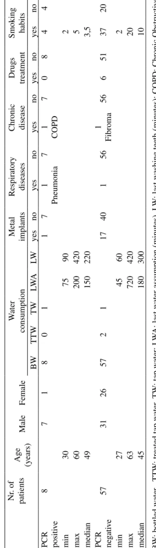
8 out of 65 plaque samples analyzed (12.3%), were positive for Legionella spp. (Table 1), one patient had a pneumonia in the past and another one suffers from chronic obstructive pulmonary disease (COPD). All these subjects drunk water and brushed their teeth 150 and 220 minutes before sampling, respectively. All the 65 patients used bottled water and only 4 drunk also tap and treated water. Most of the positive (7/8) and part of the negative (40/57) patients for Legionella spp. had no metal prosthesis. Moreover, no patient in both groups reported an infectious respiratory diseases at the moment of the sample collection.
As regards the smoking habit, the positive patients are equally distributed (4:4) with a median of 3.5 cigarettes smoked per day. Among the 57 negative patients, 37 (64.9%) were cigarette smokers (from 2 to 20 cigarettes, median 10). Some of them (n=3) also reported use of cigars and electronic cigarettes (Table 1).
389 Legionella and dental plaque
Table 1 - Positi ve (n=8) and ne gati ve (n=57) samples for Le gionella
spp. in dental plaques and main characteristics of the patients
Nr . of patients Age (years) Male Female W ater consumption Metal implants Respiratory diseases Chronic disease Drugs treatment Smoking habits BW TTW TW LWA LW yes no yes no yes no yes no yes no PCR positi ve 8 7 1 8 0 1 1 7 1 Pneumonia 7 1 COPD 7 0 8 4 4 min 30 75 90 2 max 60 200 420 5 median 49 150 220 3,5 PCR negati ve 57 31 26 57 2 1 17 40 1 56 1 Fibroma 56 6 51 37 20 min 27 45 60 2 max 63 720 420 20 median 45 180 300 10 BW : bottled w ater , TTW : treated tap w ater , TW : tap w ater; LW A: last w ater assumption (minutes), LW : last w ashing teeth (minutes); COPD: Chronic Obstructi ve Pulmonary Disease Discussion
The human oral cavity is colonized by a complex microbial community and includes several hundred different species. In the past decades, studies about the composition of the oral microbiota have been performed, especially on healthy adults or on metagenomic sequencing of microorganisms involved in gingivitis and periodontitis (4), but not well defined knowledge on the composition and the richness in species have been evidenced. It is shared opinion that oral microorganisms are in planktonic phase or organized as biofilms/dental plaque (5, 6).
The Legionnaires’ disease is a serious problem of public health. Studies on pathogenic mechanisms and way to prevent infections are continuously performed. National and international studies are widely focused on the relationship between dental practice and Legionella infections without reaching a final shared opinion (7-9).
Our study highlights the presence of
Legionella spp. in dental plaque in 12.3% of patients (8/65), with similar oral hygiene habits and risk factors except for sex, presence of past pneumonia and pulmonary chronic illnesses, which are known risk factors for legionellosis and probably influence the immune status of positive patients.
Other studies evidenced the possibility of oropharyngeal colonization by Legionella spp., mainly in the immunocompromised and cardiac patients (10), but to the best of our knowledge, this is the first study reporting the presence of Legionella spp. in human dental plaque of healthy subjects.
Further investigations are needed in order to evaluate if dental plaque could be confirmed as a new reservoir of the microorganisms. Moreover, future research studies focused on the identification of
Legionella spp. at species level, the viability of the microorganisms in the dental plaque and the possible transmission of Legionella
390 M. Tesauro et al.
through environmental monitoring, could clarify the possible role of Legionella in the dental plaque on public health aspects.
A special thank to Dr Ettore Amato for his help with the English language revision of the manuscript.
Conflicts of interest: none Funding: None
Competing interests: None declared
Ethical approval: the research has been approved by the Human Ethic Committes of the University of Milan (3 June 2015)
Riassunto
Presenza di Legionella spp. nella placca dentaria umana
Obiettivi. Lo scopo di questa ricerca è verificare la
presenza di Legionella nella placca dentaria umana.
Metodi. 65 pazienti adulti, non trattati con antibiotici
nei 2 mesi precedenti il prelievo del campione, sono stati arruolati per la raccolta della placca tra settembre 2015 e dicembre 2016. È stato somministrato un breve questionario su stile di vita e rischi per la salute. La ricerca di Legionella spp. è stata eseguita mediante semi-nested PCR.
Risultati. 8 su 65 campioni di placca (12,3%) erano
positivi per Legionella spp. Per quanto riguarda i rischi per la salute e gli aspetti legati allo stile di vita, non sono state osservate differenze rilevanti tra i pazienti coinvolti nel nostro studio, ad eccezione di due pazienti positivi che hanno riportato una BPCO in corso e una polmonite in passato.
Conclusioni. Questo studio rappresenta un passo
avanti nella conoscenza dei serbatoi del microrganismo e della ricchezza del microbiota orale.
References
1. European Centre for Disease Prevention and Control (ECDC). Legionnaires’ disease -
An-nual Epidemiological Report 2016 [2014 data]. Available from: https://ecdc.europa.eu/en/ publications-data/legionnaires-disease-annual-epidemiological-report-2016-2014-data [Last accessed 2018 Jan 16].
2. Rota MC, Caporali MG, Bella A et al. Rapporto annuale sulle legionellosi in Italia nel 2016. Not Ist Super Sanita 2017; 30(9): 3-8.
3. Huang SW, Hsu BM, Chen NH, et al. Isolation and identification of Legionella and their host amoeba from weak alkaline carbonate spring water using a culture method combined with PCR. Parasitol Res 2011; 109: 1233-41. 4. Xie G, Chain PS, Lo CC, et al.
Commu-nity and gene composition of a human dental plaque microbiota obtained by metagenomic sequencing. Mol Oral Microbiol 2010; 25(6): 391-405.
5. Arweiler NB, Netuschil L. The Oral Microbiota. Adv Exp Med Biol. 2016; 902: 45-60.
6. Wade W, Thompson H, Rybalka A, Vartoukian S. Uncultured Members of the Oral Microbiome. J Calif Dent Assoc 2016; 44(7): 447-56, 7. Petti S. Did a patient acquire Legionella
pneu-mophila from the cup filler of a dental unit or did a patient infected with L.pneumophila con-taminate the cup filler? J Hosp Infect 2017; 96: 201-2.
8. Pasquarella C, Veronesi L, Napoli C, et al. SItI Working Group Hygiene in Dentistry. Microbial environmental contamination in Italian dental clinics: A multicenter study yielding recom-mendations for standardized sampling methods and threshold values. Sci Total Environ 2012; 420: 289-99.
9. Tesauro M, Bollani M, Cesaria M, et al. Analisi delle problematiche sanitarie delle attività odon-toiatriche monospecialistiche (AOM): studio pilota nel territorio milanese. Dent Cadmos 2015; 83(3): 176-86.
10. Jaresova M, Hlozanek I, Striz I et al.
Legio-nella detection in oropharyngeal aspirates of transplant patients prior to surgery. Eur J Clin Microbiol Infect Dis 2006; 25: 63-4.
Corresponding author: Marina Tesauro, Department of Biomedical Surgical and Dental Sciences, Environmental Hygiene Lab, University of Milan, Via C. Pascal 36, 20133 Milan, Italy