Scuola Dottorale di Ingegneria
Sezione di Ingegneria dell’Elettronica Biomedica, dell’Elettromagnetismo e delle Telecomunicazioni
XXVIII CICLO DEL CORSO DI DOTTORATO
Methods for the characterization of motor control
development and its adaptation to visual biofeedback in
upright stance
Carmen D’Anna
Advisor: Prof.ssa Silvia Conforto
ii Alla mia Meravigliosa Famiglia Alla mia Cara Nonna che mi guarda da lassù Al mio unico Grande Amore
iii
K
EYWORDS
Postural control Development Vision Time to Boundary Visual Biofeedbackiv
A
BSTRACT
This PhD project deals with the study of the mechanisms underlying the development of motor control for upright stance. In particular, the contribution of vision on this development has been observed by proposing novel methods for the analysis of the phenomenon and by designing systems, based on the administration of particular visual inputs – i.e. Visual Biofeedback –, devoted to manipulate the physiological answer for enhancing the adaptation mechanisms of the motor control system.
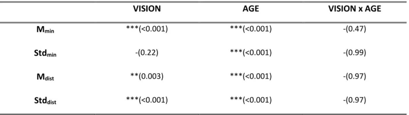
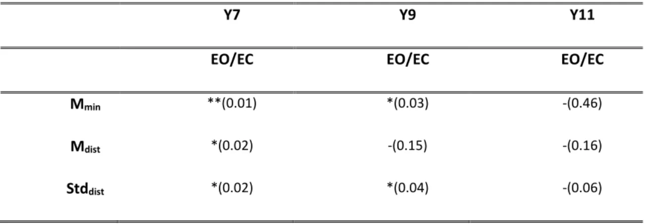
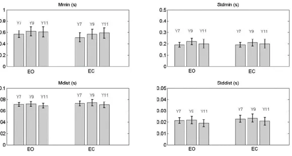
The development of postural control in children and the role of vision in this development have been studied through the analysis of the predictive measures extracted from a mathematical function called Time-to Boundary. This approach has permitted to assess several aspects of the postural control in children that traditional postural parameters do not show. In particular, some experiments based on protocols considering both static and dynamic conditions have been designed, implemented and administered to children with congenital blindness that have been compared to healthy ones. The outcomes of these experiments have highlighted some interesting results. Among the latter: i) at 11 years the healthy children show an adult-like postural control system with an effective integration of the vision input in postural control schemes; ii) in dynamic tasks the absence of vision leads the healthy children to loss the perception of the temporal limit of stability; iii) the predictive function supports the theory that excludes balance deficit in children with blindness.
The vision has then been used to manipulate the response of the postural control system and to study whether proper visual stimuli could enhance the adaptation mechanisms of that system. In
v
fact, the possibility of stimulating the adaptation processes opens interesting scenarios in the framework of the maintenance and recovery of the motor abilities. Following this rationale, different visual stimuli for biofeedback have been designed and implemented looking at the improvement of postural performance that is to the effectiveness of the Visual Biofeedback systems. In particular, the influence, which different modalities of presenting and elaborating data inside the Visual Biofeedback systems could exert on the motor performance, has been observed. Three different Visual Biofeedbacks have been designed based respectively on: i) a continuous and direct presentation of the information, ii) a discretized and indirect presentation of the information, iii) an elaboration of the centre of pressure coordinates giving raise to a predictive information. The validation of the systems, through different experimental protocols, has showed that the use of an indirect and discretized modality of presentation improves postural performance and at the same time favours a more natural postural control strategy as compared to the classical continuous Centre of Pressure presentation; moreover the Visual Biofeedback based on the predictive data elaboration improves postural performance more than the presentation of a Visual Biofeedback based on a real-time elaboration of the Centre of Pressure coordinates. By these outcomes it has been highlighted how these aspects influence in different way the postural control strategies and consequently the postural performance.
All these results extend and enhance the actual knowledge about the postural control development, with special reference to the role of vision in children and to the factors that could influence the Visual Biofeedback effectiveness, giving important suggestions for the design of tools to be spent in training and rehabilitation.
vi
A
CKNOWLEDGEMENTS
At the end of each journey we should all take some time to reflect on the moments lived. No words can fully express my gratitude to all of those who I today consider part of my second home.
Firstly, I would like to thank my advisor Prof. Silvia Conforto for her support, for her precious advice and suggestions, for giving me the possibility of developing my ideas with freedom, for conveying me the passion for biomedical engineering and research.
I would like to thank Prof. Maurizio Schmid; there has not been a single idea that I have not shared with him. His technical and moral support has allowed me to reach, gradually, every single goal that I have put in mind leading me in the right direction.
I would like to thank Prof. Tommaso D'Alessio: his teachings has been a fundamental part of my growth as a bioengineer.
A special thanks to Dr. Daniele Bibbo. He has been my mentor since the beginning of this experience. He has supported me in moments of discomfort and euphoria with his endless
encouragements always making himself available.
I would like to thank my companion in this adventure, Benish Fida, for the moments spent and shared as PhD students. Thanks to everyone in Biolab3, Ivan, Michela, Cristiano, Antonino and
Carlotta, for the moments lived in this amazing “journey”.
Last but not the least, I would like to thank my main “sponsors”: my wonderful parents, my sister Ausilia and my boyfriend Ale for supporting me every day. I wouldn’t have made it without you.
vii
TABLE OF CONTENTS
INTRODUCTION………..1
CHAPTERI:POSTURAL CONTROL: WHAT IS? INTRODUCTION ... 8
1.1THE ROLE OF THE NEUROPHYSIOLOGICAL SYSTEM IN POSTURAL CONTROL ... 10
1.2THE ROLE OF THE SENSORY SYSTEM IN POSTURAL CONTROL ... 13
1.3COMPUTATIONAL MODEL OF MOTOR CONTROL: A BRIEF OVERVIEW ... 18
BIBLIOGRAPHY ... 22
CHAPTERII:TECHNIQUES AND METHODS TO STUDY POSTURAL STABILITY INTRODUCTION ... 27
2.1TRADITIONAL APPROACH TO POSTURAL MEASURES ... 29
2.2RANDOM-WALK ANALYSIS OF THE CENTRE OF PRESSURE TRAJECTORY ... 32
2.3TIME TO BOUNDARY FUNCTION TO STUDY UPRIGHT STANCE ... 35
BIBLIOGRAPHY ... 38
PART
I:
DEVELOPMENT
OF
POSTURAL
CONTROL
SYSTEM
IN
CHILDREN
INTRODUCTION……… 41CHAPTER III: CAN TIME-TO-BOUNDARY HELP UNDERSTAND THE DEVELOPMENT OF UPRIGHT STANCE CONTROL IN CHILDREN? 3.1MATERIALS AND METHODS ... 48
3.2RESULTS ... 49
3.3DISCUSSION AND CONCLUSIONS ... 52
CHAPTER IV: TIME TO BOUNDARY FUNCTION TO ASSESS UPRIGHT STANCE IN BLIND CHILDREN 4.1MATERIALS AND METHODS ... 57
viii
4.2RESULTS ... 59
4.3DISCUSSION AND CONCLUSION ... 61
CHAPTER V: THE TIME-TO BOUNDARY FUNCTION TO ASSESS UPRIGHT STANCE IN STATIC AND DYNAMIC CONDITION: A CASE OF STUDY 5.1MATERIALS AND METHODS ... 65
5.2RESULTS ... 66
5.3DISCUSSION AND CONCLUSION ... 69
BIBLIOGRAPHY ... 71
PART II: THE EFFECT OF VISUAL BIOFEEDBACK ON POSTURAL CONTROL
INTRODUCTION……….75CHAPTER VI: THE EFFECT OF CONTINUOUS AND DISCRETIZED PRESENTATIONS OF CONCURRENT VISUAL BIOFEEDBACK ON POSTURAL CONTROL 6.1MATERIALS AND METHODS ... 82
6.2RESULTS ... 87
6.3DISCUSSION AND CONCLUSIONS ... 91
CHAPTER VII (part I): EFFICACY OF TTB-BASED VISUAL BIOFEEDBACK IN UPRIGHT STANCE TRIALS 7.I.1MATERIALS AND METHODS ... 99
7.I.2RESULTS ... 101
7.I.3DISCUSSION AND CONCLUSIONS ... 104
CHAPTER VII (part II): CAN A VISUAL BIOFEEDBACK SYSTEM BASED ON PREDICTIVE INFORMATION IMPROVE POSTURAL PERFORMANCE? 7.II.1MATERIALS AND METHODS ... 108
7.II.2RESULTS ... 111
ix BIBLIOGRAPHY ………115 GENERAL CONCLUSIONS……….. 120 APPENDIX A……….. 125 APPENDIX B……… 130 LIST OF ACRONYMS ………. 133
1
I
NTRODUCTION
The study of posture was object of interest only from the beginning of the XIX century, when the first doubts about the mechanisms related to the maintenance of upright stance began: “How does man maintain a posture upright or tilted against the wind? It’s evident that he possesses a sense by which he knows the tilting of his body and that he possesses the capacity to right it and to correct any deviation from the vertical” (Bell 1837).
After two centuries, posture is one of the most studied complex abilities in motor control and learning.
Commonly, it is defined as the relative position of parts of the body or of the whole body with respect to a reference frame, maintaining the centre of gravity of the body within its base of support.
Therefore, despite its seemingly simplicity, maintaining balance throughout postural adjustments in upright stance, is a rather complex task that involves all parts of the nervous system and that requires the integration of information coming from different sensory channels (visual, vestibular, proprioceptive and tactile). If on one hand, the main goal of the postural control is to prevent the subject from falling or losing balance, on the other the second goal is to form an interface between perception and action.
The complexity of postural control system led to the development of various techniques and methods to study upright stance. The neurophysiological studies contributed to better understand the basis of the physiological mechanisms to maintain upright stance, and the research on technical and methodological approaches contributed to assess and to analyse postural control in different conditions.
2
The classical methods of analysis, based on the extraction of the centre of pressure oscillations from data recorded from force plates, have highlighted different characteristics of sway including mean velocity, length of sway path, the area covered with the sway trajectory over a fixed time interval. The stochastic analysis of the centre of pressure oscillations during quiet stance has identified the open-loop and closed-loop components of these oscillations. Studies conducted by Loram and collegues (2011) have focussed, instead, on the biomechanical modelling to assess how the central nervous system stabilizes upright stance through two neural control strategies, which use either continuous or intermittent feedback controllers to execute sustained or ballistic movements respectively. These approaches have contributed to understand how changes in postural sway, could reflect changes in postural control strategies due to a cognitive process, to a perturbation – either internal or external –, to the presence of neuromuscular diseases, to the alteration and/or deterioration of one or more of sensory channels and more simply to the natural development of postural control with age.
About this, the age-related changes in postural control strategies are widely discussed in literature (A. Shumway-Cook and M. H. Woollacott 1985; C. Assainante 1998) When is it possible to talk about a complete development of the postural control system? How does this development depend on the evolution of the sensory systems and in particular of the vision? Which training and rehabilitation system can be used to improve postural control when a deficit occurs?
Several studies (C.L Riach and KC. Hayes 1987; K. Taguchi and C. Tada 1998; N. Kirshenbaum and colleagues 2001) have focussed their attention on the analysis of postural control from infancy to the childhood, through the study of classical postural parameters. They
3
have underlined that the development is not linearly related with age, that it follows the maturation of fine competencies in muscular coordination and that the body sway amount decreases with age.
Conflicting opinions are presented on the age at which children exhibit signs of an “adult-like” postural control strategy and at which the complete maturation of the vision system is shown. Riach et al. (1994) examined cross-sectionally the characteristics of postural sway in healthy children of different ages, by studying the spectral composition of sway, and highlighted that children until the age of 7 years use visual information to control balance in a manner different from adults; Taguchi et al. (1988) reported that the amplitude of spontaneous postural sway in children aged 9-12 with eyes open was comparable to that of adults in the same conditions; Peterson et al. (2006) suggested that children do not exhibit an adult-like sensory information use prior to age 12 years.
After having assessed the role of vision on the development and the functioning of the postural control mechanisms, even the possibility of manipulating the visual channel to adaptation and recovery phenomena has been extensively studied (M. G. Wade e G. Jones 1997; J. Laurens and collegues 2010). With the aim to improve postural control at all ages several researches have addressed the effectiveness of systems based on Visual Biofeedback: the latter has been demonstrated to provide additional information related to the human body in terms of motion and interaction with the environment, hence supplementing the natural sensory data such as, in particular, the vision.
Experiments with visual biofeedback (VBF) for postural control have been in progress since the end of XX century: typically, the subjects stand on a force plate, and watch a computer screen where a representation of the position of their centre of pressure (CoP) is supplied in
4
real-time. This type of concurrent augmented feedback can be used to control balance and to regulate body sway in either static or dynamic conditions.
The question whether visual biofeedback is beneficial to improve standing postural control is still controversial. The effect of VBF on postural control strategies, and consequently on the variation of postural sway depends on many factors. Geiger et al. (2001), in a review of patient studies, questioned the advantage of VBF therapy in bilateral standing compared with conventional therapy, whereas Dault et al. (2003) and Prosperini et al. (2013) focussed on the effect of VBF on balance training and rehabilitation, observing an improvement of the postural performance in people with disabilities or at-risk of falling.
The studies conducted by Rougier et al. (2003) and Cawsey at al. (2009) have shown that the effect and the benefit of VBF on postural stability depend not only on the instructions given to the performer but also on the adopted information representation, in particular on the scale of CoP visual display and on the time delay of the CoP presentation.
Therefore, in this complex framework, it has been decided to study in deep the role of vision on posture, trying to understand how the visual information influences the evolution and the adaptation, through the human life cycle, of the control mechanisms and whether this influence could be used for retraining. To do that this PhD project focusses on the two main following points:
the study of role of vision in the development of postural control through infancy;
the assessment of the effectiveness of VBF systems with a special emphasis on the attempt of understanding the role played by the modality of VBF presentation and/or by the modality of data elaboration.
5
To follow the first scientific goal, different experimental protocols were done with the aim to evaluate the postural control in children and the role of vision, considering static and dynamic conditions, through the study of a predictive parameter known in the literature as Time-to-Boundary. The new methodological approach has highlighted some aspects of postural control that classical posturographic parameters did not show.
The second aim was followed investigating, on one hand the impact on the control strategies of the modality of VBF presentation and on the other, how the modality of data elaboration could influence the postural performance. Three different VBFs (continuous, discretized VBF presentation and VBF based on the predictive elaboration of the CoP coordinates) were designed. The effect of VBFs was studied through different experimental protocols. The outcomes of these studies have highlighted how these aspects influence in different way the postural control strategies and consequently the postural performance.
Thesis outline
Chapter I: describes the basis of postural control mechanisms, with particular attention on the
role of neurophysiological system and on the different sensory channels.
Chapter II: describes the principal techniques and methods used to study postural control.
The thesis is then divided into two parts:
The first part describes the development of postural control in children and shows the methodological approach, based on the elaboration of the Time to Boundary function.
Chapter III: describes the development of postural control and the role the vision plays in this
development. Data have been recorded in children population from 7 to 11 years and then have been analysed through the analysis of the measures extracted from the elaboration of the Time to Boundary function.
6
Chapter IV: describes the postural control in children with blindness through the elaboration
and the analysis of the TtB. The results are compared with those obtained from data of the sighted children in the same experimental conditions.
Chapter V: describes, by the elaboration and the analysis of the TtB, the combined effect of
vision and type of the motor task – i.e. either static or dynamic – on postural control in children.
The second part introduces the Biofeedback systems in postural studies and presents the
outcomes, in terms of postural performance, obtained by Visual Biofeedback systems that have been designed and implemented in this PhD project with different specifications regarding the modalities of VBF presentation and data elaboration, respectively.
Chapter VI: shows the design of two different Visual Biofeedback systems based on the
continuous and direct presentation of the information, and on the discretized and indirect presentation of the information, respectively. A comparison of the two systems is outlined.
Chapter VII: this chapter is divided into two parts. In the first part, the design of a Visual
Biofeedback based on the real time elaboration and presentation of the Time to Boundary function is presented. The results and the comparison with the traditional Visual Biofeedback are shown and discussed. In the second part, instead, the design of a Visual Biofeedback based on the real-time elaboration of the predictive coordinates is presented. The comparison with a VBF based on the real-time elaboration of the Centre of Pressure coordinates is showed and the experimental results about the effect of this VBF on postural control are widely described.
7
CHAPTHER 1
8
Introduction
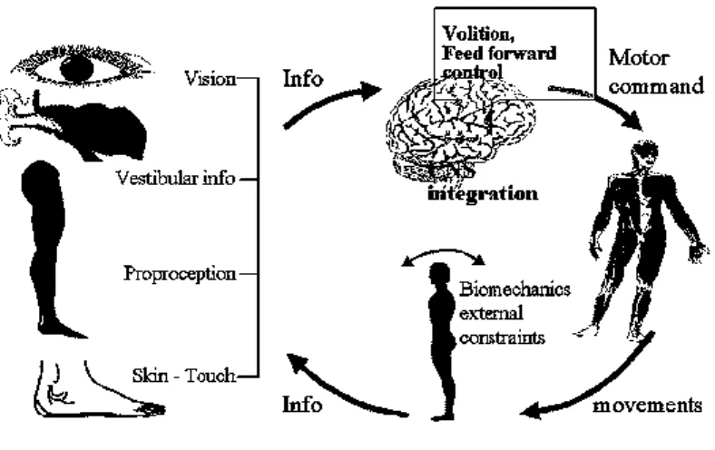
Despite the apparent simplicity, maintaining the upright stance is a very complex mechanism that requires the regulation and integration of multiple types of sensory inputs. The data from visual, vestibular and somatosensory systems are processed by the central nervous system (CNS) that puts in action the strategies needed to maintain the equilibrium (Figure 1.1).
Figure 1.1. Scheme of postural control system
(Illustration by Dr. Rolf Johansson and Prof. Måns Magnusson at Lund University, Sweden)
Two are the main functions of the postural control system: the antigravity control and the interface between perception and action [1]. The antigravity control is a mechanical function that accomplishes two processes: the first is the support of the body’s weight against gravity and ground reaction forces; the second is the equilibrium control, which requires that the projection of the centre of gravity remains inside the supporting surface under both static and dynamic conditions [2].
9
The interface between perception and action is a function that essentially controls the relationship between the external world and the body. The position and the orientation of the body segments such as the head, trunk and arms serve as a reference frame for calculating target locations in the external world and for organizing movements toward these targets.
The huge complexity of the system makes the development of the postural control system a long-term process, which is also strictly connected to the development of the central nervous system and of all sensory channels.
Therefore, if on one hand physiological theories have been developed to better understand the physiology of the postural control system, the role of each sensory channel and the complex integration of these systems; on the other the computational models, combining control theories and experimental data, have provided a powerful way to explore the biomechanical complexities of the musculoskeletal system and to infer some aspects of the control that neurophysiological approaches have not been able to highlight yet.
The role of the CNS and of the sensory system on postural control, and a brief overview about the computational model in motor control are presented in the following paragraphs.
10
1.1
T
HE ROLE OF THE NEUROPHYSIOLOGICAL SYSTEM IN POSTURAL CONTROLIn line with the functions of the postural control system, central organization includes many elements that interact to produce an appropriate motor response including sensory orientation, environment adaptation, multi-joint coordination and musculoskeletal activity. The basic circuits for stabilizing postural equilibrium are located in the high levels of CNS. The structures that have traditionally been viewed as the major players in the control of vertical posture are the cerebellum, the basal ganglia and the motor cortex [3].
Many studies demonstrated the role of the cerebellum in postural control system that seems to be devoted especially to: elaborate the visual input associated with movement; monitor the motor execution; calculate the speed of the movement and adjust the motor commands accordingly. Most of the hypotheses on cerebellar functions have been formulated from functional disruptions observed in patients. People with cerebellar damage have difficulty in keeping balance and typically they exhibit an increased postural sway [4] and a wide base of support during standing and walking [5]. Cerebellar damage in humans is also associated with hypermetric postural responses to surface displacements and with an impaired adaptation to predictable perturbations during quiet standing [6] or step initiation [7]. Lesion of the lateral hemispheres produces disorder of coordination of arm and hand without significant effect on posture [8]. Patients with restricted lesions show a normal postural response to a perturbation of the standing surface: it suggests that this response is independent from the cerebellum. The anterior lobe of the cerebellum appears critical for tuning the magnitude of postural response: typically the amplitude of the response changes on the basis of prior experience, but this does not apply to patients with deficits located in this area.
11
There are few hypotheses about the function of basal ganglia in postural control. The basal ganglia consist of many interconnected nuclei with outputs to cortical and brainstem motor system. Some basal ganglia pathways participate to descending connections to brainstem centres of locomotion in the sub-thalamus and mesencephalon and in the maintaining of postural tone; other basal ganglia pathways participate to centrally initiated motor programs including the ones devoted to control orientation and equilibrium [9]. Therefore, basal ganglia participate to different aspects of sensorimotor integration: regulation of tonic muscle activity, adaptation of motor patters to context, generation of adequate force for postural equilibrium and orientation.
The observation of patients affected by disorders of the basal ganglia and Parkinson’s disease, allows studying all these functions. Clinical Parkinson’s disease manifests itself by motor problems such as bradykinesia, flexed posture, freezing and excessive tonic activation of ankle, knee and hip flexors during quiet stance. Basal ganglia disorders are manifested by the inability to initiate voluntary movements, inability to suppress involuntary movements, abnormality in the velocity and amount of movement, and an abnormal muscle tone. Horak et al. 2005 have shown that Parkinsonian patients have difficulty in modifying the magnitude and patters of postural adjustments that are generally requested by changes of the postural demands [10]. Unlike the normal subjects, Parkinsonian patients use the same patterns of muscle activation to respond to surface displacements when standing on either narrow or wide surface. On the other hand, the basal ganglia do not appear to be essential to program the postural adjustments in voluntary movements: anticipatory postural adjustments – centrally initiated – are characterised by normal latency, standard patterns and a reduced magnitude. Furthermore the basal ganglia, influencing the spinal circuitry, contribute to decrease force generation, typical of the bradykinesia, for postural alignment and equilibrium responses.
12
The motor cortex provides a critical contribution to postural control [11]. Studies on animals have shown that the stimulation of the motor cortex in standing cats induced both a flexion movement of the contralateral forelimb and an anticipatory postural change in the supporting forelimb [12]. In addition, data from human studies demonstrate that inhibition of the motor cortex can reduce postural activity of the trunk muscles associated with voluntary limb movements [13], and can play a role in the elaboration of anticipatory postural adjustments that are necessary for the smooth and coordinated execution of the postural movements [14].
Deficits in postural control may be associated with changes in the excitability and organization of the motor cortex.
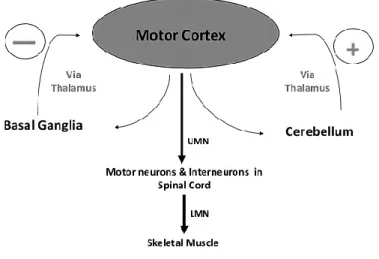
Figure 1.2 Modulation of motor activity by basal ganglia and cerebellum
Both basal ganglia and cerebellum are large collections of nuclei that modify the movement on a minute-to-minute basis. Motor cortex sends information to both, and both structures send information right back to the cortex via the thalamus (Figure 1.2). The output of the cerebellum is excitatory, while the basal ganglia are inhibitory. The balance between these two systems allows for smooth, coordinated movement, and a disturbance in either system will show up as movement disorders.
13
1.2
T
HE ROLE OF THE SENSORY SYSTEM IN POSTURAL CONTROLSensory information for postural control comes from three principle sources: the vision, the vestibular system, and the somatosensory system including muscle proprioception, joints, and cutaneous afferents.
Several studies examined the redundancy of sensory information, trying to answer the question: “Are all the channels necessary?”. The information coded by each sensory channel is unique: even if each class of receptors operates in a specific range of frequency and amplitude of the body motion, the integration of inputs from multiple channels is necessary to resolve ambiguities about postural balance and orientation. The operation of “integration” carried on by the CNS to control posture is not just a summation of the channels. The CNS needs to extract and interpret the relevant sensory information to determine the body position on the space and its orientation. The role of sensory information has been largely studied by experiments that have measured the body sway in conditions in which sensory input has been altered or limited, either by the experimenter or by pathology. The complex interaction of sensory signals during standing is reflected by changes in the postural sway. In particular standing with eyes closed gives raise to an increased sway [15], which also pops up when the vestibular system is compromised [16]. Changes in postural oscillations have been also shown in subjects who stand on a soft surface or who have a decreased sensory feedback from sensory receptors in the feet [17].
Vision is the system primarily involved in planning the locomotion and in avoiding obstacles along the way. The vestibular system is our ‘gyro’, which senses linear and angular accelerations. The somatosensory system is a multitude of sensors that sense the position and the velocity of all the body’s segments, their contact with external objects and the orientation of gravity.
14
1.2.1 The role of the Visual System
Vision provides one of the most reliable sources of information for human brain: the position of the body with respect to the environment.
The rods and cones of the retina are the visual receptors. They send to the CNS the information about the external environment. In particular, the foveal region of the retina analyses precisely the object on which attention is focussed, providing postural stability in the medio-lateral direction. Instead, the peripheral retina area sends information about the environment enhancing the postural stability in the antero-posterior direction.
Despite the visual system allows the CNS to perceive the external environment, in order to maintain the correct equilibrium, this information needs to be associated with those coming from both vestibular and proprioceptive systems. For example, the visual system cannot figure out if the flow of images on the retina is due to the whole body movement or to the head only.
Several studies showed that the control system of vertical posture strongly depends on visual information: in particular, many indexes of postural stability worsen if a person stands with eyes closed [15]. To study postural performance in case of perturbed vision, several papers used the “moving room paradigm” – room in which the subjects stand on fixed ground with the surrounding walls moving back and forth – and demonstrated that the movement of the wall induced synchronous involuntary body sway matching the room oscillations [18].
The role of the visual system has also been studied by considering the response to unexpected disturbances of balance. It has been shown that the most significant role that is played by vision in both postural orientation and equilibrium may be in the feed-forward
15
control that is critical to avoid an obstacle and to adapt to the changes of the environments [19].
1.2.2 The role of the Vestibular System
The vestibular system has been traditionally viewed as tightly linked to the postural stabilization.
The vestibular system (vestibule labyrinth) is located deeply in the temporal bone (petrous), behind the inner ear. The channel of the cochlear spiral (cochlea) is connected to a bulge – the saccule – filled with endolymph. Inside there are the microcrystals (otoliths, statoconi) and the sensory receptors (mechanoreceptors) placed in the wall of the saccule to sense the vertical acceleration.
The saccule is in communication with the vesicle, the utricle, which provides information about horizontal acceleration.
The utricle, represents the common output of the three semicircular canals of the labyrinth. The sensory receptors of the semicircular canals perceive the rotations of both head and body (angular accelerations). This receptive apparatus is very sensitive and can detect angular acceleration as small as 0.1°/s2.
All these structures contribute to provide information to the brain on the position in the space of the head and the body. In particular, it seems that only the otolithic system (saccule and utricle) participates to postural adjustment (affecting muscle tone) while the semi-circular canals intervene, only in dynamic equilibrium to detect rapid postural sway (rapid hip flexion or extension).
16
All information arrives to the vestibular nuclei, located in the brainstem that represents the true balance organ. It receives inputs from the cerebellum and from the spinal cord.
Several researches have shown that the vestibular information, in conjunction with somatosensory inputs, informs the CNS about the position and the orientation of the head to facilitate the postural orientation with respect to the gravitational force and to allow the appropriate postural response [20].
The experimental research, that typically uses an artificial stimulation of the vestibular system, showed that the postural sway changes depend on the position of the head and on the current polarity: an increase in the amplitude of the vestibular stimulation leads to an approximately linear increase of the body oscillations [21].
The studies that focussed on the role of vestibular system on falling have shown that this system is responsible for triggering the response to sudden falling: in patients with vestibular impairment, an early activation of the extensor muscles has been noticed [22]. Vice versa, vestibular inputs are not required for triggering postural responses to perturbation of the support surface: patients with bilateral loss of vestibular function show normal muscular activations in leg and trunk muscles in response to translation or rotation of surface [23] [16].
Clinical studies have shown that the patients with a deficit in the vestibular system increase the displacement and the acceleration of the head during standing; the bilateral loss of vestibular function may be associated with a forward flexed head position [24], with a more tonic activity of the neck, trunk and legs resulting in an increased stiffness.
17
Nevertheless, despite the vestibular deficit produces important effects on the motor system, characterised by changes of the postural control strategies [16], the effect on the ability to maintain quiet stance is limited.
1.2.3 The role of the Somatosensory System
The somatosensory system includes mechanoreceptors in the skin, pressure receptors in deep tissues, Golgi tendon organs, muscle spindles and joint receptors. All of these provide critical information about equilibrium and body segments configuration [2].
The receptors in the feet, legs and trunk are important to control the trunk especially when the subject is in contact with a large and stable support surface. The somatosensory system informs the CNS about the characteristics of the surface and about the forces that the body exerts against it. The cutaneous and deep mechanoreceptors are activated when there is a movement between the support surface and the feet.
Somatosensory information from the feet is important to determine the possible postural strategies that can be used under various conditions.
The importance of muscle spindles for postural orientation has been demonstrated by using mechanical vibrations to induce the illusion of leaning in standing subjects [25]. Thus, the perceived muscular stretching is interpreted as a change in the orientation of the body and it is compensated by a change of the body position in the opposite direction so resulting in vibration-induced fallings. The vibration induces also muscular activity in remote muscle groups and affects the posture depending on the interaction of the body with support surface [26].
18
The somatosensory system has an important role also to detect perturbation of stance and to trigger rapid response to maintain upright stance.
Finally, joint receptors provide information about angular displacements and consequently influence the body sway.
1.3
C
OMPUTATIONAL MODEL OF MOTOR CONTROL:
A BRIEF OVERVIEWThe studies based on the neurophysiological and biomechanical aspects of motor control have highlighted the neuroanatomical pathways, the roles of the different parts of the CNS and of the sensory systems. Nevertheless, these researches till now have not exhaustively detected the link between the functional structures of the CNS and the biomechanical architecture of the human body. Even if it is known that any gesture is the culmination of highly organized processes, which include perception schemes, anticipative planning, feedback corrections, muscular synergies and other internal elaboration systems, the all framework has not been completely understood yet.
An alternative attempt to disentangle the complexity of the motor control has been put in action by using computational approaches. In that way, the control system theory has been exploited so trying to analyse the biological motor control as a nonlinear control problem, where the CNS plays as the controller and the body as the controlled object.
The control scheme is complicated by the following elements:
the environment that is conceived not only as a reference system but also as a “provider of affordances”, which are specific information accessible during the execution of an action;
19
the “sensors” which are appointed to gather all the affordances, both from the “inside system” and the “outside world” and to supply these signals to the CNS: the sensors comprise the perception which is the process whereby sensory excitation is translated into organized experience. That experience is the joint product of the excitation and the process itself, particularly in the perception and representation of space [27].
The motor theory, illustrated by A. Berthoz (1997), is based on the concept that perception is not a passive mechanism for receiving and interpreting sensory data but is the active process to anticipate the sensory consequences of an action [28]. In computational terms, an “internal model” represents the link between the brain and the environment: it is built by sensorimotor associations between an out-going signal (the efferent copy) and the corresponding sensory re-afference. The coherence of the two representations is the basis for the stability of our sensorimotor world. These and other models have been used to explain from a mathematical point of view the ecological nature of the motor control [27].
Therefore, the human motor controller is able to manage the movements thanks to the integration of the information concerning the effectors and to the relations between the environment and the effector itself.
The question at the basis of several computational models deals with the assessment of the active neural control strategies in motor control. In other words, computational models try to suggest which types of feedback controllers the CNS utilizes to stabilize the movements in general and the posture in particular.
Maintaining balance in quiet stance or in dynamic conditions requires ongoing regulation provided through proprioceptive and exteroceptive intrinsic feedback [29]. Several studies have
20
shown that the CNS stabilizes upright stance through two neural control strategies, which use either continuous [30] or intermittent [31] [32] feedback controllers to execute sustained or ballistic movements respectively. The control of sustained movements has been explained and interpreted within the framework of the continuous control theory and it has been often modelled by using a continuous disturbance signal [30] [33]. The delay between stimulus and response is variable and reflects the fact that the responsiveness to sensory information is not constant.
Asai and colleagues [34] showed that the response might be triggered by a stimulus crossing a threshold so requiring an action; the response is constructed and executed in a serial fashion that is, even if the sensory information may be assimilated continuously, the system responds only at a particular time instant that is the one when the action is executed [35].
Loram and colleagues [32] have compared continuous and intermittent control by considering the effect of external stimuli and/or a perturbation on postural stability in quiet standing. They have further shown that event-driven intermittent control provides a framework to explain human behaviour under a wider range of conditions than continuous control, and that intermittent open loop action is a natural consequence of human physiology [31] [32]. Interestingly, Gawthrop and colleagues [36] have also demonstrated that in the presence of continuous disturbances and small event thresholds, the intermittent sampling becomes regular masquerading itself as a continuous-time control; instead, in the presence of discretized disturbances with an irregular trigger frequency, the maintenance of the natural intermittent postural control strategy is favoured.
The computational model developed by Bottaro and colleagues [31] has highlighted that continuous feedback control is potentially less efficient, from an energetic point of view, than intermittent impulsive control in quiet stance. They showed that the intermittent model could be
21
adapted in a natural way to each standing paradigm using the different sensory channels and the different effectors to evaluate the state of the pendulum (model of the body) within a stability region.
The role of each efferent system in the control model depends on the different standing tasks and on the type of applied stimulus: for example, in a postural control paradigm with a normal surface, it relies on proprioceptive information, while if the base of support is modified, it relies mostly on vestibular and visual information. In tasks of balancing a pole on a finger, vestibular information does not help whereas vision becomes predominant. The effect of the modality of sensory input on the model of control has been recently studied. A research focussed on the effect of a continuous or intermittent manual contact with a joystick to control a load through a visual biofeedback presented continually. The study showed that despite the visual information was available continuously, the non-linear process “acts intermittently” [32].
All of these computational approaches, integrated with the neurophysiological knowledge, and with the experimental research allow to better study and understand the complex nature of postural control system and to assess the strategies adopted in the interaction and adaptation to the external environment.
22
Bibliography
[1] J. Massion, A. Aleandrov e A. Frovo, «Why and how are posture and movement coordinated?,» Progress in Brain Research, pp. 13-26, 2004.
[2] F. B. Horak e J. M. Macpherson, «Postural orientation and equilibrium,» in Handbook of Physiology, Exercise: Regulation and Integration of Multiple Systems, 1996.
[3] M. L. a. M. Hadders-Algra, «what is posture and how is it controlled,» in Postural_Control_A_Key_Issue_in_Developmental_Disorders, 2008.
[4] J. Dichgans e H. C. Dinier, «Postural ataxia in late atrophy of the cerebellar anterior lobe and its different diagnosis,» in Vestibular and Visual Control on Posture and Locomotor Equilibrium, Blacks, 1985, pp. 282-289.
[5] B. P. van de Warrenburg, H. Hendriks, A. Durr e et al., «Age at onset variance analysis in spinocerebellar ataxias: a study in a Dutch-French cohort,» Ann Neurol, pp. 505-512, 2005. [6] F. B. Horak e H. C. Dinier, «Cerebellar control of postural scaling and central set in stance,»
Journal of Neurophysiology, vol. 72, n. 2, pp. 479-493, 1994.
[7] D. Timmann e F. B. Horak, «Perturbed step initiation in cerebellar subjects,» Experimental Brain Research, vol. 119, pp. 73-84, 1998.
[8] J. Dichgans e H. C. Dinier, «Different forms of postural ataxia in patients with cerebellar deseases,» in Disorders of posture and gait, Amsterdam, W.bles and T. Brandt, 1986, pp. 207-215.
[9] G. E. Alexander, M. R. Delong e P. L. Strick, «Parallel organnization of functionally segregated circuits linking basal ganglia and cortex,» Annu. Rev. Neurosci, vol. 9, pp. 357-382, 1986. [10] F. B. Horak, D. Dimitrova e J. G. Nutt, «Direction-specific postural instability in subjects with
Parkinson's disease,» Experimental Neurology, vol. 193, pp. 504-521, 2005.
[11] T. G. Deliagina, I. N. Beloozerova, P. V. Zelenin e G. N. Orl, «Spinal and supraspinal postural networks,» Brain research reviews, vol. 57, n. 1, pp. 212-221, 2008.
23
[12] Y. Gahery e A. Nicoullon, «Postural and kinetc coordinationfollowing cortical stimuli which induce flexion movements in the cat's limbs,» Brain Res, vol. 155, pp. 25-37, 1978.
[13] P. Hodges, A. Kaigle Holm, S. Holm e et al.,, «Intervertebral stiffness of the spine is increased by evoked contraction of transversus abdominis and the diaphragm: in vivo porcine studies,» Spine, vol. 28, n. 23, pp. 2594-2601, 2003.
[14] V. Gurfinkel e A. M. Elner, «Contribution of the frontal lobe secondary motor area to organization of postural components in human voluntary movements,» Neurophysiology, vol. 20, pp. 5-10, 1988.
[15] J. Doman, R. Geoff e P. J. Holliday, «Visual inputs: its importance in the control of postural sway,» Archives of physical medicine and rehabilitation, vol. 59, n. 12, pp. 586-591, 1978. [16] F. B. Horak, L. M. Nashner e H. C. Dinier, «Postural strategies associated with somatosensory
and vestibular loss,» Experimental Brain Research, vol. 82, n. 1, pp. 167-177, 1990.
[17] A. Shumway-Cook e F. B. Horak, «Assessing the influence of sensory interaction on balance suggestion from the field,» Physical Therapy, vol. 66, n. 10, pp. 1548-1550, 1986.
[18] T. M. Dijkstra, G. Schoner e C. C. Gielen, «Temporal stability of the action perception cycle for postural control in a moving visual environment,» Exp Brain ReRes, vol. 97, pp. 477-4887, 1994.
[19] T. Drew, «Visuomotor coordination in locomotion.,» Curr. Opin. Neurobiol., vol. 1, pp. 652-657, 1991.
[20] J. M. Macpherson e J. T. Inglis, «Stance and balance following bilateral labyrinthectomy,» in Natural and Artificial Control of Hearing and Balance, New York, Elseviers Science Publishers, 1993, pp. 219-228.
[21] A. C. Coats e M. S. Stoltz, «The recorded body-sway response to galvanic stimulation of the labyrinth: a preliminary study.,» Laryngoscope, vol. 79, pp. 85-103, 1969.
[22] R. Greenwood e A. Hopkins, «Muscle responses during sudden falls in men.,» J. Physiol., vol. 254, pp. 507-518, 1976.
[23] J. H. Allum, F. Honegger e H. Schicks, «Vestibular and proprioceptive modulation of postural synergies in normal subjects.,» J. Vestb. Res., vol. 3, pp. 59-85, 1993.
[24] C. L. Shpert, F. O. Black, F. B. Horak e L. M. Nashner., «Coordination of the head and body in response to support surface traslations in normals and patients with bilaterally reduced vastibular function.,» in Posture and Gait: Development, Adaptation and Modulation, New York, Elsevier Science Publishers, 1988, pp. 281-289.
24
[25] J. R. Lackner e M. Ievine, «Changes in apparent body orientation and sensory localization induced by vibration of postural muscles: vibratory myestetic illusion,» Aviat. Space Environ. Med., vol. 50, pp. 346-354, 1979.
[26] V. S. Gurfinkel, M. A. Lebedev e Y. S. Levik., «Effects of reversal in the human equilibriumregulation system.,» Neurophysiology, vol. 24, pp. 297-304, 1992.
[27] P. Morasso, «Motor Control Models: learning and performance,» in International Encyclopedia of the Social & Behavioral Sciences, Section: Mathematics and Computer Sciences, Pergamon Press, 2000.
[28] A. Berthoz, Le sens du mouvement, Édition Odile Jacob, 1997.
[29] R. Balasubramaniam e A. M. Wing, «The dynamics of standing balance.,» Trends in cognitive sciences, vol. 6, n. 12, pp. 531-536, 2002.
[30] H. Van Der Kooij e E. De Vlugt, «Postural responses evoked by platform pertubations are dominated by continuous feedback,» Journal of neurophysiology., vol. 98, n. 2, pp. 730-743, 2007.
[31] A. Bottaro, Y. Yasutake, T. Nomura, M. Casadio e et al.,, «Bounded stability of the quiet standing posture: an intermittent control model,» Human movement science, vol. 27, n. 3, pp. 473-495, 2008.
[32] I. D. Loram, H. Gollee, M. Lakie e P. J. Gawthrop, «Human control of an inverted pendulum: is continuous control necessary? Is intermittent control effective? Is intermittent control physiological?,» The Journal of physiology, vol. 589, n. 2, pp. 307-324, 2011.
[33] T. Kiemel, A. J. Elahi e J. J. Jeka, «Identification of the plant for upright stance in humans: multiple movement patterns from a single neural strategy.,» Journal of neurophysiology, vol. 100, n. 6, pp. 3394-3406, 2008.
[34] Y. Asai, Y. Tasaka, K. Nomura e et al.,, «A model of postural control in quiet standing: robust compensatin od delay-indiced instability using intermittent activation of feedback control,» Plos One, vol. 4, n. e6168, 2009.
[35] M. Lakie e I. Loram, «Mnually controlled human balancing using visual, vestibular and proprioceptive senses involves a common, low frequency neural process,» J Physiol, vol. 577, pp. 403-416, 2006.
[36] P. Gawthrop, I. Loram e M. Lakie , «Intermittent control: a computational theory of human control,» Biol Cybern, vol. 104, pp. 31-51, 2011.
26
CHAPTHER II
Techniques and Methods to study Postural
Stability
27
Introduction
How to assess and to quantify the postural stability, was the object of a lot of studies and researches. As already presented in the previous chapter, the complex interaction of sensory signals during standing is reflected in changes of the postural sway.
When executing an upright stance task, several characteristics of the vertical posture change through time. Among those: the position of the Centre of Mass (CoM), the inclination of the trunk, the variation of the Centre of Pressure (CoP). Since the CoM is in continuous motion as the body sway, the muscle forces vary continuously such as the ground reaction force [1]. However, it is commonly accepted that the oscillations of the CoP can be indirectly linked with the so-called body sway, defined as the oscillation of the lower legs, with respect to the trunk.
Therefore, to evaluate the characteristics of the postural sway and consequently of the postural stability, a variety of monitoring devices has been developed, such as video-based systems, accelerometers, and force plates. In particular, force plate data have the advantage of being directly related to the reaction forces exerted by the ground, and thus they incorporate information on both the amount of unbalance and the mechanisms that are used to counteract it.
The reconstruction of the instantaneous amplitude orientation of the resultant ground reaction force vector and its application point (CoP) is possible thanks to the insertion of load transducer elements on specific positions on the force plate pillars, and through post processing mathematical operations.
28
The force plate are today used in different clinical field of rehabilitation to monitor or re-educate standing balance in patients with hemiplegia, brain injury, amputation of the lower limb or neuromuscular diseases.
How is it possible to elaborate the data extracted from force plate (forces and torques) to quantify postural steadiness in different conditions?
The classical method of analysis has been summarized by Prieto et al. [2] It is based on the representation of the CoP coordinates in both time and frequency domains to characterize the postural steadiness. The effect of age and vision conditions has been studied by following this kind of approach. Collins and De Luca [3], instead, showed that postural sway for healthy adults exhibited power law behaviours. The CoP trajectories were analysed as a one-dimensional and two-dimensional random walk, suggesting that the CoP sway could be proficiently represented as a stochastic process and that two-control systems (long-term and short-term mechanisms) could be seen to operate during quiet standing. Slobounov et al. [4] introduced the Time to Boundary (TtB) function in the framework of postural stability by using a concept already defined in the theory of the time-to-collision in visual perception. This function, together with the relative measures extracted by it, specifies the spatiotemporal proximity of the CoP to the stability boundary.
29
2.1
T
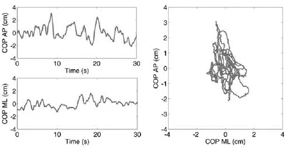
RADITIONAL APPROACH TO POSTURAL MEASURESThe classical postural measures are based on the elaboration and on the analysis of the CoP coordinates. The graphical representation of the CoP displacement is used as a tool to evaluate balance: in particular, the stabilogram is defined as the course of the medio-lateral (CoPML) and the antero-posterior component of the CoP (CoPAP) over time, and the
statokinesigram, instead, represents the movement of the CoPAP component with respect to the
movement of the CoPML component ( see Figure 2.1).
Figure 2.1. Example of Stabiligram on the left and Statokinesigram on the right.
From the CoPML and CoPAP displacements, most studies [ref ????] characterized postural
steadiness to evaluate the changes with age and with the occurrence of neurological diseases, the effect of rehabilitation interventions, pharmacologic treatments and risk of falling in the elderly.
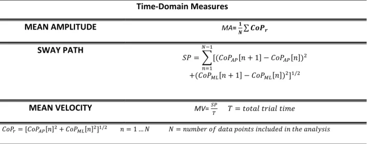
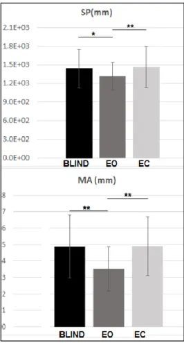
The time-domain measures are the most commonly used. These include the mean amplitude (MA), representing the average distance of the CoP displacement from its mean value,
30
the total length of the path (SP), the mean velocity the CoP (MV) calculated by dividing SP by the total trial time. The formulas are reported in Table 2.1.
Table 2.1. Time domain measures
Time-Domain Measures
MEAN AMPLITUDE MA= 𝟏𝑵∑ 𝑪𝒐𝑷𝒓
SWAY PATH 𝑆𝑃 = ∑[(𝐶𝑜𝑃𝐴𝑃[𝑛 + 1] − 𝐶𝑜𝑃𝐴𝑃[𝑛])2 𝑁−1 𝑛=1 +(𝐶𝑜𝑃𝑀𝐿[𝑛 + 1] − 𝐶𝑜𝑃𝑀𝐿[𝑛])2]1/2 MEAN VELOCITY MV= 𝑆𝑃𝑇 𝑇 = 𝑡𝑜𝑡𝑎𝑙 𝑡𝑟𝑖𝑎𝑙 𝑡𝑖𝑚𝑒 𝐶𝑜𝑃𝑟= [𝐶𝑜𝑃𝐴𝑃[𝑛]2+ 𝐶𝑜𝑃𝑀𝐿[𝑛]2]1/2 𝑛 = 1 … 𝑁 𝑁 = 𝑛𝑢𝑚𝑏𝑒𝑟 𝑜𝑓 𝑑𝑎𝑡𝑎 𝑝𝑜𝑖𝑛𝑡𝑠 𝑖𝑛𝑐𝑙𝑢𝑑𝑒𝑑 𝑖𝑛 𝑡ℎ𝑒 𝑎𝑛𝑎𝑙𝑦𝑠𝑖𝑠
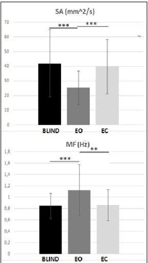
The time domain “hybrid” measures model the stabilogram as a combination of distance measures. The sway area (SA) is calculated as the area enclosed by the CoP path per unit of time. Approximately, this measure is the sum of the area of the triangles formed by two consecutive points on the CoP path and the mean of the CoP.
The mean frequency (MF) defined as the rotational frequency of the CoP if it had travelled the total excursion around a circle with radius equal to the mean amplitude. It can be considered as a combined measure of sway excursion and frequency. The formulas are reported in Table 2.2.
Table 2.2. Hybrid measures
Hybrid Measures
SWAY AREA SA= 2𝑇1 ∑𝑁−1𝑛=1|𝐶𝑜𝑃𝐴𝑃[𝑛 + 1]𝐶𝑜𝑃𝑀𝐿[𝑛] − 𝐶𝑜𝑃𝐴𝑃[𝑛]𝐶𝑜𝑃𝑀𝐿[𝑛 + 1]|
MEAN FREQUENCY 𝑀𝐹 = 𝑀𝑉
31
The frequency domain measures characterize the frequency distribution of the displacement of the CoP. From the density power spectrum of the AP and ML CoP time series, the mean power frequency in both directions is extracted (MpfML and MpfAP). The centroidal frequency
(CFreq) is calculated as the frequency where the spectral mass in concentred. Finally, the 95% power frequency is calculated as the frequency below which the total power is found. The formulas are reported in Table 2.3.
Table 2.3. Frequency Domain Measures
Frequency -Domain Measures MEAN POWER FREQUENCY
MpfML= ∫ 𝑓 𝐹𝑐 2 0 𝑃𝐶𝑜𝑃𝑀𝐿(𝑓)𝑑𝑓 ∫0𝐹𝑐/2𝑃𝐶𝑜𝑃𝑀𝐿(𝑓) MpfAP= ∫ 𝑓 𝐹𝑐 2 0 𝑃𝐶𝑜𝑃𝐴𝑃(𝑓)𝑑𝑓 ∫0𝐹𝑐/2𝑃𝐶𝑜𝑃𝐴𝑃(𝑓) CENTROIDAL FREQUENCY CFreqML=√ ∫ 𝑓2 𝐹𝑐 2 0 𝑃𝐶𝑜𝑃𝑀𝐿(𝑓)𝑑𝑓 ∫0𝐹𝑐/2𝑃𝐶𝑜𝑃𝑀𝐿(𝑓) CFreqAP= √ ∫ 𝑓2 𝐹𝑐 2 0 𝑃𝐶𝑜𝑃𝐴𝑃(𝑓)𝑑𝑓 ∫0𝐹𝑐/2𝑃𝐶𝑜𝑃𝐴𝑃(𝑓) 95% POWER FREQUENCY F95%ML=0.95 ∙ ∫0𝐹𝑐/2𝑃𝐶𝑜𝑃𝑀𝐿(𝑓)𝑑𝑓 F95%AP= 0.95 ∙ ∫0𝐹𝑐/2𝑃𝐶𝑜𝑃𝑀𝐿(𝑓)𝑑𝑓 𝑃𝐶𝑜𝑃𝐴𝑃(𝑓) = |𝐹(𝐶𝑜𝑃𝐴𝑃(𝑡))| 2 𝑃 𝐶𝑜𝑃𝑀𝐿(𝑓) = |𝐹(𝐶𝑜𝑃𝑀𝐿(𝑡))| 2
The above-mentioned measures are generally used in clinical practice and in a lot of research studies. Nevertheless, in some cases they are not very "sensitive" to highlight some pathological aspects and to allow the correct evaluation of postural control and of its development. For example, some patients with severe neurological deficits have normal sway amplitudes during quiet stance. Thus, using only one measure may not always provide a valid indication of the balance function.
32
Therefore, new different approaches to the extraction of posturographic parameters have been developed.
2.2
R
ANDOM-
WALK ANALYSIS OF THE CENTRE OF PRESSURE TRAJECTORYThe classical postural measures ignore the dynamic characteristics of stabilogram, that are, for example, the magnitude and the direction of the displacements between adjacent points, or the temporal ordering of a series of CoP coordinates. In 1993 Collins and De Luca [3] introduced a method known as stabilogram-diffusion analysis that provides a quantitative statistical measure of the apparently random variations of CoP trajectories.
They postulated that the movement of the CoP during upright stance could be modelled as a system of coupled, correlated random walks, in which the motion is considered the combination of deterministic and stochastic mechanisms. This analysis generates a stabilogram diffusion function that summarizes the mean square CoP displacement as a function of the time interval for a CoP trajectory made up of n points.
The displacement analysis of the CoP trajectories is carried out by computing the square of the displacements between all pairs of point separated in time by an interval t. The square displacements <𝑟2 >, were averaged over the number t making up the CoP time series (See Figure 2.2). The plot of <𝑟2 > versus the time interval is the stabilogram- diffusion plot.
33 Figure 2.2. Method to calculate the mean square planar displacement as a function of the time interval for a
CoP trajectory made up of N data point.
The stabilogram diffusion plot shows two parts that suggest the presence of two different control regimes: over short-term intervals of time, the postural control system utilizes an open-loop control scheme, whereas over long-time intervals closed-open-loop control mechanisms are called into play [5]. These two regions are separated by a transition period over which the slope of the plot changes (see Figure 2.3).
Figure 2.3. Schematic representation of a planar stabilogram diffusion plot
Three sets of posturographic parameters are extracted from the stabilogram-diffusion analysis, according to the laws of the classical fractional Brownian motion. The diffusion coefficients (D) reflect the level of stochastic activity and of energy of the CoP. From a
34
physiological point of view the diffusion coefficients characterize the stochastic activity of the open loop and closed loop postural control mechanism, respectively in the short-term (Drs) and long-term (Drl). They are calculated from the slopes of the resultant liner-liner plots mean square
CoP displacements versus t, in both regions (Eq. 2.1).
<𝑟2 >= 2𝐷
𝑟𝑡 Eq.2.1
The scaling exponents, H, is a real number in the range [0,1], calculated from the slopes of the resultant log-log plots of the mean square CoP displacements versus t (Eq.2.2) in both regions.
H is a representative marker of the persistence: the classical Brownian motion is characterized by H=0.5. It can be demonstrated that H>0.5 represents a persistent motion, and H<0.5 is representative of an anti-persistent motion.
< 𝑟2 > ~𝑡2𝐻𝑟 Eq.2.2
Finally, the third parameter is the critical point coordinates that approximates the transition between the two regions. This point is determined as the intersection point of the straight lines fitted to the two regions of the linear-linear version of each resultant stabilogram-diffusion plot. From a physiological point of view, it represents the temporal and the spatial characteristics of the region over which the postural control system switches from open-loop to closed-loop control. This approach was used in several studies. Peterka demonstrated that a very simple closed-loop control model of upright stance can generate realistic stabilogram diffusion function [5]; Collins’ group proved that some of these parameters highlight some dysfunctions of the postural control system, as in the Parkinson disease [6] and the difference in postural control mechanism between young and old people [7].
35
2.3
T
IME TOB
OUNDARY FUNCTION TO STUDY UPRIGHT STANCEAs highlighted in the first chapter, it is generally recognized that the goal of the postural control system is to maintain the CoP within the boundary of the base of support. It is evident that this aspect is not included in the classical approach to the study of postural stability.
Based on the Lee’s work on visual information about the time-to-collision [8], Riccio theorized that a fundamental perceptual variable in postural control is the spatio-temporal proximity to the stability boundary [9]. A measure of this is defined “Time to-Boundary function” (TtB).
This predictive variable is directly perceivable by the individual and provides information regarding the time needed to reverse a perturbation before losing balance [9]. The TtB uses current position, velocity and acceleration of the CoP trajectory to estimate the time required to the CoP coordinates to travel along the trajectory and touch the limits of the base of support (see Figure 2.4).
36
From the CoP coordinates, the TtB function is estimated as the predicted instance in time (τ) when the instantaneous CoP trajectory (𝑥𝑖, 𝑦𝑖) would cross the boundary limits, as predicted by
a parabolic motion driven by the position (𝑟𝑥, 𝑟𝑦), velocity (𝑟𝑥̇ , 𝑟𝑦̇ ) and acceleration (𝑟𝑥̈ , 𝑟𝑦̈ ) of the
CoP data at time instant ti, according to the following equations (Eq. 2.3):
𝑥𝑖(𝜏) = 𝑟𝑥(𝑡𝑖) + 𝑟𝑥̇ (𝑡𝑖) ∙ 𝜏 + 𝑟𝑥̈ (𝑡𝑖) ∙ 𝜏2 2 Eq. 2.3 𝑦𝑖(𝜏) = 𝑟𝑦(𝑡𝑖) + 𝑟𝑦̇ (𝑡𝑖) ∙ 𝜏 + 𝑟𝑦̈ (𝑡𝑖) ∙ 𝜏2 2
The TtB function has been shown to follow a pseudo-periodic behaviour, with the alternation of valleys (minima), when approaching the boundary limits, and peaks (maxima), when turning from one direction to another (See Figure 2.5). The average value of the TtB minima and its standard deviation are two of the parameters extracted from the function: the first is associated with the biomechanical constraints; the second depends on the shape of the CoP trajectory with respect to the boundary limits. The information about the temporal distance between successive minima (mean value and standard deviation) is, instead, representative of the intervention rate of the postural control: the inversion of the TtB function is a direct consequence of the ability of the control system to move the CoP away from the limits of stability [10] . Correspondingly, a lower average value of this temporal distance can be hypothesized as linked to a higher intervention rate of the control system.
37 Figure 2.5 example of TtB waveform
Several researches have suggested that TtB is more sensitive than traditional parameters in studying postural control [11] in different adult population samples (i.e young, old people) [12] [13] composed by either healthy or affected by musculoskeletal disorders [14] participants.
Recently, the parameters extracted from TtB were used to detect postural deficits that traditional parameters were unable to detect, in particular for unilateral chronic ankle instability [14] and for anterior knee pain [15].