Received: 01 May 2020 Accepted: 22 June 2020 Published: 08 July 2020 http://www.aimspress.com/journal/biophysics
Research article
Interdisciplinary approaches to the study of biological membranes
Domenico Lombardo1,*, Pietro Calandra2, Maria Teresa Caccamo3, Salvatore Magazù3 , Luigi Pasqua4 and Mikhail A. Kiselev5
1
Consiglio Nazionale delle Ricerche, Istituto per i Processi Chimico-Fisici, 98158 Messina, Italy
2
Consiglio Nazionale delle Ricerche, Istituto Studio Materiali Nanostrutturati, 00015 Roma, Italy
3
Dipartimento di Scienze Matematiche e Informatiche, Scienze Fisiche e Scienze della Terra, Università di Messina, 98166 Messina, Italy
4
Department of Environmental and Chemical Engineering, University of Calabria, Rende (CS), Italy
5
Frank Laboratory of Neutron Physics, Joint Institute for Nuclear Research, Dubna, Moscow, Russia
* Correspondence: Email: [email protected]; Tel: 3909039762222; Fax: +3909039762252. Abstract: The investigation of the structural features in biological membranes represents a central topic in many aspects of biological science. It involves the study of the collective behavior of a great number of interacting macromolecules, while the study of the structure-function relationship require the observation and calculation of a large number of key factors. The self-assembly processes involved in biomembranes represent the cornerstone of the biological systems functioning, due to the special role of the complex macromolecular assemblies containing lipids, proteins, carbohydrates, nucleic acids, and other active components. In this article, we describe the main techniques and approaches employed for the investigation of biological membranes, which are characterized by a wide range of the space-time domains. The future challenge in this research field must provide the integration of the different research models and approaches into a common background based on multi- and interdisciplinary method that combine the expertise coming from the different disciplines. Keywords: biological membranes; soft interaction; selfassembly; biotechnology; lipodomics
1. Introduction
intensive research both from the experimental and theoretical point of view, as their structure-function relationship and their related dynamic processes represents a central topic at the crossroad of different research fields, including physics, biochemistry, chemical engineering, biotechnology, and nano-medicine [1–3]. Many fundamental functions of biological membranes (such as signaling, selective transport, cell adhesion communication and recognition) are connected to processes that take place in a wide range of relevant distances and time scales, and involve the collective behavior of numerous interacting macromolecules [4–6]. Moreover, the interaction of biomolecules and nanoparticles with biomembranes strongly influences its biodistribution at the target sites within the tissues and organisms [7–9]. Therefore, the investigation of the processes occurring on the cellular and model membranes, as well as their interaction with nanocarriers in the physiological (or pathological) situations, is crucial for the development of novel therapeutic potential strategies [10–14]. For those scopes, the structural and dynamic description of biomembranes requires the calculation of a large number of simultaneous factors and development of interdisciplinary approaches in order to support the conceptual understanding and to establish fruitful cross-disciplinary connections.
2. Biomembranes: A central topic at the crossroad of different research fields
The cellular membranes of living organisms are permeable interfaces between the environment of the extracellular fluid and the internal cytoplasm. This protective barrier of the cell, which is mainly composed of lipids, proteins and carbohydrates, is involved in many important cellular functions such as sensing and signaling, transport of drug components, adhesion and recognition processes [15,16]. According to the model proposed by Singer and Nicolson (fluid mosaic model) [17], the biological membranes in living systems look like a mosaic of (highly flexible) fluid lipids bilayer structures containing both integral protein (which are anchored within the lipid bilayers by means of hydrophobic interactions), and peripheral proteins (which are attached, by weak ionic bonds, to the polar headgroups of the biomembranes lipids or to the integral proteins). Finally, the transmembrane
proteins that span the biomembrane bilayer are responsible for transporting ions or small molecules
between the two sides the cell membrane (Figure 1). The (affinity-based) assembly of amphipathic lipids and proteins confers a highly dynamic character to the fluid membranes structures, thus allowing rotation (around their individual axes perpendicular to the membrane), translation (across and parallel to the membrane), and trans-bilayer (lipids flipping between opposite external planes) movements [2]. Moreover, plasma membrane of mammalian cells exhibit an asymmetry in (internal and external) membrane leaflets, whereby phosphatidylcholine and sphingomyelin are predominantly located in the outer monolayer, whereas amino-phospholipids are mainly located in the inner monolayer [3]. Finally, (long) carbohydrate macromolecules are attached to proteins on the outside leaflet of the plasma membrane. Proteins that are embedded in the plasma membranes favors the cell interaction with its bio-environment and facilitate the carrying out of nutrients and active compounds across the biomembrane. Moreover, they favor the cell anchoring in the transmission/translation of chemical signals from outside the cell (for intracellular tasks and processes) [16,17]. Proteins also help in anchoring some fibers (cytoskeleton) to give the cell its shape, while some membrane-bound enzymes allow to perform certain reactions which are needed for some specific cellular activities.
Figure 1. Representation of the fluid bilayer model of a biological membranes in living organism (fluid mosaic model).
The cytoskeleton is composed of a network of interlinking long protein filaments present in the cytoplasm of eukaryotic cells. Those filaments, that extends from the cell nucleus to the cell membrane, create a framework that maintains the shape of the cells and give them a mechanical resistance to deformation. The cytoskeleton underlies many aspects of cell physiology, including cell division, mitosis, and extracellular matrix patterning [18,19]. The proteins of the cytoskeleton interact with membranes and regulates the cytoskeletal dynamic remodelling. The cytoskeletons contributes to the cell wall biosynthesis by regulating the transportation and deposition of the components of the cell walls. This control is achieved by the tethering of these structures to the plasma membrane. The cell wall, plasma membrane and cytoskeleton nexus (mediated by the bridging of a single or several proteins) form a scaffolding structure that may participate in signaling pathways. Cytoskeleton signals transmission across cellular membranes is far less understood [18,19]. Recent work has demonstrated that the membrane alone (or through membrane-associated proteins) can stimulate dynamic changes to the cytoskeleton [18,19]. Some mechanistic links between membranes and the cytoskeletal networks (composed of actin, microtubule, and septin) are able to integrate various signals received at the membrane, and facilitate distinct functions in response [19]. These studies highlight the membrane’s central role in coordinating these cytoskeletal systems to carry out important cell physiology processes, such as endocytosis and cellular compartmentalization. The membrane–cytoskeleton interactions are central to understand how the cytoskeletal remodelling process is integrated throughout cells and tissues. Most of the intracellular and extracellular signaling cues, in fact, have to signal through a lipid bilayer before reaching the cytoskeleton, that receives, integrates, and transmits the signals across the plasma and intracellular membranes.
proposed in order to explain the protein role in some specific cellular membrane processes [20–22]. Lipid rafts are (tightly packed) liquid-ordered phases composed of cholesterol and sphingolipids in the outer exoplasmic leaflet, that are connected to cholesterol and phospholipids (such as phosphatidylcholine, phosphatidylethanolamine, and phosphatidylserine) in the inner cytoplasmic leaflet of the lipid bilayer [22–24]. Moreover, a recent study evidenced that plasma membranes are asymmetric in lipid unsaturation, packing and protein shape, with the cytoplasmic leaflet being approximately twofold more unsaturated than the exoplasmic leaflet (which exhibit a more packed and less diffusive behavior than the inner leaflet) [25]. Because the different composition of the two monolayers of the plasma membrane the free energy of each monolayer will change differently when absorbing a cholesterol molecule. Therefore, to compensate for this inherent difference, cholesterol must distribute itself in such a way that both monolayers have the same chemical potential. Cholesterol is known to be strongly attracted to sphingomyelin because of its saturated tail. Although it is assumed to be most abundant (> 90%) in the outer mono-layer of the plasma membrane, a controversy of cholesterol distribution is still present, due to the inherent difficulty in its precise detection [26]. Recently, a study that make use of tuneable orthogonal cholesterol sensors evidenced a marked transbilayer asymmetry of plasma membrane cholesterol in various mammalian cells, with the concentration in the inner leaflet being about 12-fold lower than that in the outer leaflet [27]. The lipid rafts assemblies, that present a more ordered (fluid state) than the surrounding bilayer, are presumed to have a role in specific biological functions (such as in membrane protein sorting or the building of signal transduction complexes). When activated, they self-aggregate to form versatile larger nanostructures that favor specific cellular membrane processes, and help the proteins to perform highly functional tasks (such as signaling, processing, and transport processes) [22–24]. Moreover, anomalies in raft composition are supposed to be associated with various diseases such as atherosclerosis, muscular dystrophy, neurodegenerative disorders, Alzheimer’s disease [20–23]. Finally, it is worth mentioning that the raft hypothesis is a subject of controversy since the raft aggregation process is poorly understood and little is known about the main factors that determine the (proteins and lipids) raft association and how those nanodomains realize specific membrane functions [21–24]. In this respect the clustering process involving lipid rafts represent an interesting interdisciplinary research topic in the fields of the biomembrane science, human physiology (and physiopathology), and nanomedicine.
Apart from the lipid rafts, the co-presence of lipids and proteins stimulate specific biophysical and biochemical interaction that control and regulate a large variety of other cell tasks. Most of the assembly ability depends on the specific lipid properties such as their composition, chemical structure and morphology (headgroup size, length/saturation of acyl chain tails) [5,6]. Moreover, the lipid physical properties have a templating effect for the membrane thickness, curvature, and tension that play important roles in some crucial processes such as cell molecular recognition and signaling. For example, some lipids, that are tightly bound to cell-surface receptors (non-annular lipids) are able to bind to membrane proteins and may help membrane receptors acquire specific 3D functional nano-structures, while annular lipids, which are less tightly bound to the protein surface are exchangeable on a faster time scale with the surrounding (bulk membrane) lipids [29]. A special role is also played by the interactions of water molecules adsorbed on surfaces of lipids and proteins bio-molecules. Various investigations evidence the way in which biomolecular activity of water is involved in protein and biomembrane structures, stability and dynamics [30–33]. Critical investigation and analyses of the water properties in biological systems merging from the recent
studies, evidence the important role of the interaction at the complex interface between the biomolecular systems (such as proteins or biomembranes) and water molecules, in realizing a functional network in which the biological systems may be considered as a complex structure stabilized by water [34–37]. In this case the hydrogen bonds and hydrophobic effect play a crucial role for the establishment of the protein structure and stability within the biomembrane environment [38,39]. More specifically, the investigation of the properties of the structural water, confined at the hydration sites, and the functional water, which is located around the lipids headgroups (and modulated by the lipids hydrophobic hydrocarbon chains), represent a crucial step for the understanding of complex bio-processes in biomembranes at the level of tissues and cells [38–41]. For example, disaccharides (such as trehalose, maltose, or sucrose) have received considerable interest over the past decades for their preservation properties toward cells, or therapeutic proteins, as they can be added to biologically active solutions to overcome the limited stability of proteins (in pH, temperature, salt concentration, etc.) and to prevent the partial (or total) degradation of biological molecules caused by the dehydration or thermal stresses [42–47].
In basic research, the investigation concerning model and biological membranes are also important for the study involving their interaction with active components (such as food, drugs, enzymes and genetic material) and their drug delivery processes through the living cell membranes and other biological barriers [48,49]. For this reason the biological membranes can be considered the most multifunctional and performant cellular structure, while its study represents a central topic at the crossroad of different disciplines including biochemistry, food industry, biotechnology and nano-medicine.
3. Experimental approaches for the study of biomembranes
The structural properties of biomembranes in living systems, which encompasses rich variety of different nanostructures and morphologies, require the combination of different techniques in order to identify the basic properties of the involved association processes as well as the underling (collective) molecular phenomena involved in the resulting biological functions [50,51]. One of the main research challenges in biomembranes science is to decipher the intimate relation between the structural configuration of its main components (i.e., lipids, proteins and carbohydrates) and the interactions (at the molecular level) within the micro-environment of the biological system under study. This allows to connect the self-assembly properties (that regulate the composition-structure relationship) to the underlying biological functions of the biomembranes. In order to characterize the structure and the interactions of drugs with (model) biomembrane, from both a quantitative and qualitative point of view, the combination of complementary, experimental methods are necessary.
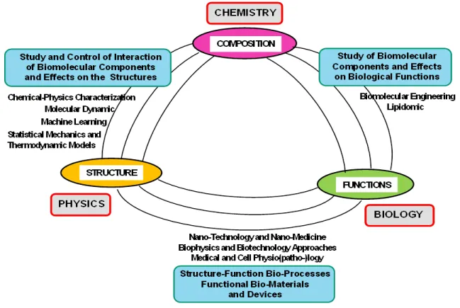
Figure 2. Schematic representation of the synergistic relationship between composition, structure and functions in a biological membrane system
As reported in Figure 2, it possible to identify a relationship between the biomembranes composition, structure and functions, while their experimental characterization methods can be grouped into three main conceptual stages.
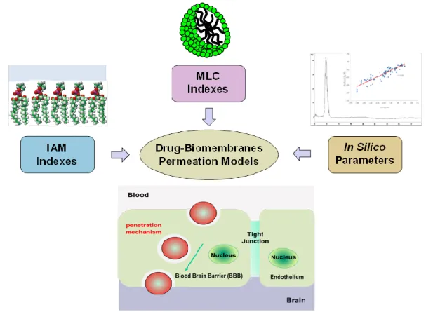
The first stage of the investigation consists in the analysis of the composition of the (model or real) biological membranes. One important (composition analysis) approach consist in the use of the high-resolution mass spectroscopy techniques [52]. Detailed information about the population distribution of the biomembrane components is also obtained by using the size-exclusion chromatography (SEC) [53]. This approach allows to separate (and quantify) the liposome populations distribution by exploiting the separation process (based on the elution velocity from a column equipped with a porous substrate) which depends on the compound hydrodynamic size. Among the chromatographic approaches, the immobilized artificial membrane (IAM) chromatography allows to efficiently discriminate the liposome/water partitioning as well as the permeation of cell membranes. [54,55]. Moreover IAM chromatography give complementary information on drug-membrane interactions (binding) and passive diffusion (permeation) of drugs, thus allowing an estimate of the passive transport through a variety of biological barriers, and the pharmacokinetic/pharmacodynamic properties of the drugs [54,55]. A further advance in this field is represented by bio-partitioning (BP) chromatography, a technique that combine the chromatographic method with biomembrane-mimetic structures (such as the phospholipid vesicles, polymer micelles, niosomes and microemulsions). This technique represents a (high-throughput) screening platform for the investigation of the drug-biomembrane permeability and interaction and their connection with the biological functions. Recently, bio-partitioning micellar liquid chromatography (MLC) and
immobilized artificial membrane (IAM) (liquid) chromatography have been used for in silico and in
reversed-phase liquid chromatographic (RPLC) technique based on the addition of surfactants (ionic or non-ionic) to aqueous mobile phases at concentrations higher than their critical micelle concentrations (CMC), under specific experimental conditions [56,57]. As in classical micellar chromatography, the retention process of compounds in MLC depends on the types of interactions between modified reversed stationary phase and micelles present in the mobile phase, and is sensitively influenced by the hydrophobic, electronic and steric properties of the included compounds. This technique can be useful in describing the biological behavior of different kinds of organic compounds and can mimic many biological processes such as blood–brain barrier penetration, skin permeability, intestinal absorption and drug partitioning process in biological systems [56,57]. Biomimetic partition chromatography systems have been successfully introduced also with the IAM chromatography method, that uses a stationary phase which consists of a monolayer of phosphatidylcholine (the major component of biological membranes) covalently immobilized on an inert silica support. IAM columns are thought to mimic very closely a membrane bilayer and are used in a high performance liquid chromatography (HPLC) system with a physiological buffer as eluent [56,57]. Furthermore, chromatographic retention coefficients of the analytes on stationary phases can be assumed as direct measures of their lipophilicity, (i.e., their affinity for lipids) [58]. The lipophilicity of a substance is an important physico-chemical property affecting the permeability and the passive transport through biological membranes (barriers) of compounds. Drug molecules possessing a sensitive affinity for lipophilic biological membranes, in fact, can be easily adsorbed and distributed within the organisms with a sensitive effect on pharmacokinetic processes and on the interactions with bioreceptors. Reversed (stationary) phase high-performance liquid chromatography has attracted great interest for lipophilicity measurement, due to its advantages, including speed, reproducibility, insensitivity to impurities and degradation products, broad dynamic range, on-line detection, and reduced handling and sizes of samples [58].
The MLC and IAM chromatographic indexes, including the calculated topological and physicochemical parameters in silico, can be used for the development of blood–brain barrier passage-predictive statistical models [56,57]. The BBB is one of the most complex and extensively studied biological membrane systems, whose main function is to preserve mammalian brain integrity against possible injurious substances. Apart from active transport mechanisms, whose occurrence is difficult to predict on a solely chemical structure basis, drugs can therefore cross the BBB by the passive transcellular route. One of the most used and reputed method of in vivo, ex vivo, and in vitro methods for measuring BBB partitioning of analytes is the determination of the quantity log
BB=log(CBrain/CBlood), expresses as a function of the concentration of the analyte is the in the brain
tissues (CBrain) and in the in the blood (CBlood). This method generally involves the use of animal
models (that may cause potential source of ethical issues), or the employment of cultured cell. Those
approaches, however, are time-consuming and do not provide any mechanistic information about the nature of the analyte passage. On the other hand, the in silico methods, although is unable to take into account all phenomena occurring in vivo, are much faster and simple to perform, as they are based on the calculation of physicochemical parameters and the screening of large libraries of compounds, thus allowing a more exhaustive investigation of the molecular mechanisms involved in membrane permeation. Moreover, in vitro methods based on the use of biomimetic stationary phases coupled with high performance liquid chromatography (HPLC) are much more reproducible and can be able to provide an in-depth understanding of the mechanisms involved in membrane barrier passage. Both IAM and MLC chromatographic indexes, combined with in silico calculated descriptors, have
demonstrated effectiveness in the prediction of BBB drug penetration [59]. The main steps of the approach is given by the following stages (Figure 3): - setup of (medium/high-throughput) methods for IAM and MLC indexes;
-validation of obtained parameters and development statistical models for the prediction of BBB llpenetration (log BB) by using the chromatographic indexes (and in silico calculated descriptors); -elucidation of the molecular mechanism involved in BBB passive diffusion of drugs
-optional molecular docking studies, to investigate the possibility the occurrence of further interaction mechanisms (such as the active efflux, or other self-assembly mechanisms).
The molecular modeling performed is simple, easy-to-perform and can be configured to run automatically in case of batch analyses [57,59]. Therefore, it can be of interest for pharmaceutical industries as a high-throughput BBB penetration screening methods.
Figure 3. A conceptual scheme for the investigation of the in vitro and in silico indexes for the modeling of the blood–brain barrier partitioning of drugs via IAM and MLC methods.
The second stage in the study of the biomembranes consists in the structural characterization methods by means of in vitro experiments. In this respect, the X-rays and Neutron small angle scattering (SAXS and SANS) and diffraction methods are among the most employed experimental techniques for the investigation, in non-invasive way, of the structural properties of biomembranes in complex biological systems [60–63]. Those non-destructive methods efficiently evidence the formation of different biomembrane structures and morphologies thus highlighting the important role of the relevant molecular conformations in many different processes of life science [64–67].
different solution conditions providing information on lipid domains (and phases), size, spacing and thicknesses in the range of a few nanometres and up to some tens of nanometres. On the same experimental set-up of the SAXS techniques (that typically operate at scattering angle between 0.1–5°) it is possible to set a further detection system allowing wide angle X-ray scattering (WAXS) experiments (at the scattering angle typically > 5°), that provides complementary information on the nature of hydrocarbon chain packing and lamellar phases (at the Å scale) when crystalline-like ordered phases exist in the biomembrane system [62]. Diffraction is a structural investigation method for the study of ordered lipid bio-membrane systems with large repeat distances and allows to resolve the existence of a variety of crystalline domain structures (such as lipid monolayers, bilayers, or multilayers) in lipid mixtures model systems as well as their hierarchical level of organization. Crystalline structure of several biomembranes composed of lipid bilayers and monolayers (with saturated alkyl chains) vary little for different lipids composition, and present similar unit cell dimensions of about 5.0 × 8.0 Å2. [61,62]. This characterization method is crucial for the investigation of biomembrane functions, thus furnishing useful information of their relevant role in important bio-processes such as phase separation, physiological and/or pathological crystallizations and protein sorting, just to name few [64]. For example, X-ray diffraction proved to be a very sensitive tool on detecting the periodical spacing and phase changes induced by antimicrobial peptides interacting with lipid membranes, such as the Alamethicin on dipalmitoyl–phosphatidyl–choline (DPPC) bilayers, as well as to predict the peptides doses necessary to trigger cell death through plasma membrane alterations [68]. Small angle neutron
scattering (SANS) shares similar principles with SAXS. While SAXS patterns (that originates
from electrons scattering) is only sensitive to the hydrophilic part of a lipid bilayer, the SANS technique (that depends on the scattering of neutrons) provides useful information on the hydrophobic tail part as they present a better “contrast” for that region. Therefore, SAXS and SANS can be used as complementary techniques for a fine structural description of the biological membranes [60–62]. Moreover neutron diffraction experiments on selectively deuterated lipids provide an important method of determining the mean conformation of a lipid molecule at different positions in the polar head group as well as in the hydrocarbon chains region [69,70].
Nuclear magnetic resonance (NMR) is a non-invasive, non-destructive, and quantitative
spectroscopy technique for the investigation of the drug-membrane interactions and provides detailed molecular level information on the structure and dynamics of (lipid) membranes (at a sub-Ångstrom resolution) [71]. As the nuclei of many atoms possess a magnetic moment (which gives rise to different energy levels and resonance frequencies in presence of an external magnetic field) many intermolecular interaction, including the drug binding to the lipid, may lead to variations in the local electronic environment surrounding the nucleus and thus cause a specific chemical shift in NMR spectrum with respect to a reference resonant frequency. More specifically multinuclear solid state NMR provides information on the nature of the membrane phase (lamellar, hexagonal, cubic, bicelle, micelle, etc.), its dynamics (fluid, gel, or liquid-ordered), and the molecular structure of embedded lipids when using the magic angle sample spinning (MAS) apparatus. Structural properties of lipid-based membranes are characterized by positional and orientational order. Moreover, the order parameters measured in solid-state NMR spectroscopy give complementary structural information on the distribution of the lipids with respect to the bilayer normal. Solid-state 2H NMR provides knowledge of segment orientations and their fluctuations at a site-resolved atomistic level. Solid state NMR has the advantage that the anisotropic magnetic or electric couplings (dipolar, quadrupolar,
chemical shift) yield segmental order parameters as model-free experimental observables. The disadvantage is that the broad spectral width requires special high-power NMR technology, and rather large amounts of sample material are needed. Relaxation time measurements are also described to measure lipid motional processes from the picosecond to the second timescale [66]. Recently, a 1H NMR Relaxometry technique provides information on the timescales of motional processes involved in the proton relaxation (on a time scale of microseconds) and allows the study of water dynamics in hydrated systems in the range of a few nanometers. The approach can provide valuable information to discriminate the different molecular mechanisms associated with water motion as a function of the hydration state, highlighting the variations of the relaxation rates connected with solvation processes, aqueous clusters and hydrogen bond network formation [72,73].
Recently, an investigation integrate the results of 2H NMR with small-angle X-ray scattering (SAXS) studies and MD simulations to achieve new insights. In the case of scattering methods (SAXS and SANS), lipid membranes are investigated as multilamellar dispersions or aligned on planar substrates, enabling comparisons with solid-state NMR spectroscopy [74].
A recent experimental method enable simultaneous measurement of optical and thermal properties of macromolecular nano-structures, such as biological membranes, by combining the
photothermal (PT) microscopy and spectroscopy with photoacoustic (PA) techniques, and the
application of broadband light sources and interferometer techniques [75,76]. Those techniques (that exploit the generation of heat due to the absorption of light) provide a broader complementary spectrum of information compared to standard spectroscopies based on to monochromators (such as the UV/VIS or IR) which require numerous separate measurements of physical quantities in different spectral ranges. A recent investigation evidenced the potential of photoacoustics experiments in the identification of the mechanisms underlying drug transport through tissue-mimicking systems and biomembranes, in a model transdermal delivery system consisting of dithranol and dodecanol-collodion (DDC) membrane samples [77]. The proposed approach may help to develop new strategies for the experimental investigation of the interactions processes in skin-mimicking membranes and may provide a better understanding of the principles of drug transport (and permeation) processes in biomembranes.
Many other complementary experimental techniques can be used, with the special scope to determine the affinity of active compounds for biomembranes inclusion, including several spectroscopy techniques such as dynamic light scattering (DLS), FT- infrared (IR), ultraviolet (UV), electron spin resonance (ESR), electron paramagnetic resonance (EPR), circular dichroism, [78,79]. Furthermore, the use of complementary highly sensitive biophysical techniques, such as the differential scanning calorimetry (DSC) [80] and isothermal titration calorimetry (ITC) [81], allow the investigation of the thermodynamics of protein–lipid (and drug-lipid) interactions (and binding). Those experimental approaches together the solid state NMR technique, allow to obtain some complementary useful thermodynamic information on the investigated systems.
Finally, (cryo-) transmission electron microscopy (TEM) experiments provide detailed insights into biomembrane packing and phase behavior, protein conformations and folding, and drug inclusion processes [82,83]. Cryo-TEM can provide high-resolution images for studying self-assembly, phase transition mechanisms, and dynamic phenomena in complex lipid-based biomembrane nanostructures. Moreover, advanced cryo-tomography can provide valuable 3D information on the structure and morphology of biomolecules, drug-loaded (and drug-free) liposomes, cubosomes, hexosomes and other biomembranes phases. As the cryo-imaging technique
is not a quantitative methodology it should be a part of an integrated analysis with quantitative biophysical techniques. For this reason, cryo-TEM is very useful when combined with SAXS or SANS techniques, and self-diffusion NMR for detailed structural and morphological characterization of self-assembled nanostructures [82,83].
Finally, the third stage is focused on the investigation of the main features of biological membrane functions. Biological membranes are involved, in fact, in many important cellular functions including signaling and sensing, (selective) transport of active compounds, cell adhesion and cell recognition. Moreover, a number of other functional processes (such as the metabolic processes, antigen-antibody recognition or DNA synthesis) take place within biomembranes systems present in the form of internal compartments inside the cytoplasmatic region, such as in lysosomes, mitochondria, chloroplasts. [15–17]. The experimental investigation of biomembranes functions within the complex environment of living cells is a complex task. Given the nanoscale dimensions and the dynamic nature of these membrane nanodomains and compartments, highly performing methods that allow the observation of lipid and protein structure and dynamics at high spatial and temporal resolution are needed. Unfortunately, no single experimental approach can combine high spatial resolution and fast image acquisition in one instrument. Among the various experimental approaches that allow the investigation of biomembrane functions, the optical far-field microscopy is a method of choice [84], since it facilitate the structural and dynamics investigation in an (almost) non -invasive way, by fluorescence labeling of the macromolecular components under observation. Until few years ago, the near-field scanning optical microscopy has been the only (optical) technique with resolution beyond the diffraction limit of light. The potential of far-field fluorescence microscopy is limited by the diffraction, to about 200 nm, for visible light (in spatial resolution) and limited by the image recording speed (in temporal resolution) [84]. High-resolution fluorescence microscopy technique is compatible with the live cell imaging. It provides excellent spectral contrast and, in combination with sensitive detectors, allows the detection of individual molecules. Recent progresses enhanced the optical microscopy resolution up to the molecular level range of the living cells (in a non-invasive way) by means of fluorescence correlation
spectroscopy (FCS) [85]. This technique allows the observation of the interaction dynamics and
molecular diffusion in the millisecond time scale resolution. In Figure 4 we report a conceptual map of the main experimental techniques for the study of biomembranes.
Figure 4. A conceptual framework of the main experimental techniques for the study of biomembranes.
4. Elaboration of models for biomembranes structure and interaction
As bio-membranes functions emerge at multi-scale hierarchical levels across a wide ranges of space and time domains, the bottom up approach of cell membranes (i.e., from molecules to cells) requires detailed information over the complete space-time range (from fast to slow and from small to large). More specifically, the combination (and intensity) of the involved soft interactions (such as electrostatic forces, H-bonds, steric hindrance and hydrophobic effects) determine the self-assembly morphology (ordered/disordered, ripple phase, liquid-crystal) and their pattern aggregation (bilayer, multi-lamellar, hexagonal/cubic phases). Those structural properties regulate the formation of energetic barriers controlling the thermally activated (diffusional, rotational and vibrational) molecular dynamics of the building-block molecules forming the biomembranes domains. Even though a considerable progress has been made with the experimental investigations of biomembrane structure and dynamics, the investigation of relevant self-assembly mechanisms is still limited by the space-time resolution of the experimental methods. Moreover, as the natural cell membranes present a great complexity of structures, with a large variety of cross-connections and functionality, the study of simplified (artificial) model systems, greatly helps the scientists to understand the effects of membrane lipids in many processes (including drug transport and uptake into cells). The use of artificial nanostructured materials as simplified models for the study of cell membranes processes has given a strong input to the understanding of the complex interactions that a biomolecule can develop toward biological membranes. For this reason, the adoption of artificial membrane models allows the carrying out of particular studies in specific experimental conditions (such as temperature, or ionic strength). In these models some great part of the cell membrane properties are obviously lost, (such as the presence of functionalized membrane proteins, receptors, endocytosis, and other active processes) and may not reproduce exactly all the aspects of the biomembrane environment. Generally speaking, three main kinds of lipid membrane models have been identified:
monolayers, vesicle-forming bilayers (liposomes), and supported bilayers [1,8]. The well-defined, simplified, composition and material configuration of these models allow to better reproduce the complex phenomena and colloidal stability that are present in vivo during drug transport, pharmacokinetic distribution.
An important modeling method in biomembrane research is given by the quantitative
structure-activity relationship (QSAR) approaches [86]. The QSAR methods allow the prediction of
the biological activity and effect of chemical compounds based on mathematical and statistical models [86]. The QSAR approach relies on the basic principle of chemistry, that states that the biological activity of any compound (such as a macromolecule, drug or ligand) is intimately connected with the structural arrangement of atoms forming its molecular structure. In this respect, structurally related molecules, that can be defined in terms of a series of parameters (called molecular descriptors), possess similar biological activities. The QSAR studies therefore involve selection of active and inactive compounds with the measurement of their biological activity, description and calculation of molecular descriptors, selection of appropriate features followed by construction of the mathematical model and its evaluation. These approaches are a great alternative to cost and intensive experiments carried out in laboratories. QSAR finds applicability in a wide range of fields including toxicology, drug design and discovery, nanomedicine [86]. The QSAR approach in biomembrane research assume that the biological activity is viewed as a sum of the various interactions (and reactions) that compounds undergoes with the sites of action (receptors) during the transport processes through bio-membranes. These interactions are assumed to be strongly regulated by the chemical structure of the compound, while a specific change in its structure can result in a change in biological response and activity [87]. An Alternative to the QSAR models, the
quantitative retention–activity relationship (QRAR) models represent other kind of modeling
techniques, in which chromatographic retention parameters are used as descriptor (or predictor) parameters of a specific biological response of chemicals [86]. Different QRAR models based on retention factors obtained using BMC and IAM chromatographic systems have been utilized to compare different physicochemical and biological parameters, in order to extimate phenols penetration across BBB [88]. For this purpose, the Linear Solvation Energy Relationship (LSER) of Abraham model [89], was applied to characterize IAM and BMC systems. Finally, the use of combined and hybrid approaches proved fast and effective in the development of highly predictive BBB permeation models, thus shedding new light into the molecular mechanism involved in the BBB uptake of therapeutics.
Many investigation concerning the study of the structure-function relationship in biological membranes make use of the concepts of the thermodynamic and statistical physics approaches. On the theoretical side, there is a large variety of analytical and computational techniques, which have shown potential usefulness in study of the collective properties of model biomembranes. Besides the methods of classical and statistical thermodynamics, new approaches have been proposed, for instance: the theories of phase transitions, the approaches of out-of-equilibrium thermodynamics, the application of the continuum elasticity, and viscoelasticity theories to lipid membranes and so on. Among them, the mean-field approximation (MFA) and the Ornstein-Zernike (O. Z.) integral equation approach are widely employed in the field of nano-colloids, biomaterials and soft matter [90–92]. Those approaches has been employed to study the range and strength of a large variety of soft interactions usually encountered in biological and nanostructured systems, including amphiphiles [93–97], proteins [98], dendrimers [99,100], hybrid and polymers-based nanostructures [101–103], and model
biomembranes [104,105].
Moreover, the progress of molecular dynamics (MD) simulations methods offers the opportunity to study of collective behavior of multicomponent systems for a wide range of biophysical processes, in great space-time resolution [106,107]. They range from highly idealized coarse-grained pictures of lipids, proteins, and water, to complete simulations at an atomistic level [108–111]. Simulations are gaining broader and broader applications because they provide, with a steady increasing level of accuracy, information on both the structural details (geometry) and the collective property of the system, including system total energy, lipid order parameters, and bilayer elastic constants.
5. System biology and the lipodomic approach
A modern interdisciplinary method in life science is given by the systems biology approach [112]. This method is based on the collection of data and information from many components in parallel, using the so called “-omics” technologies, (such as the metabolomics, proteomics and genomics approaches) [112]. This approach aims to investigate the complex interactions and functioning of the living cell at various levels, by inferring the complex pathways and intermediaries that govern every biological (physiological/pathological) process. As lipid bilayers membranes represent one of the main components of biological membranes, the lipodomics research field emerged as an important subfield of system biology method for the study of biological membranes systems at an interdisciplinary level [114]. A schematic representation of the lipidomic workflow is reported in Figure 5. More specifically, the lipidomics method investigate the pathways (and networks) of cellular lipids that attempt a large-scale mapping of the complete lipid profile within a variety of biological systems (such as a cell, tissue, or organism). Dynamics profiles of cellular lipids and the changes that occur after the perturbation of the cell system, are examined in order to identify biomarkers and elucidate the main changes in its physiological or pathological state.
Figure 5. Sketch of the lipidomic workflow. A sample of a biological systems (cell, tissue, or organism) (A), is analyzed by means of a chromatographic separations (HPLC) instrument connected to a high resolution mass spectrometer (B) in order to identify/quantify the lipid profiles (C) relevant to a specific biological (or pathological) condition. The large-scale mapping together with suitable statistical analysis and classification models (D), allow to a pathway and networks analysis to identify the relevant biomarkers associated (E).
As lipids play an important role in many metabolic diseases (such as diabetes, obesity, atherosclerosis and hypertension) the lipidomics approach is considered as a subset of the more general field of “metabolomics” which includes three other major classes of biological molecules (proteins/amino-acids, sugars and nucleic acids). Lipids bilayers biomembranes also regulate protein functions and gene transcription, and for this reasons they are also considered as a part of a dynamic “interactome” within the cell [113,114]. Those rapidly expanding fields complement the huge progress made in proteomics and genomics, within the large family of systems biology. This novel conceptual organization of disciplines facilitates the framing of the study of biomembrane within a true interdisciplinary approach at the crossroad between chemistry, informatics, mathematics, biology and medicine.
6. Strengthening the interdisciplinary and transdisciplinary aspect of biomembranes study As previously described, biomembranes in living organisms, which have the prominent role of tissues and cells protection from foreign molecules, allow the exchange of information between extra- and intra-cellular environments due to its biochemical-active surface (that include amount of associated enzymes, ion channels, receptors and signaling molecules). Supramolecular structures of biomembranes also have a crucial role in cellular homeostasis, metabolism, growth, and even cellular death. Therefore, the study of the alterations in the structure/functions relationship of relevant biological membranes is crucial for the understanding of the pathological effects that are at the origin of many diseases. The specific dynamical processes observed in a biological membrane and in bio-nanostructured assemblies depend on a rich scenario of multicomponent inter-molecular interactions which allows the formation of supramolecular structures at different levels of complexity. The traditional bottom up approach for the study of bio-membranes involves the investigation of the structural and dynamic processes of relevant nanostructured assemblies over a wide range of distances and relevant time scales, starting from the analysis of molecular interactions (~10 nm), to the assemblies of lipid-protein complexes (~100 nm) up to the meso-scale (~1 μm) and macro-scale (tissues and cells). The study of the molecular events occurring on cell membranes, as well as the multiplicity of interactions that involve bioactive compounds (in either physiological or pathological situations) requests then an interdisciplinary effort of many different field of science (Figure 4). A novel conceptual framework towards an interdisciplinary approach is therefore of paramount importance, to enlarge our knowledge of many diseases and to identify further potential therapeutic targets.
The difficulty to predict the structural behavior and functional responses of complex biomaterials is connected with the difficulty to fully describe (and model) the complex structural and dynamic processes involved in biological systems [115–117]. In this respect, none of the single experimental (or theoretical) method alone will be sufficient to fully describe the complex interplay between components, structure and function of biomembranes. Therefore, the possibility of using more experimental or theoretical methods (or a combination of them) represent a first step to integrate the main results coming from different investigations. For example, the interpretation of small angle X-ray measurements guided by molecular dynamics simulations of lipid bilayers has been recently investigated [118]. The direct comparison between the computer simulation results and experimental form factors drive the choice of the main interactions that, at the molecular level, can been directly inferred from the “best” simulated biomembrane structure [119,120]. In this respect, the integration and use of different computational (or theoretical) methods with experimental
techniques will provided valuable opportunities to extend the domain of applicability of each methods. Several example indicate the use molecular dynamic approach to identify the main inter-molecular interactions in complex biomembrane systems, by combining the information coming from different experimental techniques, including the combination of SAXS (and SANS) technique with neutron spin echo [119], atomic force microscopy (AFM) [120], or nuclear magnetic resonance (NMR) [121]. The combination of these techniques demonstrated a great potential to generate atomistic resolution dynamical models of biomembranes (and their interaction with biomolecules) in biologically relevant microenvironment. However, the main barrier that currently needs to be overcome seems to be the limited complexity of the interactions described in MD models [121].
Figure 6. A conceptual description of the main disciplines involved in the interdisciplinary study of biological membranes.
The possible choice of the appropriate combination of experimental (and/or theoretic al) approach(es) depend on the complexity of the processes under investigation and on the type (quantity and quality) of information available on those processes in the literature (or in public databases). This approach, whic have been applied especially in crucial biotechnology investigations, demands new ideas and an intense coordination effort between the expertise from different fields [122–125]. The combination of the knowledge coming from different disciplines allow to develop a conceptual framework that drive the exploration of complex processes and the related bio-physicochemical interactions at the biological interface (such as membrane surfaces, endosomal compartments, organelles and cytoplasm). However, the integration of the (sometimes heterogeneous) structural (and/or biological, chemical, clinical) data into a unified workflow is a
challenging task. For this reason, the study of a multiplicity of simultaneous factors and biological functionality may be replaced with the systematic study of the effect of a few
parameters at a time (such as surface charge density and/or nanoparticle size/topology). The
choice of a simplified version of the system allow to identify the key factors for the design of novel nanostructured systems, and represents then the fundamental (initial) step to decipher the complexity involved in complex biological processes. Future efforts should be directed toward the creation of suitable links within the complex network that relates components, structure and functions of biological membranes (Figue 6). A network-based method built upon these indications will likely provide a better integration of multidisciplinary research work.
Conclusions
The study of the biological membranes represents a central topic at the crossroad of different disciplines including biochemistry, biotechnology, and nano-medicine. Their scientific investigation involves the study of the collective behavior of numerous interacting (macro-)molecules, while their complete structural description requires the simultaneous calculation (and simulation) of a large number of parameters. The future challenge must provide an efficient integration of the different experimental research approaches and theoretical models into a common background. To achieve this objective, together with the improvement of the resolution of experimental approaches, we need an improvement of the theoretical models and computational efforts, with the aim to integrate them into a multi-scale description of the complex bio-systems. This effort requires an integration of multi- and interdisciplinary approaches that combine results coming from a wide range of sub-disciplines.
Conflict of interest
The authors declare no conflict of interest. References
1. Sackmann E (1995) Physical basis of self-organization and function of membranes: physics of vesicles. Handbook Biol Phys 1: 213–304.
2. Goni FM (2014) The basic structure and dynamics of cell membranes: an update of the Singer-Nicolson model. Biochim Biophys Acta 1838: 1467–1476.
3. Devaux PF (1991) Static and dynamic lipid asymmetry in cell membranes. Biochemistry 30: 1163–1173.
4. Tanford C (1973) The Hydrophobic Effect: Formation of Micelles and Biological Membranes 2nd Ed., New York: John Wiley and Sons Inc.
5. Ishida T, Harashima H, Kiwada H, et al. (2002) Liposome clearance. Biosci Rep 22: 197–224. 6. Allen TM, Cullis PR (2013) Liposomal drug delivery systems: from concept to clinical
applications. Adv Drug Deliv Rev 65: 36–48.
7. Bobo D, Robinson KJ, Islam J, et al. (2016) Nanoparticle-based medicines: a review of FDA-approved materials and clinical trials to date. Pharm Res 33: 2373–2387.
8. Lombardo D, Calandra P, Barreca D, et al. (2016) Soft interaction in liposome nanocarriers for therapeutic drug delivery. Nanomaterials 6: 125.
9. Ceh B, Lasic DD (1995) A rigorous theory of remote loading of drugs into liposomes. Langmuir 11: 3356–3368.
10. Katsaras J, Gutberlet T (2000) Lipid Bilayers: Structure and Interactions. Berlin: Springer-Verlag Berlin Heidelberg.
11. Moore TL, Rodriguez-Lorenzo L, Hirsch V, et al. (2015) Nanoparticle colloidal stability in cell culture media and impact on cellular interactions. Chem Soc Rev 44: 6287–6305.
12. Xing H, Hwang K, Lu Y (2016) Recent developments of liposomes as nanocarriers for theranostic applications. Theranostics 6: 1336–1352.
13. Lombardo D, Calandra P, Magazù S, et al. (2018) Soft nanoparticles charge expression within lipid membranes: the case of amino terminated dendrimers in bilayers vesicles. Colloid Surface
B 170: 609–616.
14. Lombardo D, Calandra P, Caccamo MT, at al. (2019) Colloidal stability of liposomes. AIMS
Mater Sci 6: 200.
15. Gennis RB (1998) Biomembranes: Molecular Structure and Function, New York: Springer-Verlag.
16. Singer SJ, Nicolson GL (1072) The fluid mosaic model of the structure of cell membranes.
Science 175: 720–731.
17. Dowhan W, Bogdanov M (2011) Lipid–protein interactions as determinants of membrane protein structure and function. Biochem Soc Trans 39: 767–774.
18. Liu Z, Persson S, Zhang Y (2015) The connection of cytoskeletal network with plasma membrane and the cell wall. J Integr Plant Biol 57: 330–340.
19. Bezanilla M, Gladfelter AS, Kovar DR, et al. (2015) Cytoskeletal dynamics: a view from the membrane. J Cell Biol 209: 329–337.
20. Simons K, Toomre D (2000) Lipid rafts and signal transduction. Nat Rev Mol Cell Biol 1: 31–39. 21. Simons K, Ehehalt R (2002) Cholesterol, lipid rafts, and disease. J Clin Invest 110: 597–603. 22. Jacobson K, Mouritsen OG, Anderson RGW (2007) Lipid rafts: at a crossroad between cell
biology and physics. Nat Cell Biol 9: 7–14.
23. Lingwood D, Simons K (2010) Lipid rafts as a membrane-organizing principle. Science 327: 46–50.
24. Frisz JF, Lou K, Klitzing HA, et al. (2013) Direct chemical evidence for sphingolipid domains in the plasma membranes of fibroblasts. Proc Natl Acad Sci USA 110: E613–E622.
25. Lorent JH, Levental KR, Ganesan L, et al. (2020) Plasma membranes are asymmetric in lipid unsaturation, packing and protein shape. Nat Chem Biol 16: 644–652.
26. Risselada HJ (2019) Cholesterol: The plasma membrane's constituent that chooses sides.
Biophys J 116: 2235–2236.
27. Liu SL, Sheng R, Jung JH, et al. (2017) Orthogonal lipid sensors identify transbilayer asymmetry of plasma membrane cholesterol. Nat Chem Biol 13: 268–274.
28. Sevcsik E, Schütz GJ (2016) With or without rafts? alternative views on cell membranes.
Bioessays 38: 129–139.
29. Fantini J, Barrantes FJ (2018) How membrane lipids control the 3D structure and function of receptors. AIMS Biophys 5: 22–35.
30. Disalvo EA (2015) Membrane Hydration, the Role of Water in the Structure and Function of
Biological Membranes, Switzerland: Springer International Publishing.
31. Israelachvili J, Wennerström H (1996) Role of hydration and water structure in biological and colloidal interactions. Nature 379: 219–225.
32. Branca C, Magazù S, Migliardo F, et al. (2002) Destructuring effect of trehalose on the tetrahedral network of water: A raman and neutron diffraction comparison. Phys A 304: 314–318.
33. Maisano G, Majolino D, Migliardo P (1993) Sound velocity and hydration phenomena in aqueous polymeric solutions. Mol Phys 78: 421–435.
34. Franks F (1975) Water, a Comprehensive Treatise, New York: Springher.
35. Kumar R, Keyes T (2001) The relation between the structure of the first solvation shell and the ir spectra of aqueous solutions. J Biol Phys 38: 75–83.
36. Fenimore PW, Frauenfelder H, Magazù S, et al. (2013) Concepts and problems in protein dynamics. Chem Phys 424: 2–6.
37. Minutoli L, Altavilla D, Bitto A, et al. (2008) A biophysics approach to modulate the inflammatory response during endotoxic shock. Eur J Pharmacol 589: 272–280.
38. Disalvo EA, Lairion F, Martini F, et al. (2008) Structural and functional properties of hydration and confined water in membrane interfaces. Biochim Biophys Acta Biomembr 1778: 2655–2670. 39. Roy A, Dutta R, Kundu N, et al. (2016) A aomparative study of the influence of sugars sucrose, trehalose, and maltose on the hydration and diffusion of DMPC lipid bilayer at complete hydration: investigation of structural and spectroscopic aspect of lipid–sugar interaction.
Langmuir 32: 5124–5134.
40. Cannuli A, Caccamo MT, Castorina G, et al. (2018) Laser techniques on acoustically levitated droplets. EPJ Web of Conferences 167: 5010.
41. Caccamo MT, Zammuto V, Gugliandolo C (2018) Thermal restraint of a bacterial exopolysaccharide of shallow vent origin. Int J Biol Macromol 114: 649–655.
42. Moiset G, López CA, Bartelds R, et al. (2014) Disaccharides impact the lateral organization of lipid membranes. J Am Chem Soc 136: 16167–16175.
43. Magazù S, Migliardo F, Benedetto A (2011) Elastic incoherent neutron scattering operating by varying instrumental energy resolution: principle, simulations, and experiments of the resolution elastic neutron scattering (rens). Rev Sci Instrum 82: 105115.
44. Migliardo F, Caccamo MT, Magazù S (2013) Elastic incoherent neutron scatterings wavevector and thermal analysis on glass-forming homologous disaccharides. J non-cryst solids 378: 144–151.
45. Israelachvili J, Wennerström H (1996) Role of hydration and water structure in biological and colloidal interactions. Nature 379: 219–225.
46. Kiselev MA, Lesieur P, Kisselev AM, et al. (2001) A sucrose solutions application to the study of model biological membranes. Nucl Instrum Meth A 470: 409–416.
47. Kiselev MA, Lesieur P, Kisselev AM, et al. (2001) Sucrose solutions as prospective medium to study the vesicle structure: SAXS and SANS study. J Alloys Comps 328: 71–76.
48. Bourgaux C, Couvreur P (2014) Interactions of anticancer drugs with biomembranes: what can we learn from model membranes? J Control Release 190: 127–138.
49. Helrich CS (2017) Studies of cholesterol structures in phospholipid bilayers. AIMS Biophysics 4: 415–437.
50. Contini C, Schneemilch M, Gaisford S, at al. (2018) Nanoparticle–membrane interactions. J Exp
Nanosci 13: 62–81.
51. Nel AE, Madler L, Velegol D, et al. (2009) Understanding biophysicochemical interactions at the nano–bio interface. Nat Mater 8: 543–557.
52. Mashaghi A, Mashaghi S, Reviakine I, et al. (2014) Label-free characterization of biomembranes: from structure to dynamics. Chem Soc Rev 43: 887–900.
53. Grabielle-Madelmont C, Lesieur S, Ollivon M (2003) Characterization of loaded liposomes by size exclusion chromatography. J Bioch Bioph Meth 56: 189–217.
54. Ong S, Liu H, Pidgeon C (1996) Immobilized artificial membrane chromatography: measurements of membrane partition coefficient and predicting drug membrane permeability. J
Chromatogr A 728: 113–128.
55. Barbato F (2006) The use of Immobilised artificial membrane (iam) chromatography for determination of lipophilicity. Curr Comp Aided Drug Design 2: 341–352.
56. Stępnik KE, Malinowska I (2013) The use of biopartitioning micellar chromatography and immobilized artificial membrane column for in silico and in vitro determination of blood-brain barrier penetration of phenols. J Chromatogr A 1286: 127–136.
57. Russo G, Grumetto L, Szucs R, et Al. (2017) Determination of in vitro and in silico indexes for the modeling of blood-brain barrier partitioning of drugs via micellar and immobilized artificial membrane liquid chromatography. J Med Chem 60: 3739–3754.
58. Liang C, Lian H (2015) Recent advances in lipophilicity measurement by reversed-phase high-performance liquid chromatography. TrAC Trend Anal Chem 68: 28–36.
59. De Vrieze M, Lynen F, Che K, et Al. (2013) Predicting drug penetration across the blood-brain barrier: comparison of micellar liquid chromatography and immobilized artificial membrane liquid chromatography. Anal Bioanal Chem 405: 6029–6041.
60. Fitter J, Gutberlet T, Katsaras J (2006) Neutron Scattering in Biology Techniques and
Applications. Heidelberg: Springer.
61. Nagle JF, Tristram-Nagle S (2000) Structure of lipid bilayers. BBA-Rev Biomembr 1469: 159–195.
62. Kiselev MA, Lombardo D (2017) Structural characterization in mixed lipid membrane systems by neutron and x-ray scattering. BBA-Gen Subjects 1861: 3700–3717.
63. Lesieur P, Kiselev MA, Barsukov LI, at al. (2000). Temperature-induced micelle to vesicle transition: Kinetic effects in the DMPC/NaC system. J Appl Cryst 33: 623–627.
64. Di Cola E, Grillo I, Ristori S (2016) Small angle x-ray and neutron scattering: powerful tools for studying the structure of drug-loaded liposomes. Pharmaceutics 8: 10.
65. Eicher B, Heberle FA, Marquardt D, et al (2017) Joint small-angle x-ray and neutron scattering data analysis of asymmetric lipid vesicles. J Appl Cryst 50: 419–429.
66. Kiselev MA, Janich M, Hildebrand A, et al. (2013) Structural transition in aqueous lipid/bile salt [DPPC/NaDC] supramolecular aggregates: sans and dls study. Chem Phys 424: 93–99.
67. Kiselev MA, Lombardo D, Lesieur P, et al. (2008) Membrane self assembly in mixed DMPC/NaC systems by sans. Chem Phys 345: 173–180.
68. Domenici F, Dell'Unto F, Triggiani D, et al. (2015) Vertical ordering sensitivity of solid supported dppc membrane to alamethicin and the related loss of cell viability. BBA Gen Subjects 1850: 759–768.
69. Büldt G, Gally HU, Seelig A, et al. (1978) Neutron diffraction studies on selectively deuterated phospholipid bilayers. Nature 271: 182–184.
70. Kiselev MA, Zemlyanaya EV, Ryabova NY, et al. (2014) Influence of ceramide on the internal structure and hydration of the phospholipid bilayer studied by neutron and x-ray scattering. Appl
Phys A 116: 319–325.
71. Umegawa Y, Matsumori N, Murata M (2018) Chapter two-Recent solid-state NMR studies of hydrated lipid membranes. Annu Rep NMR Spectrosc 94: 41–72.
72. Perrin JC, Lyonnard S, Guillermo A, et al. (2006) Water dynamics in ionomer membranes by field-cycling NMR relaxometry. J Phys Chem B 110: 5439–5444.
73. Kimmich R (2019) Field-Cycling NMR Relaxometry: Instrumentation, Model Theories and
Applications. London: the Royal Society of Chemistry.
74. Mallikarjunaiah KJ, Kinnun JJ, Petrache HI, et al. (2019) Flexible lipid nanomaterials studied by NMR spectroscopy. Phys Chem Chem Phys 21: 18422–18457.
75. Yang J, Maragliano C, Schmidt AJ (2013) Thermal property microscopy with frequency domain thermoreflectance. Rev Sci Instrum 84: 104904.
76. Todosijevic SZ, Soskic ZN, Galovic SP (2016) A combination of frequency photoacoustic and photoacoustic spectroscopy techniques for measurement of optical and thermal properties of macromolecular nanostructures. Opt Quant Electron 48: 300.
77. Rochowski P, Niedziałkowski P, Pogorzelski SJ (2020) The benefits of photoacoustics for the monitoring of drug stability and penetration through tissue-mimicking membranes. Int J Pharm 580: 119233.
78. Nieh MP, Heberle FA, Katsaras J (2019) Characterization of Biological Membranes Structure
and Dynamics, De Gruyter Edition.
79. Pignatello R, Musumeci T, Basile L (2011) Biomembrane models and drug-biomembrane interaction studies: involvement in drug design and development. J Pharm Bioallied Sci 3: 4–14.
80. Pignatello R (2013) Drug-Biomembrane Interaction Studies, the Application of Calorimetric
Techniques. Amsterdam: Elsevier.
81. Abraham T, Lewis RNAH, Hodges RS, et al. (2005) Isothermal titration calorimetry studies of the binding of a rationally designed analogue of the antimicrobial peptide gramicidin s to phospholipid bilayer membranes. Biochemistry 44: 2103–2112.
82. Helvig S, Azmi IDM, Moghimi SM, et al. (2015) Recent advances in cryo-TEM imaging of soft lipid nanoparticles. AIMS Biophysics 2: 116–130.
83. Almgren M, Edwards K, Karlsson G (2000) Cryo transmission electron microscopy of liposomes and related structures. Colloids Surf A 174: 3–21.
84. van Zanten TS, Cambi A, Garcia-Parajo MF (2010) A nanometer scale optical view on the compartmentalization of cell membranes. Biochim Biophys Acta 1798: 777–787.
85. Elson EL (2011) Fluorescence correlation spectroscopy: past, present, future. Biophys J 101: 2855–2870.
86. Peter SC, Dhanjal JK, Malik V, et al. (2019) Quantitative structure-activity relationship (QSAR): modeling approaches to biological applications. Encyclopedia Bioinf Comput Biol 2: 661–676. 87. Berthod A, García-Alvarez-Coque MC (2000) Micellar Liquid Chromatography, New York:
88. Bermúdez-Saldaña JM, Escuder-Gilabert L, Medina-Hernández MJ, et al. (2007) Biopartitioning micellar chromatography: an alternative high-throughput method for assessing the ecotoxicity of anilines and phenols. J Chromatogr B 852: 353.
89. Abraham MH, Platts JA, Begley M, et al. (2000) The Blood–Brain Barrier and Drug Delivery to
the CNS. New York: Marcel Dekker.
90. Hansen JP, Mc Donald IR (3013) Theory of Simple Liquids. New York: Academic Press. 91. Hunter RJ (1986) Foundations of Colloid Science. Oxford: Oxford University Press.
92. Likos CN (2001) Effective interactions in soft condensed matter physics. Phys Rep 348: 267–439.
93. Belloni L (1991) Interacting monodisperse and polydisperse spheres, In: Lindner P., Zemb T.,
Neutron X-Ray and Light Scattering, New York: Elsevier Science Publishers B. V.
94. Hamley IW (2003) Nanotechnology with soft materials. Angew Chem Int Ed Engl 42: 1692–1712.
95. Lombardo D, Munaò G, Calandra P, et al. (2019) Evidence of pre-micellar aggregates in aqueous solution of amphiphilic pdms-peo block copolymer. Phys Chem Chem Phys 21: 11983–11991.
96. Liveri VT, Lombardo D, Pochylski M, et al. (2018) Molecular association of small amphiphiles: origin of ionic liquid properties in dibutyl phosphate/propylamine binary mixtures. J Mol Liq 263: 274–281.
97. Lombardo D, Micali N, Villari V, et al. (2004) Large structures in diblock copolymer micellar solution. Phys Rev E 70: 021402.
98. Clarke J, Pappu RV, Berezovsky JN, et al. (2017) Folding and binding proteins: bridging theory and experiment. Amsterdam: Elsevier.
99. Lombardo D (2014) Modeling dendrimers charge interaction in solution: relevance in biosystems. Biochem Res Int 2014: 837651.
100. Porcar L, Hong K, Butler PD, et al. (2010) Intramolecular structural change of pamam dendrimers in aqueous solutions revealed by small-angle neutron scattering. J Chem Phys B 114: 1751–1756.
101. Li Q (2018) Functional Organic and Hybrid Nanostructured Materials: Fabrication,
Properties, and Applications, Weinheim: Wiley-VCH Verlag GmbH and Co. KGaA.
102. Bonaccorsi L, Calandra P, Kiselev MA, et al. (2013) Self-assembly in poly(dimethylsiloxane)-poly(ethylene oxide) block copolymer template directed synthesis of linde type A zeolite. Langmuir 29: 7079–7086.
103. Bonaccorsi L, Calandra P, Amenitsch H, et al. (2013) Growth of fractal aggregates during template directed sapo-34 zeolite formation, Micropor Mesopor Mat 167: 3–9.
104. Nel AE, Madler L, Velegol D, et al. (2009) Understanding biophysicochemical interactions at the nano–bio interface. Nat Mater 8: 543–557.
105. Lombardo D, Calandra P, Bellocco E, et al. (2016) Effect of anionic and cationic polyamidoamine (PAMAM) dendrimers on a model lipid membrane. Biochim Biophys Acta
Biomembr 1858: 2769–2777.
106. Saiz l, Klein ML (2002) Computer simulation studies of model biological membranes. Acc