SHORT COMMUNICATION
New Microbiologica, 41, 4, 302-305, 2018, ISN 1121-7138
Distribution of different HBV DNA forms in plasma
and peripheral blood mononuclear cells (PBMCs)
of chronically infected patients with low or undetectable
HBV plasma viremia
Ilaria Vicenti
1, Barbara Rossetti
2,3, Sara Mariano
2, Francesco Saladini
1, Francesca Montagnani
1,2,
Maurizio Zazzi
1, Andrea De Luca
21Medical Biotechnology Department, University of Siena, Siena, Italy;
2UOC Malattie Infettive, Dipartimento di Medicina Interna e Specialistica, AOU Senese, Siena, Italy; 3Clinic of Infectious Diseases, Catholic University of the Sacred Heart, Rome, Italy
Liver disease associated with persistent hepatitis B virus (HBV) infection remains an important public health prob-lem with significant morbidity and mortality. In spite of the existence of an effective vaccine, worldwide approx-imately 260 million people are chronic HBV (CHB) sur-face antigen (HBsAg) carriers and current treatment with interferon and/or nucleoside analogues (NA) is not able to achieve a complete cure (EASL, 2017; Schweitzer et
al., 2015). The key obstacle to HBV eradication is the
per-sistence of HBV DNA in the nuclei of infected hepatocytes, either integrated into the host genome or as a covalently closed circular DNA (cccDNA) episomal form (Allweiss and Dandri, 2017; Summers and Mason, 2004). While HBV integration is rare and its clinical implications still require investigation (Tu et al., 2017), cccDNA plays an essential role in the long-term persistence of HBV infec-tion and can often be detected even following NA therapy and HBsAg seroconversion (Boyd et al., 2016; EASL, 2017; Lai et al., 2017; Singh et al., 2004). Since quantification of cccDNA in infected hepatocytes (Lenci et al., 2010; Luo
et al., 2016; Werle–Lapostolle et al., 2004) requires
inva-sive liver biopsy, more accessible tissues, such as serum or peripheral blood mononuclear cells (PBMCs), have been investigated in different patient populations although
Corresponding author:
Ilaria Vicenti
E-mail: [email protected]
©2018 by EDIMES - Edizioni Internazionali Srl. All rights reserved
using non-standardized techniques (Chen et al., 2004; Li
et al., 2017). Some reports have shown that cccDNA in
patients’ sera is a marker of off-treatment virological re-lapse (Chen et al., 2004; Singla et al., 2014; Takkenberg et
al., 2009), whereas others have not found cccDNA in the
serum of CHB patients (Jun-Bin et al., 2003; Köck et al., 1996; Werle–Lapostolle et al., 2004). Some investigators speculate that PBMCs support HBV replication only par-tially, with linear and circular relaxed HBV DNA but not cccDNA formed in these cells (Köck et al., 1996; Laskus
et al., 1999; Murakami et al., 2004; Umeda et al., 2005),
while others have detected cccDNA in PBMCs and/or plasma (Cabrerizo et al., 2000; Coffin et al., 2015, 2014; Mazet-Wagner et al., 2006; Pasquinelli et al., 1990; Torii et
al., 2003) in a variable proportion of patients with chronic
and occult HBV infection. Discrepancies in HBV distribu-tion and compartmentalizadistribu-tion were observed according to the serologic profile or the coinfection status of patients analysed (Coffin et al., 2015; Loustaud-Ratti et al., 2013). While HCV coinfection seems not to be associated with a different distribution of tDNA and cccDNA in serum and/ or PBMCs of HBV positive patients, independently of their serological profile, HIV-1 seropositivity seems to influence the distribution of tDNA, which is prevalently detected in serum with respect to PBMCs in HBcAg positive and HBsAg negative patients. This profile suggests that HIV-in-duced immune dysfunction results in the maintenance of a low HBV viral load (Wagner et al., 2004).
We developed molecular methods to evaluate the distribu-tion of total HBV DNA (tDNA) and cccDNA in plasma and PBMCs of patients with low or undetectable HBV viremia. Written informed consent was obtained from each patient
Key words:
HBV, cccDNA, Real time PCR, PBMC SUMMARY
Few studies have documented hepatitis B virus (HBV) DNA in peripheral blood mononuclear cells (PB-MCs). We developed real-time PCR methods for differential amplification of covalently closed circular (cccDNA) and total HBV DNA (tDNA). The different distribution of cccDNA and tDNA in plasma and PBMCs was evaluated in 37 patients with low or undetectable viremia. Plasma tDNA measured by the Abbott reference system and the in-house assay correlated well (Spearman rho = 0.804; P<0.0001). tDNA was detected in four PBMC samples, all from patients with detectable plasma viremia (range 633-6,406 IU/ ml), cccDNA was not detected in any sample. The reasons for apparently discrepant results need further investigation but possibly include the high diversification of HBV status and plasma viremia levels.
Real-time PCR quantification of total HBV and cccDNA 303
included in the study. The study protocol conforms to the ethical guidelines of the 1975 Declaration of Helsinki (7th revision, 2013) as reflected in a priori approval by the in-stitutional human ethics committee. Plasma tDNA was quantified using the certified Abbott Real-time HBV Viral Load Assay (Abbott Diagnostics Inc.). HBsAg, HBeAg, an-ti-HBs, anti-HBe were detected by using the Elecsys sys-tem (Roche Diagnostics, Italy).
PBMCs and plasma were isolated from 8 ml of blood us-ing Ficoll gradient centrifugation (Pharmacia) and stored frozen until used. Before collection, PBMC pellets were washed twice with phosphate-buffered saline (PBS) to re-move viral particles possibly adsorbed on the cell mem-brane. Viral DNA was extracted from 200 µl of plasma or all the stored PBMCs preparation using the High Pure Viral Nucleic Acid Kit (Roche) according to the manufacturer’s instructions. Real-time quantitative PCR (qPCR) methods for differential amplification of cccDNA and tDNA in plas-ma and PBMCs samples were developed. Briefly, cccDNA was preferentially amplified with specific primers flanking the gap region and the incomplete strand of HBV DNA while tDNA primers detected all forms of HBV DNA, in-cluding relaxed circular forms and cccDNA (Table 1). The beta-globin gene was simultaneously targeted in the tDNA qPCR performed on PBMCs to estimate the number of cells in each PCR reaction. The HBV qPCR standard curve was generated using serial 10-fold dilutions of a plasmid carrying a 1,000-bp fragment of HBV genome and normal-ized on the calibration curve of the International WHO linear HBV DNA Standard (NIBSC code 10/264). Similar-ly, the beta-globin standard curve was generated using hu-man DNA derived from Sup-T1 cells (AIDS Reagent Pro-gram catalog number 100). The tDNA qPCR reaction mix-ture included 750 ng of spectrophotometrically measured PBMCs DNA or 5 µl of plasma DNA, 10 µl Premix Ex Taq (Takara Bio), 7.5 pmol of each of tDNA and beta-globin primers and 2.5 pmol of each of the tDNA universal probe and beta-globin probe in a final volume of 20 µl. Nega-tive controls for qPCR and DNA extraction were included in each run, and each sample was amplified in duplicate. To improve the specificity of cccDNA detection, 2 µg of PBMCs DNA and plasma viral DNA were digested with 10 Units of Plasmid Safe DNase (Epi centre) for 1 hour at 37°C, as indicated by the manufacturer. This step was per-formed to destroy the single stranded DNA isolated from PBMCs and plasma prior to cccDNA detection. The cccD-NA qPCR reaction mixture included 5 µl of digested DcccD-NA, 10 µl Premix Ex Taq (Takara Bio), 2% of DMSO, 7.5 pmol each of cccDNA primers and 2.5 pmol of tDNA universal
probe in a final volume of 20 µl. All the reactions were run in a Light Cycler 96 system (Roche) for 50 cycles each in-cluding 15 seconds at 95°C and 1 minute at 57°C after the first denaturation step (5 minutes at 95°C). Data acquisi-tion and handling were carried out using the Light Cycler 96 Software version 1.1.0.1320.
The association between tDNA values obtained with the Abbott Reference System and the in-house qPCR in plasma samples was evaluated by Spearman correlation analysis. The correlation between qPCR tDNA values and HbeAg status or HBV genotype were analysed by the Mann-Whitney test.
The limit of detection of qPCR was 10 IU/ml of plasma and 60 copies/106 PBMCs for tDNA and 5 IU/ml of
plas-ma and 30 copies/106 PBMCs for cccDNA. The coefficient
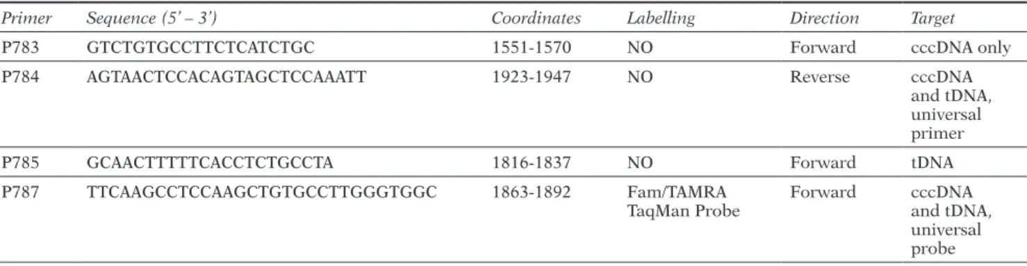
Table 1 - Primers and probe sequences used for tDNA and cccDNA differential amplification in qPCR system. Coordinates
are referred to X02763 HBV genotype A used as reference sequence (https://hbvdb.ibcp.fr/HBVdb/HBVdbNomenclature).
Primer Sequence (5’ – 3’) Coordinates Labelling Direction Target
P783 GTCTGTGCCTTCTCATCTGC 1551-1570 NO Forward cccDNA only
P784 AGTAACTCCACAGTAGCTCCAAATT 1923-1947 NO Reverse cccDNA
and tDNA, universal primer
P785 GCAACTTTTTCACCTCTGCCTA 1816-1837 NO Forward tDNA
P787 TTCAAGCCTCCAAGCTGTGCCTTGGGTGGC 1863-1892 Fam/TAMRA
TaqMan Probe Forward cccDNA and tDNA, universal probe
Table 2 - Baseline characteristics of the 37 patients included
in the study.
Population
characteristics except* median (Inter Quartile Range)Values expressed as n (%),
Patient Data
Age, years 48.4* (37.8-60.5)
Gender, males 26 (70)
Caucasian ethnicity 27 (73)
Time from diagnosis
of infection 7.3 (4.9-13.8) HIV coinfection 2 (5.4) Inactive carrier 16 (43) Serological Markers HBsAg+ 36 (97) HBsAb+ 1 (3) HbeAg+ 4 (11) HBeAb+ 33 (89) Drug Therapy Entecavir 13 (35) Tenofovir 7 (19) Virological Markers Genotype D 19 (68) A 6 (21), E 2 (7), B 1 (4) HBV DNA Undetectable 15 (40.5) HBV DNA <10 IU/ml 5 (13.5) HBV DNA ≥10 IU/ml 17 (46)
I. Vicenti, B. Rossetti, S. Mariano, et al.
304
of variability (CV) of the Ct values ranged from 5.55% to 1.37% for tDNA qPCR and 2.04% to 1.00% for cccDNA qPCR when measuring 5 to 50,000 plasmid target copies, with a linear dynamic range covering the 5 log10. The
ef-ficacy and safety of the Plasmid Safe DNase protocol was confirmed by a complete loss of PCR signal following treatment of 5,000-50,000 IU of the International linear HBV DNA Standard and no measurable loss of PCR signal following treatment of 5,000-50,000 IU of the HBV plas-mid, respectively.
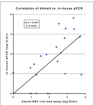
The baseline characteristics of the 37 patients recruited for the study are listed in Table 2. Plasma tDNA quanti-fied with the Abbott reference system was undetectable in 15 samples, detectable below 10 IU/ml in five samples and above 10 IU/ml in 17 (median tDNA levels 336 IU/ ml, IQR 69-793). Plasma tDNA levels measured by the Abbott reference system and the in-house qPCR correlat-ed well (Spearman rho = 0.80; P<0.0001), with mean±SD difference 0.20±0.62 log10 IU/ml (Figure 1). Discordant
results (bias of tDNA quantification larger than one log) were obtained only in 6 (16.2%) patients. Plasma tDNA levels showed no apparent association with HBeAg status (P=0.588) or HBV genotype (P=0.371). In the two HIV-1 coinfected patients, tDNA was below the limit of detection in plasma and PBMC samples. tDNA was detected in four PBMC samples, with values of 1,963, 2,017, 3,609, and 26,868 copies per 106 PBMCs, all from patients with
de-tectable plasma viremia (range 633-6,406 IU/ml). Of note, a control cccDNA qPCR performed before digestion with Plasmid Safe DNase scored positive in 13.5% of samples (2 plasma and 3 PBMCs). However after enzymatic treat-ment, cccDNA was not confirmed in any sample, suggest-ing that the digestion step must be included in cccDNA detection protocols.
Figure 1 - Correlation between in-house qPCR and Abbott
HBV viral load assay (Roche Diagnostics) in the plasma of 37 patients with low or undetectable HBV viremia.
Quantitative analysis of residual HBV DNA replication can be useful when assessing the risk of reactivation in patients with occult infection or “inactive” HBsAg car-riers, or in order to determine the potential for HBV transmission in donor individuals and the efficacy of treatment regimens on HBV reservoirs. While cccDNA can be detected in liver biopsies as the key HBV repli-cative form, less invasive blood markers have been pro-posed including quantitative HBsAg (Wong et al., 2017) and the Hepatitis B core-related antigen (HBcrAg) (Park
et al., 2012). However, these markers do not appear to
be completely representative of cccDNA levels reflecting only partially the amount of HBV DNA and cccDNA in hepatocytes (Honda et al., 2016). Quantitation of cccD-NA in blood is an attractive option but it remains contro-versial whether extrahepatic tissues contain cccDNA and whether cccDNA is released into the serum. This hypoth-esis is supported by HBV re-activation after orthotopic liver transplantation (Takaki et al., 2015).
In the small patient group with low to undetectable plas-ma tDNA levels analysed in our study, we detected no cccDNA in plasma and PBMCs while tDNA was detected only in a few cases in the PBMC compartment, all associ-ated with measurable plasma HBV DNA. Several (Cabre-rizo et al., 2000; Coffin et al., 2014; Loustaud-Ratti et al., 2013; Stoll-Becker et al., 1997; Torii et al., 2003), but not all (Köck et al., 1996; Mazet-Wagner et al., 2006; Umeda et al., 2005), studies previously documented tDNA and occasion-ally cccDNA in a proportion of patients with chronic and occult HBV infection. Our case file included low viremic patients and thus complements previous studies, support-ing the concept that PBMC tDNA and cccDNA are mostly associated with the OBI profile as recently reviewed (Joshi and Coffin, 2018). As suggested by our own control ex-periments, the lack of PBMCs/plasma pre-treatment with a DNase to digest the relaxed circular form of the HBV genome could explain the overestimation of cccDNA in some studies (Cabrerizo et al., 2000; Takkenberg et al., 2009; Torii et al., 2003). Indeed, it must be emphasized that methods for cccDNA detection are far from standard. Other reasons for the discrepancy with previous literature may be related to the high diversification of patient popu-lation analysed in terms of infection status (e.g. inactive/ active carriers or occult infection), plasma viremia levels and the proportion of treated patients. The prevalence and role of blood cccDNA remains elusive, suggesting analysis of a large and comprehensive HBV population with a stan-dardized method.
Acknowledgements
This work was partially supported by the Gilead Fellow-ship Program 2015.
Competing interests
Conflict of Interest: None. The funding organization(s) played no role in the study design; in the collection, analy-sis, and interpretation of data; in the writing of the report; or in the decision to submit the report for publication.
Compliance with Ethical Requirements
The study protocol conforms to the ethical guidelines of the 1975 Declaration of Helsinki (7th revision, 2013) as reflected in a priori approval by the institutional South East Ethical Committee (study approved on October 2015, identification code OBI-2015). Informed consent was
ob-Real-time PCR quantification of total HBV and cccDNA 305 Luo X., Huang Y., Chen Y., Tu Z., Hu J., et al. (2016). Association of Hep-atitis B Virus covalently closed circular DNA and human APOBEC3B in Hepatitis B virus-related hepatocellular carcinoma. PLoS One. 11. Mazet-Wagner A.A., Baclet M.C., Loustaud-Ratti V., Denis F., Alain S.
(2006). Real-time PCR quantitation of hepatitis B virus total DNA and covalently closed circular DNA in peripheral blood mononuclear cells from hepatitis B virus-infected patients. J. Virol. Methods. 138, 70-9. Murakami Y., Minami M., Daimon Y., Okanoue T. (2004). Hepatitis B virus
DNA in liver, serum, and peripheral blood mononuclear cells after the clearance of serum Hepatitis B virus surface antigen. J. Med. Virol. 72, 203-14.
Park Y., Hong D.J., Shin S., Cho Y., Kim H.S. (2012). Performance evalua-tion of new automated hepatitis B viral markers in the clinical labora-tory: Two quantitative hepatitis B surface antigen assays and an HBV core-related antigen assay. Am. J. Clin. Pathol. 137, 770-7.
Pasquinelli C., Melegari M., Villa I., Scaglioni P.P., Seidenari M., et al. (1990). Hepatitis B virus infection of peripheral blood mononuclear cells is common in acute and chronic hepatitis. J. Med. Virol. 31, 135-40.
Schweitzer A., Horn J., Mikolajczyk R.T., Krause G., Ott J.J. (2015). Esti-mations of worldwide prevalence of chronic hepatitis B virus infec-tion: A systematic review of data published between 1965 and 2013.
Lancet. 386, 1546-55.
Singh M., Dicaire A., Wakil A.E., Luscombe C., Sacks,S.L. (2004). Quanti-tation of hepatitis B virus (HBV) covalently closed circular DNA (cccD-NA) in the liver of HBV-infected patients by LightCycler real-time PCR.
J. Virol. Methods. 118, 159-67.
Singla B., Chakraborti A., Sharma B.K., Kapil S., Chawla Y.K., et al. (2014). Levels of hepatitis B virus replicative intermediate in serum samples of chronic hepatitis B patients. Mol. Biol. Rep. 41, 4689-96.
Stoll-Becker S., Repp R., Glebe D., Schaefer S., Kreuder J., et al. (1997). Transcription of hepatitis B virus in peripheral blood mononuclear cells from persistently infected patients. J. Virol. 71, 5399-407. Summer J., Mason, W.S. (2004). Residual integrated viral DNA after
hepad-navirus clearance by nucleoside analog therapy. Proc. Natl. Acad. Sci.
USA. 101, 638-40.
Takaki A., Yagi T., Yamamoto K. (2015). Safe and cost-effective control of post-transplantation recurrence of hepatitis B. Hepatol. Res. 45, 38-47. Takkenberg R.B., Zaaijer H.L., Molenkamp R., Menting S., Terpstra V., et al. (2009). Validation of a sensitive and specific real-time PCR for detection and quantitation of hepatitis B virus covalently closed circular DNA in plasma of chronic hepatitis B patients. J. Med. Virol. 81, 988-95. Torii N., Hasegawa K., Joh R., Hayashi N. (2003). Configuration and
repli-cation competence of hepatitis B virus DNA in peripheral blood mono-nuclear cells from chronic hepatitis B patients and patients who have recovered from acute self-limited hepatitis. Hepatol Res. 25, 234-43. Tu T., Budzinska M., Shackel N., Urban S. (2017). HBV DNA Integration:
Molecular Mechanisms and Clinical Implications. Viruses. 9. Umeda M., Marusawa H., Seno H., Katsurada A., Nabeshima M., et al.
(2005). Hepatitis B virus infection in lymphatic tissues in inactive hep-atitis B carriers. J. Hepatol. 42, 806-12.
Wagner A.A., Denis F., Weinbreck P., Loustaud V., Autofage F., et al. (2004). Serological pattern ‘anti-hepatitis B core alone’ in HIV or hepatitis C virus-infected patients is not fully explained by hepatitis B surface an-tigen mutants. AIDS. 18, 569-71.
Werle-Lapostolle B., Bowden S., Locarnini S., Wursthorn K., Petersen J., et al. (2004). Persistence of cccDNA during the natural history of chronic hepatitis B and decline during adefovir dipivoxil therapy.
Gastroenter-ology. 126, 1750-8.
Wong D.K.H., Seto W.K., Cheung K.S., Chong C.K., Huang F.Y., et al. (2017). Hepatitis B virus core-related antigen as a surrogate marker for covalently closed circular DNA. Liver Int. 37, 995-1001.
tained from all individual participants included in the study. This article does not contain any studies with ani-mals performed by any of the authors.
References
Allweiss L., Dandri M., (2017). The Role of cccDNA in HBV Maintenance.
Viruses. 9 (6).
Boyd A., Lacombe K., Lavocat F., Maylin S., Miailhes P., et al. (2016). Decay of ccc-DNA marks persistence of intrahepatic viral DNA synthesis un-der tenofovir in HIV-HBV co-infected patients. J. Hepatol. 65, 683-91. Cabrerizo M., Bartolomé J., Caramel C., Barril, G., Carreno V. (2000).
Molecular analysis of hepatitis B virus DNA in serum and peripher-al blood mononuclear cells from hepatitis B surface antigen-negative cases. Hepatology. 32, 116-23.
Che Y., Sze, J., He M.L. (2004). HBV cccDNA in patients’ sera as an indica-tor for HBV reactivation and an early signal of liver damage. World J.
Gastroenterol. 10, 82-5.
Coffin C.S., Mulrooney-Cousins P.M., Osiowy C., van der Meer F., Nishi-kawa S., et al. (2014). Virological characteristics of occult hepatitis B virus in a North American cohort of human immunodeficiency virus type 1-positive patients on dual active anti-HBV/HIV therapy. J. Clin.
Virol. 60, 347-53.
Coffin C.S., Osiowy C., Gao S., Nishikawa S., Van Der Meer F., et al. (2015). Hepatitis B virus (HBV) variants fluctuate in paired plasma and pe-ripheral blood mononuclear cells among patient cohorts during dif-ferent chronic hepatitis B (CHB) disease phases. J. Viral Hepat. 22, 416-26.
European Association for the Study of the Liver. (2017). EASL 2017 Clini-cal Practice Guidelines on the management of hepatitis B virus infec-tion. J. Hepatol. 67, 370-98.
Honda, M., Shirasaki, T., Terashima, T., Kawaguchi, K., Nakamura, M., et al. (2016). Hepatitis B virus (HBV) core-related antigen during nucle-os(t)ide analog therapy is related to intra-hepatic HBV replication and development of hepatocellular carcinoma. J. Infect. Dis. 213, 1096-106. Joshi S.S., Coffin C.S. (2018). Hepatitis B virus lymphotropism: emerging
details and challenges. Biotechnol Genet Eng Rev. 34, 139-51. Jun-Bin S., Zhi C., Wei-Qin N., Jun F. (2003). A quantitative method to
de-tect HBV cccDNA by chimeric primer and real-time polymerase chain reaction. J. Virol. Methods. 112, 45-52.
Köck J., Theilmann L., Galle P., Schlicht H.J. (1996). Hepatitis B virus nu-cleic acids associated with human peripheral blood mononuclear cells do not originate from replicating virus. Hepatology. 23, 405-13. Lai C.L., Wong D., Ip P., Kopaniszen M., Seto W.K., Fung J., et al. (2017).
Reduction of covalently closed circular DNA with long-term nucleos(t) ide analogue treatment in chronic hepatitis B. J. Hepatol. 66, 275.81. Laskus T., Radkowski M., Wang L.F., Nowicki M., Rakela J. (1999).
Detec-tion and sequence analysis of hepatitis B virus integraDetec-tion in peripher-al blood mononuclear cells. J. Virol. 73, 1235-8.
Lenci I., Marcuccilli F., Tisone G., Di Paolo D., Tariciotti L., et al. (2010). Total and covalently closed circular DNA detection in liver tissue of long-term survivors transplanted for HBV-related cirrhosis. Dig. Liver
Dis. 42, 578-84.
Li X., Zhao J., Yuan Q., Xia N. (2017). Detection of HBV Covalently Closed Circular DNA. Viruses. 9.
Loustaud-Ratti V., Wagner A., Carrier P., Marczuk V., Chemin I., et al. (2013). Distribution of total DNA and cccDNA in serum and PBMCs may reflect the HBV immune status in HBsAg+ and HBsAg- patients coinfected or not with HIV or HCV. Clin. Res. Hepatol. Gastroenterol.