This work was supported by a fellowship from the International PhD Program in Neuropharmacology of the University of Catania and by Fondation pour la Recherche Médicale (FRM).
The work in this thesis has been carried out as a joint-PhD program in the laboratories of:
Home Institute Guest Institute
Prof. Filippo Drago Drs. Giovanni Marsicano and BIOMETEC Federico Massa
Department of Biomedical and Neurocentre Magendie – INSERM U862 Biotechnological Sciences University of Bordeaux
School of Medicine 146 Rue Leo Saignat University of Catania 33000 Bordeaux Via S. Sofia 64 France
95125 Catania Italy
TABLE
OF
CONTENTS
A
BSTRACT 5R
ÉSUMÉ 7L
ONGR
ÉSUMÉ 10L
IST OFA
BBREVIATIONS 16C
HAPTERI
-
G
ENERALI
NTRODUCTION1.1 Neuronal networks: definition and importance of their understanding 18 1.2 The Hippocampus as the brain structure to study neuronal networks 21 1.3 Functional connectivity in the hippocampal formation 24
1.4 The CA1 region of hippocampus 26
1.5 Excitatory and inhibitory transmissions in the hippocampus 35 1.6 The excitatory/inhibitory balance in hippocampal networks 47
1.7 Voltage sensitive dye imaging as a tool to record neuronal networks activity 49
A
IM OF THE THESIS 53C
HAPTERII
-
Quantitative assessment of CA1 network dynamics by combining voltage sensitive dye imaging and optical flow methods56
C
HAPTERIII
-
Layer-specific potentiation of network GABAergic inhibition in thehippocampus 90
C
HAPTERIV
-
G
ENERALD
ISCUSSION ANDC
ONCLUSION 130A
BSTRACTIn order to better understand brain functioning we need to investigate all the structural domains present in it, from single cell to interconnected entire brain regions. However, while our knowledge in terms of single/few cells functioning is vast, very little is known about neuronal networks, which are interacting collections of neurons functionally related to the same task. Moreover, the balanced and concerted activity of excitatory and inhibitory networks plays a key role for proper cortical computations. However, while exist several tools to record excitatory networks activity, this is not the case for inhibitory networks. Voltage sensitive dye imaging (VSDI) is a technique that allows the recording of neuronal activity by mean of proportional emission of fluorescence according to changes in membrane potential. The advantage of using VSDI over other recording techniques using electrodes is that VSDI allows not invasive recording of neuronal activity from hundreds of sites at the same time.
During the last decades, VSDI has been widely used both in vitro and in vivo and to investigate both single cells and excitatory network activities. However, by using VSDI, investigations on excitatory networks activity have been mainly performed by quantifying fluorescence emission in defined regions of interest at time-fixed points, while inhibitory activity has been evaluated only at single cell level. The former approach misses several information of the dynamics of spreading of glutamatergic transmission because does not consider for example how fast a signal propagates and in which direction. The latter approach instead, does not allow the monitoring of network inhibitory events, which would be very important considering the extensive spatial spreading of interneurons within cortical areas.
During my doctoral course I aimed at studying in detail excitatory and inhibitory neuronal networks in the CA1 area of mouse hippocampus with VSDI.
To study excitatory networks more comprehensively, in collaboration with a team of mathematicians, we developed a mathematical algorithm that allowed measuring the velocity and the direction of spreading of the VSDI signal and it represents a new method to determine an optical flow. After successful validation of the algorithm with surrogate data to test its accuracy, we analysed two set of experiments in which network excitatory activity has been manipulated either by increasing Schaffer’s collaterals stimulation intensity or by blocking GABAergic transmission with the GABAA receptor antagonist picrotoxin in order to increase the depolarization in the CA1 region of the hippocampus. The results of these manipulations significantly decreased signal velocity whereas picrotoxin application significantly modified the direction of spreading, making the depolarization-mediated VSDI signal less dispersed compared to control.
Using VSDI I was able to fully characterize GABAA receptor-mediated hyperpolarizing signals in all the CA1 sublayers (field IPSPs), thus providing a new way of monitoring inhibitory events at network level. Moreover, I found that the activation of mGluR5 receptors induced an increase in a long-lasting manner of the VSDI-recorded field IPSPs, with duration and magnitude that relied on the specific CA1 sublayer considered.
Overall, my work shows new methodologies and new findings that may represent a step forward in the quest for a better understanding of neuronal networks, both excitatory as well as inhibitory, which hopefully can contribute to reduce the gap of knowledge between single cell activity and behaviour.
R
ÉSUMÉDans le but de mieux comprendre le fonctionnement du cerveau nous devons examiner les domaines structuraux qui le composent, de la simple cellule à des régions entières du cerveau interconnectées. Cependant, bien que le fonctionnement d’une ou plusieurs cellules est relativement bien connu, il n’y a que peu d’informations concernant les groupements de neurones interagissant fonctionnellement dans une même tâche, les réseaux neuronaux. De plus, l'activité équilibrée et concertée des réseaux excitateurs et inhibiteurs joue un rôle clé pour les intégrations corticales appropriées. Par ailleurs, il existe plusieurs outils afin d’enregistrer l’activité des réseaux excitateurs, ce qui n’est pas le cas pour les réseaux inhibiteurs. δ’imagerie du colorant sensible au voltage (VSDI) est une technique permettant l’enregistrement de l’activité neuronale en moyennant une émission de fluorescence proportionnelle au changement de potentiel de membrane. Par rapport aux autres techniques employant des électrodes, le VSDI permet l’enregistrement non évasif de l’activité de centaines de sites en même temps.
Au cours des dernières décennies, le VSDI a été largement utilisé tant in vitro qu’in vivo pour étudier l’activité d’unique cellule et des réseaux excitateurs. Néanmoins, en utilisant le VSDI, les recherches quant à l’activité des réseaux excitateurs ont été principalement réalisées par quantification d’émission de fluorescence en définissant des régions d’intérêts à des temps fixes, alors que l’activité inhibitrice n’a été évalué uniquement qu’à l’échelle cellulaire. δa première approche ne permet pas l’obtention de toutes les informations de la dynamique de propagation de la transmission glutamatergique du fait qu’elle ne prenne pas en considération la vitesse à laquelle le signal se propage ni dans quelle direction. En revanche, la seconde approche n’offre pas la possibilité d’étudier l’activité du réseau inhibiteur ce qui serait toutefois important de
définir du fait de la propagation spatiale extensive des interneurones au sein des aires corticales.
Durant mon doctorat, le but de mon travail a été d’étudier en détail les réseaux neuronaux excitateurs et inhibiteurs de l’aire CA1 de l’hippocampe de souris à l’aide du VSDI.
Pour étudier les réseaux excitateurs de façon plus compréhensive, en collaboration avec une équipe de mathématicien, nous avons développé un algorithme mathématique permettant de mesurer la vitesse ainsi que la direction de propagation du signal VSDI, ce qui représente une nouvelle méthode pour analyser le flux optique. Après la validation réussie de l’algorithme avec des données de substitution pour tester sa précision, nous avons analysé deux séries d’expériences dans lesquelles l’activité des réseaux excitateurs a été manipulée soit par augmentation de l’intensité de stimulation passant de 10 à 30 Volts ou en bloquant la transmission GABAergique avec la picrotoxine, un antagoniste du récepeteur GABAA. Les résultats de ces manipulations montrent une diminution significative de la vitesse alors que l’application de picrotoxine modifie de façon significative la direction de propagation, ce qui rend le signal de dépolarisation médié par le VSDI moins dispersées par rapport au contrôle.
δ’utilisation du VSDI a permis l’entière caractérisation des signaux hyperpolarisants médiés par les récepteurs GABAA dans toutes les sous-couches de CA1 (champ IPSP), offrant ainsi une nouvelle façon d’étudier les évènements inhibiteurs à l’échelle d’un réseau. De plus, j’ai montré qu’en activant les récepteurs mGluR5, j’étais capable d’augmenter de façon durable le champ IPSP du VSDI, avec la durée et l’ampleur au niveau des sous-couches spécifiques de CA1.
Globalement, je présente dans cette thèse de nouvelles méthodes et nouveaux résultats qui peuvent représenter une avancée dans la quête d’une meilleure
compréhension des réseaux neuronaux, excitateurs et inhibiteurs, ce qui espérons, pourra contribuer à réduire l’écart de connaissance entre l’activité d’une seule cellule et celle du comportement.
L
ONGR
ESUMEDepuis l'antiquité, la compréhension du fonctionnement du cerveau a été un des objectifs majeurs de l'humanité. L'énorme progrès dans cette quête au cours des derniers siècles nous a permis de démontrer jusqu'à maintenant, que le cerveau fonctionne selon un niveau d’organisation multi-échelle, de l'activité de cellules individuelles à l'ensemble interconnecté de régions du cerveau, ce qui se traduit en fin de compte par des réponses comportementales. Cependant, entre activité de cellules individuelles et le comportement se trouvent les réseaux neuronaux, qui correspondent à un ensemble de cellules fonctionnellement liés à une même tâche et permettent la communication entre les différentes régions du cerveau. Néanmoins, alors que nous savons relativement beaucoup en termes de pathophysiologie à l’échelle de la cellule et que d'autre part, nous pouvons qualitativement et quantitativement étudier le comportement, il est plus difficile d’évaluer le fonctionnement des réseaux neuronaux. Par conséquent, si nous voulons mieux comprendre le comportement, nous devons améliorer la connaissance des réseaux neuronaux sous-jacents.
L'hippocampe, avec sa forme d’ampoule très conservée chez les mammifères, a été une zone du cerveau d’objet de recherches intensives au cours du temps. La principale raison à cela est sa structure en couches bien organisées, avec des entrées synaptiques bien définies au niveau des lamina dendritiques qui, avec le développement de la préparation de tranche d’hippocampe, à faciliter énormément les investigations électrophysiologiques et biochimiques. En fait, la plupart des informations connues concernant les mécanismes fonctionnels du cerveau ont été découvertes dans l'hippocampe, comme par exemple la plasticité synaptique activité-dépendante, les
mécanismes de les transmissions excitateurs et inhibiteurs et les processus d'absorption de neurotransmetteurs et d’excitotoxicité.
Dans l'hippocampe, comme dans d'autres régions du cerveau, les réseaux neuronaux utilisent principalement le glutamate et le GABA comme neurotransmetteur. Les réseaux glutamatergiques constitués de cellules principales sont le substrat structurel du flux d'information entre et à l'intérieur des régions du cerveau, tandis que les neurones GABAergiques limitent localement l'activité glutamatergique excessive et coordonnent la sortie de l’information de la cellule principale. En effet, alors que les cellules principales représentent un type cellulaire plutôt homogène, les interneurones GABAergiques diffèrent remarquablement en termes de morphologie, propriétés électrophysiologiques, d'expression de marqueur neurochimique et d'innervation fournis aux cellules principales. En effet, l'activité concertée et coordonnée des cellules glutamatergiques et GABAergiques assurent le bon fonctionnement du cerveau.
Bien que les techniques d’enregistrement des réseaux excitateurs soient déjà disponibles et populaires, la possibilité d'enregistrer l'activité GABAergique à grande échelle est très limitée à ce jour, du fait de l’utilisation d’une ou très peu électrodes ce qui ne permet pas de fournir des informations spatiales suffisantes. Les dernières décennies ont permis une grande amélioration de la technologie pour enregistrer l'activité neuronale, dans lequel les photons remplacent les électrons, avec le résultat d'un accès plus facile aux neurones en raison du manque d'électrodes d'enregistrement. Une des techniques les plus représentatives qui informe une activité neuronale au moyen de lumière est l’imagerie du colorant sensible au voltage (voltage sensitive dye imaging) (VSDI). A travers l'utilisation d'un colorant, le VSDI émet une fluorescence proportionnellement aux changements du potentiel de membrane. Le VSDI a été utilisé au cours des années pour évaluer l'activité neuronale in vitro et in vivo, tant au niveau de
cellules individuelles que de réseaux excitateurs. Avec les colorants, le VSDI a besoin d'un équipement supplémentaire afin d'être exécutées, comme l'optique avec une ouverture numérique élevée et un faible grossissement, une caméra très sensible pour l’acquisition de faibles variations de fluorescence émises par le colorant suivant l'activité neuronale et une source de lumière pour exciter le colorant lui-même.
Le travail présenté dans cette thèse vise à étudier en détail les réseaux excitateurs et inhibiteurs dans la région CA1 de l'hippocampe de souris en introduisant de nouvelles méthodes développées par le VSDI.
Une analyse détaillée des réseaux excitateurs a été réalisée par élaboration d'un algorithme avec la collaboration d'une équipe de mathématiciens, ce qui représente une nouvelle méthode pour estimer un flux optique à partir des données VSDI. Le flux optique dans le traitement de l'image est la mesure de la tendance du mouvement apparent des objets, des surfaces et des bords dans une scène visuelle causée par le mouvement relatif entre un observateur (un œil ou une caméra) et la scène. L'algorithme est basé sur un ancien problème mathématique conçu à l'origine par le mathématicien français Gaspard εonge à la fin du 18ème siècle et mis en œuvre plus tard par le mathématicien russe Leonid Kantorovich. Ce problème consiste à trouver une solution pour transporter une certaine quantité de masse à partir d'une configuration initiale à une configuration finale, en minimisant un coût donné fonctionnel. Dans notre cas, la sortie de cet algorithme est un champ vectoriel dans lequel chaque vecteur représente la distance minimale parcourue par la dépolarisation neuronale chaque 2,2 millisecondes, ce qui est la résolution temporelle des enregistrements VSDI utilisés dans cette étude. Particulièrement, ces vecteurs fournissent deux importantes informations quantitatives : la distance (quantifiée par le nombre de pixels couverts au cours de la propagation de la dépolarisation neuronale) et la direction générale de propagation à l'intérieur de chaque
région d'intérêt (représentée par la convergence / divergence). Après nous avons validé avec succès l'algorithme avec des données de substitution afin de tester sa précision, nous avons analysé deux séries d'expériences dans lesquelles nous avons manipulé l'activité du réseau excitateur dans CA1, soit en augmentant l'intensité de stimulation de 10 à 30 volts ou en bloquant la transmission GABAergique avec l'antagoniste du récepteur GABAA, la Picrotoxine. Ce que nous avons constaté est le suivant :
- les deux manipulations ont augmenté significativement la dépolarisation neuronale (représenté par l'augmentation émission de fluorescence, ΔF*F-1) dans CA1 en général et en particulier dans ses sous-couches. Ce qui diffère entre ces deux expériences résulte du fait qu’en bloquant l'inhibition GABAergique, l'activité excitatrice est uniquement prolongée et n’est pas sensiblement affectée au cours des toutes premières étapes de propagation de signal (~ 10 millisecondes), ce qui suggère que les interneurones sont recrutés principalement pour empêcher excitation excessive et prolongée de l’activité excitatrice.
- les deux manipulations ont diminué de manière significative la vitesse du signal de dépolarisation (quantifié du rapport de la distance par le temps) uniquement lors de la phase intermédiaire/tardive de la propagation. Cette constatation peut être surprenante et paradoxale, mais peut être expliquée par la persistance du réseau dans un état plus dépolarisée par rapport aux conditions de contrôle, soit après augmentation de l'intensité de stimulation (30 Volts) ou après le blocage des récepteurs GABAA.
- l'inhibition GABAergique bloquée influe considérablement la direction générale de propagation du signal de VSDI, ce qui le rend plus ciblée et moins divergent par rapport au contrôle. Cela démontre comment les interneurones participent activement à l'acheminement de signaux excitateurs le long du réseau de CA1.
En ce qui concerne les réseaux inhibiteurs, j’ai caractérisé pour la première fois avec le VSDI des champs de potentiels post-synaptiques inhibiteurs (fIPSP) évoqués, médiés par les récepteurs GABAA, s’étendant dans l'ensemble CA1 et dans toutes ses sous-couches. Ceci démontre la possibilité d'utiliser le VSDI comme outil pour l'étude directe de l’activé inhibitrice au niveau du réseau et pas seulement à la résolution d'une seule cellule. En particulier, j’ai montré que le fIPSP se produit principalement dans la couche pyramidale et à une distance inférieure à ~ 500 µm par rapport à l'électrode de stimulation. Ces résultats sont compatibles avec le fait que la majorité des synapses GABAergiques sont dans la région périsomatique de cellules pyramidales dans CA1 et que la stimulation active des populations d'interneurones est locale. De façon intéressante, lors de l’application d’une brève période de temps (10 minutes) de l’agoniste des récepteurs du groupe I mGluR, le (S) -3,5-dihydroxyphénylglycine (DHPG), j’ai observé un phénomène de plasticité GABAergique caractérisé par une augmentation durable de la force synaptique, dont la durée et l'amplitude dépend des différentes couches de CA1, avec une plus longue durée (60 minutes) et plus élevé dans le stratum radiatum proximal par rapport à la couche pyramidale. Des expériences complémentaires ont montré que le récepteur mGluR5 est responsable de cette plasticité grâce à l'activation intracellulaire ultérieure du récepteur IP3. Les phénomènes de plasticité à long terme des synapses GABAergiques sont déjà connues dans l'hippocampe, mais c’est la première fois qu’il est rapporté une potentialisation de longue durée des mGluR5 dépendant de l'inhibition GABAergique, et que celle-ci est couche spécifique concernant la durée et l’amplitude.
Globalement, ces données fournissent de nouveaux aperçus sur les mécanismes à travers lesquels les transmissions excitatrices et inhibitrices coopèrent étroitement dans la région de CA1. Les données de l'investigation mathématique des réseaux excitateurs
ont permis de mettre en évidence en particulier, la façon dont l'inhibition par les récepteurs GABAA est importante dans la propagation normale de l'activité glutamatergique, à un niveau neuroarchitectural supérieur comme au niveau du réseau, au lieu de quelques simples cellules. De plus, l'algorithme que nous avons développé pourrait potentiellement être utilisé pour analyser les données de diverses techniques d'imagerie optique, compte tenu leur large utilisation dans tous les domaines de recherche sur la santé et les maladies, ce qui pourrait accroitre considérablement les connaissances actuelles. Les données de l'inhibition du réseau en effet ont démontré la possibilité d'utiliser le VSDI avec une grande résolution spatiale sans précédente pour l'étude de la transmission et de la plasticité des phénomènes GABAergiques, contrairement aux enregistrements avec électrodes. En addition, ces données apportent de nouvelles connaissances quant au rôle neuromodulateur de la signalisation GABAergique sur la transmission glutamatergique.
LIST OF ABBREVIATIONS
AMPA = α-amino-3-hydroxy-5-methyl-isoxazole-propionic acid CA = Cornu Ammonis
CCK = cholecystokinin DG = Dentate Gyrus
DHPG = (S)-3,5-Dihydroxyphenylglycine EPSC = excitatory post synaptic current EPSP = excitatory post synaptic potential GABA = -Aminobutyric acid
IP3 = inositol 1,4,5-trisphosphate IPSC = inhibitory post synaptic current IPSP = inhibitory post synaptic potential mGluR = metabotropic glutamate receptor
MKP = Monge – Kantorovich mass transport problem ms = milliseconds
NMDA = N-methyl-D-aspartate PDE = partial differential equations PTX = picrotoxin
PV = parvalbumin ROI = region of interest VSD = voltage sensitive dye
CHAPTER I
GENERAL INTRODUCTION
“I have never had reason, up to now, to give up the concept which I have always stressed, that nerve cells, instead of working individually, act together, so that we must think that several groups of elements exercise a cumulative effect on the peripheral organs through whole bundles of fibres. It is understood that this concept implies another regarding the opposite action of sensory functions. However opposed it may seem to the popular tendency to individualize the elements, I cannot abandon the idea of a unitary action of the nervous system, without bothering if, by that, I approach old conceptions.”
Camillo Golgi Nobel Lecture, 11 December 1906
1.1 – Neuronal networks: definition and importance of their understanding
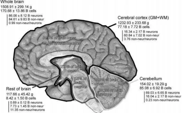
One of our greatest challenges nowadays is comprehend how brain works. To give a rough idea of its complexity, let’s just consider the estimated amount of cells that compose the human brain: approximately 171 billion! (see Figure 1, from Azevedo et al., 2009).
Figure 1 - Absolute mass, numbers of neurons, and numbers of nonneuronal cells in the entire adult human brain. Values are mean±SD and refer to the two hemispheres together. B, billion. From Azevedo et al., 2009.
Each neuron can connect to thousands of other neurons and these networks of neurons influence all behaviours, including perception, movement, memory and language (Parker, 2006). Moreover and actually surprising, some practical applications of ideas in neurobiology are not supported by strong scientific evidences. For example, lawyers
might propose brain scans as evidence of their clients’ lack of responsibility and governments plan to scan the brains of employees, despite the lack of evidence that the scans predict behaviour. Another example is children to whom are given amphetamines to correct disruptive behaviour, despite the lack of evidence for disturbances in brain chemistry, while children with no obvious learning disabilities take cognitive ‘enhancing’ smart drugs (‘Viagra for the brain’), with little evidence of any beneficial effects (Caplan, 2002; Parker, 2006; Rose, 2002). In addition, psychiatric and neurological treatments often lack insight into their mechanisms of action. For example, deep brain stimulation is used as a treatment for several disorders, including Parkinson’s disease, but it is unclear how it alleviates symptoms (Greenberg, 2002; McIntyre et al., 2004; Parker, 2006), and the potential benefits and underlying mechanisms of electroshock therapy, psychosurgery and psychopharmacology are uncertain at best (Parker, 2006; Schloss and Henn, 2004).
The 1990s were declared the decade of the brain by the US congress (Parker, 2006). The Dana Alliance, an organization of neuroscientists, listed 10 objectives to be attained during the decade. These were: identifying the genes defective in Alzheimer’s disease, Huntington’s disease, hereditary blindness, deafness and manic depression; developing strategies for reducing nerve cell death and promoting regeneration after injury; developing drugs to alleviate chronic pain, multiple sclerosis, Alzheimers’s disease, motor neuron disease, Parkinson’s disease and epilepsy; developing treatments for manic depression, anxiety and schizophrenia; and understanding the mechanisms of addiction, learning and memory. In 2010 a Dana Alliance report from representative scientists of the organization highlights how our knowledge in basic neuroscience research improved significantly in all the domains thanks to the dramatic advance in available technology, which will certainly lead to the identification of treatments (2010). This optimism, however, is not shared by people affected by the disorders listed
above, and is not shared by all neuroscientists (Parker, 2006). Torsten Wiesel, who won the Nobel prize for his work on visual cortex, claimed that ‘we need a century, maybe a millennium’ to comprehend the brain, and that beyond understanding a few simple mechanisms ‘we are at a very early stage of brain science’ (Horgan, 1999). Caution about our potential for understanding was earlier raised by the Nobel prize-winning neurophysiologist Charles Sherrington who said that ‘physiology has not enough to offer about the brain in relation to mind to lend the psychiatrist much help’ (Horgan, 1999).
We know a lot about the cellular properties of the nervous system and continuously increase our knowledge in order to identify molecular, developmental and functional properties of neurons and synapses. At the opposite end of the scale, we can characterize and quantify behaviours and correlate them with activity imaged with increasing sophistication in different regions of the brain (Parker, 2010). However, between these two levels there is an ‘explanatory gap’ that has prevented us from explaining behaviours directly in terms of their underlying cellular and synaptic mechanisms. We are thus data-rich but lack knowledge of how to integrate these data into a coherent picture of brain function (Parker, 2010).
Specific behaviours result from the activity in assemblies of interconnected nerve cells (‘neuronal networks’). Neuronal networks process sensory inputs, perform cognitive functions, and programme specific outputs. These networks assemble interacting groups of neurons that act together to generate behaviours, making the network the interface between the physiological (cellular) and behavioural levels (Parker, 2006). Understanding these networks is thus an essential component to our understanding of normal and abnormal behaviour. There are obvious reasons for wanting to understand the brain: increasing knowledge of our thoughts and behaviours, to correct the effects of injury or disease and finally to have the opportunity to apply insight obtained
on the nervous system to technology, which may significantly improve our daily life (Parker, 2006).
1.2 – The Hippocampus as the brain structure to study neuronal networks
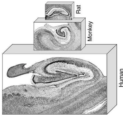
The hippocampal formation, comprised of the hippocampus itself (divided in dentate gyrus, CA3, CA2 and CA1), the subicular complex (subiculum, presubiculum, and parasubiculum) and the entorhinal cortex, has a bulb-like shape which protrudes into the lateral ventricles. The basic layout of cells and fiber pathways of the hippocampal formation is very similar in all mammals (Andersen P., 2007, Figure 2).
Figure 2 - Nissl-stained coronal sections through the rat, monkey, and human hippocampal formation. Note the general similarity of this brain region across species. From Andersen P. et al., 2007.
During the last decades the hippocampus has attracted the interest of scientists from many disciplines in neuroscience, from basic neurophysiology to cognitive and systems neuroscience. Thirty years ago, the most widely studied cell in the nervous system was the alpha motoneuron of the ventral horn of the spinal cord, today is the pyramidal cell of the hippocampus (Andersen P., 2007). One reason for the high amount of investigations carried out in hippocampus has been the peculiar anatomy of this brain area, with all principal cells oriented in a single layer and very organized synaptic inputs to well defined dendritic lamina. This simplified architecture facilitated electrophysiological and biochemical investigations, leading to ground-breaking discoveries about the mechanisms of excitatory and inhibitory transmissions, about the many neurotransmitter uptake mechanisms, about activity-dependent synaptic plasticity, and about the deleterious consequences of excitotoxicity for brain cells (Andersen P., 2007). These discoveries were dramatically facilitated by the development of the in vitro hippocampal slice preparation, which allowed the investigators to easily access and study cells on an unprecedented scale and to expand knowledge obtained in the hippocampus to other brain regions. An overview of some areas in neuroscience that have benefited from studies carried out on hippocampus is summarized in Table 1:
TABLE 1 (from Andersen P. et al., 2007):
• First use of microelectrodes for extracellular neuronal studies • Development of tetrodes for unit recording from behaving animals • Interpretation of field synaptic potentials and population spikes as tools for analysis of extracellular signals
• Pioneering use of intracellular recording for central nervous system neurons • Isolated slices of cortical tissue for neuroscience studies
• Development of histochemical methods for localization of neurotransmitters and receptor types
• Transplantation studies
• Pharmacological studies of central neurons and synapses • εolecular biological analysis of synaptic function
• Formulation of computational models to explain ways in which neural networks can implement learning and memory
Another very interesting feature of the hippocampus is that the granule cells of the dentate gyrus are one of the rare types of neurons that regenerate throughout life, whose mechanistic findings offer potential benefits in neuronal repair research and for possible therapeutic interventions (Andersen P., 2007).
In addition, different studies in the hippocampus showed why and when pyramidal cells are activated in the living brain. Recordings from freely moving animals while they navigate in a familiar space have shown that individual hippocampal pyramidal cells fire in particular locations. These findings led to the development of new behavioural tools to study the neural mechanisms of memory in animals (Andersen P., 2007).
The hippocampal formation is involved in a plethora of neurological disorders such, as among others, epilepsy, Alzheimer’s disease, and cerebrovascular disease (Andersen P., 2007). One of the hallmark features of epilepsy is the loss of neurons in several hippocampal fields and the pathological changes that occur in Alzheimer’s disease manifest initially in the entorhinal cortex and then spread to the hippocampus proper and ultimately to the entire cerebral cortex (Andersen P., 2007). Such findings
have led to the development of model systems in which pathophysiological events like these may be studied and, hopefully, alleviated by treatment (Andersen P., 2007).
1.3 – Functional connectivity in the hippocampal formation
As defined in the previous paragraph, the hippocampus, the subicular complex and the entorhinal cortex compose the so-called hippocampal formation. The main reason for including the aforementioned areas under an unique hippocampal complex is that they are linked, one to the next, by largely unidirectional (functional) neuronal pathways (Amaral D., 2007).
The entorhinal cortex is considered the first step in the intrinsic hippocampal circuit because much of the neocortical input reaching the hippocampal formation does so through the entorhinal cortex. Cells in the layer II of the entorhinal cortex give rise to axons that project, among other destinations, to the dentate gyrus. The projections from the entorhinal cortex to the dentate gyrus form part of the major hippocampal input pathway called the perforant path. Although the entorhinal cortex provides the major input to the dentate gyrus, the dentate gyrus does not project back to the entorhinal cortex. This pathway is therefore nonreciprocated, or unidirectional (Amaral D., 2007; Neves et al., 2008).
Likewise, the principal cells of the dentate gyrus, the granule cells, give rise to axons called mossy fibers that connect with the proximal apical dendrites of pyramidal cells of the CA3 field of the hippocampus. The CA3 cells, however, do not project back to the granule cells, have substantial associative ipsilateral interconnections between them and can receive direct inputs from layer II of the entorhinal cortex. The pyramidal cells of CA3, in turn, are the source of the major input to the CA1 hippocampal field (the Schaffer
collateral axons). Following the pattern of its predecessors, CA1 does not project back to CA3 and distal apical dendrites of CA1 pyramidal neurons can receive a direct input from layer III cells of the entorhinal cortex. The CA1 field of the hippocampus then projects unidirectionally to the subiculum, providing its major excitatory input. Again, the subiculum does not project back to CA1 (Amaral D., 2007; Neves et al., 2008).
From the CA1 and the subiculum, other unidirectional projections close the hippocampal processing loop by making synapses with the deep layer of entorhinal cortex, the layer V. A schematic representation of the information flow throughout the hippocampal formation is in Figure 3.
Figure 3 - Schematic representation of the information flow throughout the hippocampal formation. From Neves G. et al., 2008.
1.4 – The CA1 region of hippocampus
The vastness of the literature on CA1 and related cellular types, rather than other hippocampal areas, is largely attributable to a combination of structural considerations, cell viability, and historical accidents (Spruston N., 2007). Generally it is easier to keep cells in this region alive and healthy in slice preparations compared for example to the CA3, together with the fact that Schaffer collateral axons from CA3 form a homogeneous pathway that is easily activated to study synaptic transmission and plasticity (Spruston N., 2007).
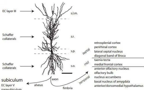
The principal cellular layer is called the pyramidal cell layer, where the somata of pyramidal cells are tightly packed. Below the pyramidal cell layer is the stratum oriens, which is a narrow, relatively cell-free layer that contains the basal dendrites of the pyramidal cells, several classes of interneurons and is the sub-region in which some of the CA3 to CA3 associational connections and the CA3 to CA1 Schaffer collateral connections are located. Deep to the stratum oriens is the thin, fibre-containing alveus. The stratum radiatum is located immediately above the pyramidal cell layer and is the region in which the CA3 to CA1 Schaffer collateral connections are located. Above the stratum radiatum is the stratum lacunosum-moleculare, where the projections from the layer III of the entorhinal cortex terminate and where are present afferents from other regions, such as the nucleus reuniens of the midline thalamus (Spruston N., 2007, see Figure 4 for a schematic representation of the position and the connectivity of a pyramidal cell along the different CA1 layers).
Figure 4 - CA1 dendritic morphology, spines, and synaptic inputs and outputs, respectively. Camera lucida drawing of a CA1 pyramidal neuron from an adult rat, showing the cell body in the stratum pyramidale (s.p.), basal dendrites in the stratum oriens (s.o.), and apical dendrites in the stratum radiatum (s.r.) and stratum lacunosum-moleculare (s.l.m.). The major excitatory inputs in each layer and the major outputs are also indicated. EC = entorhinal cortex. For the fimbrial projection, the septo-temporal positions noted indicate the source of CA1 cells projecting to different target regions. For the alveus projection, the subiculum is the major target. Bar = 100 μm. Adapted from Spruston N. et al., 2007.
Pyramidal cells of CA1: From the pyramid-shaped soma of rat CA1 neurons emerge two elaborately branching dendritic trees. The basal dendrites occupy the stratum oriens, and the apical dendrites occupy the stratum radiatum (proximal apical) and stratum lacunosum-moleculare (distal apical). Both the apical and basal dendritic trees occupy a roughly conical (sometimes ovoid) volume (Pyapali et al., 1998; Spruston N., 2007). The combined length of all CA1 dendritic branches is 12.0 to 13.5 mm: basal dendrites contribute about 36% of the total length, apical dendrites in the stratum radiatum contribute about 40%, and apical dendrites in the stratum
lacunosum-moleculare contribute the remaining 24% (Bannister and Larkman, 1995; Ishizuka et al., 1995; Megias et al., 2001; Spruston N., 2007; Trommald et al., 1995).
Along the length of the primary apical dendrite, several dendritic ramifications emerge obliquely in the stratum radiatum, which branch no more than a few times, with a typical branch bifurcating just once at a location close to its origin from the apical trunk (Spruston N., 2007). Despite their limited branching, however, oblique dendrites constitute most of the dendritic length in the stratum radiatum (Bannister and Larkman, 1995; Megias et al., 2001; Spruston N., 2007). After the primary apical trunk enters the stratum lacunosum-moleculare the apical dendrites continue to branch, forming a structure referred to as the apical tuft, which has an average of about 15 terminal branches (Bannister and Larkman, 1995; Spruston N., 2007; Trommald et al., 1995). Emerging from the base of the pyramidal soma are two to eight dendrites (a mean of five). Most of these dendrites branch several times (maximum 15 branch points), forming a basal dendritic tree with about 40 terminal segments (Bannister and Larkman, 1995; Pyapali et al., 1998; Spruston N., 2007).
Interneurons of CA1: Whereas pyramidal cells have their cell bodies organized into a highly structured layer (i.e., the pyramidal layer), the somata of GABAergic inhibitory interneurons show no such apparent organization. In fact, the somata of this highly diverse population of neurons are scattered throughout almost all subfields and strata of the hippocampus (Spruston N., 2007). Moreover, despite representing only ~11% of the total hippocampal neuronal population (Bezaire and Soltesz, 2013), each interneuron can connect with several hundreds of pyramidal cells (Freund and Buzsaki, 1996; Li et al., 1992; Sik et al., 1995) and other interneurons (Chamberland and Topolnik, 2012; Freund and Buzsaki, 1996) and represent perhaps one of the most diverse cell
populations. Because of this high heterogeneity, over the last decades numerous efforts have been made in trying to classify interneurons based on anatomical, neurochemical markers and connectivity patterns to pyramidal cells.
In classifying interneurons population based on their functional innervation to pyramidal cells, can be recognised two main classes: interneurons that innervate the soma and the axon initial segment and interneurons that innervate specifically dendrites of pyramidal cells (Freund and Buzsaki, 1996; Freund and Katona, 2007; Klausberger, 2009).
The class of interneurons which connect to the perisomatic region of pyramidal cells in the CA1 is represented by:
Basket cells = the predominant dendritic morphology of basket cells is pyramidal-shaped or bitufted. One to three dendrites originate from the apical pole of the triangular or fusiform soma, which then branch proximally, ascend through stratum radiatum, and often penetrate stratum lacunosum-moleculare. They also branch close to the soma and fan out toward the alveus, spanning the entire depth of stratum oriens (Freund and Buzsaki, 1996). All dendrites are spine-free, but occasionally a small number of short spine-like appendages can be observed. Basket cells in CA1 are likely to receive input from all major sources of excitatory afferents, such as Schaffer collaterals, commissural and entorhinal afferents, and recurrent collaterals of local principal cells in stratum oriens (Freund and Buzsaki, 1996). Basket cell axons instead fill the entire depth of stratum pyramidale and proximal stratum oriens (Freund and Buzsaki, 1996).
Basket cells do express several neurochemical markers such as the calcium-binding protein parvalbumin (PV) and the peptides cholecystokinin (CCK) and vasoactive intestinal polypeptide (VIP) (Freund and Buzsaki, 1996).
The main types of basket cells, the PV- and the CCK-expressing, have a functional dichotomy that is associated with characteristically different electrophysiological features and expression patterns for receptors, transmitters, and modulators (Freund and Katona, 2007). Several lines of research support the hypothesis that the PV-containing basket cells operate as clockworks for cortical network oscillations, whereas CCK-containing interneurons function as a plastic fine-tuning device (Freund and Katona, 2007). The latter cells modulate synchronous ensemble activities as a function of subcortical inputs that carry information about motivation, emotions, and the autonomic state of the animal, whereas the former have only a few receptor types for subcortical modulatory signals, but are efficiently and faithfully driven by local principal cells, as expected from an ‘‘oscillator’’ (Freund and Katona, 2007).
Chandelier cells = named also axo-axonic cells. The major distinguishing feature of chandelier cells is the characteristic termination of their axon, which run horizontally above the pyramidal cell layer and give rise to collaterals descending into the pyramidal layer, where they form characteristic bouton rows aligned parallel to the trajectory of axon initial segments of pyramidal cells (Freund and Buzsaki, 1996). The cell bodies are located within or immediately adjacent to the pyramidal cell layer and possess radially oriented dendrites spanning all layers. The dendrites are smooth, often varicose, and spines are only rarely present on a few branches. There is a rich arbor of basal dendrites in stratum oriens, which extends up to, or occasionally penetrates, the alveus. Thus, according to the distribution of the dendritic tree, chandelier cells are in a position to receive excitatory input from all major sources of afferents in the CA1 (Freund and Buzsaki, 1996). Chandelier cells contain mainly parvalbumin (Howard et al., 2005) and contribute to synchrony and oscillations in the hippocampus such as the theta rhythm (4–
8 Hz), which occurs during environmental exploration and REM sleep, and fast ripples (120–200 Hz), which occur during slow-wave sleep (Howard et al., 2005).
The class of interneurons which specifically contact the dendrites of pyramidal cells in the CA1 is represented by:
Oriens lacunosum-moleculare (O-LM) cells = they are the most studied interneurons that target dendrites of pyramidal cells. In the CA1 area, O-LM cells somata are located in stratum oriens and have horizontally extending dendrites with hairy spines on distal segments. The axons of O-LM cells give few collaterals in stratum oriens but project mainly through the strata pyramidale and radiatum to branch heavily in stratum lacunosum-moleculare (Klausberger, 2009), matching the glutamatergic input from the entorhinal cortex and thalamus. The axons give some collaterals also in the deep stratum radiatum but do not cross the fissure to the dentate gyrus. O-LM cells are often regarded as providing a classical example of GABAergic feedback inhibition. They express the neuropeptides somatostatin and parvalbumin (Klausberger, 2009).
Bistratified cells = the axonal arborization of bistratified cells overlaps with the glutamatergic input from CA3 pyramidal cells in stratum radiatum and oriens. This two-layered axonal arrangement gives the cell its name. Bistratified cells make GABAergic synapses with basal and oblique dendrites of CA1 pyramidal cells. Their somata are mainly located in stratum pyramidale, but have also been reported oriens-bistratified cells with somata and horizontally running dendrites in stratum oriens. The dendrites of bistratified cells in stratum pyramidale extend widely in the strata oriens and radiatum and form connexin36-containing gap junctions with other interneurons. Bistratified cells express parvalbumin to a similar extent to basket and axo-axonic cells and they also express somatostatin and neuropeptide Y (Klausberger, 2009).
Schaffer collateral-associated cells = the somata of Schaffer collateral-associated cells are located mainly in stratum radiatum with dendrites spanning all layers. The axons of these cells innervate the oblique and to a lesser extent basal dendrites of CA1 pyramidal cells and interneurons in strata radiatum and oriens, matching the excitatory input from CA3 pyramidal cells, giving the cell its name. In contrast to bistratified cells, the axons of Schaffer collateral-associated cells are concentrated more in stratum radiatum than in stratum oriens. Schaffer collateral-associated cells express CCK and the calcium-binding protein calbindin (Klausberger, 2009).
Perforant path-associated cells = the cell bodies of perforant path-associated cells are often located at the stratum radiatum - lacunosum moleculare border and their dendrites can either cover all layers or remain in stratum lacunosum moleculare and adjacent stratum radiatum. The axons of this cell type are concentrated in stratum lacunosum moleculare, overlapping with the excitatory perforant path input from the entorhinal cortex, giving the cell its name. Thus, they innervate the apical tuft of CA1 pyramidal cells. Interestingly, whereas the axons of O-LM cells always remain within the CA1 area, the axons of perforant path-associated cells often cross the fissure and also innervate the dendrites of granule cells in the dentate gyrus. Perforant path-associated cells express CCK and calbindin (Klausberger, 2009).
Neurogliaform cells = the cell bodies of neurogliaform cells are often located in stratum lacunosum moleculare and they have relatively short and numerous dendrites, giving the cell its name. The axons of neurogliaform cells are extremely dense, especially in stratum lacunosum moleculare. Similar to CCK-expressing perforant path-associated cells but in contrast to O-LM cells, the axons of neurogliaform cells often cross the fissure into the dentate gyrus. Many neurogliaform cells express neuropeptide Y and α-actinin-2 and are connected by gap junctions (Klausberger, 2009).
Ivy cells = in contrast to neurogliaform cells innervating the apical tuft of pyramidal cells, the very dense axons of ivy cells cover strata oriens and radiatum, making synapses onto the basal and oblique dendrites of pyramidal cells (Fuentealba et al., 2008; Klausberger, 2009). The cell bodies of ivy cells are located in strata pyramidale and radiatum and the usually short dendrites can cover all layers. Ivy cells express neuropeptide Y, neuronal nitric oxide synthase and a high level of GABAA receptor containing the α1 subunit (Klausberger, 2009).
In addition to the aforementioned interneuron types, other and less characterized interneurons exist in the CA1 region of hippocampus (Freund and Buzsaki, 1996; Klausberger, 2009). There are long-range projecting interneurons (i.e. trilaminar cells, backprojecting cells, radiatum retrohippocampal projection neurons, oriens retrohippocampal projection cells and double projection cells) which have their soma in the CA1 field and can send projections to the subiculum, to the CA3, the dentate gyrus and in areas outside the hippocampal formation as well (Freund and Buzsaki, 1996; Klausberger, 2009). There are also interneurons which are making synapses specifically with other interneurons (i.e. the interneuron-selective cells) and not with pyramidal cells, suggesting how an inhibitory control over other interneurons is essential.
In Figure 5 is schematically represented the overall interneuron population within the CA1 region.
1.5 – Excitatory and inhibitory transmissions in the hippocampus
Excitatory transmission: The main excitatory transmitter in the hippocampus, as elsewhere in the mammalian central nervous system is glutamate (Kullmann, 2007). Although the earliest evidence for this was principally obtained in the spinal cord, many important insights came from work on hippocampal tissue: for example, glutamate depolarizes hippocampal neurons (Biscoe and Straughan, 1966; Kullmann, 2007), and electrical stimulation in the CA1 evokes glutamate release (Dolphin et al., 1982; Walker et al., 1995). Importantly, glutamate activates the three principal types of receptors that mediate ionotropic excitatory transmission: α-amino-3-hydroxy-5-methyl-isoxazole-propionic acid (AMPA), kainate, and N-methyl-D-aspartate (NMDA) (Kullmann, 2007), which take their names from the exogenous agonists that activate them in a relatively selective fashion (Watkins and Evans, 1981;Nakanishi et al., 1998; Ozawa et al., 1998). At most excitatory hippocampal synapses, EPSCs are mediated by AMPA and NMDA receptors, which have strikingly different biophysical and pharmacological properties. Instead, kainate receptors play a relatively poorly understood role in synaptic transmission (Kullmann, 2007).
In addition to ionotropic transmission, glutamate activates also metabotropic receptors (mGluRs), which are coupled to several G protein types, are typically outside of the pre- and postsynaptic domains and modulate glutamate release, GABA release, and neuronal excitability (Anwyl, 1999; De Blasi et al., 2001; Osten, 2007). The mGluR family subdivides into three groups based on their pharmacological and functional properties: group I (mGluR1, mGluR5), II (mGluR2, mGluR3), and III (mGluR4, mGluR6, mGluR7, mGluR8) (Osten, 2007; Shigemoto et al., 1997).
AMPA receptors = they are composed of different combinations of four subunits (GluR1-4, also known as GluRA-B) and are present at almost all excitatory synapses in the hippocampus, gating a cation-selective channel (Kullmann, 2007). At resting membrane potentials, Na+ influx accounts for most of the current, but the channel is also permeant to other small monovalent cations, so K+ efflux can also occur at depolarized potentials. Most AMPA receptors in pyramidal neurons of the adult hippocampus (at least in rodents) are thought to be GluR1-2 or GluR2-3 tetramers (Kullmann, 2007; Wenthold et al., 1996). When a membrane patch taken from the soma or proximal dendrite of a hippocampal neuron is exposed to a pulse of 1 mM glutamate (roughly corresponding to the synaptic glutamate transient) (Kullmann, 2007) a current is generated with a rapid rise time (100–600 µs at physiological temperature). Native AMPA receptors deactivate rapidly following clearance of synaptic glutamate (with a time constant of 2.3–3.0 ms) (Colquhoun et al., 1992), the latter being a faster phenomenon then the termination of AMPA receptor-mediated EPSCs (Kullmann, 2007). If glutamate is not cleared, however, AMPA receptors close rapidly and enter a desensitized state from which they recover relatively slowly, with a decay time constant of the order of 5–10 ms (Kullmann, 2007; Mosbacher et al., 1994). The time course of desensitization depends on the subunit composition of the receptors and is affected by alternative splicing of two exons encoding a 38-amino-acid segment of the GluR2 subunit, with the consequence of having each subunit as either a “flip” or a “flop” variant, depending on which exon is retained the subunit mRNA and with flip forms that desensitize with slower kinetics compared to flop forms (Osten, 2007).
AMPA receptors can even desensitize in the presence of glutamate concentrations that are insufficient to open them or if the glutamate concentration rises sufficiently slowly (Kullmann, 2007). This form of desensitization may be an adaptation that prevents
excessive receptor activation under pathological conditions where extracellular glutamate accumulates. Depending on their subunit composition, AMPA receptors can also show significant permeability to Ca2+ ions. This permeability is determined by the presence or absence of a critical aminoacid (arginine, R) in a pore-lining segment of the GluR2 subunit. This subunit undergoes post-transcriptional RNA editing resulting in a change of the aminoacid at this position from glutamine (Q) to arginine (R) (Kullmann, 2007; Sommer et al., 1991). The presence of the edited form of GluR2 ensures that the receptor is impermeable to Ca2+, which is the case for most of the glutamate receptors in principal cells. If the GluR2 subunit is absent, the receptor has significant Ca2+ permeability and those receptors are present in some hippocampal interneurons (Geiger et al., 1995; Kullmann, 2007).
NMDA receptors = they consist of heteromultimers of subunits belonging to two relatively distinct subtypes, NR1 and NR2A-D (Kullmann, 2007; McBain and Mayer, 1994). The NR1 subunit is encoded by one gene but exists in several alternatively spliced isoforms. It does not bind glutamate but, instead, contains an important binding site for aminoacids such as glycine and D-serine, which act as co-agonists. The NR2A-D subunits, on the other hand, contain the glutamate-binding site (Kullmann, 2007; Laube et al., 1997). They are encoded by four genes and are variably expressed in different regions of the brain and at different stages of development. NMDA receptors have very slow kinetics and can continue to mediate an ion flux for several hundreds of milliseconds after the glutamate pulse has terminated (activation time constant is approximately 7 ms; deactivation time constants are approximately 200 ms and 1–3 s) (Kullmann, 2007). The slow kinetics are explained by an extremely slow receptor unbinding rate (Lester et al., 1990), that is, once glutamate binds to NMDA receptors, they remain bound for a long
time, during which time the ionophore can undergo repeated opening (Kullmann, 2007). In addition to their slow kinetics, NMDA receptors have three other important features. First, a second agonist-binding site (the “strychnine-insensitive glycine site”) must be occupied before glutamate is able to activate them (Johnson and Ascher, 1987; Kleckner and Dingledine, 1988; Kullmann, 2007), even though some estimates of the tonic extracellular glycine concentration in the brain suggest that the glycine-binding site is normally occupied (Kullmann, 2007). Alternatively, D-serine can substitute for glycine, and it has been proposed that this aminoacid plays a physiological role in regulating NMDA receptor function (Baranano et al., 2001; Schell et al., 1995). Second, NMDA receptors are highly permeable to Ca2+ ions and monovalent cations (Ascher and Nowak, 1988; Kullmann, 2007). Ca2+ influx via NMDA receptors plays a central role in several forms of long-term synaptic plasticity and NMDA receptor activation has been shown to trigger further release of Ca2+ from intracellular stores (Emptage et al., 1999; Kullmann, 2007). Third, Mg2+ ions block the ionophore in a voltage-dependent manner (Mayer et al., 1984; Nowak et al., 1984). Thus, at resting membrane potentials (more negative than approximately –50 mV), NMDA receptors are unable to mediate an EPSC even if glutamate and glycine (or D-serine) are present (Kullmann, 2007). They mediate an ion flux only when the membrane is depolarized. The Ca2+ permeability and Mg2+ blockade of NMDA receptors explain their role as synaptic coincidence detectors: Ca2+ influx occurs only if there is a conjunction of presynaptic glutamate release and postsynaptic depolarization, a situation that arises when pre- and postsynaptic activity occur together (Kullmann, 2007; Wigstrom and Gustafsson, 1986).
Kainate receptors = they share many features with AMPA receptors (Ben-Ari and Cossart, 2000; Kullmann, 2007; Kullmann, 2001; Lerma et al., 1997; Lerma et al., 2001)
because they are heteromultimeric as well, made up of different combinations of five subunits: GluR5-7 and KA1-2. However, not all of these subunits have the same status because receptors made up of KA1 or KA2 alone are non-functional (Kullmann, 2007). GluR5-6 undergoes Q/R editing with similar consequences as for the AMPA GluR2, although the proportion of edited subunits is much less spread. All this features of kainate receptors means that the Ca2+ permeability, single-channel conductance, and rectification of the receptor cannot be inferred easily from the subunit composition (Kullmann, 2007). The biophysical properties of recombinant kainate receptors are similar to those of AMPA receptors because they open and desensitize rapidly, and they have single-channel conductances, rectification properties, and Ca2+ permeabilities that depend on Q/R editing (Bowie and Mayer, 1995; Kamboj et al., 1995; Kullmann, 2007). However, there are some unexplained discrepancies between the relatively low affinity and rapid kinetics of kainate receptors studied in isolated cells and the finding that synaptic kainate receptor-mediated signals exhibit very slow kinetics (Castillo et al., 1997; Kullmann, 2007; Lerma et al., 1997; Lerma et al., 2001). Some kainate receptor-mediated EPSCs can last more than 100 ms, at which time they would have been expected to have deactivated following clearance of glutamate or desensitized if glutamate persisted (Kullmann, 2007). Several explanations have been proposed for this discrepancy, including the possibility that synaptic kainate receptors differ from nonsynaptic receptors in their subunit composition because of the actions of accessory proteins or because of site-specific phosphorylation (Garcia et al., 1998; Kullmann, 2007). Among other possible explanations for the slow kinetics of kainate receptor-mediated EPSCs is that glutamate needs to diffuse a long distance to reach the receptors, perhaps because they are relatively remote from the site of exocytosis (Kullmann, 2007). In addition, synaptic co-release of a modulatory substance might alter the response of kainate receptors to
glutamate. Another puzzle is that, although kainate receptors are abundant in the hippocampus, synaptic responses mediated by them are very small and generally require trains of high frequency stimuli to be detected (Kullmann, 2007). Recently, however, relatively fast, apparently monoquantal kainate receptor-mediated EPSCs have been described in interneurons and CA3 pyramidal neurons (Cossart et al., 2002), further complicating the picture about the physiological role played by these receptor types. Evidence has emerged for a major role of presynaptic kainate receptors in modulating transmitter release (Chittajallu et al., 1996; Cossart et al., 2001; Kullmann, 2007; Rodriguez-Moreno et al., 1997; Vignes et al., 1998), axon excitability (Semyanov and Kullmann, 2001) and synaptic plasticity (Contractor et al., 2001; Kullmann, 2007; Lauri et al., 2001). Surprisingly, kainate receptors at various synapses, activated by different concentrations of agonists, either enhance or depress transmitter release (Kullmann, 2007). The mechanisms underlying these phenomena are not completely understood (Kullmann, 2001) and may include depolarization, Ca2+ influx via permeable receptors, and coupling to a metabotropic cascade (Rodriguez-Moreno and Lerma, 1998). Although presynaptic kainate receptors exert a powerful influence on synaptic function, the adaptive significance of the enhancement and depression of transmitter release mediated by kainate receptors remains a subject of speculation (Kullmann, 2007).
Metabotropic glutamate receptors (mGluRs) = mGluRs are seven transmembrane receptors coupled to G proteins, which mediate most of their actions. Metabotropic glutamate receptors fall into three classes, although eight genes have been identified (Kullmann, 2007; Pin and Duvoisin, 1995; Schoepp, 2002). Group I receptors, which includes mGluR1 and 5, have a selective somatodendritic perisynaptic distribution
in principal neurons and are typically located at the outer edge of postsynaptic densities of dendritic spines (Baude et al., 1993; Lujan et al., 1996; Shigemoto et al., 1997).
mGluR1 mRNA is present in all principal cells, with the order of expression level DG>CA3>CA1, and in somatostatin-positive but not parvalbumin-positive interneurons in the CA1 stratum oriens and the stratum oriens and radiatum of CA3 (Kerner et al., 1997; Shigemoto et al., 1997). mGluR-5 mRNA is abundant in the hippocampus, expressed strongly in CA pyramidal cells, dentate granule cells, many types of GABAergic interneurons (somatostatin-positive and parvalbumin-positive) and in astrocytes (Ferraguti and Shigemoto, 2006; Fotuhi et al., 1994; Kerner et al., 1997).
Both mGluR1 and 5 are coupled to Gq heterotrimeric G proteins, thus leading to activation of phospholipase C and subsequent mobilization of inositol 1,4,5-trisphosphate (IP3), which in turn increases cytosolic Ca
2+
via activation of IP3 receptors on the endoplasmic reticulum (Fagni et al., 2000).
Group II (mGluR 2, 3) and group III (mGluR4, 6, 7, 8) receptors tend to be located in presynaptic membranes (Kullmann, 2007). Several group III receptors, on the other hand, tend to be located in synapses, that is, very close to or even within active zones (Ferraguti and Shigemoto, 2006; Shigemoto et al., 1997). mGluR3 mRNA is abundant in dentate granule cells but absent from pyramidal cells (Kullmann, 2007) and is also expressed in hippocampal white matter tracts, the fimbria, and the fornix (Ohishi et al., 1993). mGluR4 and mGluR7 mRNAs are both expressed in CA1 and CA3 pyramidal cells, dentate granule cells, and scattered interneurons (Ohishi et al., 1995) with the mGluR4 markedly present in CA2 pyramidal cells (Osten, 2007). The mGluR2 and mGluR7a proteins are on axons and terminals of the medial perforant path, and at mossy fibers mGluR2 receptors are located relatively far from glutamate release sites (in axonal membranes) implying that they detect only glutamate molecules that have escaped from
the synaptic cleft (Kullmann, 2007; Yokoi et al., 1996). A prominent immunoreactivity for mGluR8 is present, instead, in the lateral perforant path in the dentate gyrus and the CA3 area (Osten, 2007). Both Group II and III mGluRs inhibit adenylate cyclase activity via Gi proteins.
The physiological roles of metabotropic receptors are not fully understood (Kullmann, 2007). The perisynaptic postsynaptic group I receptors may preferentially respond to trains of action potentials that result in the prolonged presence of glutamate in their vicinity and indeed, such stimulus patterns successfully evoke postsynaptic currents and Ca2+ signals mediated by group I receptors (Heuss et al., 1999; Kullmann, 2007; Yeckel et al., 1999). It is widely documented that activation of group I mGluRs does increase cells excitability in hippocampus (Anwyl, 1999). In particular, on pyramidal cells has been shown a direct depolarization mediated by mGluR1 as well as a decrease of the slow afterhyperpolarization and a potentiation of NMDA currents, both mediated by mGluR5 (Mannaioni et al., 2001). In addition to pyramidal cells, group I mGluRs excite interneurons as well (McBain and Mayer, 1994; van Hooft et al., 2000), leading to mGluR1-induced increased frequency but not amplitude of spontaneous IPSCs recorded on pyramidal cells (Mannaioni et al., 2001), suggesting a pre-synaptic mechanism. However, in addition to the effects mentioned above which are short-lasting and ascribable to acute actions of the group I mGluRs agonists used, activation of those receptors in the adult hippocampus may in turn induce long-lasting forms of excitatory and inhibitory synaptic plasticity (Anwyl, 1999; Castillo et al., 2011). Regarding excitatory transmission, is well characterized a form of long-term depression following pharmacological activation of group I mGluRs (Gladding et al., 2009), while concerning inhibitory transmission is known a long-term depression mediated by retrograde endocannabinoids signaling (Chevaleyre and Castillo, 2003), and long-term potentiation
(Patenaude et al., 2003), the latter being expressed cooperatively with GABAB receptors and group II mGluRs activation.
Regarding group II receptors, their predominantly extrasynaptic presynaptic location implies that they detect the extracellular build-up of glutamate and that they therefore act as autoreceptors that regulate neurotransmitter release as a function of the volume-averaged excitatory traffic (Kullmann, 2007; Scanziani et al., 1997). The intrasynaptic presynaptic location of some group III receptors prompts the speculation that they act as autoreceptors on a smaller spatial scale (Kullmann, 2007). However, they are also present at some GABAergic terminals (Shigemoto et al., 1997), which are not known to release glutamate (Kullmann, 2007). Moreover, there is evidence that they detect glutamate released from neighboring synapses (Semyanov and Kullmann, 2000), so their role may be akin to that of group II receptors. Nevertheless, evidence has been put forward for an occlusion of presynaptic Ca2+ channels, an activation of presynaptic K+ channels, and a direct inhibition of exocytosis in the group II/III mGluR- mediated decrease of neurotransmitters release (Anwyl, 1999).
Inhibitory transmission: The major inhibitory transmitter in the hippocampus as well as in the brain is -Aminobutyric acid (GABA), which acts on the ionotropic GABAA receptor and the metabotropic GABAB receptor (Fishell and Rudy, 2011).
GABAA receptor = they are heteropentameric, and consist of a combination of 7 different subunits: α1-6, 1-3, 1-3, , , π and θ (Fishell and Rudy, 2011; Mehta and Ticku, 1999). Of these, α6, , π and θ, appear to be excluded from the rodent hippocampus or to occur at very low levels (Kullmann, 2007). Most hippocampal GABAA receptors contain