i TUSCIA UNIVERSITY OF VITERBO
DEPARTMENT OF
AGRICULTURE AND FORESTRY SCIENCE (DAFNE)
Ph.D. RESEARCH PROGRAM IN PLANT BIOTECHNOLOGY-XXVIII
SINGLET OXYGEN GENERATED BY PSII REACTION CENTERS OF CHLAMYDOMONAS REINHARDTII MUTANTS IN RELATION TO BIOSENSORISTIC PURPOSES
SCIENTIFIC SECTOR-DICIPLINARY (s.s.d. BIO/13)
Ph.D. Dissertation of: Mehmet Turemis
Coordinator Tutor
Prof. Stefania Masci Dr. Maria Teresa Giardi
ii UNIVERSITA DEGLI STUDI DELLA TUSCIA DI VITERBO
DIPERTIMENTO DI
SCIENZE AGRARIE E FORESTALI (DAFNE)
CORSO DI DOTTORATO DI RICERCA IN BIOTECNOLOGIE VEGETALI – XXVIII CICLO
SINGLET OXYGEN GENERATED BY PSII REACTION CENTERS OF CHLAMYDOMONAS REINHARDTII MUTANTS IN RELATION TO BIOSENSORISTIC PURPOSES
SETTORE SCIENTIFICO DISCIPLINARE (s.s.d. BIO/13)
Tesi di dottorato di: Mehmet Turemis
Coordinatore del corso Tutore
Prof. Stefania Masci Dott. Maria Teresa Giardi
iii To my loving family
iv Acknowledgements
This study was carried out in the Biosensor s.r.l (ITALY/Rome) during the years 2013-2016 within the Marie Curie ITN project, “The Singlet Oxygen Strategy: sustainable oxidation procedures for applications in material science, synthesis, wastewater treatment, diagnostics and therapeutics. The research leading to these results has received funding from the European Union’s Seventh Framework Program (FP7/2007-2013)/Marie Curie ITN Grant Agreement No. 316975 to Biosensor Srl. Thus, I would like to express my sincere gratitude to Marie Curie research fellowship program for their generous research funding which gave me the possibility to carry out this PhD research in an international environment and gain experience abroad in the private sector. This prestigious grant allowed me to learn new techniques and add valuable publications to my CV.I express my special thanks to my advisor Dr. Maria Teresa Giardi for her supervision, guidance, support and encouragement during my study. With her creative ideas and guidance, I was able to learn how to approach a problem and solve it. It was a great opportunity for me to work with Dr. Maria Teresa Giardi. I would like to thank my PhD coordinator Prof. Stefania Masci and Prof. Renato D’Ovidio, Prof. Carla Perrotta, Dr. Guido Cipriani and Dr. Fabiana Arduini for kindly taking part and precious contributions in my thesis comitee. I want to thank Prof. Dr. Joao Tome and Dr. Venkatramaiah Nutalapati for their help, guidance, and kind encouragement and for everything I learned from them at all stages of my secondment study at University of Aveiro. I would also to thank Dr. Giuseppina Rea for her advices in experiments and her critical views throughout my study.
I am especially thankful to Gianni Basile the general manager of Biosensor s.r.l for his help, support and motivation.
I would like to thank all my friends and colleagues in Biosensor s.r.l. I especially thank to Dr. Gianni Pezzotti, Dr. Giuseppe Rodio, Dr. Laura Moro, Dr. Ivano Manfredonia and Dr. Silvia Silletti for their participation and contribution in my project. I am also thankful to Dr. Laura Moro for always being helpful and support throughout writing my articles and the thesis.
Last, my warmest thanks go to my family for their endless support, patience, helps and encouragement throughout my whole life.
v INDEX/TABLE OF CONTENTS
Acknowledgements ... iv
LIST OF ABBREVIATIONS AND ACRONYMS ... ix
LIST OF SYMBOLS ... xii
ABSTRACT ... xiii
CHAPTER 1 ... 1
Introduction ... 1
1. Biosensors ... 2
Classification of biosensors ... 3
Different types of bioreceptors ... 7
Biosensor characteristics ... 14
Considerations in biosensor development ... 14
2. Singlet oxygen production in photosystem II ... 15
3. Genetic engineering to improve sensitivity and selectivity of biomediators for biosensor development 17 4. Microalgae PSII based biosensors as useful bioanalytical tools for herbicide detection ... 18
References ... 23
CHAPTER 2 ... 26
“An optical biosensor based on microalga-paramecium symbiosis for improved marine monitoring” ... 26
Abstract ... 27
1. Introduction ... 28
2. Materials and methods ... 31
2.1. Chemicals and solutions ... 31
2.2. Strains, culture conditions and production of mutants ... 31
2.3. Lipid analyses method ... 32
2.4. Fluorescence and optical instrumentation and parameters ... 32
2.5. Design and production of microfluidic chips ... 35
2.6. Preparation of immobilized algae bio-reporter for fluorescence measurements ... 35
2.7. Fluorescence analyses ... 36
2.8. Validation with LC-MS ... 36
3. Results and discussion ... 39
3.1. Selection of salinity resistant algal biomediators based on PSII fluorescence ... 39
3.2. Optimization of algae cells entrapment ... 44
3.3. Design of microfluidic chips and integration of the algae bio-reporter ... 45
3.4. Response of the fluorescence biosensor ... 45
3.5. Analyses in real world samples ... 47
3.6. Long-term stability and reproducibility of the biosensor ... 49
vi
References ... 52
CHAPTER 3 ... 56
“A Novel optical/electrochemical biosensor for real time measurement of physiological effect of astaxanthin on algal photoprotection” ... 56
Abstract ... 57
1. Introduction ... 58
2. Materials and methods ... 60
2.1. Culture growth conditions of Chlamydomonas reinhardtii and production of mutants ... 60
2.2. Oxygen evolution and pigments analyses ... 61
2.3. Astaxanthin extraction from Haematococcus pluvialis cell cultures and UV-Vis quantification 61 2.4. Immobilization of algal cells on the screen printed electrodes ... 62
2.5. Biosensor experimental set-up ... 62
2.6. Statistical analyses and limit of detection ... 64
3. Results and discussion ... 64
3.1. Characterization of D1 mutant strains ... 66
3.2. Fluorescence-based biosensor for analysis of the alga photoprotection by astaxanthin ... 69
3.3. Amperometric-based biosensor for analysis of the alga photoprotection by astaxanthin ... 70
3.4. Analysis of algal extracts ... 73
3.5. Repeatability and stability of the biosensor ... 75
3.6. Comparison of biosensors ... 75
Conclusions ... 76
References ... 78
CHAPTER 4 ... 83
“An optical biosensor based on a multiarray of enzymes for monitoring a large set of chemical classes in milk” ... 83
Abstract ... 84
1. Introduction ... 85
2. Materials and methods ... 87
2.1. Chemicals ... 87 2.2. Algal biomediator ... 88 2.3. Acetylcholinesterase ... 89 2.4. Tyrosinase ... 90 2.5. Urease ... 90 2.6. β-galactosidase ... 90 2.7. D-lactic dehydrogenase ... 91
2.8. Statistical analyses and LODs ... 91
2.9. Multi-array biosensor-setup ... 91
vii
3.1. Algal mechanism for detection of herbicides ... 100
3.2. Acetylcholinesterase mechanism for detection of organosphosphorus pesticides ... 101
3.3. Tyrosinase mechanism for the detection of phenolic contaminants ... 103
3.4. Quality of milk production checked as content of urea, lactose and lactic acid ... 103
3.5. Performance of the biosensor platform ... 107
Conclusions ... 108
References ... 109
CHAPTER 5 ... 115
“Performance Analysis for Steady Flow Generation and Improved Readout Signal in Amperometric Biosensors” ... 115
Abstract ... 116
1. Introduction ... 117
2. The amperometric signal ... 118
3. New fluidic microsystems ... 123
3.1. Peristaltic pump ... 123
3.2. Mariotte pump ... 125
4. Experimental setup construction ... 130
5. Flow rate assessment ... 131
6. Amperometric signal enhancement ... 133
7. Future development ... 139
Conclusion ... 139
References ... 141
CHAPTER 6 ... 143
“Automatic Photosystem II based Biodevices to reveal the effect of herbicides on Mutants of Photosynthetic Oxygenic Microorganisms” ... 143
1. Brief introduction ... 144
2. Materials and methods ... 147
2.1. Chemicals and materials ... 147
2.2. Culture growth conditions of C. reinhardtii and production of mutants ... 147
2.3. The fluorescence sensor: Monitoring of chlorophyll a fluorescence induction kinetics ... 148
2.5. Sample preparation ... 150
2.6. Determination of total phenolic content ... 150
2.7. Determination of total lipid content ... 151
2.8. Determination of in-vitro and in-vivo antioxidant activity ... 151
3. Results and discussion ... 152
3.1. Physiological characterization of Chlamydomonas mutants ... 152
3.2. Oxidative stress... 161
viii
Conclusion ... 163
CHAPTER 7 ... 168
“Development of biomimetic D1 peptides as novel photosynthetic based-biosensors for environmental monitoring”... 168
1. Brief introduction ... 169
2. Materials and methods ... 171
2.1. Reagents and solutions ... 171
2.2. Instrumentation ... 171
2.3. Conjugation of biomimetic D1 peptide to QDs nanoparticles ... 172
2.4. Assay procedure and fluorescence measurement ... 173
3. Results and discussion ... 174
Conclusion ... 176
References ... 177
APPENDIX A ... 180
“Better together: strategies based on magnetic particles and quantum dots for improved biosensing” ... 180
Abstract ... 181
1. Introduction ... 182
2. Immunoassays ... 186
2.1. Sandwich format immunoassays ... 187
2.2. Competitive format immunoassays ... 195
3. Nucleic acid-based bioassays ... 202
3.1. Sandwich format nucleic acid-based bioassays ... 203
3.2. Competitive format nucleic acid-based bioassays ... 206
4. From the bench to the market: concerns and potential solutions for bioassay commercialization .... 210
5. One step further: towards innovative approaches ... 213
Conclusions ... 214
References ... 215
ix LIST OF ABBREVIATIONS AND ACRONYMS
Abs: Absorbance
AC: Alternating current ATP: Adenosine triphosphate
BBM: Bolds basal medium
CA: Chronoamperometry
Chl: Chlorophyll
3Chl: Triplet chlorophyll
CNT: Carbonnanotube
CSR: Complex space radiation
DMSO: Dimethyl sulfoxide
DNA: Deoxyribonucleic acid DOL: Degree of labeling
DPPH: 1,1-Diphenyl-2-picrylhydrazyl radical dsDNA: Double stranded deoxyribonucleic acid
DW: Dry weight
ECL: Electrochemiluminescance
EU: European Union
FET: Field effect transistor
FITC: Fluorescein 5 (6)-Isothiocyanate
Fm: Maximum fluorescence (dark-adapted chlorophyll fluorescence)
Fv: Difference between maximum fluorescence and minimum fluorescence or variable fluorescence
Fv/Fm: The maximum quantum efficiency of photosystem II photochemistry GC-MS: Gas chromatography-mass spectrometry
GOD: Glucose oxidase
HL: High light
HPLC: High performance liquid chromatography HRP: Horseradish peroxidase
x ICP-MS: Inductively coupled plasma mass spectrometry
Ig: Immunoglobulin
ISS: International space station
LC-MS: Liquid chromatography mass spectrometry LED: Light emitting diodes
LL: Low light
LOD: Limit of detection
LPPS: Liquid phase peptide synthesis
MALDI-TOFMS: Matrix-assisted laser desorption/ionization with time-of-flight mass spectroscopy
MIP: Molecularly imprinted polymers
NA: Nucleic acid
NADPH: Nicotinamide adenine dinucleotide phosphate NMR: Nuclear magnetic resonance
O2: Oxygen
1O2: Singlet oxygen
ODNs: Oligodeoxyribonucleotides OEC: Oxygen evolving complex PCR: Polymerase chain reaction
PSI: Photosystem I
PSII: Photosystem II
PTFE: Polytetrafluoroethylene TAP: Tris-acetate-phosphate
TEAC: Trolox equivalent antioxidant capacity QCM: Quartz crystal microbalance
QA: Plastoquinone A
QB: Plastoquinone B
QD: Quantum dot
R2: Coefficent of variation
RC: Reactive center
xi ROS: Reactive oxygen species
RSD: Relative sdard deviation
SE: Standard error
SPE: Screen-printed electrode SPPS: Solid phase peptide synthesis SPR: Surface plasmon resonance
ssDNA: Single strand deoxyribonucleic acid
UV: Ultraviolet
xii LIST OF SYMBOLS %: Percentage oC: Degree Celsius cm: Centimeter cm2: Square centimeter Da: Dalton g: Gram h: Hour Hz: Hertz kHz: Kilohertz
ml/s: Milliliter per second
mm: Millimeter µg: Microgram µL: Microliter µm: Micrometer µmol: Micromol mM: Millimolar µM: Micromolar
µA: Micro ampere
µg/L: Microgram per liter
m: Meter
nm: Nanometer
ppb: Parts per billion
s: Second
w/v: Weight per volume
v/v: Volume per volume
xiii ABSTRACT
SINGLET OXYGEN GENERATED BY PSII REACTION CENTERS OF CHLAMYDOMONAS REINHARDTII MUTANTS IN RELATION TO BIOSENSORISTIC PURPOSES
With the advent of the 21st century, public awareness on issues related to human and animal health, food safety, or environmental pollution has increased considerably and a great deal of research and substantial investments have focused on the development of novel tools for continuous in situ environmental monitoring. In this respect, the use of microalgae as a biological tool for monitoring and assessment of environmental toxicants is an application that has attracted much interest. Among photosynthetic organisms sensitive to pesticide classes, the unicellular green alga Chlamydomonas reinhardtii and Chlorella vulgaris are some examples revealed to be a smart bio-sensing element useful for the realization of analytical devices. Intact algae cell-based biosensors preserve photosystem functionality, and perform sensitive detection at parts per billion (ppb) levels of herbicide. However, they provide a slow response and sometimes-low sensitivity. Beside, their analytical application is sometimes hampered, among other problems, by their relative instability in non-physiological or extreme environmental conditions. The main challenge is the preservation of the algal photosynthetic functionality when integrated with non-biological electronic components or operated under fluctuating environmental conditions. In fact, photodynamic reactive oxygen species (ROS) reactions are determinative in reactions damaging the photosynthetic apparatus of Chlamydomonas reinhardtii, causing a short half-life that is a problem for biosensor applications. High light illumination of photosynthetic organisms stimulates the production of singlet oxygen (1O2)
by photosystem II and causes photooxidative stress. It is known that photo-inhibitory damage of Photosystem II (PSII) under various conditions can result in the formation not only of 1O2 but also of
other ROS. In Chlamydomonas reinhardtii, reducing the formation of 1O2 and ROS thus lessening
xiv sensitivity for biosensor applications is of special interest. The ability to produce new algal biomediators, with a broad structural stability, can be expected to lead biosensing for food control or environmental monitoring.
The main goal of this PhD thesis was increasing the stability and sensitivity of PSII from algae for the detection of different subclasses of pollutants under extreme environmental conditions (high light, salinity etc.) using various ways such as symbiosis, site directed mutagenesis of D1 protein, UV mutagenesis and synthetic biomimetic approach.
Firstly, an optical biosensor based on algal biomediators for monitoring of marine pollutants was developed. The algae-protozoa symbiotic association between Chlorella vulgaris and the ciliate Tetrahymena pyriformis was deeply studied as a potential biomediator for biosensor development, showing enhanced resistance to the salinity of marine water when compared with free-living algae strains. The symbiosis was adapted to grow into microfluidic flow cells with integrated detectors for real-time detection of marine pollutants by fluorescence analysis of photosynthetic photosystem II. An integrated symbiotic association of Paramecium-Chlorella-based biosensor was successfully developed for real-time monitoring of marine water quality and evaluation of biotoxicity.
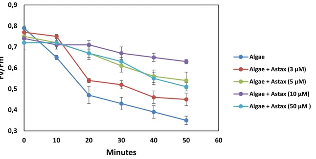
Secondly, an optical/amperometric biosensor based on algal cells immobilized in calcium alginate gel was developed. Various Chlamydomonas reinhardtii strains mutated at the level of photosynthetic D1 protein were used as biomediators to quantify the capacity of the carotenoid xanthophylls to protect the photosynthetic apparatus from photoinhibition. The highly sensitive and selective biosensor was used for studies on cell physiology aimed to determine the antioxidant and light filtering effects of the xanthophyll astaxanthin. The biosensor was proved to be suitable for the determination of the exogenous supplied astaxanthin showing in a short time a reliable response with a detection limit of 3 µM. This technique revealed the photo-protective effect of astaxanthin-enriched extracts of Haematococcus pluvialis in algal cells. The results suggest that in algae astaxanthin exploits both filtering and antioxidant effects at different light intensities. This bioinspired approach
xv can provide new insights into biological, biomedical, environmental and agricultural research applications and nutraceutical studies.
We also projected and realized a biosensor platform, which combines an optical system, intimately, integrated with an array of biomediators able to provide a helpful tool for safety management of the milk. Optical feature selection of various enzymes was performed for monitoring compounds of various chemical classes and metabolic markers of cow’s wellness. The detection of selected analytes was evaluated through biomediators of Chlamydomonas reinhardtii cells, and acetylcholinesterase, tyrosinase, urease, β-galactosidase and D-lactate dehydrogenase enzymes. The analyses were performed by fluorescence that for the algal cells was based on chlorophyll a fluorescence emission while for enzymes was guaranteed by the use of fluorescein 5(6)-isothiocyanate or 5(6)-carboxynaphthofluorescein.
Furthermore, a novel fluidic setup based on simple fluid mechanics laws has been realized with no moving components when pump is on. It yields a fluctuation-free outcome signal that we compare with those produced by commercial peristaltic pumps. Evidence of the readout amperometric enhanced signal is shown thus confirming the successful implementation of more reliable biosensors and electrochemical signal transduction devices. Such stable and oscillation free amperometric signals are very desirable in view of biosensors integration in environmental monitoring platforms equipped with advanced communication technologies.
In addition, two set of C. reinhardtii mutants, able to quench 1O2 and other ROS produced
under extreme conditions, were produced in collaboration with U. Johanningmeier (Uni of Halle). First set was mutated by site-directed mutagenesis (antiox-mutants): antioxidant peptides able to quench 1O2 and variety of free radicals introduced in the D1 protein of Photosystem II. The second
set was produced by UV-mutagenesis. The mutated strains were characterized to select more resistant strains to be used as biological element for environmental monitoring under extreme conditions.
xvi In this study, total lipid and phenol content of two set of Chlamydomonas reinhardtii mutants (Antiox and UV mutants) were determined. The in-vivo and in-vitro antioxidant capacity of the antiox-mutants were evaluated using different assay and in the presence of 1O2 precursors. The mutants have
reduced lipid and phenol content compared to the wild type Chlamydomonas reinhardtii. However, they showed better resistant to stress conditions such as high light, 1O2 and H2O2. UV mutants have
showed longer half-life. The produced mutants revealed to be a promising candidate for improved environmental monitoring on earth.
We also described the use of biomimetic peptides of the photosynthetic plastoquinone binding niche of the green alga Chlamydomonas reinhardtii for pesticide measurement in environmental samples. Three biomimetic peptides containing the plastoquinone-binding site in a loop shaped by two alpha helices were designed and characterized. Natural sequence of 70 aminoacid-peptide was modified to increase the solubility in aqueous solvents by adding two histidine in the N- and C-terminus. A cysteine was included in two of the modified peptides (S264C or S268C) and labeled with commercial carboxylated quantum dots (peak emissions 710 nm) by carbodiimide reaction coupling and fluorometric detection was performed. The results confirmed that mutation S264C conferred resistance to atrazine and diuron, while the change S268C increased the sensitivity.This work demonstrates the interest to replace whole microalgae cells or their photosynthetic apparatus (thylakoids, isolated reaction centers) by much smaller fragments such as D1 peptide mimics. This approach might improve the system in terms of sensitivity, but also in terms of stability (oligopeptides being much more stable than the whole three-dimensional protein molecule) and facilitate the commercialization for environmental monitoring.
English is an international language that allows us to communicate with scientists from all over the world and writing a manuscript is important since it is a form of written communication in science. In this occasion, as a part of my training to improve my writing skills and enhance my knowledge on biosensors, we presented a review focuses on bioassay development based on the
xvii simultaneous use of quantum dots and magnetic beads. Due to the outstanding characteristics of both particles for biosensing applications and the large number of publications using a combined approach, we aim to provide a comprehensive overview of the literature on different bioassays. The improvement of current approaches together with novel multiplex detection systems and nanomaterials-based research, including the use of multimodal nanoparticles, will contribute to simpler and more sensitive bioanalyses.
The optical and electrochemical biosensors, that were constructed and optimized within this doctoral thesis, have the advantage of being user friendly, low cost and portable analytical tools, which facilitates a future transfer to the market of environmental and agrifood monitoring.
1
CHAPTER 1
2 1. Biosensors
Over the past decade, many important technological advances have provided us with the tools and materials needed to construct biosensor devices. Since the first invention of the Clark Oxygen Electrode sensor, there have been many improvements in the sensitivity, selectivity, and multiplexing capacity of the modern biosensor. Before the various types of biosensor technologies and application are discussed, it is first important to understand and define ‘‘biosensor’’ (Perumal, 2014).
A biosensor is a self-contained integrated device that is capable of providing specific quantitative or semi-quantitative analytical information using a biological recognition element (biochemical receptor) that is retained in direct spatial contact with a transduction element. However, a common working definition is that a biosensor is an analytical device that uses biological macromolecules to recognize an analyte and subsequently activate a signal that is detected with a transducer (Cullum and Sumner, 2010). Once the interaction of the biochemical receptor with the analyte is converted into a signal detectable by the transduction system, the signal is send to a readout or display by appropriate electrical equipment (Farré et al., 2009, Thévenot et al., 2001, Justino et al., 2010).The biosensor consists of three parts: the first element is the biomediator a biologically derived material e.g. tissue, microorganisms, organelles, cell receptors, enzymes, antibodies, nucleic acids, and biological sensitive elements created with genetic engineering, or a biomimic system e.g. aptamers, MIPs, etc. The second element is the transducer e.g. physicochemical, optical, piezoelectric, electrochemical, etc. that transforms the signal resulting from the analytes interaction with the biological element into a signal that can be measured and quantified. The third element is the associated electronics or signal processor, responsible of the visualization results in a user-friendly way. There is a need for a simple, rapid and reagentless method for specific determination, both qualitative and quantitative, of various compounds in various applications. Hence, it is paramount to have fast and accurate chemical intelligence, which is particularly conspicuous in human health care.
3 Fig. 1. The Principle of a biosensor
Classification of biosensors
Biosensors can be classified according to either the biorecognition principle used for sensing (e.g. enzymes, nucleic acids, antibody or whole cells) or the transduction element employed (e.g., electrochemical, optical, piezoelectrical or thermal). Some authorities subdivide biosensors into affinity biosensors and catalytic biosensors based upon the activity of the biorecognition element. Thus, affinity biosensors have as their fundamental property the recognition (binding) of the analyte by the biorecognition element (for example, antibody-antigen), whereas catalytic biosensors have as their biorecognition element proteins (or microorganisms) that not only bind the analyte but also catalyze a reaction involving the analyte to produce a product (for example, glucose biosensors) (Cullum and Sumner, 2010).
According to the transducer type, biosensors can be classified in four different groups (Frachiolla et al., 2013)As described above, the transducer is the portion of the biosensor responsible for converting the biorecognition event into a measurable signal. It is possible to exploit a change in a number of physical and chemical properties (mass, temperature, electrical properties, and optical properties) to allow for different transduction formats. Although a variety of transducer methods have
4 been feasible toward the development of biosensor technology, the most common methods are electrochemical and optical followed by piezoelectric.
1.1.1. Optical detection
Due to a number of advantages, optical transduction is one of the most widely used biosensor transduction formats. For example, optical transduction can be very rapid where the limiting factor for the speed of detection is often a diffusion-limited process of the biomolecular recognition event, rather than the optical transducer. Another advantage of optical transduction is that the interferences that can hinder electrochemical transduction measurements (such as voltage surges, harmonic induction, corrosion of electrode elements, and radio frequency interferences) are not present. Some of the disadvantages of using optical transduction formats include detection challenges when analyzing turbid samples and the cost associated with detection system components.
A wide variety of optical transduction formats have been employed, where changes of the interaction of light with the biomolecular system are used to produce a measurable signal. These changes can be based on differences in refractive index, production of chemiluminescent reaction products, fluorescence emission, fluorescence quenching, radiative and nonradiative energy transfer, temporal changes in optical emission properties, and scattering techniques, well as other optical effects.
These effects can be monitored using a variety of optical platforms including total internal reflectance and evanescent wave technologies, interferometric, resonant cavities, and biochip devices. The following paragraphs review the most common as well as most popular emerging optical transduction formats: fluorescence, chemiluminescence, and surface plasmon resonance (Cullum and Sumner, 2010).
5 1.1.1.1. Fluorescence
Fluorescence is the most popular form of optical transduction due to the high sensitivity that is fundamental to this type of optical process. Another advantage of fluorescence-based methods is that they generally do not have the interference issues that SPR and other refractive index based methods possess. However, in most cases, the intrinsic sample fluorescence is not sufficient for analysis, and a fluorogenic reporter is used to label an affinity reagent to create a bioreporter. By monitoring the intensity of the fluorogenic reporter, it is possible to determine the presence and concentration of the target analytes, as illustrated in the bioassay techniques already described. It is also possible to monitor shifts in the wavelength of the fluorophore reporter, as well as energy transfer phenomena, and time dependence of the fluorescence emission, all of which can be related to binding interactions depending on the assay employed. The distinguishing features between biosensors, besides the above mentioned properties that can be monitored, include the optical detection format used. For example, it is possible to utilize fiber-optic probes to immobilize bioreceptors at the tip of the fiber and use total internal reflectance properties of the fiber to transmit excited and emitted light. Total internal reflectance can also be employed in an evanescent wave format, where a residual amount of (evanescent) light at the reflectance point that escapes is used to excite immobilized bioreceptors only in close proximity to the surface, rather than in bulk solution. This format allows for controlled excitation and can allow for minimal fluorescence background.
However, a key disadvantage is the lack of evanescent excitation power and sometimes poor coupling of the emission when using similar collection geometries. Fluorescence detection can be used with a wide variety of detection formats. For example, it is routinely coupled with flow cytometry and microfluidic platforms or imaging array systems such as biochips that utilize spatial patterning of biological recognition elements to match fluorescence location to target species (Cullum and Sumner, 2010).
6 1.1.2. Electrochemical
Electrochemical transduction is one of the most popular transduction formats employed in biosensing applications. One of the main advantages of biosensors that employ electrochemical transduction is the ability to operate in turbid media and often in complex matrices. Another distinct advantage of electrochemical transduction is that the detection components are inexpensive and can be readily miniaturized into portable, low-cost devices.
In general, electrochemical-based sensing can be divided into three main categories: potentiometric, amperometric, and impedance. Potentiometric sensors typically rely on a change in potential caused by the production of an electroactive species that is measured by an ion selective electrode. For a biosensor system, this change in electroactive species concentration is usually brought about by an enzyme. In an amperometric sensor system, a change in current is directly measured. Electrochemical sensors based on impedance most commonly utilize impedance spectroscopy since controlled AC electrical stimulus over a range of frequencies is used to detect variations in the sensor surface properties (that is, charge transfer and capacitance at the interface layer). In this way, the resistance to flow of an alternating current is measured as voltage/current. For example, metabolic changes (for example, growth) have been shown to correspond to an increase or decrease in impedance. Some of the many variations of potentiometric, amperometric, and impedance biosensors that provide for improved biosensor performance include field effect transistors (FET) and electrochemiluminescence (ECL) (Cullum and Sumner, 2010; Chaplin, 2000).
1.1.3. Mass-sensitive
Biosensors that are based on mass-sensitive measurements detect small mass changes caused by chemical binding to small piezoelectric crystals. Initially, a specific electrical signal can be applied to the crystals to cause them to vibrate at a specific frequency. This frequency of oscillation depends on the electrical signal frequency and the mass of the crystal. As such, the binding of an analyte of
7 interest will increase the mass of the crystal and subsequently change its frequency of oscillation, which can then be measured electrically and used to determine the mass of the analyte of interest bound to the crystal (Morrison, 2008).The main advantage to the piezoelectric transduction (that is, mass sensor) approach includes the ability to perform label-free measurements of the binding events, including real-time analysis of binding kinetics (Cullum and Sumner, 2010).
1.1.4. Thermal/Calorimetric
Calorimetric sensors utilize thermistor probes to monitor changes in temperature due to exothermic chemical reactions. This change in temperature can be correlated to the amount of reactants consumed or products formed. Many biological reactions are exothermic (for example, enzyme reactions), and hence calorimetric detection allows for a near-universal transduction format. One key disadvantage of this approach is that environmental temperature fluctuations must be shielded from the sensor system. Calorimetric biosensors traditionally have been large and bulky, although advances in silicon microfabrication technologies and microfluidics have allowed for miniaturization and improved performance.
Different types of bioreceptors
The key to specificity for biosensor technologies involves bioreceptors. They are responsible for binding the analyte of interest to the sensor for the measurement. These bioreceptors can take many forms and the different bioreceptors that have been used are as numerous as the different analytes that have been monitored using biosensors. However, bioreceptors can generally be classified into five different major categories. These categories include: 1) antibody/antigen, 2) enzymes, 3) nucleic acids/DNA, 4) cellular structures/cells and 5) biomimetic (Dinh, 2007).Enzymes have been the most widely used bioreceptor molecules in biosensor applications, with antibodies and
8 protein receptor molecules increasingly incorporated in biosensors. The specificity of a biosensor comes from the specificity of the bioreceptor molecule used (Lee and Mutharasan, 2005).
Enzymes: Enzyme based biosensors were the earliest biosensors, introduced by Clark and Lyons in 1962 – an amperometric enzyme electrode for a glucose sensor which used a ‘‘soluble’’ enzyme electrode. Since the first biosensor, enzyme based biosensors have faced a massive growth in usage for various applications up to the present. Enzymes are very efficient biocatalysts, which have the ability to specifically recognize their substrates and to catalyze their transformation. These unique properties make the enzymes powerful tools to develop analytical devices (Perumal and Hashim, 2014).
In biocatalytic recognition mechanisms, the detection is amplified by a reaction catalyzed by macromolecules called biocatalysts. The lock and key and induced fit hypothesis can apply to explain the mechanism of the enzyme action, which is highly specific for this type of biosensor.This high specificity of enzyme–substrate interactions and the usually high turnover rates of biocatalysts are the origin of sensitive and specific enzyme-based biosensor devices. This specific catalytic reaction of the enzyme provides these types of biosensor with the ability to detect much lower limits than with normal binding techniques (Perumal and Hashim, 2014).
The catalytic activity of enzymes depends upon the integrity of their native protein conformation. If an enzyme is denatured, dissociated into its subunits, or broken down into its component amino acids, its catalytic activity is destroyed (Dinh, 2007). Ideally, enzyme catalytic action can be influenced by several factors such as the concentration of the substrate, temperature, presence of a competitive and non-competitive inhibitor and pH. Essentially the Michaelis– Menten equation can be used to further explain the detection limit of the enzyme based biosensor. Some recent studies have shown that enzyme based biosensors can be used to detect cholesterol, food safety and environmental monitoring, heavy metals and also pesticides. (Perumal and Hashim, 2014).
9 Antibodies: An antibody is a complex biomolecule, made up of hundreds of individual amino acids arranged in a highly ordered sequence. They represent one of the major classes of proteins. They constitute about 20% of the total plasma protein and are collectively called immunoglobulins (Ig).An antigen can be any macromolecule that induces an immune response. The antibody binds reversibly with a specific antigen. Unlike the enzyme proteins, the antibodies do not act as catalysts.The way in which an antigen and its antigen-specific antibody interact may be understood as analogous to a lock and key fit, by which specific geometrical configurations of a unique key enables it to open a lock. In the same way, an antigen-specific antibody “fits” its unique antigen in a highly specific manner. This unique property of antibodies is the key to their usefulness in immunosensors where only the specific analyte of interest, the antigen, fits into the antibody-binding site.
DNA/nucleic acid: The use of a nucleic acids sequence for a specific diagnostics application was developed in the early 1953 and is still growing widely. Biosensors based on DNA, RNA and peptide nucleic acid gain their high sensitivity and selectivity from the very strong base pair affinity between complementary sections of lined — up nucleotide strands (Borgmann et al. 2011). Nucleic acid (NA) — based biosensors integrate an NA (natural and biomimetic forms of oligo- and polynucleotides) as the biological recognition element. Nowadays, mainly synthetic oligodeoxyribonucleotides (ODNs) are used as probes in the DNA hybridization sensors (Monosik, 2012). The highly specific affinity binding's reaction between two single strand DNA (ssDNA) chains to form double stranded DNA (dsDNA) is utilized in the nucleic acids based biosensor which appoints the nucleic acids as the biological recognition element. This biosensor possesses a remarkable specificity to provide analytical tools that can measure the presence of a single molecule species in a complex mixture (Perumal and Hashim, 2014).
Biomimetic receptors: An artificial (man-made) receptor that is fabricated and designed to mimic a bioreceptor is often termed a biomimetic receptor. Several different methods have been developed over the years for the construction of biomimetic receptors. These methods include genetically
10 engineered molecules, artificial membrane fabrication and molecular imprinting. Recombinant techniques, which allow for the synthesis or modification of a wide variety of binding sites using chemical means, have provided powerful tools for designing synthetic bioreceptors with desired properties (Dinh, 2007).
Cells: These bioreceptors are based on biorecognition either by an entire cell/microorganism or by a specific cellular component that is capable of specific binding to certain species (Monosik, 2012; Dinh, 2007). Microorganisms offer a form of bioreceptor that often allows a whole class of compounds to be monitored. Generally, these microorganism biosensors rely on the uptake of certain chemicals into the microorganism for digestion. Often, a microorganism ingests a class of chemicals therefore allowing a class-specific biosensor to be created. Microorganisms such as bacteria and fungi have been used as indicators of toxicity or for the measurement of specific substances. For example, cell metabolism (e.g., growth inhibition, cell viability, and substrate uptake), cell respiration or bacterial bioluminescence have been used to evaluate the effects of toxic heavy metals. Whole mammalian tissue slices or in vitro cultured mammalian cells are used as biosensing elements in bioreceptors.
Photosytnhetic organisms such as plant tissues and microalgae are also used in plant/algae-based biosensors since they are effective catalysts because of the enzymatic pathways they possess. In general, their use is associated to the environmental monitoring and in particular to pesticides, herbicides and heavy metals (Scognamiglio et al. 2010;Giardi and Pace, 2005).
It is know that photosynthesis in green microalgae and higher plants occurs into chloroplasts that are called cellular organelles, which contain membrane-made closed structures (thylakoids) tightly leant one to the other to form piles named grana. Within thylakoid membranes, the chlorophyll is associated in complexes containing up to 250 molecules among which only a few number are directly involved in the photochemical reactions producing ATP all the other ones are used as light-harvesting antennae. The antennae have the function to collect and convey the light to the producing
11 molecules of the ATP. The Photosynthetic apparatus is composed of two photosystems named Photosystem I (PSI) and II (PSII), present in the thylakoids (Fig. 2).As shown in the figure, PSI and PSII are constituted by the ensemble of several proteins and pigment protein complexes that work in concert in order to build up functional units of metabolic importance. These interactions lead to the generation of a transmembrane proton gradient necessary to ATP synthesis and to the production of the reducing power necessary to NADPH biosynthesis (Foyer et al 2012). The chlorophylls of the light-harvesting complex of PSII absorb the light radiation and transfer it to the pigment P680 (primary electron donor of PSII). A group of pigments that are excitonically coupled constitutes this pigment. In fact when they absorb a photon act as a single molecule. P680 is the strongest biological oxidizing agent known at present (Giardi and Pace, 2005).
Fig. 2. The Photosynthetic apparatus is arranged in the Photosystem I (PSI) and II (PSII) present in the thylakoids.PSII composed by D1 and D2 proteins and Oxygen Evolving Complex (OEC), QA
12 When the light arrived to PSII, chlorophyll-protein harvesting complexes absorb it then it is funneled to the photochemically active reaction center composed by the subunits D1, D2 and Oxygen Evolving Complex (OEC). Here photoexcitation converts P680 into the oxidized form P680+ thus triggering a single electron transfer first to a pheophytin cofactor then to a plastoquinone molecule QA located in
D2 and finally to a second plastoquinone QB in the D1 protein as shown in the Figure 2.
As mentioned above, using photosynthetic organisms and organelles it is possible to detect herbicides presence in waters, environment or agrifood products because herbicides act directly on the photosynthetic system inhibiting the photosynthetic process. About 30% of herbicides including phenylurea, triazine and phenolic herbicides inhibit photosynthetic electron flow by blocking the PSII quinone-binding site and thus modifying chlorophyll fluorescence. Various amperometric and some optical biosensors are developed to detect various kind of herbicides. (Campas et al. 2008; Scognamiglio et al. 2010).
The phenomenon of autofluorescence in photosynthetic organisms is particularly important for our present work. The capture of light energy for photosynthesis is enhanced by networks of pigments in the chloroplasts arranged in aggregates on the thylakoids. These aggregates are called antenna complexes. Evidence for this kind of picture came from research by Robert Emerson and William Arnold in 1932 when they measured the oxygen released in response to extremely bright flashes of light. They found that some 2500 molecules of chlorophyll was required to produce one molecule of oxygen, and that a minimum of eight photons of light must be absorbed in the process. The model that emerges is that of some 300 chlorophyll molecules and 40 or so betacarotenes and other accessory pigments acting as a light harvesting antenna surrounding one chlorophyll a molecule that is a part of an action center. A photon is absorbed by one of the pigment molecules and transfers that energy by successive fluorescence events to neighboring molecules until it reaches the action center where the energy is used to transfer an energetic electron to an electron acceptor. The fluorescence model would suggest that each transferred photon has a longer wavelength and lower
13 quantum energy with some energy being lost to heat. When a photon reaches the chlorophyll a in the reaction center, that chlorophyll can receive the energy because it absorbs photons of longer wavelengths than the other pigments. Two types of chlorophyll centers have been identified, and are associated with two protein complexes identified as Photosystem II and Photosystem I (Fig. 2).
Essentially, the living cell based biosensor is a unique biosensor in contrast to other types of biosensor that contain materials extracted from living things. These types of biosensor have a number of pros and cons. The detection limit of this biosensor is mainly determined by the natural environmental conditions in which the cell can stay alive for a long period in order to control the physical and chemical parameters of the environment. However, the major limitations of the cell based biosensor are the stability of the cell, which depends on various conditions such as sterilization, lifetime, biocompatibility and etc. Another issue that governs the success of cell-based sensors depends primarily on selectivity, in which the cell based sensor has poor selectivity due to the multireceptor behaviour of the intact cells (Belkin and Gu, 2010). Despite these pitfalls, the cell-based biosensor is still favoured by researchers due to the advantages over the enzyme-based biosensor. The cell-based biosensors are less sensitive to inhibition by solutes and are more tolerant of suboptimal pH and temperature values than the enzyme based biosensor, though they must not exceed a narrow range because of the possibility of cell death. A longer lifetime can be expected than with the enzymatic sensors and they are much cheaper because the active cells do not need to be isolated (Struss et al., 2010). The literature indicates that cell based sensors have become emerging tools for medical diagnostics (i.e., such as disease detection), environmental analysis, food quality control, chemical–pharmaceutical industry and drugs detection. Similarly, it was reported that the cell based biosensor is envisioned to be an emerging frontier in the area of nano-diagnotics due to their attractive characteristics.
14 Biosensor characteristics
Biosensors are characterized by eight parameters (Lee and Mutharasan, 2005). These are:
(1) Sensitivity is the response of the sensor to per unit change in analyte concentration.
(2) Selectivity is the ability of the sensor to respond only to the target analyte. That is, lack of response to other interfering chemicals is the desired feature.
(3) Range is the concentration range over which the sensitivity of the sensor is good. Sometimes this is called dynamic range or linearity.
(4) Response time is the time required for the sensor to indicate 63% of its final response due to a step change in analyte concentration.
(5) Reproducibility is the accuracy with which the sensor’s output can be obtained.
(6) Detection limit is the lowest concentration of the analyte to which there is a measurable response.
(7) Life time is the period over which the sensor can be used without significant deterioration in performance characteristics.
(8) Stability characterizes the change in its baseline or sensitivity over a fixed period.
Considerations in biosensor development
Once a target analyte has been identified, the major tasks in developing a biosensor involve:
1. Selection of a suitable bioreceptor or a recognition molecule
2. Selection of a suitable immobilization method
15 4. Design of biosensor considering measurement range, linearity, and minimization of interference, and enhancement of sensitivity
5. Packaging of the biosensor into a complete device
The first item above requires knowledge in biochemistry and biology, the second and third require knowledge in chemistry, electrochemistry and physics, and the fourth requires knowledge of kinetics and mass transfer. Once a biosensor has been designed, it must be packaged for convenient manufacturing and use. The current trend is miniaturization and mass production. Modern IC (integrated circuit) fabrication technology and micromachining technology are used increasingly in fabricating biosensors, as they reduce manufacturing costs. Therefore, an interdisciplinary research team, consisting of the various disciplines identified above, is essential for successful development of a biosensor (Lee and Mutharasan, 2005).
2. Singlet oxygen production in photosystem II
When higher plant, algae and cyanobacteria are exposed to highlight intensity illumination, PSII activity is inhibited in a process called photoinhibition. Photo-inactivation of PSII is considered to be caused by damage to the D1 protein, one of the two proteins, which formed a heterodimer with the D2 protein. It is widely accepted that D1 damage is caused by two distinct mechanisms of photoinhibition i.e. the so called acceptor and donor side mechanism. In the acceptor side photoinhibition, over-reduction of the primary electron acceptor QA leads to its release from the binding site in the D2 protein. In donor side photoinhibition, the formation of long-lived highly oxidizing molecules P680•+ /TyrZ• leads to the oxidation of the organic components such as proteins and lipids (Yadav and Pospisil et al., 2012).
It has been reported that different types of reactive oxygen species (ROS) are formed in both the acceptor and the donor side photoinhibition. In the acceptor-side photoinhibition, singlet oxygen
16 (1O2) is considered the main ROS responsible for PSII damage.It is generated by the interaction of
molecular oxygen and triplet chlorophyll formed by the charge recombination of the triplet radical pair 3[P680•+ Pheo•-]. Trebst and co-workers provided evidence that 1O2 is the important damaging
species during photoinhibition of Chlamydomonas reinhardtii cells (Trebst et al., 2002).A special case is when herbicides are bound to PSII. Under these conditions electron transfer can take place only as far as QA-• and charge recombination is the main outcome of light-induced charge separation. It was pointed out recently that the stimulated charge recombination occurring in herbicide-treated PSII could be an important factor in the herbicidal action mediated by 1O2. This suggestion was based
on a number of experimental observations and theoretical considerations (Fufezan et al., 2002):
1. Different herbicide classes have different influences on the photoinhibition of PSII. Phenolic herbicides increase photodamage, while with 3-(3,4-dichlorophenyl)- 1,1-dimethylurea (DCMU) and other urea- and triazine-type herbicides photodamage is less marked although all of these herbicides bind to the QB-binding site and inhibit forward electron transport.
2. Different classes of herbicides influence the charge recombination measured by differential changes in the thermoluminescence emission temperature. Phenolic herbicides decrease the emission temperature while urea- and triazine type herbicides increase it.
3. Herbicide binding influences the midpoint potential (Em) of the QA/QA-• redox couple in PSII with phenolic herbicides lowering the Em by 45 mV, while DCMU raises the potential by 50 mV (Krieger-Liszkay et al., 1998).
Correlating these factors it was suggested that herbicides modulate the recombination pathway within PSII and thus the degree of 1O2-mediated photodamage. It is predicted that 1O2 will be
generated in herbicide-treated PSII and, importantly, that the yield will be greater when phenolic herbicides are used compared to other classes of herbicide. It is known that photoinhibitory damage of PSII under various conditions can result in the formation not only of 1O2 but also of other reactive
17 lessening the photooxidative membrane damage and increasing the stability and sensitivity for biosensor applications is of special interest. The ability to produce new algal biomediators, with a broad structural stability, can be expected to lead biosensing for food control or environmental monitoring.
3. Genetic engineering to improve sensitivity and selectivity of biomediators for biosensor development
Nowadays, genetic engineering allows the modification of specific nucleotide sequences of an organism genome to obtain proteins with novel improved properties, and innovative biotechnological approaches make it possible to integrate these systems, or their functional sub-structures, into artificial assemblies for specific applications such as environmental monitoring. Several biomediators have been already developed exploiting molecular biological techniques to produce enzymes and/or protein with improved features in the detection of specific analytes (Wang et al., 2009).
In the context of the photosynthesis-based biosensors, activities in different research areas allowed the design and development of engineered photosynthetic microorganisms with improved sensitivity and stability features to be used as bio-recognition elements for the detection of environmental contaminants. Different approaches, such as space research and physical elicitations, have been applied to select microorganisms with improved tolerance to extreme environmental conditions. The newly selected organisms generated for biosensor purposes were able to maintain a stable photosynthetic efficiency and an increased oxygen evolution capacity (Rea et al., 2008).
In particular, researchers carried out modifications of the D1 reaction center proteins, as they play a crucial role in electron tunneling-mediated charge separation and transmembrane electric field generation, acting principally on reduction, release and migrations of (plasto) quinones. Random mutagenesis targeted to the D1-encoding psbA gene was exploited as a directed evolution strategy to
18 produce a huge mutant library of Chlamydomonas carrying novel D1 proteins with different aminoacidic composition. In addition, thanks to the support of bioinformatics studies, site-directed mutagenesis was also exploited to generate specific point mutations in the D1 protein, in order to modify the properties of the PQ/atrazine binding affinity.
Various mutant strains has been produced and used for biosensing approach so far. Chlamydomonas D1 random and site-directed mutants were produced by particle gun bombardments of the chloroplast genome (Przibilla et al., 1991). The Del1 Chlamydomonas strain was used as a recipient host for the mutant’s generation (Preiss et al., 2001). This strain has a defined deletion in the chloroplast-encoded psbA gene and is unable to grow photoautotrophically, as it cannot produce a functional D1 protein. Acetate is needed as carbon source as minimal media do not support its growth. Minimal media were used to select photosynthetically active colonies generated after the integration of the psbA variant produced both by random and site-directed PCR (Dauvillee et al., 2004). Selected mutants were then characterized by analyzing their photosynthetic performance and the sensitivity and/or resistance to different classes of herbicides assessed (Tibuzzi et al., 2007; Rea et al., 2009; Giardi et al., 2009; Scognamiglio et al., 2009). After the characterization, the best performing mutants were immobilized on screen-printed electrodes and integrated in amperometric or optic transduction systems. Both electrochemical and optical devices were arranged in multi-arrayed setups.
4. Microalgae PSII based biosensors as useful bioanalytical tools for herbicide detection
The exploitation of herbicides for weed control is vital to increase the yields and productivity in agriculture. Without the use of herbicides, it would have been impossible to fully mechanize the production of cotton, sugar beets, grains, potatoes, and corn. Therefore, given the harmful economic implications of poor harvesting, herbicide production is the principal driver of the farming industry.
19 However, the continuous and massive application of these compounds can negatively affect human health and ecosystems. These consequences result in an increased demand for risk assessment and prompt the regulatory agencies to update legislation aimed at controlling environmental contaminations. In this scenario, it is essential to develop analytical devices able to detect the low levels of herbicide contaminants defined by the EU directives, and to distinguish among different classes of compounds.
A major difficulty in estimating environmental quality related to herbicides contamination is due to seasonal change of field application and the extremely low levels of the maximum admissible concentrations set by the European commission. Several methods, which partially satisfying these requirements have been already developed. In this context, chromatographic techniques, such as gas chromatography (GC) and high performance liquid chromatography (HPLC) with UV and/or mass spectrometry (MS) detection, surely represent the most trustworthy and common techniques used to monitor the presence of herbicides. The classical analytical techniques are unlikely to provide adequate sensitivity, while advanced instrumental methods are highly sensitive, but generally expensive, require skilled operators and are not easily amenable to on-site field-testing. Herbicides usually represent a very small fraction of the whole sample under investigation, so pre-treatments such as clean up and/or pre-concentration steps are required to make their identification possible. Consequently, the qualitative and quantitative analysis of herbicide residues is time consuming and involves high costs. Because of the large numbers of samples to be measured, the necessity of expensive equipment, organic solvents and laborious sample preparation, the development of a fast, automated and inexpensive test is of great interest. These concerns encouraged researchers to seek out alternative methods providing the desired analytical information.
Studies in the framework of the development of biosensors for the detection of environmental pollutants exploit photosynthetic microorganisms or parts of them, such as thylakoids. Photosynthetic systems are naturally occurring anisotropic supramolecular arrangements of proteins and small
20 molecules that are able to harvest light energy and funnel it towards building up biomass and releasing oxygen. For these purposes, photosynthetic organisms are equipped with multi-enzymatic complexes embedded in thylakoid or free membranes known as photosystems. The hierarchical organization of these pigment-protein complexes is at the basis of their unique efficiency. Functional and structural knowledge of photosynthetic systems has been steadily increasing, and as a result, fundamental and applied research have made it possible to integrate biological photosystems or their functional sub-structures into artificial assemblies in order to get them to carry out their tasks in a controlled environment for specific applications.
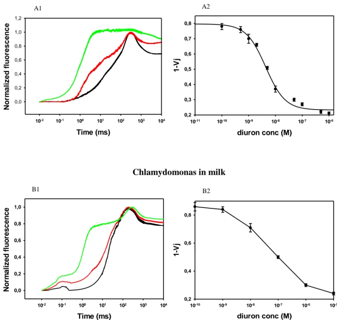
In PSII if the electron transfer from the reaction center to the quinone pool is blocked, such as during the binding of the photosynthetically active pesticides, these parameters change dramatically and can be monitored by electro-optical analysis in a pesticide concentration dependent manner. Currently, in parallel to traditional analytical methods, novel detection systems have been already developed based on biosensor technology, which provides rapid, inexpensive and reliable tools for herbicide monitoring and screening analyses, to answer the concern on this issue. An optical biosensor based on the green photosynthetic alga Chlamydomonas reinhardtii described by Tibuzzi and coworkers was employed to monitor several classes of herbicides, such as atrazine, diuron, ioxynil, terbuthylazine, prometryn and linuron, in a low concentration range (10-8-10-10 M) (Tibuzzi et al., 2007). In particular, a miniaturized optical biosensor instrument was designed and produced using multiarray fluorescence measurements of several biomediators in series, for applications in environmental monitoring and agrofood analysis. In the work by Rea and coworkers, a computational study was performed to design and construct a set of mutant strains from the green photosynthetic alga C. reinhardtii, with higher sensitivity towards several classes and subclasses of herbicides (Rea et al., 2009). An in silico study was performed to predict mutations within the D1-D2 heterodimer which improve its specificity, sensitivity, and binding affinity for atrazine. In detail, taking advantage of the high sequence homology observed between Thermosynecococcus elongatus D1 and D2
21 proteins and the corresponding proteins from C. reinhardtii (87% and 89% amino acid sequence identity, respectively), the three-dimensional structure of the latter proteins was homology modelled. Based on this model, a series of D1 and D2 mutants were generated in silico and the atrazine affinity of wild type and mutant proteins was predicted by binding energy calculations to identify mutations able to increase PSII affinity for atrazine.
New advances in the same context were achieved in amplifying the range of recognition elements and measurement of a significant number of different classes of environmental pollutants. These advances occurred through the development of a biosensing system, which uses sets of mutant organisms with different affinities towards pesticides. Giardi and coworkers presented a library of functional mutations in the unicellular green alga C. reinhardtii for preparing biomediators (Giardi et al., 2009). Exploiting bioinformatics to design new mutant strains resulted in the construction of microorganisms, which showed different limits of detection for diazines, triazines and urea herbicides, underlined the high potential of bioinformatics and molecular biology in the design of desired biological material suitable for biosensor use.
Scognamiglio and coworkers (Scognamiglio et al., 2009) assembled a multi-biomediator fluorescence biosensor based on a new versatile portable instrument. The biosensor instrument was composed of a 24-cell array configuration able to host different mutant strains for the detection of a variety of herbicide classes such as triazines, diazines and ureas. As we can observe from the described advances in biosensor technology, the main features of a successful biosensor are characterized by the interchangeable recognition elements, which provide the versatility to measure large numbers of analytes. Buonasera and coworkers was constructed a biosensing platform to provide an analytical tool applicable to the daily pre-screening of a broad spectrum of samples (Buonasera et al., 2010). The platform combined the most used transduction systems for biosensors, amperometric and optical systems, and used genetically modified microorganisms as versatile biomediators, allowing detection of different subclasses of herbicides. It represented a sensitive,
22 reliable, and low-cost system able to detect water pollutants such as atrazine, diuron, linuron, and terbuthylazine down to 10-8-10-10 M. Combining the amperometric and optical detection systems, the platform was able to determine the toxicological potential of samples, through the determination of the biomediator physiological activity inhibition. Fluorescence modification and current reduction were related to the concentration of herbicide quantified by a dedicated data acquisition software. In addition, the opportunity to use a wide range of biological materials made the platform a good candidate for the development of a biosensor with required features.
In this thesis, we focused on the development of new types of bio-sensing elements for building up a platform of modular biosensors with the best features in terms of versatility, stability, life-time and long-term activities, sensitivity and selectivity, detection limits, linear range, reproducibility and low cost, which can be easily adopted for the simultaneous detection of several herbicides. We developed an array of novel whole-cell biosensors based on the activity of engineered photosystem II with improved sensitivity and stability features, and biomimetic peptides as sensing elements. The new complex biosensors array based on both optical and electronic transduction for multi-parameter detection. It will be able to monitor the herbicide levels and to diagnose their biological impact. This improvement should provide the impetus for the technological transfer from laboratory devices to in-field operation systems. The new devices will lead to a tremendous breakthrough in the detection of contaminants and quality control in risk assessment sectors by providing a rapid broad-spectrum screening tool.
23 References
Belkin, S., Gu, M.B., 2010. Whole Cell Sensing Systems I: Reporter Cells and Devices. Springer, Heidelberg. Buonasera, K., Pezzotti, G., Scognamiglio, V., Tibuzzi, A., Giardi, M.T., 2010 New platform of biosensors for prescreening of pesticide residues to support laboratory analyses., J. Agric. Food Chem. 58, 5982–90. doi:10.1021/jf9027602.
Campas M., Carpentier, R., Rouillon, R., 2008. Plant tissue-and photosynthesis-based biosensors. Biotechnology Advances 26, 370-378.
Cavalcanti, A., Shirinzadeh, B., Zhang, M. & Kretly, L.C. (2008). Nanorobot hardware architecture for medical defense. Sensors. 8(5), 2932–2958.
Chaplin, M.F., 2000. Biosensors, in: Walker, J.M., Raply, R. (Eds.), Molecular Biology and Biotechnology. RCS Publishing, pp. 513-546.
Cullum, D.S., Sumner, J., 2010. Biosensors and Bioelectronics, in: Armstrong R.E., Drapeau, M.D., Loeb, C.A., Valdes, J.J. (Eds.), Bio-Inspired Innovation and National Security Biosensors. National Defense University Press, pp. 77-103
Dauvillee, D.; Hilbig, L.; Preiss, S., Johanningmeier, U. (2004). Minimal extent of sequence homology required for homologous recombination at the psbA locus in Chlamydomonas reinhardtii chloroplasts using PCR-generated DNA Fragments. Photosynthesis Research, 79(2): 219-224.
Dinh, T.V., 2007. Biosensors and Biochips, in: Ferrari, M., Bashir, R., Wereley, S. (Eds.), BioMEMS and Biomedical Nanotechnology. Springer US, pp. 1-20. DOI: 10.1007/b136241
Farré, M., Kantiani, L., Pérez, S., Barceló, D. 2009. Sensors and biosensors in support of Eu directives. Trends Anal Chem. 28 (2): 170-85.
Foyer, C.H., Neukermans, J., Queval, G., Noctor, G., Harbinson, J., 2012. Photosynthetic control of electron transport and the regulation of gene expression. Journal of Experimental Botany 63 (4), 1637-1661.