DOTTORATO DI RICERCA IN
“SCIENZE BIOMEDICHE"
CICLO XXVII
COORDINATORE Prof. SILVANO CAPITANI
ROLE OF novel PKCs IN PLATELET
PRODUCTION AND FUNCTION
Settore Scientifico Disciplinare BIO/16
Tutore
Prof. Silvano Capitani
Dottorando Co-tutore
Dott. Cecilia Carubbi Prof. Marco Vitale
Co-tutore
Prof. Giuliana Gobbi
ROLE OF novelPKCs IN PLATELET
PRODUCTION AND FUNCTION
Part of the results that will be presented here have been published in the international scientific journal:
Carubbi C, Mirandola P, Mattioli M, Galli D, Marziliano N, Merlini PA, Lina D, Notarangelo F, Cozzi MR, Gesi M, Ardissino D, De Marco L, Vitale M, Gobbi G. PROTEIN KINASE C ε EXPRESSION IN PLATELETS FROM PATIENTS WITH ACUTE MYOCARDIAL INFARCTION. PLoS One. 2012;7(10).
INDEX
1
INTRODUCTION
1
1.1 MEGAKARYOCYTOPOIESIS 2
1.1.1 Haematopoiesis 2
1.1.2 Megakaryocytopoiesis and thrombopoiesis 6
1.1.3 Bcl-2 family in megakaryocytopoiesis 23
1.1.4 Platelet production for infusion 29
1.2 PLATELETS 33
1.2.1 Platelet structure 33
1.2.2 Platelet trascriptome and proteome 44
1.2.4 Platelets and atherosclerosis 49 1.3 PKC FAMILY 57 1.3.1 PKC structure 57 1.3.2 PKC epsilon 64 1.3.3 PKC delta 69 1.3.4 PKCε and PKCδ in megakaryocytopoiesis 74
1.3.5 PKCε and PKCδ in platelet function 75
2
AIM OF THE THESIS
80
3
MATERIALS AND METHODS
83
3.1 PART I: PKCε IN PLATELET FUNCTION 84
3.1.3 Platelet purification 85
3.1.4 RNA isolation 86
3.1.5 Amplification of RNA for PKCε gene expression analysis 86
3.1.6 Quantitative analysis for PKCε gene expression by real time PCR 87
3.1.7 Protein extraction and western blot 88
3.1.8 PKCε protein expression in platelets by flow cytometry 89
3.1.9 Platelets transfection with PKCε protein 89
3.1.10 Platelet adhesion analysis in shear rate system 91
3.1.11 Statistical analysis 92
3.2 PART II: PKCδ/ε BALANCE IN HUMAN THROMBOPOIESIS 93
3.2.2 shrna cell infection 94
3.2.3 Pharmacological inhibition and activation of PKCδ and PKCε 94
3.2.4 Morphological evaluation of MKs differentiation 95
3.2.5 Flow cytometric analysis 96
3.2.6 Protein expression analysis 97
3.2.7 Statistical analysis 98
4
RESULTS
99
4.1 PART I: PKCε IN PLATELET FUNCTION 100
4.1.1 MI patients carry hyper-responsive platelets 100
4.1.2 Platelets from MI patients contain mRNA for PKCε and express PKCε
101
4.1.3 Platelets from MI patients during follow up became negative for PKCε mRNA
4.1.4 PKCε-expressing platelets are hyper-responsive 104
4.1.5 PKCε-overexpressing platelets are hyper-responsive and show enhanced adhesion to collagen
105
4.2 PART II: PKCδ/ε BALANCE IN HUMAN THROMBOPOIESIS 108
4.2.1 PKCδ/PKCε and Bax/Bcl-xl expression levels are differently modulated during MK differentiation
108
4.2.2 PKCδ down-regulation reverses the normal expression of Bcl-xl and Bax
109
4.2.3 PKCδ down-regulation impairs MK differentiation and platelet formation
111
4.2.4 PKCδ/PKCε balance regulates human thrombopoiesis 112
5
6
DISCUSSION
REFERENCES
LIST OF SCINTIFIC PAPERS PRODUCED DURING
DOCTORAL FELLOWSHIP
116
122
140
1.1 MEGAKARYOCYTOPOIESIS
1.1.1 HAEMATOPOIESIS
Blood cells are traditionally categorized into two separate lineages: lymphoid and myeloid lineage. The lymphoid lineage consists of T, B, and NK cells, which carry out adaptive and innate immune responses. The myeloid lineage includes granulocytes (neutrophils, eosinophils, mast cells, and basophils), monocytes, erythrocytes, megakaryocytes and platelets. Because these mature blood cells are predominantly short lived, ranging from few hours for granulocytes to a couple of weeks for erythrocytes, it is demand a continued process through immature cells become differentiated blood cells which are functionally competent.
This process is named hematopoiesis, it takes place primarily in the bone marrow, and it is organized as a cellular hierarchy with a common precursor, the hematopoietic stem cell (HSC), at the apex, terminally differentiated cells on the bottom, and a series of progenitor cell intermediates that undergo a gradual fate restriction to assume the identity of a mature blood cell (Figure 1.1.1)(Doulatov S et al. 2012).
Figura 1.1.1. The diagram summarize human haematopoiesis showing lineage relationship between the different subtypes, from HSC, to mature cells; the main surface markers are displayed. (Doulatov S et al. 2012)
In human bone marrow only 1 in 106 cells is a transplantable HSC (Wang et al. 1997), and the first marker used to identify this population is CD34 (Civin et al. 1984), actually, the generally accepted phenotype of human HSCs is CD34+CD38–Thy1+CD45–, emerged over the past decades (Bhatia et al. 1997; Conneally et al. 1997; Lansdorp et al. 1990). HSCs are critical for lifelong blood production and are uniquely defined by their capacity to durably self-renew, or generate daughter stem cells, while still contributing to the pool of differentiating cells. The multi potent progenitor (MPP), derived from HSC, shows high proliferative and self-renewal capacity and starts the differentiation process in hematopoiesis leading to oligopotent and later on to lineage-committed progenitors with a diminished proliferation but increased differentiation (Notta et al. 2011). The contemporary model of hematopoiesis assumes that the decision for differentiation into the lymphoid/myeloid or megakaryocyte/erythrocyte lineages probably occurs very early in
lymphoid-primed multipotent progenitor (LMPP) retain only minor megakaryocyte/erythrocyte lineage potential, whereas the vast majority of progenitors appears to be committed to the granulocyte/monocyte as well as the lymphoid lineage. In the next step of ongoing differentiation oligopotent progenitors with differentiation capacity for several hematopoietic lineages develop from an ancestor, the common lymphoid progenitor (CLP) and the common myeloid progenitor (CMP). The CLP is the earliest population in the lineage-negative fraction that upregulates the receptor for interleukin 7 (IL-7), an essential cytokine for T and B cell development. Furthermore, the CLP carries differentiation potential for all types of lymphoid cells including B cells, T cells and NK cells.
The megakaryocyte-erythrocyte progenitor (MEP) is restricted to megakaryocytes and erythrocytes.
Still a matter of dispute is the dendritic cell (DC) development, because DC mainly are the progeny of GMP, but can also be generated from lymphoid progenitors such as CLP and pro T cells under certain conditions. However, the majority of plasmacytoid DC (pDC) and conventional or myeloid DC (mDC) develop successively by several commitment steps downstream of the GMP in the bone marrow. The first step is the development of the monocyte/macrophage and DC precursor (MDP) out of the GMP that has lost differentiation potential for granulocytes. Further differentiation of MDP, which is accompanied by the loss of monocyte potential, leads to the common DC precursor (CDP) that can only give rise to pDC and mDC.
Besides the characterization of MDP and CDP by several studies, further progenitor populations for eosinophils, basophils and mast cells have been isolated downstream of the GMP and their position in the hematopoietic hierarchy is depicted.
Moreover, the monopotent megakaryocyte lineage-committed progenitor (MKP) and erythroid progenitor (EP) have been described downstream of the MEP.
Only for the monocyte/macrophage lineage and the neutrophil granulocytes, a putative committed precursor downstream of the GMP has not been identified to date.
With regard to lymphoid development one committed precursor downstream of the CLP is the bipotent T/NK cell progenitor that resides in the bone marrow and is able to generate thymic- and bone marrow-dependent NK cells as well as T cells.
Lineage commitment is to a large part mediated by small soluble extracellular protein cytokines or growth factors, but also by other extracellular factors, including direct cell-cell or cell-cell-extracell-cellular matrix interactions. Alternatively, it could be induced by intrinsic mechanisms, such as the stochastic up-regulation of transcription factors, or other regulatory molecules, such as microRNAs (Laiosa CV et al. 2006).
Whether cytokines/growth factors can actively instruct the lineage choice of hematopoietic stem and progenitor cells or merely create a selective environment in which numbers of intrinsically committed cells are amplified by receiving survival and proliferation signals has been disputed for many years. Activation of the receptors leads to common downstream signaling events and ultimately to lineage-characteristic gene expression, depending on the intracellular context of the target cell. It is proved that most cytokine receptors activate very similar if not essentially identical downstream signaling pathways and, at the same time, cytokines can have pleiotropic effects on different cell types and even different maturation stages within the identical lineage (Endele et al. 2012).
Defining the exact cocktail of cytokines required to commit HSC into lymphoid cellular subtypes is complex because several Interleukins (IL) are required: the most important and better defined cytokines involved in this process are IL-2, 4, 7, 15. IL-7 is fundamental in
the early lymphoid commitment and, with IL-2, it commits lymphoid progenitors to the T lineage; IL-4 and IL-15 are needed to commit lymphoid precursors to B and NK lineages, respectively.
Instead, stem cell factor (SCF) and Thrombopoietin (TPO) are important cytokines that enhance proliferation and of HSCs and their early commitment in CMPs; with IL-3, these cytokines also drive CMPs to differentiate into MEPs, which will be committed to the erythrocytic lineage in presence of erythropoietin (EPO) or to the megakaryocytic lineage in presence of TPO. On the other hand, granulo-monocyte colony stimulating factor (GM-CSF) pushes CMPs to differentiate in GMPs; monocyte colony stimulating factor (M-CSF) is needed to differentiate GMPs into monocytes while granulocytes colony stimulating factor (G-CSF) to differentiate GMPs into granulocytes (Kaushansky K et al.
2006).
1.1.2 MEGAKARYOCYTOPOIESIS
Megakaryocytopiesis is the process by which a hematopoietic stem cell differentiates into a large progenitor cell in the bone marrow, called megakaryocyte (MK), which eventually release platelets by the extension of long, cytoplasmic branching processes, designated proplatelets, into the circulation.
Regulation of megakaryocytopoiesis
MKs develop from haematopoietic stem cells in the bone marrow and the process of differentiation is mainly regulated by thrombopoietin (TPO).
The discovery of thrombopoeitin (TPO), and its MK-specific receptor c-Mpl revolutionized the field of MK and platelet biology. In 1994, several groups discovered and cloned c-Mpl, the receptor that promotes the growth and development of MKs from their haematopoietic stem cell precursors (Bartley TD et al. 1994; Kaushansky K et al.
1994; Kuter DJ et al. 1994; Lok S et al. 1994; de Sauvage SJ et al. 1994; Sohma Y et al. 1994; Wendling F et al. 1994). This discovery facilitated development of in vitro cell
culture systems that reconstituted MK differentiation, proplatelet extension and platelet production, and allowed study of the mechanisms that regulate these processes (Cramer
EM et al, 1997; Lecine P et al, 1998). In vitro studies revealed that binding of TPO to
c-Mpl results in dimerization of the receptor and activation of a number of downstream signalling pathways, including the phosphatidyli- nositide 3-kinase (PI3K), Akt, MAPK and ERK1/ERK2 path- ways (Kaushansky K. 2005).
TPO was emerged as the primary regulator of thrombopoiesis. It has been demonstrated that elimination of either c-Mpl or TPO leads to severe thrombocytopenia, due to reduced MK progenitors and mature MKs as well as reduced maturation in the remaining MKs
(Solar GP et al. 1998). Interestingly, in most cases of thrombocytopenia, plasma TPO
concentration varies inversely with platelet number (Nichol JL et al. 1995). This is due to an autoregulatory loop. Platelets have c-Mpl receptors that bind to and remove TPO from plasma, and as platelet numbers rise, more TPO is removed from circulation, driving TPO levels down. Conversely, in thrombocytopenic states there are fewer platelets to adsorb TPO, allowing levels to rise and drive increased thrombopoiesis. While at first seemingly counterintuitive, this regulatory loop explains why high blood platelet levels are associated with low TPO levels, and vice versa.
moderator of the osteoblastic haematopoietic stem cell niche expansion (Olson TS et al.
2013). After clinical stem cell transplantation, haematopoietic stem cells preferentially
engraft in specialized areas of bone marrow known as haematopoietic stem cell niches. Olson et al. performed radioablative conditioning in mice, and showed that host MKs are subsequently recruited to the haematopoietic stem cell niche, where they stimulate expansion and facilitate stem cell engraftment after transplantation. Interestingly, the migration of MKs to the niche is dependent on TPO signalling through the c-Mpl receptor on MKs. Likewise, inhibition of c-Mpl-dependent MK migration and function results in significantly decreased donor stem cell engraftment in murine transplantation assays
(Olson TS et al. 2013). This is consistent with older data showing that TPO-overexpressing
mice develop a myeloproliferative phenotype similar to idiopathic myelofibrosis described in humans, with symptoms including thrombocytosis, leucocytosis and bone marrow myelofibrosis (Yan XQ et al. 1996; Villeval JL et al. 1997).
Still undefined, however, is the precise cellular and molecular pathways by which TPO modulates the bone marrow environment in a way that enhances stem cell proliferation and maturation.
Other cytokines, in addition to TPO, are involved in MKs differentiation. In particular IL-3, a cytokine produced by both mast cells and T lymphocytes, can independently stimulate the early stages of megakaryocyte development up to the endomitotic phase (Segal GM et
al. 1988; Yang YC et al. 1986). Kit ligand, also known as stem cell factor, Cytokines such
as IL-6, IL-11, and stem cell factors, also regulate stages of megakaryocyte development at multiple levels but appear to function only in concert with TPO or IL-3 (Flanagan JG,
Megakaryocyte commitment
MKs develop from haematopoietic stem cells that reside mainly in the bone marrow, but are also found in the yolk sac, fetal liver and spleen in early development (Long MW et al.
1982; Ogawa M.1993). Various classification schemes based on morphological features,
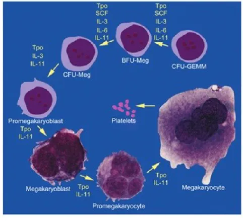
histochemical staining, and biochemical markers have been used to categorize different stages of megakaryocyte development. In general, three types of morphologies can be identified in bone marrow (Figure 1.1.2)
.
Figura 1.1.2 A schematic representation of megakaryocyte commitment. (Italiano JE and Hartwing JH. Platelets 3rd edition).
The promegakaryoblast is the first recognizable megakaryocyte precursor. The megakaryoblast, or stage I megakaryocyte, is a more mature cell that has a distinct morphology (Long MW et al. 1992).The megakaryoblast has a kidney-shaped nucleus with
two sets of chromosomes (4N). It is 10 to 50μm in diameter and appears intensely basophilic in Romanovsky-stained marrow preparations as a result of the large quantity of ribosomes, although the cytoplasm at this stage lacks granules. The megakaryoblast displays blebbing of the plasma membrane, a high nuclear-to-cytoplasmic ratio and, in rodents, is acetylcholinesterase-positive. The promegakaryocyte, or stage II megakaryocyte, is 20 to 80μm in diameter with a polychromatic cytoplasm. The cytoplasm of the promegakaryocyte is less basophilic than the megakaryoblast and contains developing granules.
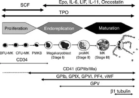
In humans, MK progenitors have been characterized by clusters of differentiation markers on their surface also present in other hematopoietic progenitors, such as the CD34, CD31, and the CD133. The expression of HLA-DR enables BFU-MK (HLA-DRlow) to be distinguished from CFU-MK (HLA-DRhigh) (Briddell RA, et al. 1989). Most studies have focused on the main platelet glycoproteins, the GPIIb/IIIa (aIIbb3 integrin or CD41b) and the GPIb complex (GPIba, GPIbb, GPIX and GPV or CD42a, b, c and d) (Mazur EM et al.
1981; Vinci G et al. 1984). CD41a (GPIIb) or CD41b (GPIIb/IIIa complex) appear
relatively specific for the MK lineage, especially in adults, although mast cells can express these integrins (Kirshenbaum AS et al. 2005). CD41 is present on a small fraction of marrow CD34+ cells (about 3%). These CD34+ CD41+ cells are enriched in MK progenitors, but they do not contain all the CFU-MK. CD41 expression precedes the detection of other major platelet proteins including CD42. However, CD42 is present on a fraction of CD34+ CD41+ cells (Debili N et al.1992). Thus, CD34+ CD41+ CD42- cells correspond to true CFU-MK whereas CD34+ CD41+ CD42+ cells give rise to single MKs or clusters of less than 4 MKs. The surface expression of CD42 corresponds to a late differentiation step and is associated with a marked increase in the expression of Mpl, GPVI (collagen receptor), the a2b1 integrin (collagen receptor), CD36 and in the detection
of proteins contained in the a granules, such as PF4 or vWF. Thereafter, expression of these different platelet proteins markedly increases whereas the CD34 antigen disappears during the endomitotic process (Figure 1.1.3) (Chang Y et al. 2007).
Figure 1.1.3. Expression of differentiation markers along the human megakaryocytic differentiation. (Chang Y et al. 2007)
During development, MKs increase in size, became polyploid and full of granules, expand their cytoplasmic content of cytoskeletal proteins and develop a highly invaginated membrane system.
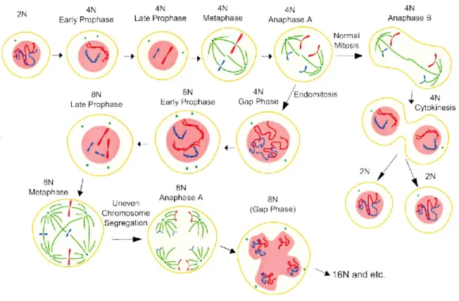
Endomytosis and polyploidization
Endomitosis in MKs is a TPO-dependent process by which MKs become polyploid through cycles of DNA replication without cell division (Ebbe S. 1976; Gurney AL et al. 1994). During their life cycle, MKs first undergo a proliferative 2n stage in which their progression through the cell cycle is identical to other haematopoietic cells. Following which, MKs begin endomitosis and accumulate a DNA content of 4n, 8n, 16n, 32n, 64n, and even 128n in a single polylobulated nucleus before proceeding with their final
maturation and subsequent proplatelet formation (Zimmet J, Ravid K. 2000). Although the number of endomitotic cycles can range from two to six, the majority of megakaryocytes undergo three endomitotic cycles to obtain a DNA content of 16N.
Endomitosis does not result from a prematurely terminated mitosis (Raslova H et al. 2003;
Nagata Y et al. 1997; Vitrat N et al 1998) (Figure 1.1.4).
Figure 1.1.4. A schematic representation of endomitotic cell cycle in megakaryocytes. (Ravid et al. 2002)
Megakaryocyte progenitors initiate the cycle and undergo a short G1 phase, a typical S phase for DNA synthesis, a short G2 phase, followed by endomitosis (Odell T et al. 1968). Megakaryocytes begin the mitotic cycle and proceed from prophase to anaphase A, but do not enter anaphase B or telophase or undergo cytokinesis. The nuclear envelope breaks down and an abnormal spherical mitotic spindle forms, each spindle attaches chromosomes that align to a position equidistant from the spindle poles (metaphase). Sister chromatids
segregate and begin to move toward their respective poles (anaphase A). However, the spindle poles fail to move apart and do not undergo the separation typically observed during anaphase B. Individual chromatids are not moved to the poles, and subsequently a nuclear envelope reassembles around the entire set of sister chromatids, forming a single enlarged, but lobed, nucleus with multiple chromosome copies. The cell then skips telophase and cytokinesis to enter G1. This failure to separate sets of daughter chromosomes fully may prevent the formation of a nuclear envelope around each individual set of chromosomes (Nagata Y et al. 1997; Vitrat N et al 1998).
Much work has focused on identifying the signals that regulate polyploidization, highlighting a critical role for mitosis promoting factor (MPF) activity, a multiprotein complex consisting of Cdc2 and cyclin B (Wang Z et al. 1995; Gu XF et al. 1993), Rho family (Geddis AE, Kaushansky K. 2006), and protein-tyrosine phosphatases Shp1 and Shp2 (Mazharian A et al. 2013), however a complete and definitive description of endomitosis regulator is not currently available.
Polyploidization is essential for efficient platelet production. It is theorized that MKs are polyploid in order to support cytoplasmic maturation characterized by a great production of mRNA and proteins that are packaged into granules and platelets and by a formation of invaginated membrane system (IMS) generation from which proplatelets derive (Zimmet
J, Ravid K. 2000; Machlus KR, Italiano JE. 2013).
Cytoplasmic maturation
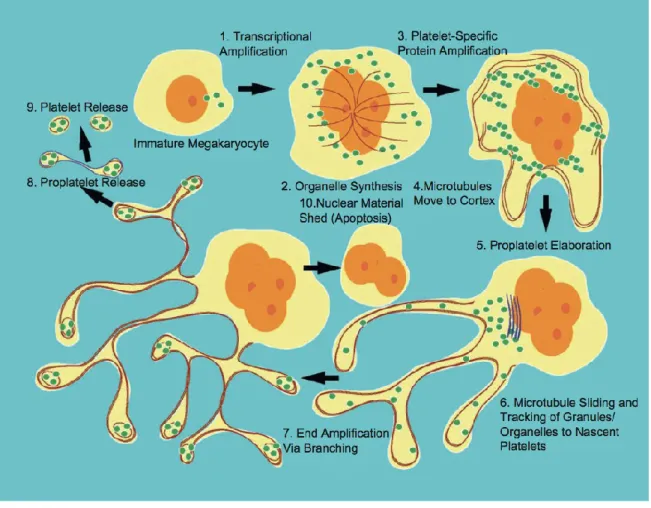
After the process of endomitosis is completed, the megakaryocyte begins a maturation stage in which the cytoplasm rapidly fills with platelet-specific proteins, organelles, granules and membrane systems that will ultimately be subdivided and packaged into platelets.
In the MK, granules are derived from the budding of small vesicles containing granule cargo from the trans-Golgi network (Blair P, Flaumenhaft R. 2009). Vesicles budding from the trans-Golgi network may be delivered directly to multivesicular bodies, where proteins are sorted and eventually pack- aged into granules (Heijnen HF et al. 1998;
Youssefian T, Cramer EM. 2000). Organelles and granules are then individually
transported from the MK cell body, along the proplatelet shaft where they move bidirectionally until they are captured at proplatelet tips (Richardson JL et al. 2005).
The most abundant are α granules, which contain proteins essential for platelet adhesion during vascular repair. These granules are typically 200 to 500 nm in diameter and have spherical shapes with a dark central core. They are present in early-stage megakaryocytic
(Jones OP. 1960) and they acquire their molecular contents both from endogenous protein synthesis and by uptake and packaging of plasma proteins by receptor-mediated endocytosis and pinocytosis (Handagama P et al. 1987). Endogenously synthesized proteins such as platelet factor 4, β-thromboglobulin, and von Willebrand factor are detected in megakaryocytes before endocytosed proteins such as fibrinogen. In addition, synthesized proteins predominate in the juxtanuclear Golgi area, whereas endocytosed proteins are localized in the peripheral regions of the cell (de Larouziere V et al. 1998). It has been well documented that uptake and delivery of fibrinogen to α granules is mediated by integrin αIIβ3 (Coller BS et al. 1991; Handagama P et al. 1993; Handagama P et al.
1993-2). Several membrane proteins critical to platelet function are also packaged into α granules, including integrin αIIbβ3, P-selectin (CD62P), and CD36. MKs also contain dense granules, approximately 250 nm in size, identified in electron micrographs by virtue of their electron-dense cores, contain a variety of hemostatically active substances that are released upon platelet activation, including serotonin, catecholamines, ADP, adenosine 5’ -triphosphate (ATP), and calcium (Youssefian T et al. 2000).
The maturation of MKs is characterized by the progressive formation of an elaborate membrane system that is continuous with the plasma membrane and permeates the cytoplasm. Originally, the IMS was called the demarcation membrane system (DMS) because it was thought to divide, or ‘demarcate,’ the MK cytoplasm into small territories from which platelets were formed (Behnke O. 1969; Radley JM, Haller CJ. 1982). However, since establishing the proplatelet model of platelet release, it is more accurately referred to as the IMS to reflect its unique characteristics; the IMS is derived from the plasma membrane, retains contact with the extracellular environment and functions as a membrane reservoir for proplatelet formation (Behnke O. 1969; Nakao K, Angrist AA.
1968). The IMS has also been proposed to mature into the open canalicular system (OCS)
of mature platelet, which functions as a channel for the secretion of granules contents
(Nakao K, Angrist AA. 1968).
Platelet formation
Although it has been universally accepted that platelets derive from megakaryocytes, the mechanisms by which platelets are formed and released by these precursor cells are still largely unclear and several models of platelet formation have been proposed. (Figure 1.1.5).
Figure1.1.5. Mechanisms proposed for platelet production (Italiano JE and Hartwing JH. Platelets 3rd edition)
The first model is the cytoplasmic fragmentation via the DMS. Microscopists recognized that maturing megakaryocytes became filled with membranes and platelet-specific organelles and postulated that these membranes formed a system that defined territories or fields for developing platelets (Shaklai M, Tavassoli M. 1978). Release of individual platelets was proposed to occur by a massive fragmentation of the megakaryocyte cytoplasm along DMS fracture lines residing between these fields. The DMS model predicts that platelets form through an extensive internal membrane reorganization process
(Kosaki G. 2005). However this model has lost support because of several inconsistent
observations. For example, if platelets are delineated within the megakaryocyte cytoplasm by the DMS, then platelet fields should exhibit structural characteristics of platelets, which is not the case (Radley J, Hatshorm M. 1987).Platelet territories within the megakaryocyte cytoplasm lack marginal microtubule coils, the most characteristic feature of resting platelet structure. In addition, there are no studies directly demonstrating that platelet fields shatter into mature, functional platelets.
According to the second model, named platelet budding, platelets pinch off from blebs protruded at the megakaryocyte periphery. Examination of these structures by thin-section electron microscopy, however, revealed that these blebs did not contain platelet organelles, an observation inconsistent with the concept of platelet budding as a mechanism for platelet release (Djaldetti M et al. 1979; Ihzumi T et al 1977).
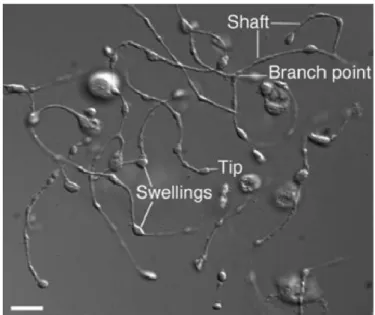
Actually the most accreditate theory for platelet formation is the proplatelet model.
Proplatelet model
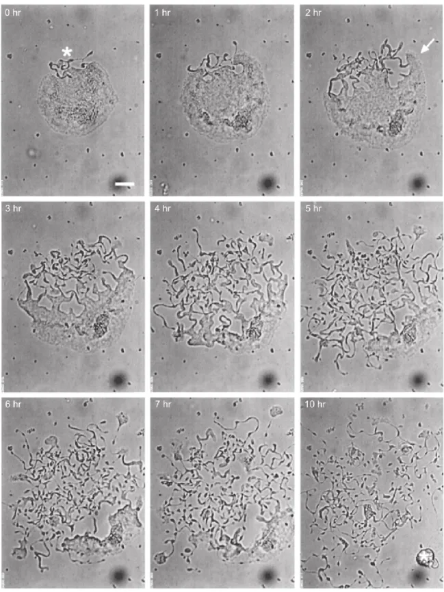
Mature MKs extend long, branching processes called proplatelets into the sinusoidal blood vessels of the bone marrow. The process commences at a single site on the MK plasma membrane. From this region, a pseudopod forms, elongates, and tapers into a tubule with an average diameter of 2– 4 um, called the proplatelet shaft. Proplatelets function as the assembly lines of platelet production, and are comprised of platelet-sized swellings in tandem arrays along the shaft (Italiano JE et al. 1999). The multi-lobed nucleus remains in the MK cell body as the rest of the MK is transformed into proplatelets. After the entirety of the MK cell body is converted into proplatelets, the nucleus is extruded and degraded (Figure 1.1.6 and Figure 1.1.7).
Figura 1.1.6. Video-enhanced light microscopy of mouse megakaryocyte forming proplatelet in vitro (Italiano JE and Hartwing JH. Platelets 3rd edition).
Figure 1.1.7. Proplatelet process (Patel SR et al.2005).
Proplatelet formation is dependent on the function of microubules. Microtubules, polymers assembled from dimers of alpha and beta tubulin, are the major components of the machine that drive proplatelet extension. Visualization of the microtubule cytoskeletons of proplatelet-producing MKs provides insights into how microtubules power platelet production. The microtubule cytoskeleton in MKs undergoes a remodelling during proplatelet production. (Figure 1.1.8).
Figure 1.1.8. Propatelet model detailing some of the cytoskeletal mechanisms of platelet biogenesis (Italiano JE and Hartwing JH. Platelet 3rd edition)
In immature megakaryocytes, that have not formed proplatelets, microtubules extend out from the cell centre to the cortex. As thick pseudopodia develop during the initial stages of proplatelet formation, cortical microtubules organize into thick bundles situated beneath the plasma membrane of these processes. When pseudopodia begin to elongate (at an average rate of 0.85 um/min), microtubules form thick arrays that line the entire length of the proplatelets (Italiano JE et al. 1999). The microtubule bundles are thickest in the region of the proplatelet near the body of the MK, but narrow to bundles of 5–10 microtubules near proplatelet tips. The distal end of each proplatelet always has a microtubule bundle that loops just beneath the plasma membrane and re-enters the shaft to form a teardrop-shaped or tennis racket-shaped structure. Because microtubule coils
similar to those observed in blood platelets are detected only at the ends of proplatelets and not within the platelet-size beads found along the length of proplatelets, mature platelets are formed only at the tips of proplatelets (Italiano JE et al. 1999). Moreover, the ends of proplatelets are amplified in a dynamic process that repeatedly bends and bifurcates the proplatelet shaft (Italiano JE et al 1999).End amplification initiates when a proplatelet shaft is bent into a sharp kink that then folds back on itself, forming a loop in the microtubule bundle. The new loop eventually elongates, forming a new proplatelet shaft branching from the side of the original proplatelet. Loops lead the proplatelet tip and define the site where nascent platelets will assemble and where platelet-specific contents are trafficked. In marked difference from the microtubule-based motor that elongates proplatelets, actin-based force is used to bend the proplatelet in end amplification.
In addition to playing a crucial role in proplatelet elongation, the microtubules lining the shafts of proplatelets serve a second important function—tracks for the transport of membrane, organelles, and granules into proplatelets and assembling platelets at proplatelet ends. Individual organelles are sent from the cell body into the proplatelets, where they move bidirectionally until they are captured at proplatelet ends (Kelley MJ et
al. 2000).
The final step of platelet formation is represented by proplatelets extension into bone marrow vascular sinusoids, where they are released and enter the bloodstream (Figure 1.1.9)
Figure 1.1.9. Megakaryocyte protruding proplatelets into bone marrow vessel. (Geddis AE et al. 2007)
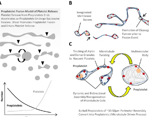
Notably, it has been observed that these enucleate fragments typically exceed platelet dimensions, suggesting that platelet morphogenesis continues in the circulation (Junt T et
al. 2007). In line with these observations, Thon and co-workers, have recently identified a
previously unrecognized intermediate stage in platelet formation and release, which they termed the preplatelet (Thon JN et al, 2010). Preplatelets, which appear as “giant platelets,” are defined as discoid cells (3-10 microns) that retain the capacity to convert into barbell-shaped proplatelets and undergo fission into platelets. (Figure 1.1.10)
Figure 1.1.10. Proplatelet fission model of platelet release (Thon JL et al. 2010)
1.1.3 BCL-2 FAMILY IN MEGAKARYOCYTOPOIESIS
There is an ongoing debate about the role of apoptosis in platelet formation, and considerable evidence has been accumulated suggesting that apoptosis is necessary for MKs to start proplatelet formation (De Botton et al, 2002; Clarke et al, 2003).
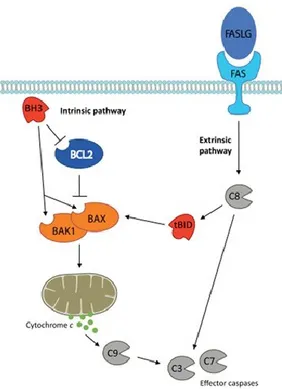
Apoptosis refers to two convergent pathways of programmed cell death: intrinsic and extrinsic (Youle & Strasser, 2008) (Figure 1.1.11).
Figure 1.1.11. A schematic representation of the two pathways of apoptosis. (Kile BT. 2014)
The critical regulators and effectors of the former are proteins of the BCL2 family, which are characterized by the presence of one or more BCL2 homology (BH) domains. They are divided into three groups. The first comprises the multi-domain killers BAK1 and BAX, which are the essential effectors of the intrinsic pathway. The second group are the pro-survival proteins (BCL2, BCL-XL, BCL2L2, MCL1 and BCL2A1), whose function is to prevent the activation of BAK1 and BAX. While the precise mechanisms are still being clarified, it is believed they achieve this by physically restraining BAK1 and BAX and by sequestering a third group of pro- apoptotic BCL2 family members known as ‘BH3-only’ proteins (BIM (BCL2L11), BID, BAD, PUMA (BBC3), NOXA (PMAIP1), BMF, BIK and HRK). In a healthy cell, BCL2 family pro-survival activity is sufficient to keep BAK1 and BAX in check. Apoptotic signals, such as DNA damage, trigger the BH3-only proteins, either by transcriptional upregulation or post-translational modification, thereby
allowing them to overwhelm the function of the pro-survival proteins and activate BAK1 and BAX. This is thought to be the result of BH3-only proteins binding to and inhibiting pro-survivals, but also by directly binding and activating BAK1 and BAX (Willis SN et al.
2007; Ren D et al. 2010). Once unleashed, the latter oligomerize and induce mitochondrial
outer membrane permeabilization (MOMP), facilitating the efflux of apoptogenic factors, such as cytochrome c and SMAC (DIABLO), into the cytoplasm, and eventually leading to the dissipation of mitochondrial potential. Once MOMP has occurred, the apoptotic caspase cascade is engaged, beginning with the ‘initiator’, or ‘apical’ caspase, caspase-9. Together with APAF1 and cytochrome c, the inactive caspase-9 zymogen is recruited to form the apoptosome complex, the function of which is to proteolytically activate caspase-9. Active caspase-9 then triggers the rest of the caspase cascade, culminating in the activation of the ‘effector’ caspases, caspase-3 and caspase-7. Caspases are the executioners of the intrinsic apoptosis pathway, mediating many of the hallmarks of apoptosis, such as DNA fragmentation, phosphatidylserine (PS) exposure and membrane blebbing. The other apoptotic pathway, the extrinsic, is activated by ligands binding to cell surface death receptors that belong to the tumour necrosis factor (TNF) receptor family, such as FAS or TNF receptor-1 (TNFR1). Once activated, the adap- tor protein FAS-associated death domain (FADD) and caspase-8 are recruited to the receptor complex. Caspase-8 is the essential mediator of the extrinsic apoptosis pathway. Following proteolytic cleavage, the activated form of caspase-8 directly triggers activation of caspase-3/7. In so-called Type I cells, such as lymphocytes, this is sufficient to induce death (Scaffidi C et al. 1998). In Type 2 cells, such as hepatocytes, the additional recruitment of the intrinsic pathway is required to induce killing. This is achieved via caspase-8-mediated activation of the BH3-only protein BID, which then triggers the activation of BAK1 and BAX (Yin MB et al. 1999; Jost PJ et al. 2009).
In the last twenty years, the identification of thrombopoietin (TPO) and development of primary megakaryocyte culture systems led to the observation that peak platelet production by mature megakaryocytes ‘corresponded to the onset of apoptosis’ (Zauli G et al. 1997). When mice lacking the pro-death BH3-only protein BIM (Bouillet P et al. 1999), or overexpressing pro-survival BCL2 were found to be thrombocytopenic, the idea that megakaryocytes might undergo apoptosis deliberately in order to facilitate platelet shedding was born (Ogilvy S et al. 1999).
Seminal work by Debili and colleagues cemented this hypothesis (De Botton S et al. 2002). They reported the presence of active apoptotic caspases in the cytoplasm of cultured primary human CD34+-derived megakaryocytes, and showedhat proplatelet formation was impaired in the presence of the fluoromethylketone caspase inhibitors VAD.fmk, Z-LEDH.fmk or Z-DEVD.fmk. Moreover, transgenic megakaryocytes expressing BCL-XL under the control of the Platelet Factor 4 (PF4) promoter, produce fewer proplatelets in culture (Kaluzhny Y et al. 2002).
Collectively, these data point to a productive role for the intrinsic apoptosis pathway in platelet production and, in particular, Bcl-xL expression is critical to allow the survival of the cells during the early endomitotic phase of megakaryocytic differentiation, but it must decrease to allow the platelet release process to happen, which can be considered a peculiar and productive apoptotic-like process (Kile BT. 2014).
In recent years, the role of the intrinsic apoptosis pathway in megakaryocytes has been explored in detail, through studies conducted in genetically modified mice.
Spurred on by the data linking the pathway to the regulation of platelet life span, several groups have examined the requirement for BCL2 family proteins and apoptotic caspases in megakaryocyte life and death. The results to date clearly indicate that megakaryocytes, like
their anucleate progeny, possess a fully functional intrinsic apoptosis pathway. In addition to BCL-XL, megakaryocyte growth and development requires MCL1 (Josefsson et al.
2011; Debrincat et al. 2012; Kodama et al. 2012). Megakaryocyte-specific deletion of
BCL-XL results in severe thrombocytopenia, a reflection not only of dramatically reduced platelet life span, but aberrant platelet shedding by dying megakaryocytes (Josefsson EC et
al. 2011). It appears that at least in mice, megakaryocytes become dependent on BCL-XL
just at the point at which they are ready to produce proplatelets. While BCL-XL-deficient megakaryocytes develop normally, they are unable to form proplatelets in vitro, and manifest striking ultrastructural defects in vivo, with large vacuolated fragments of cytoplasm being sloughed off into the sinusoids. Loss of both MCL1 and BCL-XL results in a failure of megakaryopoiesis, systemic haemorrhage and embryonic lethality, indicating that the combined functions of both pro-survivals are required for megakaryocyte growth and development (Debrincat MA et al. 2012; Kodama T et al.
2012). If their role is to prevent the activation of the intrinsic apoptosis pathway, then it
would be expected that deletion of BAK1 and BAX would completely rescue the defects caused by loss of MCL1 and BCL-XL, and this is indeed the case: quadruple MCL1/BCL- XL/BAK1/BAX knockout mice are healthy and exhibit normal megakaryopoiesis
(Kodama T et al. 2012).
A corollary of these studies is that there is little to suggest megakaryocytes require the intrinsic apoptosis pathway to produce platelets. Deletion of BAK1 and BAX does not impair thrombopoiesis, in fact platelet counts are significantly elevated due to the extended platelet life span that loss of BAK1 confers. Rather than be activated to facilitate cytoskeletal rearrangements, the intrinsic pathway must be restrained in order to survive and proceed safely through the process of platelet shedding. Treatment of mature wild- type mouse megakaryocytes in culture with ABT-737 induces mitochondrial damage and
effector caspase activation (Josefsson EC et al. 2011). The result is failure of proplatelet formation and death. These effects are mediated by the intrinsic apoptosis pathway, since Bak1-/- Bax-/- megakaryocytes are resistant to ABT-737. Other apoptotic stimuli, such as the topoisomerase II inhibitor etoposide and the broad-spectrum kinase inhibitor staurosporine (STS), also trigger mitochondrial damage and caspase activation. Interestingly, however, while deletion of BAK1 and BAX can prevent etoposide- induced mitochondrial damage and caspase activation, it does not rescue proplatelet formation, suggesting that DNA damage signals suppress platelet shedding independently of the apoptotic pathway being engaged. While some differences are observed in vivo, the basic premise is the same. ABT-737 does not appear to kill wild-type megakaryocytes in situ, but in mice with a megakaryocyte-specific deletion of MCL1, it induces the fulminant apoptotic death of megakaryocytes within 3 h (Debrincat MA et al. 2012; Kodama T et al.
2012). These data suggest there might be subtle differences in MCL1 expression or
protein-protein interactions in cultured megakaryocytes versus their counterparts in vivo, potentially the result of the latter’s exposure to a range of pro-megakaryocytic cytokines and growth factors. Regardless, activation of apoptosis is accompanied by a failure of platelet shedding and severe thrombocytopenia. As expected, deletion of BAK1 and BAX renders MCL1-deficient megakaryocytes entirely refractory to the effects of ABT-737 in vivo (Kodama T et al. 2012).
Finally, data obtained with transgenic mice are in part contradictory in respect to previous observations on cultured primary human CD34+-derived megakaryocytes. This may reflect differing experimental procedures and types of MKs used in the studies, and so the role of Bcl-2 family in megakaryocytopoiesis and platelet production needs to be clarified.
1.1.4 PLATELET PRODUCTION FOR INFUSION
Platelets are essential for haemostasis, accordingly, patients with low numbers of circulating platelets or functionally hyporeactive platelets are at increased risk of spontaneous bleeding or hemorrhage following traumatic injuries or during surgical procedures. Thrombocytopenia (defined as platelet counts <150000/ul) is a major clinical problem encountered across a number of conditions, including immune thrombocytopenic purpura, myelodysplastic syndromes, chemotherapy, aplastic anaemia, human immunodeficiency, virus infection, complications during pregnancy and delivery, and surgery.
The therapeutic strategy to prevent severe bleeding is platelet transfusion. The first evidence supporting the use of platelet transfusion was reported in 1910 and since 1950 was developed the ability to prepare platelet concentrate that are currently used in patients with severe thrombocytopenia (Duke WW. 1983; Dillard GH et al. 1951). The use of transfused platelets has increased more than other blood components in the last two decades (Lu SJ et al. 2011), they derive entirely from human donors however, there are several limitations and challenges with platelet preparation and storage technologies. One of these limitations is the dependence upon a large donor pool. Because of the limited donor supply, there is often a shortage of available blood products. Compounding the challenges of availability is the half-life of transfused platelets (3 days), which is significantly shorter than the half-life of platelets in vivo (approximately 8-12 days)
(Dumont L et al. Rossi’s principles of transfusion medicine. 4th ed. AABB/Blackwell; 2009). Transfused platelet units also have approximately 50% recovery (Filip DJ, Aster RH. 1978). Therefore, approximately twice as many platelets need to be given to achieve
among the mechanisms implicated are platelet activation and apoptosis (Ohto H, Nollet
KE. 2011; Egidi MG et al. 2010). This poor recovery and decreased platelet half-life and
reactivity with time is termed the “platelet storage lesion”, and it is reduced by room temperature storage (Ohto H, Nollet KE. 2011). On the other hand, because of the need to store at room temperature, bacterial contamination of platelets is a significant issue10 with the result that platelets have a short shelf life of 5 days. In addition, over 10% of multiply transfused patients develop decreased responsiveness to platelet transfusion with time (platelet refractoriness). (Bordin JO et al. 1994).
On these basis, a long-term goal for the field of hematology is to obtain an unlimited supply of platelets for transfusion making platelet ex vivo and several strategy are under study.
The ability to grow ex vivo of human megakaryocytes derived from peripheral blood stem cells was first described in 1990 when Mazur and colleagues used serum from aplastic dogs to stimulate megakaryocyte development (Mazur EM et al. 1990). With the isolation and identification of thrombopoietin in 1994 (de Sauvage FJ et al. 1994; Wendling F et al.
1994) and the subsequent cloning and generation of recombinant TPO from both humans and mice (Sohma Y et al 1994; Lok S et al. 1994; Kuter DJ et al. 1994), it became possible to culture sufficient megakaryocytes both for studies of their biology and also to contemplate harvesting ex vivo-generated platelets for clinical application. The first report of functional human platelets obtained by a two-step culture process was in 1995 (Choi ES
at al. 1995), showing that megakaryocytes, larger proplatelets, and smaller, platelet-sized
particles could be generated in vitro from CD34+ peripheral blood cells in the presence of appropriate cytokines. These ex vivo-generated platelets have normal platelet morphology, surface expression of glycoproteins, and ultrastructural characteristics, including the presence of alpha, dense, and lysosomal granules and are responsive to agonist stimulation
(Guerriero R et al. 1995; Tao H et al. 1999). However it has been difficult to assess the in
vivo half-life, functionality, ability to significantly raise platelet counts, and potential to reverse a bleeding diathesis, in large part due to the difficulty harvesting these ex vivo- generated platelets. One strategy for large-scale production of non- donor-derived platelets would be to have a continuous supply of homogenous progenitors that, upon appropriate stimulation, differentiate into megakaryocytes. The development of human embryonic stem cells (hESCs) (Thomson JA et al.1998) and induced pluripotent stem cells (iPSCs)
(O’Malley J et al. 2009) provides a renewable source of cells with the potential to expand
indefinitely in culture and eliminate the need for human donors. Platelet produced from hESCs, for the first time in 2006, and from iPSCs, for the first in 2010, show morphological features comparable to plasma-derived human platelet, the ability to respond to platelet agonists, and to be incorporated into a developing thrombus in vivo
(Gaur M et al. 2006; Zingariello M et al. 2010; Takayama N et al. 2008; Shattil SJ et al. 1985; Lu SJ et al. 2011). Despite encouraging first steps in generating clinically useful
platelets beginning with hESC/iPSCs, two major roadblocks have been identified. The first is that the number of megakaryocytes generated per initial progenitor cell is orders of magnitude too low, beginning with one progenitor cell <10 megakaryocytes are obtained
(Reems JA. 2011). The second roadblock is the efficiency of platelets produced per
megakaryocyte. In order to increase in vitro platelets production yields, recently a microfluidic platelet bioreactor has been developed: it recapitulates bone marrow stiffness, extracellular matrix composition, micro-channel size, hemodynamic vascular shear stress, and endothelial cell contacts, and supports high-resolution live-cell microscopy and quantification of platelet production. Physiological shear stresses triggered proplatelet initiation, reproduced ex vivo bone marrow proplatelet production, dramatically increasing the amount of producing platelets MKs (Thon JN et al. 2014).
Nonetheless, due to the clinical and economical interests, further studies on megakaryocytopoiesis and platelets production biology need to be performed in order to discover optimal conditions of culture and achieve an increased platelets yields.
Beside bioreactors, other different approaches must be tested, such gene therapy or pharmacological approaches that are rationally designed to interact with pathways involved in the megakaryocytopoiesis and platelets production.
1.2 PLATELETS
In 1862, Giulio Bizzozero, the so-called ‘father of the platelet’, described a novel ‘morphological element’ in the blood with important roles in haemorrhage and thrombosis
(de Gaetano G. 2001). These elements became known as platelets, and they are the
smallest, circulating, anucleate cells that are derived from megakaryocytes within the bone marrow. There are 150–400 × 109 platelets per litre of blood and because the lifespan of an individual platelet is only 8–10 days, 100 billion new platelets must be produced daily from bone marrow megakaryocytes in order to maintain normal platelet counts
(Kaushansky K. 2006).
Their primary physiological role is to sense damaged vessel endothelium and accumulate at the site of the vessel injury, where they initiate blood clotting to block the circulatory leak. Platelets circulate at high-shear rates and are activated following binding to the collagen substratum or to other extra- cellular matrix proteins that are exposed during vascular injury. Stable adhesion to collagen promotes the release of many soluble mediators from the platelet’s intracellular stores, leading to further platelet recruitment and activation. These events are regulated by complex interactions involving several families of molecules, including various selectins, integrins, lipids and cytokines. Together with leukocytes and red blood cells, the activated platelets create a thrombus that arrests blood loss (Gawaz M. 2006).
1.2.1 PLATELET STRUCTURE
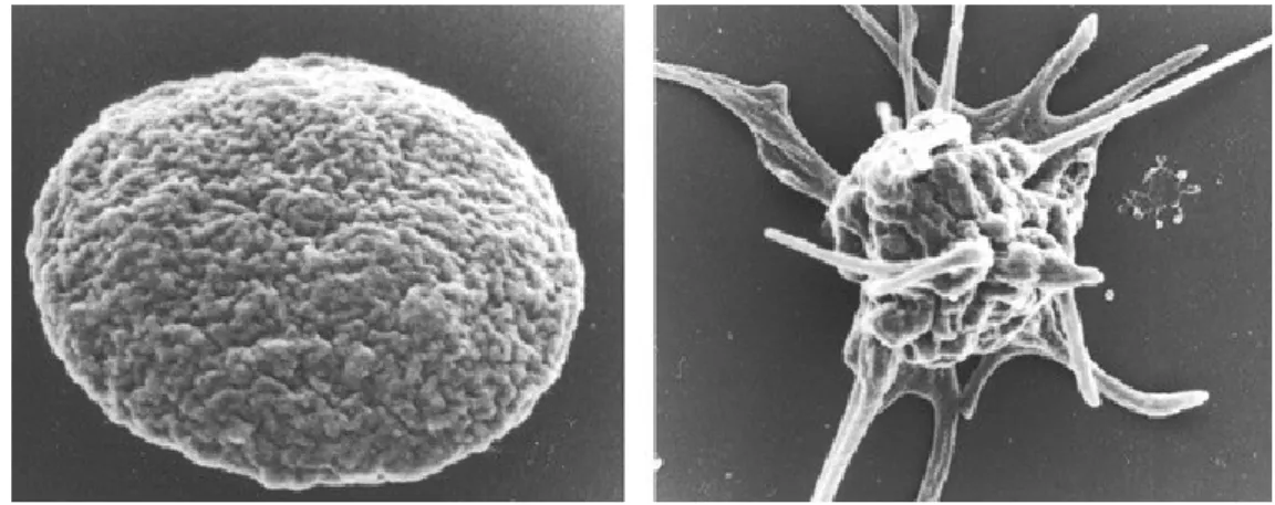
Platelets circulate in blood with a typical discoid shape, the surface of each disc is featureless, lacking protrusions. Plug formation requires platelets to undergo rapid morphological changes as they convert from their resting discoid forms to their active
shapes. The first event that is observed as the platelet makes contact with the surface, is that the discoid shape is lost and becomes rounded or spheroid in form. Next, fingerlike projections grow from the cell periphery, then the platelet flattens over surfaces and broad lamellae are extended. (Figure 1.2.1).
Figura 1.2.1. Platelet shape change in resting and activated states (White JC. Platelets 3rd edition)
Platelet structure should be divided in:
• peripheral zone
• sol-gel zone
• platelet membrane systems
Pheripheral zone
The peripheral zone is formed by an exterior glicocalix and a lipid bilayer.
The lipid bilayer is a typical unit membrane that does not differ in appearance from the membrane covering other cells (White JG, Conrad WJ. 1973), it is incompressible and cannot stretch, but it is significantly different because of its role in the acceleration of blood coagulation participating to thrombin generation. Clotting is initiated at the site of vascular injury by tissue factor (TF), which serves as a cofactor for the coagulant protein,
factor VIIa, in converting factor X to Xa. Factor Xa, in association with factor Va, converts prothrombin to thrombin on the surface of the anionic phospholipid, phosphatidylserine. Platelet lipid bilayer plays a key role in this process because phosphatidylserine is provided by surface of activated platelets, and, on the other hand, platelets also contain TF encrypted with the cholesterol-rich lipid islands in the resting platelet surface membrane (Heijnen HF
et al. 1998).
The platelet glicocalix is thicker then in other blood cells and this exterior surface does not serve merely as a barrier to separate internal contents of platelets from the external milieu; rather, it is a very dynamic structure that serves as the site of first contact and senses changes in the vascular compartment requiring the hemostatic response of platelets at sites of vessel injury. The glycocalyx is covered with major and minor glycoprotein receptors necessary to facilitate platelet adhesion to a damaged surface, trigger full activation of the platelet, promote platelet aggregation and interaction with other cellular elements, and accelerate the process of clot retraction (Clemetson KJ et al. 1985; Carrell NA et al. 1985;
Kunicki TJ. 1988).
Platelet receptors include integrins, Leucine-Rich repeat family, Seven Trans-membrana Receptors and Immunoglobulin superfamily.
Among integrins, the GpIIbIIIa complex, also named αIIbβ3, is the only integrin expressed uniquely on platelets and megakaryocytes. αIIbβ3 represents 3% of the total platelet protein and 17% of the platelet membrane protein masswith 50000 to 100000 copies per platelet (White JG, Krumwiede M. 1985; White JG et al. 1986; White JG, Sauk JJ. 1984). Platelet α-granule membranes also contain αIIbβ3, and this pool can become accessible and is functional upon platelet stimulation (White JG, Krivit W. 1967; Whie JG. 1968). Central to the function of integrin αIIbβ3 is its capacity to undergo activation, a transition
cellular ligands during platelet activation and clot formation. When platelets encounter any one of a number of agonists, a series of intracellular signaling events are triggered that converge at the cytoplasmic tails (CT) of αIIbβ3. Changes in the CT are then transmitted across the platelet membrane and transform the extracellular domain of αIIbβ3 into a high-affinity state for its extracellular ligands. As a consequence, αIIbβ3 can now bind divalent fibrinogen or multivalent von Willebrand factor (VWF) (Hynes RO. 1992), which can bridge platelets together to form aggregates. Recognition of other ligands, such as vitronectin, fibronectin, and thrombospondin (Hynes RO. 1992; Plow EF et al. 2000; Plow
EF, Shattil SJ. 2001; Reheman A et al. 2005),by αIIbβ3 may mediate platelet adhesion to components of the subendothelial matrix and regulate platelet aggregation.
The α2β1 is an integrin also known on platelets as GPIa-IIa and on lymphocytes as VLA2
(Staatz WD et al. 1989). There are 2000 to 4000 copies per platelet. α2β1 is well
established as a major collagen adhesion receptor on platelets and a wide range of other cells.
Other platelet integrins are α5β1 and α6β1, that represent respectively fibronectin and laminin receptor, they have a supplementary role in in platelet adhesion at injury sites
(Piotrowicz RS et al. 1988; Sonnenberg A et al. 1988).
The leucine-rich repeat (LRR) family is represented in platelets mainly by the GPIb-IX-V complex. The GPIb-IX-V is the second most common receptor, in platelet with approximately 50000 copies per platelet. Functions of GPIb-IX-V include regulating platelet adhesion to subendothelial matrix, endothelial cells or leukocytes, assembly of procoagulant activity on activated platelets, and signaling, mediated in part by association of GPIb-IX-V with accessory signaling receptors and localization in lipid rafts. GPIb-IX-V is the most important receptor for VWF and exposure of vascular subendothelium under
high shear results in the immediate attachment of GPIb-IX to von Willebrand factor (VWF) covering collagen fibers in the wound (White JG. 1987).
In platelet, Seven Trans-membrana Receptors include thrombin receptors, ADP receptors, tromboxane and prostaglandines receptors.
Thrombin receptors (PARs) are major representatives of the seven-transmembrane receptor group on platelets, because thrombin is a critical platelet agonist. Human platelets have about 2,500 copies of the PAR1 receptor, which responds to levels of thrombin of about 1 nM and a lower number of copies of PAR4 (Andersen H et al. 1999),which is sensitive to concentrations of thrombin 10 times higher than PAR1.
P2Y1 and P2Y12 are platelet ADP receptors. ADP is one of the earliest identified primary platelet agonists. It has also a critical autocrine role, via secretion from dense granules,. Recently, ADP receptors have been definitively identified.
One of the major member of platelet immunoglobulin superfamily, is GPVI. This protein is exclusively expressed on platelets and megakaryocytes with 4000-6000 copies per platelet. It is a low affinity collagen receptor and it actives a second collagen receptor, integrin α2β1. The binding of collagen to integrin α2β1 contributes to stable adhesion and reinforces binding to GPVI (Furihata K et al. 2001; Best D et al. 2003; Samaha FF et al.
2005; Senis YA et al. 2009). The immunoglobulin superfamily also includes:
• FcγRIIa (CD32), which is a low-affinity receptor for the IgG Fc domain in platelets, has a role in heparin-induced thrombocytopenia (Carlsson LE et al. 1998). Antibodies are developped against the heparin/platelet α-granule chemokine (platelet factor 4) complex, inducing platelet activation via FcγRIIa and, eventually, leading to thrombocytopenia.
• FcεRI, the high-affinity receptor for IgE, is a platelets activator (Joseph M et al.
1997; Hasegawa S et al. 1999).
Integrin-associated protein (CD47) is a receptor for the cell-binding domain of thrombospondin (TSP). TSP can mediate platelet activation and aggregation.
The endothelial cell-selective adhesion molecule (ESAM), that localizes to platelet-platelet contacts, regulates thrombus formation in vivo (Stalker TJ et al. 2009).
Sol-gel zone
The sol-gel zone consist of a meshwork of fibrous material in which formed organelles are imbedded.
Fibrous material represents platelet cytoskeleton that serves as a system of molecular struts and girders that defines the discoid shape of the resting platelet and maintains cell integrity as platelets encounter high fluid shear forces generated by blood flow over the endothelium. Critical components of this system are, from the plasma membrane inward, a spectrin-based skeleton (Boyles J et al.1985; Fox J et al. 1988), a microtubule coil and a rigid network of crosslinked actin filaments. (Figures 1.2.2, 1.2.3, 1.2.4).
Figure 1.2.2. Microtubule coil isolated from human platelets after simultaneous fixation and detergent extraction in suspension. The coil has many loops but appears to consist of one microtubule. Mag X
13000. (White JC. Platelets 3rd edition)
Figure 1.2.3. Thin section of a discoid platelet cytoskeleton fixed after detergent extraction in presence of lysine and phalloidin. The microtubule is well preserved (T). Remnants of α granules (G) are suspended in a matrix of microfilaments (MF) resistant to detergent extraction. Mag X 22000. (White
Figure 1.2.4. Cross-section of a discoid platelet cytoskeleton. The cell is from a sample of C-PRP treated with Taxol (1024 M) before extraction. Microtubule (T) profiles are evident at the polar ends of
the cell. A meshwork of microfilaments (MF) replaces the cytoplasmic matrix. Although the surface membrane is gone, a fine amorphous layer remains in its place, probably submembrane filaments.
Mag.X22,000. (White JC. Platelets 3rd edition)
A spectrin-based skeleton is a two-dimensional assembly of spectrin strands that is adherent to the cytoplasmic side of the plasma membrane. It shows both binding sites for membrane glicoprotein and for actin, connecting platelet membrane to the actin network
(Glenney J, et al. 1982; Gratzer W. 1978; Wasenius VM et al. 1989; Winkelmann J et al. 1990).
Platelets contain a long microtubule, composed by tubulin molecules, that is approximately 100 μm in length and 25nm in diameter. To fit the long microtubule inside the resting platelet, it is wound from 8 to 12 times into a coil. The coil sits in the cytoplasm, just beneath the plasma membrane, along the thin edge of each disc. This microtubule coil is a cytoskeletal support system, responsible for distorting the cell into a discoid shape.
The third components of platelet cytoskeleton is a network of crosslinked actin filaments, also named microfilaments. Actin is the most abundant of all the platelet proteins. The concentration of actin in a platelet is 0.55 mM, which translates into approximately two million copies per platelet (Nachmias VT. 1980).Of these molecules, 800000 assemble to
form the 2000 to 5000 linear actin polymers that form the cytoskeleton of the resting cell
(Hartwig J, DeSisto M. 1991). All evidence indicate that the filaments of the resting platelet are interconnected at various points to form a mechanically rigid cytoplasmic network. Indeed, platelets express high concentrations of actin cross-linking proteins, including filamin (FLNa and FLNb) and α-actinin (Feng Y, Walsh CT. 2004; Stossel T et
al. 2001). In the resting cell, the microfilament network serves as the matrix on which all
organelles and other structural components are suspended and maintained separate from each other (Escolar G et al. 1986). Following platelet activation, a large amount of actin molecules polymerize, and microfilaments have a unique role in contractile physiology. They constrict the circumferential microtubule coils and drives the α granules and dense bodies into close association in platelet centers. If stimulation is strong enough, granule and dense body contents are secreted to the exterior via channels of the OCS (Escolar G et
al. 1986; White JG, Krumwiede M. 1987), leaving behind a dense, central mass of actin.
All platelet organelles, such as mitochondria, ribosome and Golgi complex, are imbedded in this network of microtules and microfilaments; in particular platelet cytoplasm is rich of three major types of secretory organelles, α granules, dense bodies (δ granules) and lysosomes.
α-Granules are unique to platelets and are the most abundant platelet granules. There are approximately 5 α-granules per platelet, ranging in size from 200 to 500 nm (Frojmovic
MM, Milton JG. 1982).They constitute approximately 10% of the platelet volume and the total α-granule membrane surface area per platelet is 14 μm. Morphologic features, that have historically defined α-granules, include: (1) the peripheral membrane of the granules, (2) an electron-dense nucleoid that contains chemokines and proteoglycans, (3) a less electron-dense area adjacent to the nucleoid that contains fibrinogen, and (4) a peripheral
contain both membrane bound proteins, that become expressed on the platelet surface after membrane fusion, and soluble proteins, that are released into the extracellular space. This granule content include integral membrane proteins (αIIbβ3, GPIbα-IX-V, GPVI, P-selectin), coagulant and anticoagulants proteins (Factor V, factor IX, factor XIII, plasminogen, plasminogen activator inhibitor), adhesion proteins (Fibrinogen, von Willebrand factor, thrombospondin), chemokines, growth factors, angiogenic factors.
(Berger G et al. 1996; Maynard DM et al. 2007; Nurden P et al.2004).
Human platelet dense bodies (DB) are smaller than the α-granules (diameter of 150nm), are fewer in number (3-8 per platelet), and have high morphological variability (Berger G
et al. 1996). Their most distinguishing feature is the electron-opaque spherical body within
the organelle, which is however separated from the enclosing membrane by an empty space. This feature led to the term “bull’s-eye” being used to describe the dense body. However, some DB are irregular in shape, have filaments extending from the dense inner core to the enclosing membrane, or contain a granule-like substance filling the usually empty space. Platelet dense granules contain Adenine nucleotides, ADP and ATP, polyphosphates,bioactive amines, such as serotonin and histamine (Holmsen H, Weiss HJ.
1979; Ruiz FA et al. 2004). Dense granule membrane proteins include those that are
typically sorting into lysosome-related organelles such as CD63 (granulophysin) and LAMP-2. Several platelet plasma membrane have also been identified in dense granule membranes, including GPIb and αIIbβ3 (Youssefian T, et al. 1997).
Platelets contain few primary and secondary lysosomes, approximately 3 lysosmes per platelet with a diameter of 200-250nm. Platelet lysosome function is not well studied. It is possible that lysosomes may have a role in endosomal digestion, as observed in nucleated cells. Indeed, lysosomes contain protein-degrading enzymes (Cathepsins, elastase, collagenase, carboxypeptidase, proline carboxypeptidase), carbohydrate-degrading