ISSN 2372-5087 (Print) 2372-5095 (Online) Copyright © The Author(s). All Rights Reserved. Published by American Research Institute for Policy Development DOI: 10.15640/ijmp.v7n1a2 URL: https://doi.org/10.15640/ijmp.v7n1a2
Men with Breast Cancer: 15 Cases
Garipoli Claudia
1, Dottore Alessia
2, & Massafra Marco
2Abstract
This study aims to understand the clinical features, treatment, and prognosis of patients with male breast cancer (MBC) in Medical Oncology Unit-University of Messina- from January 2013 to December 2017. Data for 15 patients with MBC were collected for analysis. Immunohistochemistry, pathological results, and other data such as demographic characteristics (age, smoking history, and family history of cancer) as well as clinical data were investigated by retrieving information from the patients’ medical records. A total of 355 patients were diagnosed with breast cancer between January 2013 and December 2017 , and 15 were patients with MBC among them. The median diagnosis age of patients with MBC was 65 years (range 37–75 years). The most common complaint was a painless lump in the breast, and the pathological type in MBC was infiltrating ductal carcinoma (100 %). In terms of hormone receptors, all the patients were positive: 81% (12/15) of patients with MBC were estrogen and progesterone receptor positive, 14 % (3/15) of patients were estrogen receptor positive only, and no patient was HER2 over expressing. Keywords: Male Breast Cancer, Late Diagnosis, Tumor Stage, Poor Prognosis
1. Introduction
Compared to female breast cancer (FBC),male breast cancer (MBC) is a rare disease.[1] [ 2]Breast cancer in men is a very rare cancer, accounting for 1% of all breast cancers with a ratio of 1: 100 between men and women and about 1% of all malignancies in men. [3] [4]. It also represents <0.2% of all cancer-related deaths among men.[5]Invasive ductal carcinoma is the most common histological subtype while invasive lobular carcinoma is responsible for only 1.5% of the total cases. Precisely because it is considered a rare disease, it has been the focus of limited research. Because the incidence of male breast cancer is increasing, [6] there has been a growing interest in this disease. The risk factors of MBC include age, race, family history, genetics, obesity, hormone exposure, other diseases (Cowden syndrome and Klinefelter syndrome) and environmental factors.[7] [8-11] In contrast to the bimodal age distribution of FBC, MBC shows a unimodal age distribution,[ 9] [12] and the probable reason is different hormonal exposures.[12] The median diagnosis age of MBC is 68 years and it is 5–10years later than FBC.[6] [8]While the mortality rate of MBC improved in the last 30 years.[13]Due to insufficient data and understanding of the disease, the treatment of MBC is usually based on the guideline of FBC in clinical practice.[14] [15] Mastectomy, radiotherapy, adjuvant chemotherapy, HER2-targeted therapy, and adjuvant endocrine with tamoxifen were the main treatments of breast cancer in men.[16-19]Our study analyzed MBC data from Medical Oncology Unit-University of Messina- for 5 years period, to evaluate the clinical characteristics, treatment, and prognosis of MBC.
2. Material and Method
We conducted a study on 15 male patients in our Medical Oncology Unit, from January 2013 to December 2017, with histologically confirmed breast cancer and aged over 18 years. The variables studied were data concerning the general characteristics of the patients (age, family history, result of the initial mammogram, tumor histopathology, tumor stage (TNM), treatment modalities (surgery, chemotherapy, radiotherapy, hormone therapy, target therapy) and evolution.
1Researcher/Professor of Oncology, Medical Oncology Unit of Messina University, General Pathology Department of Adult
and Child ,Avenue Gazzi-Messina.
2Medical Doctor in training for Oncology, Medical Oncology Unit of Messina University, General Pathology Department of
Our goal was:
• Report the epidemiological, clinical, radiological and histological features of male breast cancer. • Report the therapeutic management, survival rates and factors of this disease.
2.1 General characteristics of patients:
Of a total of 355 cases of breast cancer with histological confirmation (female and male) admitted in the period January 2013-December 2017, 340(95%) cases were female and 15 (5%) male. The frequency of MBC was 3 cases per year. The medianage at diagnosis was 65 years (range 37-75 years) Table 1.
Table 1. Age Case 1 37 Case 2 57 Case 3 72 Case 4 75 Case 5 58 Case 6 65 Case 7 71 Case 8 66 Case 9 70 Case 10 68 Case 11 74 Case 12 65 Case 13 58 Case 14 38 Case 15 51
Only 2 patients (13%), the youngest (37 and 38 years), had a family history of neoplasia (mother with colon cancer and father with liver cancer the first and mother with breast cancer and father with prostate cancer the second) and, therefore, have been sent to genetic counseling. Of the 15 patients, 6 (38%) were smokers. None of our patients had previously taken hormone therapy. The main symptom was the presence of a solid mass at the breast which, in 9 (63%) cases, was associated with mastodynia. The skin lesions were found in 6 (38%) cases. The average time for consultation in our Unit was 3 months (range 1 - 12 months).Table 2
Table 2. Time for consultation
Case 1 2 months Case 2 1 month Case 3 12 months Case 4 2 months Case 5 1 month Case 6 3 months Case 7 3 months Case 8 2 months Case 9 3 months Case 10 4 months Case 11 2 months Case 12 1 month Case 13 4 months Case 14 3 months Case 15 3 months 2.2 Clinical features:
The clinical examination revealed, in the 15 cases, the presence of a breast mass; this mass was localized to the left breast in 11 cases (75%) and in the right in 4 (25%) cases, there was no case of bilateralism. The average size of the tumor at the time of diagnosis was about 3 cm (range 2-9 cm)Table 3.
Table 3. Size Case 1 2 cm Case 2 2 cm Case 3 9 cm Case 4 6 cm Case 5 3 cm Case 6 5 cm Case 7 4 cm Case 8 3 cm Case 9 2 cm Case 10 2 cm Case 11 3 cm Case 12 2 cm Case 13 2 cm Case 14 3 cm Case 15 2 cm
There were 9(62%) patients who experienced inflammatory signs in the breast. Palpable lymph nodes in the axillary region were objected in 8 cases (53%): in 5 patients, the locally advanced disease was classified as N1, while in 3 patients was classified as N3. Skin changes associated with a nipple retraction were found in 8 (53%) cases. Of the 15 patients, 9 (62%) performed a mammogram, followed by ultrasound, which showed a left poster lateral plane in 2 cases, the upper right quadrant in 3 cases and the upper left quadrant in 4 cases. While 6 (38%) patients performed only ultrasound of the breast that showed a hypo-ecogenic mass with irregular limits: in 5 cases at the left breast and in 1 case at the right breast. Micro calcifications were not found in any examination. In 12 (80%) patients the diagnosis was confirmed by a transcutaneous mammary micro biopsy under ultrasound guidance. Invasive ductal carcinoma is the histological type found in all patients. Hormone receptors were positive in all patients. Her 2 neu was not over-emphasized under any circumstances. Ki 67 was evaluated: in 8 (53%) patients it was about 8%, in 2 (13%) patients 15% and in 5 (34%) about 30%. The first dose of CA 15-3 was performed in 13 (87%) patients. Table 4.
Table 4. Initial dosageCA15-3 (U/mL)
Case 1 29.2 Case 2 1100 Case 3 1297 Case 4 900 Case 5 255 Case 6 200 Case 7 55 Case 8 28.2 Case 9 54 Case 10 1000 Case 11 210 Case 12 198 Case 13 100 2.3 Histopathologicalcharacteristics
According to the TNM classification, tumors were classified as T1: 2 cases (13%), T2: 6 (40%) cases, T4: 7 (47%)cases.Table 5.
Table 5.Histopathological characteristics
13%
40%
47% T1
T2 T4
To 8 (53%) patients were diagnosed with metastatic disease (pulmonary, cutaneous, lymph node, brain and bone metastases). The 7 (47%) patients who were not diagnosed with metastatic disease had a mastectomy with axillary lymph node dissection. The median number of lymph node dissection was 13 (range 9 - 19 lymph nodes). 6 (38%) patients had a lymph node invasion without capsular rupture. There was no vascular or lymphatic involvement
2.4 Therapy administered:
Adjuvant chemotherapy was administered in 6 (38%) patients who had lymph node involvement. The protocol used was the one with Anhtracyclines(FEC: Fluorouracil, Epidoxorubicin, Endoxan) followed by Taxanes.The number of cycles was 8 (4 FEC + 4 Taxotere). All patients undergoing adjuvant chemotherapy experienced some side effects such as: alopecia, oral-mucositis, nausea and vomiting. Of the 8 patients with metastatic disease, 6 were treated with palliative chemotherapy: 4 patients received two lines of chemotherapy while 2 received only one line of palliative chemotherapy. The side effects of palliative chemotherapy were mainly: nausea, vomiting, myalgia, arthralgia, alopecia, oral-mucositis.
Locoregional radiation therapy was performed in 6 (38%) patients: 3 patients received 52.2 Gy while the other 3 patients received 66.6 Gy. The median radiotherapy time was 5 months (2 to 7 months).
Hormone therapy with tamoxifen was administered in 14 (93%) patients,1 refused to undergo hormonal treatment. For bone metastases, 6 (38%) patients received ZoledronicAcid.
3. Results
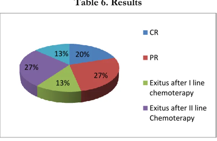
3 (20%) patients are in complete remission (CR) of disease; 4 (27%) in partial remission (PR) of disease; 2 (13%) patients died after the first treatment with first-line chemotherapy, due to an important pulmonary embolism, with an overall survival of 2 months one, and for myocardial infarction, with a 3-month overall survival, the other ; 4 (27%) patients died after the second line of chemotherapy with a 10-month overall survival, 2 (13%) patients died not due to the tumor. Table 6
Table 6. Results
4. Discussion
MBC has some epidemiological, genetic, and biological differences from FBC.[20] [21]In our study,5%(15/355) of the total patients with breast cancer were diagnosed with MBC between January 2013 and December 2017 from Complex Operating Unit Medical Oncology-University of Messina. The median age at diagnosis of MBC in our study was 65 years, which was in accordance with other literature reports that stated the median age of MBC to be between 62 and69 years.[6] [ 22] [23]MBC tumors express ER and PR more often,[7] and lower HER2 over expression than FBC.[23]Infiltrating ductal carcinoma is reported to be the predominant histological type, which accounts for 90% of all MBCs. [7]Our study confirmed this data.
5. Conclusion
Male breast assessment is essential during any physical examination for early detection and reduction of mortality and morbidity related to male breast cancer. Often, and this work was an example, the patient does not realize the importance of seeking prompt medical advice when the mammary nodule is at an early stage of development. When most of the patients came to our observation, the mammary mass was already at the advanced stage of breast cancer. Thus, an in-depth evaluation and a good education of the patient is made desirable and necessary, promoting the awareness that the male breast cancer is constantly increasing and that the early identification of the disease can lead to a significant reduction of the deaths related to it.
20% 27% 13% 27% 13% CR PR
Exitus after I line chemoterapy Exitus after II line Chemoterapy
However, an improvement in the prognosis linked to a higher survival rate may be with an increase in awareness that breast cancer is becoming more and more a disease that, although in a small percentage, can affect the male sex correlating, in 90% of cases, to a poor prognosis due to an inadequate diagnosis.
References
Ravandi-Kashani F, Hayes TG. Male breast cancer: a review of the literature. EurJ Cancer 1998;34:1341–7. Giordano SH, Buzdar AU, Hortobagyi GN. Breast cancer in men. AnnInternMed2002;137:678–87.
La Pinta M, Fabi A, Ascarelli A, Ponzani T, Di Carlo V, Scicchitano F, et al. Male breast cancer: 6-year experience.Minerva Chir 2008;63:71-8
Yoney A, Kucuk A, Unsal M. Male breast cancer: A retrospective analysis. Cancer Radiother 2009;13:103-7 Weiss JR, Moysich KB, Swede H. Epidemiology of male breast cancer. Cancer Epidemiol Biomarkers
Prev2005;14:20-6
Giordano SH, Cohen DS, Buzdar AU et al. Breast carcinoma in men: a population-based study. Cancer2004;101:51–57
Fentiman IS, Fourquet A, Hortobagyi GN. Male breast cancer. The Lancet2006;367:595–604. Ferzoco RM, Ruddy KJ. The epidemiology of male breast cancer. CurrOncol Rep2016;18:1.
Patten DK, Sharifi LK, Fazel M. New approaches in the management of malebreast cancer. Clin Breast Cancer2013;13:309–14.
Tariq KB, Al-Saffar F, Ibrahim S, et al. Male breast cancer and hyperestrogenemia: a thirteen-year review. World JOncol 2014;5:55–61.
Speirs V, Shaaban AM. The rising incidence of male breast cancer. Breast Cancer Res Treat 2009;115:429–30. Anderson WF, Althuis MD, Brinton LA, et al. Is male breast cancer similar or different than female breast
cancer?Breast Cancer Res Treat 2004;83:77–86.
Korde LA, Zujewski JA, Kamin L, et al. Multidisciplinary meeting on male breast cancer: summary and researchrecommendations. J ClinOncol 2010;28:2114–22.
Gómez-Raposo C, ZambranaTévar F, Sereno Moyano M, et al. Male breast cancer. Cancer Treat Rev 2010;36:451–7.
Yu XF, Wang C, Chen B, et al. The effect of adjuvant chemotherapy in male breast cancer: 134 cases from aretrospective study. ESMO Open 2017;2:e000134.
Cardoso F, Bartlett JMS, Slaets L, et al. Characterization of male breast cancer: results of the EORTC10085/TBCRC/BIG/NABCG International Male Breast Cancer Program. Ann Oncol 2018;29:405–17. Giordano SH. Breast cancer in men. N Engl J Med 2018;378:2311–20.
Leone JP, Leone J, Zwenger AO, et al. Locoregional treatment and overall survival of men with T1a,b,cN0M0breast cancer: A population-based study. Eur J Cancer 2017;71:7–14.
Aisner J, Ross DD, Wiernik PH. Tamoxifen in advanced male breast cancer. Arch Intern Med 1979;139:480–1. Giordano SH, Cohen DS, Buzdar AU, et al. Breast carcinoma in men: a population-based study.
Cancer2004;101:51–7.
Nahleh Z, Girnius S. Male breast cancer: a gender issue. Nat ClinPractOncol 2006;3:428–37.
Hong JH, Ha KS, Jung YH, et al. Clinical features of male breast cancer: experiences from seven institutions Over 20 Years. Cancer Res Treat2016;48:1389–98.
Masci G, Caruso M, Caruso F, et al. Clinicopathological and immunohistochemical characteristics in male breast cancer: a retrospective case series. Oncologist 2015;20:586–92.