DOI: 10.3748/wjg.v21.i28.8508 © 2015 Baishideng Publishing Group Inc. All rights reserved.
REVIEW
Gastroesophageal reflux and congenital gastrointestinal
malformations
Lucia Marseglia, Sara Manti, Gabriella D’Angelo, Eloisa Gitto, Carmelo Salpietro, Antonio Centorrino,
Gianfranco Scalfari, Giuseppe Santoro, Pietro Impellizzeri, Carmelo Romeo
Lucia Marseglia, Gabriella D’Angelo, Eloisa Gitto, Neonatal Intensive Care Unit, Department of Pediatrics, University of Messina, 98125 Messina, Italy
Sara Manti, Carmelo Salpietro, Unit of Paediatric Genetics and Immunology, Department of Paediatrics, University of Messina, 98125 Messina, Italy
Antonio Centorrino, Gianfranco Scalfari, Pietro Impellizzeri, Carmelo Romeo, Unit of Pediatric Surgery, Department of Pediatrics, University of Messina, 98125 Messina, Italy
Giuseppe Santoro, Department of Biomedical Sciences and Morpho-Functional Images, University of Messina, 98125 Messina, Italy
Author contributions: Marseglia L coordinated the initial draft of this publication; Manti S, D’Angelo G, Gitto E and Salpietro C wrote the paper; Centorrino A, Scalfari G and Santoro G prepared the final draft; Impellizzeri P and Romeo C approved the final version.
Conflict-of-interest statement: The authors declare no conflicts of interest.
Open-Access: This article is an open-access article which was selected by an in-house editor and fully peer-reviewed by external reviewers. It is distributed in accordance with the Creative Commons Attribution Non Commercial (CC BY-NC 4.0) license, which permits others to distribute, remix, adapt, build upon this work non-commercially, and license their derivative works on different terms, provided the original work is properly cited and the use is non-commercial. See: http://creativecommons.org/ licenses/by-nc/4.0/
Correspondence to: Lucia Marseglia, MD, Neonatal Intensive Care Unit, Department of Pediatrics, University of Messina, Via Consolare Valeria, 1, 98125 Messina, Italy. [email protected] Telephone: +39-90-2213100
Fax: +39-90-2213876 Received: January 21, 2015
Peer-review started: January 22, 2015 First decision: March 26, 2015
Revised: April 24, 2015 Accepted: May 27, 2015 Article in press: May 27, 2015 Published online: July 28, 2015
Abstract
Although the outcome of newborns with surgical congenital diseases (e.g., diaphragmatic hernia; esophageal atresia; omphalocele; gastroschisis) has improved rapidly with recent advances in perinatal intensive care and surgery, infant survivors often require intensive treatment after birth, have prolonged hospitalizations, and, after discharge, may have long-term sequelae including gastro-intestinal comorbidities, above all, gastroesophageal reflux (GER). This condition involves the involuntary retrograde passage of gastric contents into the esophagus, with or without regurgitation or vomiting. It is a well-recognized condition, typical of infants, with an incidence of 85%, which usually resolves after physiological maturation of the lower esophageal sphincter and lengthening of the intra-abdominal esophagus, in the first few months after birth. Although the exact cause of abnormal esophageal function in congenital defects is not clearly understood, it has been hypothesized that common (increased intra-abdominal pressure after closure of the abdominal defect) and/or specific (e.g., motility disturbance of the upper gastrointestinal tract, damage of esophageal peristaltic pump) pathological mechanisms may play a role in the etiology of GER in patients with birth defects. Improvement of knowledge could positively impact the long-term prognosis of patients with surgical congenital diseases. The present manuscript provides a literature review focused on pathological and clinical characteristics of GER in patients who have undergone surgical treatment for congenital abdominal malformations.
Key words: Gastroesophageal reflux; Congenital diaph-ragmatic hernia; Esophageal atresia; Omphalocele; Gastroschisis
© The Author(s) 2015. Published by Baishideng Publishing
Group Inc. All rights reserved.
Core tip: Although the outcome of newborns with surgical congenital diseases has improved rapidly with recent advances in perinatal intensive care and surgery, infant survivors often may have long-term sequelae including, above all, gastroesophageal reflux (GER). Common or specific pathological mechanisms may play a role in the etiology of GER in patients with birth defects. The improvement of knowledge of long-term outcome and follow-up could positively impact the long-term prognosis of newborns with surgical congenital diseases. The present manuscript provides a literature review focused on pathological and clinical characteristics of GER in patients who have undergone surgical treatment for congenital abdominal malformations.
Marseglia L, Manti S, D’Angelo G, Gitto E, Salpietro C, Centorrino A, Scalfari G, Santoro G, Impellizzeri P, Romeo C. Gastroesophageal reflux and congenital gastrointestinal malformations. World J Gastroenterol 2015; 21(28): 8508-8515 Available from: URL: http://www.wjgnet.com/1007-9327/full/ v21/i28/8508.htm DOI: http://dx.doi.org/10.3748/wjg.v21. i28.8508
GASTROESOPHAGEAL REFLUX
Gastroesophageal reflux (GER) is defined as the involuntary retrograde passage of gastric content into the esophagus, with or without regurgitation or vomiting[1]. GER is a well-recognized condition in
infants, which usually resolves after physiological maturation of the lower esophageal sphincter (LES) and lengthening of the intra-abdominal esophagus in the first few months after birth[2-4]. The incidence
of GER in infants is 85%, occurring 1.6 times more frequently in males than in females[1]. Prevalence of
GER decreases to 18% in childhood[1]. This condition is
markedly less common in adult subjects[5,6]. Generally,
it is classified into: (1) “primary”; (2) “secondary”; and (3) “acquired” GER. Primary GER results from a functional disorder of the proximal digestive tract, whereas secondary GER includes structural, infectious, metabolic, neurological and allergic disorders which cause the passage of gastric contents into the esophagus[7]. Despite the fact that the majority of GER
cases are independent of congenital gastrointestinal malformations, acquired GER can also be due to, or favoured by congenital anomalies[8] (Table 1); it has
been reported that GER is commonly associated with birth defects. Although the exact cause of abnormal
esophageal function in congenital defects is not clearly understood, it has been hypothesized that common (increased intra-abdominal pressure after closure of the abdominal defect) and/or specific (e.g., motility disturbance of the upper gastrointestinal tract, damage of esophageal peristaltic pump) pathological mechanisms may play a role in the etiology of GER in patients with birth defects. Moreover, associated anomalies, mental retardation or neurological impairment after prematurity or chromosome abnormality may further increase the risk of GER in these pediatric populations[9]
. In accordance with these data, a high frequency of gastroesophageal long-term complications (e.g., dysphagia, esophageal strictures, esophagitis, esophageal metaplasia and esophageal carcinoma), failure to thrive, as well as respiratory symptoms has been observed, compared to healthy controls[10,11]. Additionally, pathological findings were
found in a high number of patients through endoscopy, manometry and pH-measurements[10,12,13]. However,
it must also be remembered that literature results are heterogeneous and often report opposing outcomes. This is likely due to different inclusion criteria of patients, lack of correlation in neonatal variables, non systematic assessments, and heterogenous therapeutic protocols used[14].
Hence, we wish to summarize what is currently known on pathological and clinical characteristics of GER in surgically-treated patients with congenital gastrointestinal malformations.
GER And cOnGEniTAL
diAPHRAGmATic HERniA
Congenital diaphragmatic hernia (CDH) is a rare (1 per 2500 births) anomaly of the diaphragm, typically characterized by abdominal organ herniation into the chest cavity[15]
. In 84% of cases, the defect is located on the left side of the diaphragm[16]
. Right-sided and bilateral CDH, which occur in 14% and 2% of cases, respectively, are associated with a worse prognosis[16]
. Several hypothesis have been proposed to explain the embryologic events leading to CDH, and several genetic (e.g., structural abnormalities of chromosomes, aneuploidies, genetic syndromes)[17] and environmental
(impaired retinoic acid pathway)[11] factors may play a
role in the development of CDH. CDH can present as an isolated defect or in combination with other congenital anomalies[18] and includes pulmonary, neurological
and gastro-intestinal, especially GER, comorbidities. GER is a common complication in CDH survivors and incidence is 20%-84% during the first year of life[19]. A prevalence of 63% was reported in adult
survivors of CDH, co-existing with Barrett’s esophagus in 54%[20,21]. The mechanisms responsible for GER
in CDH survivors have not been clarified and several theories have been proposed. Esophageal dysmotility and ectasia, maldevelopment or weakness of the
crura, shortening of the esophagus, disruption of the angle of His, intestinal malrotation, and a higher intra-abdominal pressure after surgical repair are possible explanations of GER in CDH[22]. Additionally, the
diaphragmatic sling may be malformed or absent, and repair changes the anatomy of the region. Moreover, abnormality of the esophageal dimensions might also contribute to development of GER. This pathological condition might be congenital or acquired, resulting from extrinsic pressure on the mediastinum exercised by the herniated viscera[23]. Furthermore, malrotation
may delay gastric emptying, and the abnormal balance of pressures in the thorax and abdomen, during respiratory cycle, facilitates retrograde passage of gastric contents to the esophagus[24]. There is evidence
of abnormal enteric innervation in CDH and it is likely that esophago-gastric peristalsis is impaired[25]. Finally,
studies reported that preoperative thoracic position of the stomach and/or an inappropriate intra-abdominal position or volvulus of the stomach after surgical treatment could cause intragastric stasis of food and, therefore, GER[26,27].
Several authors have attempted to define clinical variables that may predict GER. A significant relationship has been found between duration of ventilatory support and GER[20,28]. Postoperative intolerance of enteral
feedings and prolonged hospitalization were also found predominantly in patients with GER[20]. Finally, size
of diaphragmatic defect and use of prosthetic patch could influence incidence of GER in CDH patients[29].
GER can complicate pre-existing respiratory disease and, in the light of abovementioned pathological pathways, responds poorly to medical treatment[25].
However, authors reported satisfactory improved clinical parameters in patients treated conservatively by maintaining supine position, by frequent administration of small amounts of oral nutrition, and by medical treatment[30-32]. Antireflux surgery may be an option
for patients after failed medical therapy, although long-term success rate of this procedure has yet to be proven[33]. A prophylactic fundoplication has also
been proposed in patients with CDH[34]. Up to 23% of
affected newborns exhibiting an intrathoracal position of the liver need such surgical correction[14]. However,
it is difficult to accurately compare results of studies conducted on GER-related CHD as many aspects of the disease are still unknown and, additionally, each study varies regarding CDH severity, postoperative CDH management and follow-up period. Furthermore, predictive factors for late GER could not be identified and screening for early GER does not protect from future GER; therefore, long-term follow-up for GER in CDH survivors is mandatory[30,35-37].
GER And ESOPHAGEAL ATRESiA
Esophageal atresia (EA) is the most common esophageal malformation; incidence is 1 in 3500 live births[38,39].
Classification of EA anomalies is determined by the location of the atresia and by the presence of any associated fistula to the trachea. Five different variants have been clinically described. The primary types of congenital EA are: EA with distal tracheoesophageal fistula (TEF) (85%, Vogt Ⅲb, Gross C), isolated EA without TEF (8%, Vogt Ⅱ, Gross A), TEF without atresia or H-type TEF (4%, Gross E), EA with proximal TEF (3%, Vogt Ⅲ, Gross B) and EA with proximal and distal TEF (< 1%, Vogt Ⅲa, Gross)[40,41]. The
outcome following EA/tracheoesophageal fistula (EA/TEF) repair is variable. Some patients have an uneventful postoperative period, while others reported several complications that can significantly affect their health through adulthood, including GER. GER is so often recognized in survivors of neonatal repair of esophageal atresia and tracheoesophageal fistula (EA/TEF) that it has become a component of this malformation: approximately 50% of these patients had GER during infancy[42], and this proportion
tends to increase over time[43]
. The presence of GER is generally believed to be due to an intrinsic deficiency in the motor function of the stomach and/or esophagus[44]. Authors assessed that delayed gastric
emptying and antral hypomotility, important factors predisposing to GER, are frequent in long-term follow-up of patients operated on for EA-TEF[45,46]. Esophageal
dysfunction has also been reported after successful repair of EA in 20 newborns with EA by recording intraluminal pressure of both proximal and distal segments[47]. In addition, abnormal motility might
affect esophageal clearance, resulting in abnormal bolus clearance, decreased clearance of refluxed materials, dysphagia, severe mucosal damage, esophagitis and, possibly, esophageal carcinoma[48]. It
is likely that GER is exacerbated by surgical repair and gastrostomy, causing an alteration of the anatomical gastro-esophageal junction and the angle of His[49-51]
. Esophageal anastomosis under tension displaces the gastroesophageal junction upwards, diminishing the efficiency of the antireflux barrier[52]. The esophageal
peristaltic pump, another antireflux barrier, is also
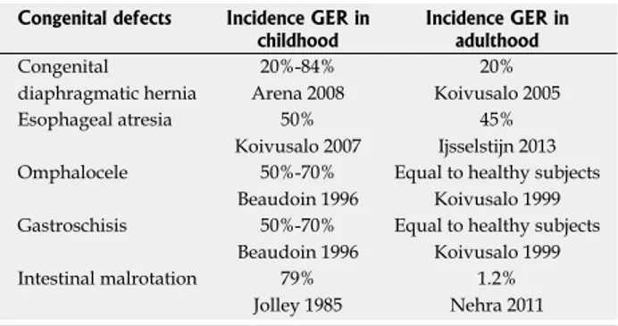
Table 1 Incidence of gastroesophageal reflux in children and adults affected by gastrointestinal congenital defects
Congenital defects Incidence GER in
childhood Incidence GER in adulthood
Congenital diaphragmatic hernia 20%-84% Arena 2008 20% Koivusalo 2005 Esophageal atresia 50% Koivusalo 2007 45% Ijsselstijn 2013 Omphalocele 50%-70% Beaudoin 1996
Equal to healthy subjects Koivusalo 1999 Gastroschisis 50%-70%
Beaudoin 1996
Equal to healthy subjects Koivusalo 1999 Intestinal malrotation 79%
Jolley 1985
1.2% Nehra 2011 GER: Gastroesophageal reflux.
in infants with congenital abdominal wall defect (CAWD), including omphalocele and gastroschisis[64,65].
Incidence of GER in these patients has been reported to be between 50% and 70%[66]. Moreover, associated
anomalies (e.g., esophageal atresia, duodenal atresia and diaphragmatic hernia), chromosome abnormality, and neurological impairment can strongly increase the likelihood of GER[9]. Although it has been hypothesized
that the etiology of GER in patients with CAWD is both related to increased intra-abdominal pressure after the closure of the abdominal defect and gastrointestinal motility disturbance[67], the etiology of GER related to
omphalocele or gastroschisis is still under study. Omphalocele, a ventral defect of the umbilical ring resulting in herniation of the abdominal viscera, is one of the most common congenital anterior abdominal wall defects. It occurs in 1 in 3000 to 10000 live births[68]
and is characterized by the absence of abdominal muscles, fascia, and skin due to the defective closure of the abdominal wall in the embryo before 9 weeks of gestation. The malformation causes herniation of the abdominal contents, covered by a membranous sac consisting of peritoneum and amnion, into the base of the umbilical cord[69]. In infants with omphalocele, the
incidence of GER considerably exceeds that of control group children[70]. It is hypothesized that in patients
with a large omphalocele and severely underdeve-loped abdominal cavity, GER is favored by the high postoperative intra-abdominal pressure after repair of the defect[71]. Furthermore, Beaudoin et al[66] reported
that infants affected by large omphaloceles had an increased risk of GER, especially where primary closure with skin or silo only was possible. A high incidence of GER in omphalocele patients was described above all during the first few years of life; it appears that GER clearly improves after infancy and, later childhood symptoms are less marked and easily controlled with medication[70]. In light of these data, routine diagnostic
workup (endoscopy and pH monitoring) for GER is warranted for children with omphalocele during the first year of life. If GER is diagnosed, medical treatment should be started and long-term follow-up might be arranged if it persists. Additionally, older patients with omphalocele should undergo GER evaluation in the presence of GER-related symptoms[70].
Gastroschisis is defined as an extraumbilical herniation of bowel without covering sac through an anterior abdominal wall defect. It occurs in about one in 600 live births[71]. Gastroschisis could be
the result of amniotic damage, possibly from some as yet unidentified toxin. Further bowel damage can be explained by the subsequent mesenteric injury[72]. Infant mortality with gastroschisis has been
markedly reduced in the last decade by new surgical techniques, improved metabolic monitoring, and total parenteral nutrition. On the other hand, to date, late complications of gastroschisis are emerging as clinical problems, with the longer survival of affected newborns. Some gastrointestinal complications, such damaged in EA/TEF due to deficient arrangement of
the muscle layers and damage to the innervation of the repaired esophagus. Moreover, extrinsic innervation by branches of the vagus and laryngeal nerves, and also intrinsic innervation, are congenitally deficient in EA[53]. In addition, gastrostomy, sometimes used
in the treatment of EA/TEF, facilitates GER[54]. Finally,
tracheomalacia and/or tracheal stenosis, which are relatively common in EA/TEF newborns, cause airway obstruction and induce GER by exaggerating negative inspiratory force and increasing the abdomino-thoracic pressure gradient[53]. However, to date, it is unclear if
these abnormalities exist immediately after EA repair or if they develop over time. Although studies suggest that motility disorders are also present in esophageal atresia before surgery[47], published studies do not
allow us to conclude whether GER is congenital (abnormal innervation)[55] or acquired due to inevitable
damage during surgical reconstruction[56].
Although GER manifests itself by vomiting, failure to thrive, and respiratory symptoms, it has been shown that symptoms of GER and histologic findings are poorly correlated, raising doubts regarding correct management. Generally, a post-operative long-term endoscopic and pH-metric follow-up of all patients is warranted. Endoscopic follow-up is recommended for all surgically treated patients irrespective of symptoms. Endoscopic follow-up of children with completely normal esophageal biopsies can be discontinued at 3 years of age. In patients with mild esophagitis, routine follow-up should be extended to at least 6 years of age[43]. As EA/TEF newborns with GER do not benefit
from postural, dietary or prokinetic treatments[57],
primarily surgery may be required, especially in the case of refractory anastomotic stenosis, pure and long-gap EA, and duodenal atresia[58]. Patients who have
undergone anti-reflux surgery should also be followed long-term[43,59]. Nevertheless, some authors refuse to
perform fundoplication as esophageal motility, in these children, is poorly known. Moreover, to date, there is no randomized trial that has evaluated treatment of GER in children with EA with fundoplication vs. acid suppressive medication[60]. Certainly, several studies
have shown that the persistence of GER might increase the risk of long-term complications, such as dysphagia, esophageal strictures, esophagitis, esophageal meta-plasia and esophageal carcinoma in children and adults born with EA compared to healthy controls[61].
In conclusion, there are no well-outlined algorithms for medical and/or surgical therapy. Standardized protocols and reliable reporting are necessary to develop guidelines to better manage postoperative GER in EA/TEF patients[62,63].
GER And cOnGEniTAL AbdOminAL
wALL dEFEcTS: OmPHALOcELE And
GASTROScHiSiS
if evidence of reflux stems from failure of the lower esophageal sphincter (LES) or from problems that “look like” GER[71]. However, Koivusalo et al[70] reported
that, in infants with gastroschisis, when normal bowel motility was surgically restored, the prevalence of GER did not exceed that of healthy children. In gastroschisis, GER may be secondary to operative reduction of the bowel which, in turn, can distort the stomach and the angle of His, suggesting one cause for a major incidence of reflux in these infants[73]. In
addition, clinical and experimental models supported evidence that intestinal dysmotility may contribute to GER and to formation of hiatal hernia. Recently, a study has described a high incidence of associated hiatal hernia in gastroschisis patients. The presence of large associated hiatal hernia is correlated with severe GER, delayed feeding, requirement for antireflux surgery, and a prolonged hospital stay. Although the role of dysmotility in GER-related gastroschisis has not been clearly defined[74-76], Jadcherla et
al[77] reported impaired feeding milestones, basal
pharyngoesophageal peristaltic failure and less frequent peristaltic reflexes of the upper and lower esophageal sphincters in neonates with gastroschisis. In an experimental model, a decreased number of interstitial cells of Cajal (intestinal pacemaker cells) have been found, and neuronal abnormalities might further justify intestinal dismotility[77,78]. Additionally,
authors assessed that animal models, with carb-oxypeptidase-like protein deficiency, had a very similar abdominal wall defect to human gastroschisis and also exhibited absence of intestinal pacemaker cells and delayed neuromuscular development[79].
The clinical course of GER-related to gastroschisis is still unclear[80]. Patients with gastroschisis should
undergo GER evaluation only in the presence of GER-related symptoms[70]
. Koivusalo and collaborators reported that in CAWD patients, compared to a general population, the quality of life and morbidity from acquired disorders are similar and rarely cause serious problems[71]. Conversely, in the presence of large
hiatal hernia (Ⅱ or Ⅲ type), with severe, medically refractory GER, this selected patient population might require antireflux surgery[81,82].
GER And inTESTinAL mALROTATiOn
Intestinal malrotation (IM) is defined as an anomaly of rotation and fixation of the midgut[83]. Malrotation
of the intestinal tract is a product of a well-defined aberrant embryology. It usually occurs due to incomplete rotation or a complete failure of rotation of the primitive intestinal loops around the superior mesenteric artery axis in the fetal period[84]. IM is
found either alone or in combination with other congenital anomalies, including CHD and CAWD[85].
IM is a rare disorder, found in only approximately one per 10000 individuals. However, the incidence
of IM in infants and children beyond the neonatal period is difficult to estimate, as such anomalies may remain asymptomatic and undetected throughout life. Approximately 50% to 70% of cases are diagnosed during the neonatal period; about 50% present in the first week of life, and over 60% present before the end of the first month[86,87].Male infants are more frequently
affected than female infants[88]. Wang et al[89] classified
IM into four categories: non-rotation, malrotation, reversed rotation, and paraduodenal hernia. Due to the lack of normal intestinal mesenteric fixation, IM may present with acute or chronic symptoms. Acute symptoms are due to duodenal obstruction, with or without midgut volvulus, and sudden infant death. Chronic symptoms, due to intermittent intestinal obstruction, include abdominal pain, bloating or constipation, postprandial fullness, epigastric discomfort, early satiety, and GER[90]. The association
between GER and IM has been well described. In a series of 74 children undergoing Nissen fundoplication for persistent GER unresponsive to intensive medical treatment, there was an unexpectedly high incidence of IM[91]. Moreover, the high incidence of GER and
the significant improvement after correction of malrotation further show the relationship between delayed gastric emptying and GER[92]. In light of
these data, delayed or impaired gastric emptying in IM are thought to be contributing factors in the development of GER. Using gastric emptying studies and esophageal pH monitoring, authors assessed that gastric dysmotility and esophageal pH abnormalities were highly prevalent in children with malrotation compared with children only affected by isolated GER[93]. It has been hypothesized that, in infants
with IM, reversed gastric peristaltic waves, especially during fetal development lead to defects of the plexus myentericus, favouring GER[85]. In addition, the high
prevalence of GER in infants affected by IM suggests that an impaired gastroesophageal junction may result from the same embriological defect that leads to the interference of normal intestinal rotation and fixation during fetal development[85]
. Conversely, no sufficient literature data supported the mechanical gastric-outlet obstruction as a factor promoting GER; a high GER prevalence persisted despite the relief of gastric-outlet obstruction with a successful Ladd operation[90,94].
Generally, the course of treated infants with GER and IM depends on the presence or absence of symptoms to suggest GER. The course in symptomatic newborns is similar to infants with GER without IM, thus, their outcomes are related to reflux pattern type[67]
. Conversely, asymptomatic patients with GER have a better prognosis. However, these latter might continue to have GER, during follow-up, favouring Barrett esophagus in children[85,95]. Therefore, the high
prevalence of GER in infants with IM requires careful evaluation and follow-up.
cOncLUSiOn
Despite the fact that the majority of GER cases are independent of congenital gastrointestinal malformations, GER is most common in patients with congenital gastrointestinal diseases[5,6]
. GER can further worsen birth defects and favour the onset of related comorbidities, constituting the most prevalent complication in long-term follow-up[96,97]
. However, to date, long-term outcome and follow-up data after surgical repair are limited. Improvement of knowledge could positively impact the long-term prognosis of these patients who, benefiting from a more intensive follow-up program, would avoid gastroenterological symptoms and/or comorbidities during adulthood. Additionally, this might lead to an era of “individualised” management, identifying specific selection criteria for medical and/or surgical treatment, especially for younger children with longer life expectancy[98-100]
.
REFEREncES
1 Czinn SJ, Blanchard S. Gastroesophageal reflux disease in
neonates and infants: when and how to treat. Paediatr Drugs 2013;
15: 19-27 [PMID: 23322552 DOI: 10.1007/s40272-012-0004-2]
2 Boix-Ochoa J, Canals J. Maturation of the lower esophagus. J Pediatr Surg 1976; 11: 749-756 [PMID: 993945]
3 Park KY, Chang SH. Gastro-esophageal reflux disease in healthy
older children and adolescents. Pediatr Gastroenterol Hepatol Nutr 2012; 15: 220-228 [PMID: 24010091 DOI: 10.5223/ pghn.2012.15.4.220]
4 Schatzlein MH, Ballantine TV, Thirunavukkarasu S, Fitzgerald
JF, Grosfeld JL. Gastroesophageal reflux in infants and children. Diagnosis and management. Arch Surg 1979; 114: 505-510 [PMID: 435065]
5 Chait MM. Gastroesophageal reflux disease: Important
considerations for the older patients. World J Gastrointest Endosc 2010; 2: 388-396 [PMID: 21191512 DOI: 10.4253/wjge. v2.i12.388]
6 El-Serag HB. Time trends of gastroesophageal reflux disease:
a systematic review. Clin Gastroenterol Hepatol 2007; 5: 17-26 [PMID: 17142109]
7 Costa AJ, Silva GA, Gouveia PA, Pereira Filho EM. [Prevalence
of pathologic gastroesophageal reflux in regurgitant infants]. J Pediatr (Rio J) 2004; 80: 291-295 [PMID: 15309230]
8 Ottolenghi A, Camoglio FS, Valletta E, Giacomello L, Pasquini A.
[Primary ad secondary gastro-esophageal reflux in pediatric age]. Minerva Pediatr 2004; 56: 91-96 [PMID: 15249918]
9 Fonkalsrud EW, Ament ME. Gastroesophageal reflux in
childhood. Curr Probl Surg 1996; 33: 1-70 [PMID: 8536488] 10 van Wijk M, Knüppe F, Omari T, de Jong J, Benninga M.
Evaluation of gastroesophageal function and mechanisms underlying gastroesophageal reflux in infants and adults born with esophageal atresia. J Pediatr Surg 2013; 48: 2496-2505 [PMID: 24314193 DOI: 10.1016/j.jpedsurg.2013.07.024]
11 Kumar V, Mathai SS, Kanitkar M. Preliminary study in to the incidence of gastroesophageal reflux (GER) in high risk neonates admitted to NICU. Indian J Pediatr 2012; 79: 1197-1200 [PMID: 22002315 DOI: 10.1007/s12098-011-0569-8]
12 Al Dulaimi D. Recent advances in oesophageal diseases. Gastroenterol Hepatol Bed Bench 2014; 7: 186-189 [PMID: 25120902]
13 Triadafilopoulos G. Stretta: a valuable endoscopic treatment modality for gastroesophageal reflux disease. World J Gastroenterol 2014; 20: 7730-7738 [PMID: 24976710 DOI: 10.3748/wjg.v20. i24.7730]
14 van Manen M, Hendson L, Wiley M, Evans M, Taghaddos S, Dinu
I. Early childhood outcomes of infants born with gastroschisis. J Pediatr Surg 2013; 48: 1682-1687 [PMID: 23932607 DOI: 10.1016/j.jpedsurg.2013.01.021]
15 Lally KP. Congenital diaphragmatic hernia. Curr Opin Pediatr 2002; 14: 486-490 [PMID: 12130916]
16 de Buys Roessingh AS, Dinh-Xuan AT. Congenital diaphragmatic hernia: current status and review of the literature. Eur J Pediatr 2009; 168: 393-406 [PMID: 19104834 DOI: 10.1007/s00431-008-0904-x]
17 Kantarci S, Casavant D, Prada C, Russell M, Byrne J, Haug LW, Jennings R, Manning S, Blaise F, Boyd TK, Fryns JP, Holmes LB, Donahoe PK, Lee C, Kimonis V, Pober BR. Findings from aCGH in patients with congenital diaphragmatic hernia (CDH): a possible locus for Fryns syndrome. Am J Med Genet A 2006; 140: 17-23 [PMID: 16333846]
18 Montedonico S, Sugimoto K, Felle P, Bannigan J, Puri P. Prenatal treatment with retinoic acid promotes pulmonary alveologenesis in the nitrofen model of congenital diaphragmatic hernia. J Pediatr Surg 2008; 43: 500-507 [PMID: 18358289 DOI: 10.1016/ j.jpedsurg.2007.10.030]
19 Arena F, Romeo C, Baldari S, Arena S, Antonuccio P, Campennì A, Zuccarello B, Romeo G. Gastrointestinal sequelae in survivors of congenital diaphragmatic hernia. Pediatr Int 2008; 50: 76-80 [PMID: 18279210 DOI: 10.1111/j.1442-200X.2007.02527.x] 20 Vanamo K, Rintala RJ, Lindahl H, Louhimo I. Long-term
gastrointestinal morbidity in patients with congenital diaphragmatic defects. J Pediatr Surg 1996; 31: 551-554 [PMID: 8801311] 21 Hammoud GM, Hammad H, Ibdah JA. Endoscopic assessment
and management of early esophageal adenocarcinoma. World J Gastrointest Oncol 2014; 6: 275-288 [PMID: 25132925 DOI: 10.4251/wjgo.v6.i8.275]
22 Su W, Berry M, Puligandla PS, Aspirot A, Flageole H, Laberge JM. Predictors of gastroesophageal reflux in neonates with congenital diaphragmatic hernia. J Pediatr Surg 2007; 42: 1639-1643 [PMID: 17923189]
23 Van Meurs KP, Robbins ST, Reed VL, Karr SS, Wagner AE, Glass P, Anderson KD, Short BL. Congenital diaphragmatic hernia: long-term outcome in neonates treated with extracorporeal membrane oxygenation. J Pediatr 1993; 122: 893-899 [PMID: 8501565]
24 Qi B, Soto C, Diez-Pardo JA, Tovar JA. An experimental study on the pathogenesis of gastroesophageal reflux after repair of diaphragmatic hernia. J Pediatr Surg 1997; 32: 1310-1313 [PMID: 9314250]
25 Tovar JA. Congenital diaphragmatic hernia. Orphanet J Rare Dis 2012; 7: 1 [PMID: 22214468 DOI: 10.1186/1750-1172-7-1] 26 Stolar CJ, Levy JP, Dillon PW, Reyes C, Belamarich P, Berdon
WE. Anatomic and functional abnormalities of the esophagus in infants surviving congenital diaphragmatic hernia. Am J Surg 1990;
159: 204-207 [PMID: 2405729]
27 Kieffer J, Sapin E, Berg A, Beaudoin S, Bargy F, Helardot PG. Gastroesophageal reflux after repair of congenital diaphragmatic hernia. J Pediatr Surg 1995; 30: 1330-1333 [PMID: 8523237] 28 Kamiyama M, Kawahara H, Okuyama H, Oue T, Kuroda
S, Kubota A, Okada A. Gastroesophageal reflux after repair of congenital diaphragmatic hernia. J Pediatr Surg 2002; 37: 1681-1684 [PMID: 12483629]
29 Diamond IR, Mah K, Kim PC, Bohn D, Gerstle JT, Wales PW. Predicting the need for fundoplication at the time of congenital diaphragmatic hernia repair. J Pediatr Surg 2007; 42: 1066-1070 [PMID: 17560222]
30 Koivusalo A, Pakarinen M, Vanamo K, Lindahl H, Rintala RJ. Health-related quality of life in adults after repair of congenital diaphragmatic defects--a questionnaire study. J Pediatr Surg 2005;
40: 1376-1381 [PMID: 16150336]
31 Di Pace MR, Caruso AM, Farina F, Casuccio A, Cimador M, De Grazia E. Evaluation of esophageal motility and reflux in children treated for congenital diaphragmatic hernia with the use of combined multichannel intraluminal impedance and pH
monitoring. J Pediatr Surg 2011; 46: 1881-1886 [PMID: 22008321 DOI: 10.1016/j.jpedsurg.2011.04.093]
32 Caruso AM, Di Pace MR, Catalano P, Farina F, Casuccio A, Cimador M, De Grazia E. Gastroesophageal reflux in patients treated for congenital diaphragmatic hernia: short- and long-term evaluation with multichannel intraluminal impedance. Pediatr Surg Int 2013; 29: 553-559 [PMID: 23571823 DOI: 10.1007/s00383-013-3303-y]
33 American Academy of Pediatrics Section on Surgery; American Academy of Pediatrics Committee on Fetus and Newborn, Lally KP, Engle W. Postdischarge follow-up of infants with congenital diaphragmatic hernia. Pediatrics 2008; 121: 627-632 [PMID: 18310215 DOI: 10.1542/peds.2007-3282]
34 Dariel A, Rozé JC, Piloquet H, Podevin G. Impact of prophylactic fundoplication on survival without growth disorder in left congenital diaphragmatic hernia requiring a patch repair. J Pediatr 2010; 157: 688-90, 690.e1 [PMID: 20633898 DOI: 10.1016/ j.jpeds.2010.06.009]
35 Peetsold MG, Kneepkens CM, Heij HA, IJsselstijn H, Tibboel D, Gemke RJ. Congenital diaphragmatic hernia: long-term risk of gastroesophageal reflux disease. J Pediatr Gastroenterol Nutr 2010; 51: 448-453 [PMID: 20512059 DOI: 10.1097/ MPG.0b013e3181d1b149]
36 Kawahara H, Okuyama H, Nose K, Nakai H, Yoneda A, Kubota A, Fukuzawa M. Physiological and clinical characteristics of gastroesophageal reflux after congenital diaphragmatic hernia repair. J Pediatr Surg 2010; 45: 2346-2350 [PMID: 21129542 DOI: 10.1016/j.jpedsurg.2010.08.029]
37 Koziarkiewicz M, Taczalska A, Piaseczna-Piotrowska A. Long-term follow-up of children with congenital diaphragmatic hernia--observations from a single institution. Eur J Pediatr Surg 2014; 24: 500-507 [PMID: 24163197 DOI: 10.1055/s-0033-1357751] 38 Shaw-Smith C. Oesophageal atresia, tracheo-oesophageal
fistula, and the VACTERL association: review of genetics and epidemiology. J Med Genet 2006; 43: 545-554 [PMID: 16299066] 39 Martinucci I, de Bortoli N, Giacchino M, Bodini G, Marabotto
E, Marchi S, Savarino V, Savarino E. Esophageal motility abnormalities in gastroesophageal reflux disease. World J Gastrointest Pharmacol Ther 2014; 5: 86-96 [PMID: 24868489 DOI: 10.4292/wjgpt.v5.i2.86]
40 Spitz L. Oesophageal atresia. Orphanet J Rare Dis 2007; 2: 24 [PMID: 17498283]
41 Olbers J, Gatzinsky V, Jönsson L, Friberg LG, Abrahamsson K, Sillén U, Gustafsson P. Physiological Studies at 7 Years of Age in Children Born with Esophageal Atresia. Eur J Pediatr Surg 2014; Epub ahead of print [PMID: 25302538]
42 Okada A, Usui N, Inoue M, Kawahara H, Kubota A, Imura K, Kamata S. Esophageal atresia in Osaka: a review of 39 years’ experience. J Pediatr Surg 1997; 32: 1570-1574 [PMID: 9396528] 43 Koivusalo A, Pakarinen MP, Rintala RJ. The cumulative incidence
of significant gastrooesophageal reflux in patients with oesophageal atresia with a distal fistula--a systematic clinical, pH-metric, and endoscopic follow-up study. J Pediatr Surg 2007; 42: 370-374 [PMID: 17270551]
44 Lemoine C, Aspirot A, Le Henaff G, Piloquet H, Lévesque D, Faure C. Characterization of esophageal motility following esophageal atresia repair using high-resolution esophageal manometry. J Pediatr Gastroenterol Nutr 2013; 56: 609-614 [PMID: 23343933 DOI: 10.1097/MPG.0b013e3182868773] 45 Romeo C, Bonanno N, Baldari S, Centorrino A, Scalfari G,
Antonuccio P, Centonze A, Gentile C. Gastric motility disorders in patients operated on for esophageal atresia and tracheoesophageal fistula: long-term evaluation. J Pediatr Surg 2000; 35: 740-744 [PMID: 10813339]
46 Ijsselstijn H, van Beelen NW, Wijnen RM. Esophageal atresia: long-term morbidities in adolescence and adulthood. Dis Esophagus 2013; 26: 417-421 [PMID: 23679035 DOI: 10.1111/ dote.12059]
47 Romeo G, Zuccarello B, Proietto F, Romeo C. Disorders of the esophageal motor activity in atresia of the esophagus. J Pediatr
Surg 1987; 22: 120-124 [PMID: 3820005]
48 Deurloo JA, Aronson DC. Possibility that esophageal atresia (EA) carries an increased risk for esophageal carcinoma. J Pediatr Surg 2006; 41: 876-87; author reply 877 [PMID: 16567217]
49 Mortell AE, Azizkhan RG. Esophageal atresia repair with thoracotomy: the Cincinnati contemporary experience. Semin Pediatr Surg 2009; 18: 12-19 [PMID: 19103416 DOI: 10.1053/ j.sempedsurg.2008.10.003]
50 Taylor AC, Breen KJ, Auldist A, Catto-Smith A, Clarnette T, Crameri J, Taylor R, Nagarajah S, Brady J, Stokes K. Gastroesophageal reflux and related pathology in adults who were born with esophageal atresia: a long-term follow-up study. Clin Gastroenterol Hepatol 2007; 5: 702-706 [PMID: 17544997] 51 Maynard S, Bouin M. Follow-up of adult patients with repaired
esophageal atresia: how, when, and for how long? Dis Esophagus 2013; 26: 422-424 [PMID: 23679036 DOI: 10.1111/dote.12060] 52 Nagaya M, Kato J, Niimi N, Tanaka S, Iio K. Proposal of a novel
method to evaluate anastomotic tension in esophageal atresia with a distal tracheoesophageal fistula. Pediatr Surg Int 2005; 21: 780-785 [PMID: 16177921]
53 Nakazato Y, Landing BH, Wells TR. Abnormal Auerbach plexus in the esophagus and stomach of patients with esophageal atresia and tracheoesophageal fistula. J Pediatr Surg 1986; 21: 831-837 [PMID: 3783365]
54 Isch JA, Rescorla FJ, Scherer LR, West KW, Grosfeld JL. The development of gastroesophageal reflux after percutaneous endoscopic gastrostomy. J Pediatr Surg 1997; 32: 321-32; discussion 321-32; [PMID: 9044145]
55 Pederiva F, Burgos E, Francica I, Zuccarello B, Martinez L, Tovar JA. Intrinsic esophageal innervation in esophageal atresia without fistula. Pediatr Surg Int 2008; 24: 95-100 [PMID: 17962964] 56 Shono T, Suita S. Motility studies of the esophagus in a case of
esophageal atresia before primary anastomosis and in experimental models. Eur J Pediatr Surg 1997; 7: 138-142 [PMID: 9241497] 57 Burjonrappa S, Thiboutot E, Castilloux J, St-Vil D. Type A
esophageal atresia: a critical review of management strategies at a single center. J Pediatr Surg 2010; 45: 865-871 [PMID: 20438915 DOI: 10.1016/j.jpedsurg.2010.02.004]
58 Tovar JA, Fragoso AC. Gastroesophageal reflux after repair of esophageal atresia. Eur J Pediatr Surg 2013; 23: 175-181 [PMID: 23720211 DOI: 10.1055/s-0033-1347911]
59 Tovar JA, Fragoso AC. Anti-reflux surgery for patients with esophageal atresia. Dis Esophagus 2013; 26: 401-404 [PMID: 23679031 DOI: 10.1111/dote.12063]
60 Pedersen RN, Markøw S, Kruse-Andersen S, Qvist N, Hansen TP, Gerke O, Nielsen RG, Rasmussen L, Husby S. Esophageal atresia: gastroesophageal functional follow-up in 5-15 year old children. J Pediatr Surg 2013; 48: 2487-2495 [PMID: 24314192 DOI: 10.1016/j.jpedsurg.2013.07.019]
61 Castilloux J, Bouron-Dal Soglio D, Faure C. Endoscopic assessment of children with esophageal atresia: Lack of relationship of esophagitis and esophageal metaplasia to symptomatology. Can J Gastroenterol 2010; 24: 312-316 [PMID: 20485706]
62 Shawyer AC, D’Souza J, Pemberton J, Flageole H. The management of postoperative reflux in congenital esophageal atresia-tracheoesophageal fistula: a systematic review. Pediatr Surg Int 2014; 30: 987-996 [PMID: 25011995 DOI: 10.1007/ s00383-014-3548-0]
63 Sistonen SJ, Pakarinen MP, Rintala RJ. Long-term results of esophageal atresia: Helsinki experience and review of literature. Pediatr Surg Int 2011; 27: 1141-1149 [PMID: 21960312 DOI: 10.1007/s00383-011-2980-7]
64 Corey KM, Hornik CP, Laughon MM, McHutchison K, Clark RH, Smith PB. Frequency of anomalies and hospital outcomes in infants with gastroschisis and omphalocele. Early Hum Dev 2014;
90: 421-424 [PMID: 24951080 DOI: 10.1016/j.earlhumdev.2014.0
5.006]
65 Puri A, Bajpai M. Gastroschisis and omphalocele. Indian J Pediatr 1999; 66: 773-789 [PMID: 10798139]
Gastroesophageal reflux in neonates with congenital abdominal wall defect. Eur J Pediatr Surg 1995; 5: 323-326 [PMID: 8773221] 67 Jolley SG, Tunell WP, Thomas S, Young J, Smith EI. The
significance of gastric emptying in children with intestinal malrotation. J Pediatr Surg 1985; 20: 627-631 [PMID: 2935609] 68 McNair C, Hawes J, Urquhart H. Caring for the newborn with
an omphalocele. Neonatal Netw 2006; 25: 319-327 [PMID: 16989131]
69 Ledbetter DJ. Gastroschisis and omphalocele. Surg Clin North Am 2006; 86: 249-60, vii [PMID: 16580922]
70 Koivusalo A, Rintala R, Lindahl H. Gastroesophageal reflux in children with a congenital abdominal wall defect. J Pediatr Surg 1999; 34: 1127-1129 [PMID: 10442606]
71 Koivusalo A, Lindahl H, Rintala RJ. Morbidity and quality of life in adult patients with a congenital abdominal wall defect: a questionnaire survey. J Pediatr Surg 2002; 37: 1594-1601 [PMID: 12407546]
72 Oh KS, Dorst JP, Dominguez R, Girdany BR. Abnormal intestinal motility in gastroschisis. Radiology 1978; 127: 457 [PMID: 148068]
73 Bargy F, Beaudoin S. Comprehensive developmental mechanisms in gastroschisis. Fetal Diagn Ther 2014; 36: 223-230 [PMID: 25171094 DOI: 10.1159/000360080]
74 Blane CE, Wesley JR, DiPietro MA, White SJ, Coran AG. Gastrointestinal complications of gastroschisis. AJR Am J Roentgenol 1985; 144: 589-591 [PMID: 3871569]
75 Curry JI, Lander AD, Stringer MD. A multicenter, randomized, double-blind, placebo-controlled trial of the prokinetic agent erythromycin in the postoperative recovery of infants with gastroschisis. J Pediatr Surg 2004; 39: 565-569 [PMID: 15065029] 76 Hillemeier AC, Grill BB, McCallum R, Gryboski J. Esophageal
and gastric motor abnormalities in gastroesophageal reflux during infancy. Gastroenterology 1983; 84: 741-746 [PMID: 6825985] 77 Jadcherla SR, Gupta A, Stoner E, Fernandez S, Caniano D,
Rudolph CD. Neuromotor markers of esophageal motility in feeding intolerant infants with gastroschisis. J Pediatr Gastroenterol Nutr 2008; 47: 158-164 [PMID: 18664867 DOI: 10.1097/MPG.0b013e318162082f]
78 Midrio P, Faussone-Pellegrini MS, Vannucchi MG, Flake AW. Gastroschisis in the rat model is associated with a delayed maturation of intestinal pacemaker cells and smooth muscle cells. J Pediatr Surg 2004; 39: 1541-1547 [PMID: 15486901]
79 Vannucchi MG, Midrio P, Flake AW, Faussone-Pellegrini MS. Neuronal differentiation and myenteric plexus organization are delayed in gastroschisis: an immunohistochemical study in a rat model. Neurosci Lett 2003; 339: 77-81 [PMID: 12618304] 80 Fasching G, Huber A, Uray E, Sorantin E, Mayr J. Late follow-up
in patients with gastroschisis: Gastroesophageal reflux is common. Pediatr Surg Int 1996; 11: 103-106 [PMID: 24057527 DOI: 10.1007/BF00183736]
81 Danzer E, Layne MD, Auber F, Shegu S, Kreiger P, Radu A, Volpe M, Adzick NS, Flake AW. Gastroschisis in mice lacking aortic carboxypeptidase-like protein is associated with a defect in neuromuscular development of the eviscerated intestine. Pediatr Res 2010; 68: 23-28 [PMID: 20386491 DOI: 10.1203/00006450-2 01011001-00040]
82 Tsai J, Blinman TA, Collins JL, Laje P, Hedrick HL, Adzick NS, Flake AW. The contribution of hiatal hernia to severe gastroesophageal reflux disease in patients with gastroschisis. J Pediatr Surg 2014; 49: 395-398 [PMID: 24650464 DOI: 10.1016/ j.jpedsurg.2013.09.005]
83 Stringer DA. Small bowel. In: Stringer DA, editor. Pediatric
Gastrointestinal Imaging. Philadelphia: BC Decker Inc, 1989: 235-239
84 Zissin R, Rathaus V, Oscadchy A, Kots E, Gayer G, Shapiro-Feinberg M. Intestinal malrotation as an incidental finding on CT in adults. Abdom Imaging 1999; 24: 550-555 [PMID: 10525804] 85 Jolley SG, Lorenz ML, Hendrickson M, Kurlinski JP. Esophageal
pH monitoring abnormalities and gastroesophageal reflux disease in infants with intestinal malrotation. Arch Surg 1999; 134: 747-752; discussion 752-753 [PMID: 10401827]
86 Millar AJW, Rode H, Brown RA. The deadly vomit: malrotation and midgut volvulus. Pediatr Surg Int 1987; 2: 172-176
87 Cywes S, Millar AJW. Embryology and anomalies of the intestine. Bockus Gastroenterology. 5th ed. Philadelphia: WB Saunders, 1995: 899-929
88 Millar AJ, Rode H, Cywes S. Malrotation and volvulus in infancy and childhood. Semin Pediatr Surg 2003; 12: 229-236 [PMID: 14655161]
89 Wang CA, Welch CE. Anomalies of intestinal rotation in adolescents and adults. Surgery 1963; 54: 839-855 [PMID: 14087118]
90 Nehra D, Goldstein AM. Intestinal malrotation: varied clinical presentation from infancy through adulthood. Surgery 2011; 149: 386-393 [PMID: 20719352 DOI: 10.1016/j.surg.2010.07.004] 91 Mazziotti MV, Strasberg SM, Langer JC. Intestinal rotation
abnormalities without volvulus: the role of laparoscopy. J Am Coll Surg 1997; 185: 172-176 [PMID: 9249085]
92 Kumar D, Brereton RJ, Spitz L, Hall CM. Gastro-oesophageal reflux and intestinal malrotation in children. Br J Surg 1988; 75: 533-535 [PMID: 3395817]
93 Tiboni SG, Patel Y, Lander AD, Parikh DH, Jawaheer G, Arul GS. Management of gastroesophageal reflux associated with malrotation in children. J Pediatr Surg 2011; 46: 289-291 [PMID: 21292075 DOI: 10.1016/j.jpedsurg.2010.11.005]
94 Demirbilek S, Karaman A, Gürünlüoğlu K, Akin M, Taş E, Aksoy RT, Kekilli E. Delayed gastric emptying in gastroesophageal reflux disease: the role of malrotation. Pediatr Surg Int 2005; 21: 423-427 [PMID: 15912364]
95 Fouad YM, Mostafa I, Yehia R, El-Khayat H. Biomarkers of Barrett’s esophagus. World J Gastrointest Pathophysiol 2014; 5: 450-456 [PMID: 25400988 DOI: 10.4291/wjgp.v5.i4.450] 96 Davies I, Burman-Roy S, Murphy MS. Gastro-oesophageal reflux
disease in children: NICE guidance. BMJ 2015; 350: g7703 [PMID: 25591811 DOI: 10.1136/bmj.g7703]
97 Yadlapati R, Gawron AJ, Bilimoria K, Keswani RN, Dunbar KB, Kahrilas PJ, Katz P, Richter J, Schnoll-Sussman F, Soper N, Vela MF, Pandolfino JE. Development of quality measures for the care of patients with gastroesophageal reflux disease. Clin Gastroenterol Hepatol 2015; 13: 874-83.e2 [PMID: 25460560 DOI: 10.1016/ j.cgh.2014.11.012]
98 Tighe M, Afzal NA, Bevan A, Hayen A, Munro A, Beattie RM. Pharmacological treatment of children with gastro-oesophageal reflux. Cochrane Database Syst Rev 2014; 11: CD008550 [PMID: 25419906 DOI: 10.1002/14651858.CD008550]
99 Badillo R, Francis D. Diagnosis and treatment of gastroesophageal reflux disease. World J Gastrointest Pharmacol Ther 2014; 5: 105-112 [PMID: 25133039 DOI: 10.4292/wjgpt.v5.i3.105] 100 Worrell SG, Greene CL, DeMeester TR. The state of surgical
treatment of gastroesophageal reflux disease after five decades. J Am Coll Surg 2014; 219: 819-830 [PMID: 25241236 DOI: 10.1016/j.jamcollsurg.2014.05.018]
P- Reviewer: Lakatos PL, Shouman S, Zamir DL S- Editor: Ma YJ L- Editor: A E- Editor: Zhang DN