UNIVERSITÀ DEGLI STUDI DI CATANIA
Dipartimento di Scienze Bio-Mediche
DOTTORATO DI RICERCA INTERNAZIONALE IN DISCIPLINE MICROBIOLOGICHE
Ciclo XXV
Dott. Giulio Petronio Petronio
Study of fluoroquinolone resistance in
Lactobacillus spp.
_______________________ TESI DI DOTTORATO _______________________
Coordinatore: Tutor:
Prof.ssa Adriana Garozzo Prof. Pio Maria Furneri
Una vita senza ricerca non vale la pena di essere vissuta Platone
Abstract
Introduction. The genus Lactobacillus and, more generally, the commensal organisms, which colonize the human gastro-intestinal and genital tracts, can potentially serve as reservoirs of resistance genes(1). The main danger associated with such condition regards products made of viable Lactobacillus, such as probiotics and fermented foods, that can transfer resistance genes to commensal or pathogens bacteria; this set-up can potentially result in a spread of antibiotic resistance among microorganism.
Several studies have confirmed the presence of genes encoding antibiotic resistance in Lactobacillus (2)(3)(4)(5)(6)(7)(8)(9)(10)(11). Some of these genes are localized in plasmids and / or trasposonics that could be transferred horizontally between lactobacilli and other species of the intestinal microbiota, increasing significantly their pathogenic potential. Probiotics and fermented foods are a vehicle for massive amounts of living bacteria, which could represent human reservoirs of antibiotic resistance genes. However, many phenomena of resistance are not due to the presence of mobile genetic elements, but tothe onset of mutated clones that give rise to resistant strains. In particular the resistance to quinolones in lactobacilli has been described since 2003 by Fukaoo et al., which have demonstrated the absence of changes in the Lactobacillus gyrA and parC genes (12).
Aims of the study and results. 244 strains previously classified as Lactobacillus spp., isolated from women‟s vagina and belonging to the collection of Department of Bio-Medical Science section of Microbiology, University of Catania, have been characterized to the species level using a polyphasic approach. This approach provides both isolation on selective media, and the use of genotyping techniques: 16S-RFLP (13),two steps multiplex PCR (14) and tuf gene species-specific primer for L. paracesei-L.rhamnusus discrimination(15).
The susceptibility profiles for ciprofloxacin, levofloxacin, ofloxacin and ulifloxacin have been determined (16).
In particular, we have studied the mechanisms of genotypic resistance of four strains of L. fermentum that showed reduced in vitro susceptibility or resistance to the fluoroquinolone ciprofloxacin (assuming as resistant strains with MIC ≥ 4 mg/mL).
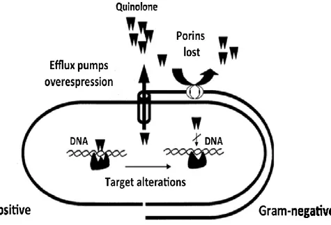
The first hypothesized mechanism of resistance involves mutations in QRDR regions (Quinolone stance made Determining Regions) of the DNA gyrase and topoisomerase IV subunits genes. In order to identify these mutations, QRDR of the parC and gyrA genes were amplified (17). The sequencing results revealed the presence of nucleotide mutations, which, however, did not result in changes of the amino acid sequence. These results are consistent with those obtained by Fukaoo et all. in 2003 (12).
The quinolone resistance mechanisms mediated by efflux pumps MDR (Multi Drug Resistance) was also investigated. The trend of the intracellular concentrations of ciprofloxacin in an interval between zero and four hours has been measured; ciprofloxacin concentrations were analyzed by exploiting the values of maximum absorption at 275 nm which give rise to an emission peak at 447 nm (18)(19)(20).
Further studies, conducted with phenotypic uncouples (CCC carbonyl-cianil-chlorophenyl hydrazone) and with MDR channel blockers (Verapamil and reserpine) have revealed a reduction of ciprofloxacin MIC values (2 fold reduction) (21).
The comparative genomic analysis performed on GenBank showed that in L.
fermentum ATCC 14931 there are two hypothetical proteins: one (GenBank
ref.ZP_03944345.1) belonging to the MFS (Major Facilitator Superfamily) family which has a homology of 98% with Nora (GenBank ref.CCE58495.1), the protein responsible for quinolones efflux in S. aureus; the other one (GenBank ref.ZP_03944509.1) belonging to the ABC (ATP Binding Cassette) family, which has a sequence homology of 90% with LmrA (GenBank ref.YP_005868060.1) responsible for quinolones efflux in L. lactis.
Future outlooks Studies of characterization of this protein in collaboration with Professor Patrizia Brigidi and Dr. Beatrice Vitali University of Bologna are currently ongoing.
Introduzione. I lattobacilli e, più in generale, i microrganismi commensali che nell‟uomo colonizzano il tratto gastro-intestinale e le vie genitali, possono potenzialmente servire da serbatoi di geni di resistenza (1). Il pericolo maggiore, associato ad una tale condizione, è quello che i prodotti in cui si fa uso di lattobacilli vitali, quali i probiotici ed i cibi fermentati, possano trasferire geni di resistenza a batteri commensali o potenzialmente patogeni con conseguente aumento del fenomeno della resistenza agli antibiotici.
Diversi studi hanno confermato la presenza, nei lattobacilli, di geni codificanti la resistenza antibiotica (2)(3)(4)(5)(6)(7)(8)(9)(10)(11). Alcuni di questi geni sono a localizzazione plasmidica e/o trasposonica per cui potrebbero essere trasferiti orizzontalmente fra lattobacilli e altre specie commensali del microbiota intestinale, incidendo notevolmente sul loro potenziale patogeno. Probiotici e alimenti fermentati sono veicolo di enormi quantità di batteri viventi i quali potrebbero rappresentare nell‟uomo dei serbatoi di geni di resistenza agli antibiotici. Tuttavia molti fenomeni di resistenza non sono dovuti alla presenza di elementi mobili ma all‟insorgenza di cloni mutati che danno a ceppi resistenti.
In particolare la resistenza ai chinoloni nei lattobacilli è stata descritta sin dal 2003 da Fukaoo et al i quali hanno dimostrato l‟assenza di modificazioni a carico dei geni gyrA e parC in Lactobacillus (12).
Obiettivi della ricerca e risultati. 244 ceppi in precedenza classificati come Lactobacillus spp., di origine vaginale e appartenenti alla batterioteca del Dipartimento di Scienze Bio-Mediche sez. Microbiologia dell'Università degli studi di Catania, sono stati caratterizzati a livello di specie mediante un approccio di tipo polifasico. Tale approccio prevede sia l'isolamento su terreni selettivi, sia l'uso di tecniche genotipiche:16S-RFLP (13), two steps multiplex PCR (14) e tuf gene PCR per la discriminazione di L. paracasei-L.rhamnosus. Sono stati determinati i profili di sensibilità per quattro fluoroquinoloni: ciprofloxacina, levofloxacina, ofloxacina e ulifloxacina (16).
In particolare, sono stati studiati i meccanismi di resistenza genotipica di quattro ceppi di L. fermentum che hanno mostrato ridotta sensibilità in vitro o
resistenza verso ciprofloxacina (assumendo come resistenti, i ceppi con MIC ≥ 4 µg/mL).
Il primo meccanismo di resistenza ipotizzato coinvolge le mutazioni presenti a livello delle regioni QRDR(Quinolone Resistance Determining Regions) dei geni delle subunità della DNA girasi e della topoisomerasi IV , per individuare tali mutazioni sono state amplificate le QRDR dei geni parC e gyrA, delle rispettive subunità della Topoisomerasi IV e della DNA girasi, bersagli farmacologici dei chinoloni. I risultati del sequenziamento hanno evidenziato la presenza di mutazioni nucleotidiche, che però non hanno determinato variazioni nella sequenza aminoacidica (17). Tale risultato è in linea con quanto descritto nel 2003. da Fukaoo et al. (12), i quali hanno dimostrato l‟assenza di modificazioni a carico dei geni gyrA e parC , quindi lo studio si è orientato alla ricerca di meccanismi di efflusso mediate da pompe MDR (Multi
Drug Resistance). A tal fine è stato misurato l‟andamento delle concentrazioni
intracellulari in un intervallo di tempo compreso tra zero e quattro ore; la variazione delle concentrazioni di ciprofloxacina è stata analizzata sfruttando i valori di assorbimento massimo a 275 nm da cui scaturisce un picco di emissione a 447 nm (18)(19)(20).
Lo studio fenotipico condotto sia con disaccoppianti (CCC carbonil-cianil-clorofenil idrazone), sia con bloccanti dei canali tipo MDR (Verapamil e reserpina), hanno rivelato una riduzione dei valori di MIC per ciprofloxacina (riduzione di due diluizioni) (21).
L'analisi genomica comparata condotta su GenBank ha mostrato che in L.
fermentum ATCC 14931 sono presenti due proteine ipotetiche: una (GenBank
ref.ZP_03944345.1) appartenente alla famiglia MFS (Major Facilitator Superfamily) che presenta un‟omologia del 98% con NorA (GenBank ref.CCE58495.1) proteina responsabile dell'efflusso dei chinoloni in S. aureus; un‟altra (GenBank ref.ZP_03944509.1), appartenente alla famiglia ABC, che presenta una omologia del 90% con LmrA (GenBank ref.YP_005868060.1) responsabile dell‟efflusso dei chinoloni in L. lactis.
Prospettive future Sono attualmente in corso gli studi di caratterizzazione di questa proteina in collaborazione con la Prof. Patrizia Brigidi e la Dott.ssa Beatrice Vitali dell‟Università di Bologna.
I
Summary
1 The genus Lactobacillus... 1
1.1 Cell morphology ... 2
1.2 Metabolism ... 4
Obliged homofermentative lactobacilli. ... 5
Facoltative heterofermentative lactobacilli. ... 5
Obliged heterofermentative lactobacilli. ... 6
1.3 Nutritional requirements and cultural characteristics ... 7
1.4 Habitat ... 10
1.4.1 Gastro-Intestinal (GI) Tract ... 10
1.4.2 Vaginal microbiota ... 12
1.5 The salutary effects of lactobacilli... 15
1.5.1 General mechanisms of the action of probiotic lactobacilli ... 16
1.6 Probiotics side effects ... 19
2 Taxonomy ... 25
2.1 Classification ... 27
2.2 Comparative genomic analysis ... 35
3 Molecular methods of identification ... 37
3.1 Macromolecules as “molecular clock" of microbial diversity ... 43
3.2 The Choice of the 16S rRNA as Sequencing Gene ... 44
3.3 Other phylogenetic markers ... 48
3.3.1 Elongation factor Tu and GTPs superfamily ... 49
II
4 Antibiotic resistance in lactic acid bacteria (non-enterococcal) ...
... 57
4.1 LAB, Lactic Acid Bacteria ... 57
4.2 Antibiotics resistance: acquisition and dissemination ... 59
4.3 Intestinal bacteria as reservoirs of antibiotic resistance ... 63
4.4 Lactobacilli susceptibility/resistance profiles ... 64
4.5 Antibiotic susceptibility/resistance profiles determination in LAB .. ... 67
5 Quinolones ... 69
5.1 Structure and classification ... 69
5.2 Ciprofloxacin ... 76
5.3 Levofloxacin ... 77
5.4 Fluoroquinolones mechanism of action ... 79
5.4.1 DNA gyrase and topoisomerase IV ... 79
Structure ... 79
Function ... 80
Ternary complex formation ... 81
6 Mechanisms of resistance ... 83
6.1 Fluoquinolones resistance ... 84
6.1.1 Target Alteration ... 85
DNA gyrase Alterations ... 86
Topoisomerase IV Alterations ... 90
6.1.2 Are there alterations of target in quinolone-resistant lactobacilli? ... ... 93
6.2 Decreased uptake ... 94
III
6.2.2 Efflux Pumps ... 96
ABC (ATP-binding cassette) ... 98
MFS (Major Facilitor Superfamily) ... 100
NorA ... 102
LmrA ... 103
MDR inhibitors ... 104
7 Materials and methods ... 106
7.1 Cultivation ... 106
7.2 Susceptibility testing ... 107
7.3 Molecular identification... 108
7.3.1 DNA Extraction ... 108
Spectrophotometric analysis ... 109
7.3.2 PCR/RFLP analysis of the 16s rDNA [13] ... 109
7.3.3 Two-steps multiplex PCRs:16S-ITS-23S and 23S rDNA flanking region (Song e coll., 2000) [14] ... 117
7.3.4 tuf gene amplification [15] ... 122
7.4 Mechanisms of resistance to ciprofloxacin in L. fermentum ... 125
7.4.1 QRDR amplification in gyr A and parC [17] ... 125
Sequencing protocol... 127
7.4.2 Fluoroquinolones accumulation essay [18] [19] [20] ... 128
Reading fluorescence spectrophotometer ... 129
Calibration curve ... 129
7.4.3 Inibitors influence on fluoroquinolone MICs [224] ... 130
8 Results ... 132
8.1 Molecular identification of Lactobacillus species ... 132
IV
8.3 Mechanisms of resistance to ciprofloxacin in L. fermentum ... 138
8.3.1 Sequence analysis of gyrA and parC ... 138
8.3.2 Ciprofloxacin intracellular accumulation ... 139
8.3.3 Inibitors influence on fluoroquinolones MICs ... 140
8.3.4 L. fermentum ATCC 14931 genome Analysis ... 141
9 Discussion ... 145
9.1 Strains identification... 145
9.2 Lactobacilli resistance profiles distribution ... 148
9.3 Possible mechanism of ciprofloxacin resistance ... 152
10 Future outlooks ... 157
1
1 The genus Lactobacillus
The genus Lactobacillus includes microorganisms Gram-positive, catalase negative, nonspore-forming, with a shape that can vary from long and thin (rod shaped) to short and curved (cocco-bacillary, coryneform); they are generally facultative anaerobic, or microaerophilic almost always motionless (22)
2
1.1 Cell morphology
The degree of curvature and the length of the rods depend on the age of the culture, the composition of the medium (availability of esters of oleic acid) and the oxygen pressure. Some species of gas-producing lactobacilli (L. fermentum, L. brevis) are represented as rods long and short together. The morphological differences between species are still evident and discriminating in the case of lactobacilli coconut-bacillary form, these may appear so short as to be incorrectly identified in the genus Leuconostoc (eg L. confusus, originally considered as a species of the genus Leuconostoc) or in the genus Streptococcus (eg L. xylosus and L. hordniae classified as lactobacilli and only recently reclassified as streptococci).
Lactobacilli tend to arrange themselves in chains: this characteristic is variable among species, sometimes from strain to strain of the same species. This variability depends on the growth phase and on the pH of the medium.
The asymmetric development of coryneform lactobacilli during cell division leads to the formation of corrugated chains or even rings. Forms wrapped in an irregular manner can be observed in the case of symbiotic growth (in kefir grains)or under the high
3 concentrations of glycine, amino acids or antibiotics active on the cell wall.
The motility in lactobacilli is very rare and, if present, it is due to the presence of peritrichous flagella, and it is dependent on cultivation parameters (medium components and age of the culture). It can be observed during isolation, and it is lost during subsequent transplantation in artificial medium.
Some strains show bipolar bodies with internal granulation that let them appear striped after Gram staining with methylene blue, especially the rods of homofermentative species. The large bipolar structures likely to contain polyphosphates and appear very electron dense to an electron microscope (22).
4
1.2 Metabolism
Lactobacilli are microorganisms witch are obligately saccharo-clastic and at least half of the final product of carbon is lactate; additional products may be acetate, ethanol carbon dioxide, formate and succinate. Volatile acids with more than two carbon atoms are not produced.
The reduction of nitrate is unusual and present only when the pH is increased up to 6.0. The gelatin is not liquefied and casein is not digested, but small amounts of soluble nitrogen are made from most of the strains. They are not indole or hydrogen sulfide producers. Overall lactobacilli are catalase negative due to lack of cytochromes (porphyrins are absent), but some strains can decompose the peroxide through a pseudo-catalase; they give a negative reaction with benzidine. The production of pigments is rare and if present it becames yellow orange-rust or red-brick.
Species belonging to the genus Lactobacillus are divided into three metabolic groups according to the presence of enzymes responsible for sugars omofermentation (fructose 1-6 diphosphate) or hetero-fermentation (aldolase and phosphoketolase):
5 Obliged homofermentative lactobacilli.
Belong to this group species that ferment hexoses carbohydrates almost exclusively producing lactic acid through the glycolytic pathway of Embden-Meyerhof-Parnas (EMP) and which are not able to ferment the pentose and the gluconate. From a morphological point of view they are generally like isolated long cells or arranged in very long or coiled chains. The species of the group live in different habitats and are phylogenetically related. The group also includes the most acidifying species (2.7% of lactic acid) and can be subdivided into two subgroups: the homofermentative psychrophilic that grow at a low temperature (~15°C) and thermophilic homofermentative that grow at high temperature (~45°C).
Facoltative heterofermentative lactobacilli.
The species of the group ferment hexoses through the route of EMP and produce almost exclusively lactic acid, in the presence of small quantities of glucose, lactate acetate, ethanol and formic acid can also be produced; they are able to ferment pentoses to lactate and acetate through an inducible phosphoketolase due to the presence of pentoses.
6 Facultative heterofermentative lactobacilli are usually mesophilic, with the exception of some species; the cells morphology is variable from court to stubby curve most often arranged in very long chains. They also have vegetables and fermented meats as habitats.
Obliged heterofermentative lactobacilli.
Species belonging to this group ferment hexoses to lactate, acetate (or ethanol) and carbon dioxide through the phosphogluconate pathway, the pentoses are also fermented to lactate and acetate, again through the action of the enzyme phosphoketolase. This heterofermentative lactobacilli are characterized by their ability to produce volatile aromatic substances and by their poor acidifier power (0.5% lactic acid). Cells are very short and generally isolated; they tend to develop in association with other species of lactic acid bacteria or other microorganisms both in fermented foods and in the digestive tract (22) (23)
7
1.3 Nutritional requirements and cultural
characteristics
The majority of lactobacilli species are very demanding from a nutritional point of view with specific characteristics for different species. Complex substrates are required for growth such as sources of carbon, nitrogen, phosphorus and sulphur compounds, also growth factors, vitamins, amino acids and trace elements. In order to supply their nutritional needs, culture media must contain fermentable carbohydrates, peptones, meat extract and yeast extract, in addition to substances and elements such as tomato juice, manganese, and acetate esters of oleic acid as growth promoters. Substances such as pantothenic acid and nicotinic acid are essential for all species while compounds such as thiamine are necessary for the growth of heterofermentative lactobacilli. Even riboflavin is a compound frequently required while the biotin and vitamin B12 are necessary to only some strains.
Lactobacilli grow well in slightly acid medium with an initial pH of 6.4-4.5, the growth stops when it reaches a pH of 4.0-3.6, this is a condition variable between species and between individual strains. Since they are acidophilous, the optimum pH is generally 5.5-6.2;
8 the speed of growth is often reduced when the initial pH is neutral or alkaline. Many strains are weakly aerotolerant so optimal growth occurs in anaerobic or microaerobic conditions; the increase of the concentration of carbon dioxide (5% approx.) can stimulate the growth itself. The growth temperature ranges from 2 to 53 °C and the optimum is generally between 30 °C-40 °C.
Colonies on agar are very small (2-5mm in diameter), with well-defined margins, convex, smooth, translucent or opaque, in rare cases, pigmented yellow or red. Some species form characteristically irregular colonies and thin distinct colonies are formed only by L. confusus.
When growth occurs on agar containing dispersed proteins or fat, there are no light areas generated by exoenzymes. However, many strains show a weak proteolytic activity, which is made by proteases and peptidases released from the cell wall and aweak lipolytic activity, carried out mostly by intracellular lipases.
Growth in liquid medium generally occurs in suspension and, once completed, the cells fall, with a smooth and even sediment, witch is rarely granular or gelatinous. Lactobacilli do not develop characteristic odours as they grow in common culture media. When they represent as the predominant microbiota, MRS agar (DeMan
9 Rogosa Sharpe) can be used; while, if they represent only a part of a complex microbial population, selective media such as, acetate Rogosa SL2, commonly called Rogosa, are needed. Although this is not completely selective, other lactic acid bacteria, for example,
Leuconostoc, Pediococcus, Enterococcus, and Bifidobacterium as
well as yeasts can grow in this culture medium. In addition, some lactic acid bacteria, mostly from unusual environments, do not grow on Rogosa SL. Depending on the source from which they were isolated, minor changes in Rogosa SL, such as the addition of specific growth factors [meat extract, tomato juice, yeast extract, malt extract, ethanol, or even some of the mevalonate natural substrates (beer, juices)] may facilitate the isolation of lactobacilli that have adapted themself to the growing conditions of their ecological niche (22).
10
1.4 Habitat
Lactobacilli colonize some specific parts of the human body: at these sites they play specific functions.
1.4.1 Gastro-Intestinal (GI) Tract
Lactobacilli are part of the normal microbiota of the mouth and intestines of healthy people and animals. Species composition and their amount depend on individuals, host age and GI area considered (24). It is difficult to distinguish from those indigenous (oral cavity) to allochthonous one (fermented foods)(25).
Lactobacilli form only a small part of the fecal microbiota of the adult (0.01% -0.6% of total bacteria) (26) (27). L. gasseri, L.
reuteri, L. crispatus, L. salivarius and L. ruminis are considered
more prevalent as indigenous, while L. acidophilus, L. fermentum,
L. casei, L. rhamnosus, L. johnsonii, L. plantarum, L. brevis, L. delbrueckii, L. curvatus and L. sakei are present in variable
amounts (25).
In comparison to the adult microbiota, the one present in children is highly unstable and contains lactobacilli in varying amounts. The number of lactobacilli in infants varies from 105 CFU/g of to 106
11 CFU/g feces, while in a newborn of one month old or more, this interval increases to 106-108 CFU/ of feces (28).
The presence of lactobacilli as indigenous microbiota is guaranteed by their ability to adhere to the mucous membranes of the districts concerned. In the case of L. plantarum is a mannose binding adesine to ensure colonization (29).
Lactobacilli appear in the mouth during the first year of neonatal life. Their presence depends on numerous factors including the existence of ecological niches, for example the natural cavities of the teeth. In many cases, lactobacilli may play a beneficial role by inhibiting the proliferation of cariogenic bacteria. Ahumada et al. (30) have shown that 36% of lactobacilli isolated from the tongue are able to prevent the growth of S. mutans. The homofermentative ones produce a greater number of inhibitory substances, compared to those belonging to the group of heterofermentative. Many authors have studied the role of lactobacilli as probiotics for oral health. In agreement with Busscher et al. (31), L. acidophylus and
L. casei, present in yogurt, can colonize the oral cavity due to their
12
1.4.2 Vaginal microbiota
Unlike the G.I. tract, the presence of lactobacilli is very pronounced in the female urogenital tract. The role of lactobacilli in these districts is potentially important because of the protective role against pathogenic microorganisms.
Aerobic and anaerobic microorganisms, generally constitute the microbiota of a healthy woman. Lactobacilli are present in the absolute majority in the vaginal fluid of healthy not menopauses women. Their metabolic products, such as hydrogen peroxide (H2O2), lactic acid, bacteriocins, play an important role in the maintenance of normal vaginal microbiota inhibiting the colonization of pathogenic bacteria. The predominant species detected by molecular biology studies are L. crispatus, L. jensenii and L. gasseri (33) (34). In recent studies, L. iners (L. 1086V) described by Anthony et al. (35) has been identified among species colonizing the human vagina (36) (34) (37)(38).
The high levels of estrogen present during the reproductive age cause the storage of large quantities of glycogen vaginal epithelium (39), which can be metabolized by vaginal microbiota in organic acids (40). Since the vaginal pH in women of reproductive age is
13 around 3.5/4.5, it is believed that these drastic conditions allow the proliferation of acidophilic species like lactobacilli. Lactic acid and other acids produced by lactobacilli inhibit the proliferation of pathogenic microorganisms such as E. coli, C. albicans, G.
vaginalis. Also, the hydrogen peroxide is toxic to fungi, viruses,
etc. (41).
Bacterial Vaginosis (BV) is a disorder of the vaginal microbiota, where the normal lactobacilli colonizers, are overwhelmed by an abnormal growth of different anaerobic bacteria (Gardnerella
vaginalis, Mycoplasma hominis, Mobiluncus spp.,
Peptostreptococcus, Prevotella, Bacteroides, etc.) (42). This
condition is common in women of reproductive age (43)(44) and can cause foul-smelling vaginal secretions, although many women are asymptomatic (45). In pregnant women this may represent a risk factor for the occurrence of perinatal complications, including preterm labor and chorioamnionitis (43)(44)(45) (46)(47).
BV may be associated with different behaviours of women, including sexual ones (relations with multiple partners, use of intrauterine devices for contraception, etc.) (31). The incidence of BV varies markedly between different ethnic groups: about 6% in Asian women, 9% in the white, 16% in spanish and 23% in african
14 americans. The rationalization of this phenomenon lies in the socio-demographic characteristics, sexual behaviour, and personal hygiene (48).
15
1.5 The salutary effects of lactobacilli
It has been demonstrated that the lactobacilli carry healthy effects when applied under various conditions. The best results have been obtained in the treatment and prevention of enteric infections and in post-antibiotic syndromes. Some lactobacilli are able to reduce the diarrhea associated with Clostridium difficile (49) and preventing necrotizing enterocolitis in preterm infants (50).
The GI tract is certainly the district where it is believed that lactobacilli exert major effects on the health of host; nevertheless probiotic applications of some species, in other districts, seem to be promising, for example, in the prevention and treatment of urogenital infections and bacterial vaginosis (37), in the prevention of atopic disease, in food hypersusceptibility (51) and in the prevention of dental caries (52). The probiotic lactobacilli must possess the status of GRAS (Generally Regarded as Safe) and so must be well tolerated. There have been rare cases of infection, presumably caused by probiotics in immunocompromised individuals or in patients with severe disease (53) (54).
16
1.5.1 General mechanisms of the action of
probiotic lactobacilli
Abilities of lactobacilli to carry salutary effects for the host, can be recognized in one or more of the following mechanisms (Figure 2) (55)(56):
i. Bacterial pathogens inhibition and homeostasis restoration through microbe-microbe interaction;
ii Enhancement of epithelial barrier function; iii Modulation of immune responses.
The ability of lactobacilli to inhibit pathogenic microorganisms is well-known, given that they have been used for centuries for the preservation of foods. Subsequently the immunostimulating and immunomodulating capacity of these microorganisms were investigated by molecular studies.
Different strains of probiotic lactobacilli, have been associated with different effects in relation to specific properties such as the ability to express particular surface molecules, to secrete specific proteins and metabolites that can interact directly with host cells (24).
Molecular characterization of probiotic strains is extremely important and has two main objectives:
17 a. Define the best conditions that determine the "performance"
of the best probiotic strains;
b. Select well-defined molecular criteria for new probiotic strains.
Figure 1-2 Probiotic lactobacilli mechanistic view. Molecular studies on probiotics lactobacilli in order to identify factors that promote survival, as a result of adaptation, and host colonization factors (adaptation) and factors that directly promote the health benefits (on probiotic factors )(24)
There are two main categories of factors that contribute to optimize the activity of probiotic lactobacilli: factors that promote optimum adaptation to ecological niches that the bacteria met temporarily in the host (adaptation factors) and factors that directly contribute to
18 the promotion of the beneficial effects (probiotic factors) (figure 1-2). Probiotic Factors include three main mechanisms: the maintenance of microbial balance, epithelial protection and immunomodulation. Adaptation Factors include resistance to stress, adaptation to host metabolism and adherence to intestinal mucosa (24).
Specific metabolic and physiological features of lactic acid bacteria play a key role to adaptation in host environment. In Gram-positive bacteria, the cell wall is made up of unique structures: a thin, multi-layer peptidoglycan (PG), surrounded with protein, teichoic acid and polysaccharides, in some species (L. acidophilus, L. gasseri, L
johnsonii, L. brevis L. crispatus L. helveticus) there is a shell
protein packed in a cristalline layer (S layer) (57).
Wall macromolecules determine the specific properties of the strain including the ability to adapt to environmental changes and interaction with host epithelial cells and receptors of immune responses (58).
19
1.6 Probiotics side effects
Probiotics are not selected among pathogens, and the theoretical risk of infection is thus very low. The risk of their passage in blood, eventually by translocation, is, however, important to determine. Bacterial translocation is defined as the passage of micro-organisms from the gastrointestinal to extra-intestinal sites such as the mesenteric lymph nodes, liver, spleen and bloodstream. Indigenous bacteria are continuously translocating in low numbers but are rapidly killed in the lymphoid organs. Bacterial translocation is a major cause of severe infection in immunosuppressed, trauma and post-surgical patients. This may result from three mechanisms: intestinal bacterial overgrowth, increased permeability or damage of the intestinal mucosal barrier, and immunodeficiency (59).
Rare cases of infection, including septicaemia and endocarditis caused by lactobacilli, bifidobacteria or other lactic acid bacteria, have been reported (60). Enterococcus faecium and E. faecalis are
more frequently involved in clinical infection, and there is concern over the emergence of vancomycin-resistant strains. In most cases of infection, the organism appeared to have come from the patient‟s own microbiota, but, in a few cases, the recent consumption of
20 probiotics was proposed as a potential cause. About 30 cases of fungaemia have been reported in patients treated with
Saccharomyces boulardii(61)(62), and two cases of infection have
been traced back to food-borne L.rhamnosus (63)(64). Nearly all subjects who had fungaemia involving Saccharomyces boulardii had an indwelling vascular catheter (61)(62).
Infection caused by L. rhamnosus similar to the probiotic GG strain was observed ia 74-year-old woman with non-insulin-dependent diabetes, who suffered from a liver abscess, which proved to contain Lb. rhamnosus, and pleuropulmonary infection(63). No cause for this infection was found, but the woman reported a regular consumption of dairy drinks containing L. rhamnosus GG. The clinical strain appeared to be indistinguishable from the GG strain. The other case of infection occured in a 67-year-old man with mild mitral valve regurgitation who habitually chewed a probiotic mixture and had carious teeth to be removed and suffered after a few days from an endocarditis. Lactobacillus rhamnosus was isolated from his blood, further analysis showing that it was indistinguishable from one of the organisms present in the probiotic preparation. (64) Saxelin and colleagues (65)(66) studied the prevalence of bacteriaemia caused by Lactobacillus species in
21 Southern Finland and compared the characteristics of the blood culture isolates and the probiotic dairy strains. In their first study, lactobacilli were identified in eight of 3317 blood culture isolates, none of the isolates corresponding to a dairy strain. In the second study, 5912 blood cultures were analysed, none of the 12 lactobacilli isolated being identical to any of the commercial Lactobacillus strains. To summarise, there is no evidence that ingested probiotic lactobacilli or bifidobacteria pose any greater risk of infection than do commensal strains, but there is insufficient knowledge on the risks or benefits of probiotics in immunodeficiency. Other risk factors for opportunist infection, such as extremes of age, pregnancy and digestive lesions, have not been identified as risk factors for probiotic infections.
According to EFSA (European Food Saefety Authority) since the 2010 only few reports have been published concerning lactobacilli and clinical infections (67)
One article from Turkey (68) detected a „Lactobacillus acidophilus or Lactobacillus jensenii‟ strain in clinical specimen amongst other species in an immunocompromised patient. The clinical relevance of this isolate was not clear. In addition, the taxonomic identification was done with the API system and therefore no clear
22 attribution to a taxonomic unit can be done. Several reports related to the well known association of lactobacilli with dental caries (69) (70)(71). Kneist et al. (2010) (69) found five species from carious dentine: Lactobacillus paracasei subsp. paracasei, Lactobacillus
paracasei subsp. tolerans, Lactobacillus rhamnosus, Lactobacillus gasseri, and Lactobacillus alimentarius.
They concluded that Lactobacillus rhamnosus and Lactobacillus
paracasei subsp. paracasei occurred in all caries progression
stages, whilst the other species were found only sporadically. There is a connection to endocarditis via caries which has been reported on several occasions earlier. There is also a possibility of lactic acid bacteria (LAB) being involved in endocarditis after dental surgery(72).
Caries is a multifactorial disease, including bacteria from the oral cavity, eating and drinking habits (high sugar amounts) and insufficient oral and dental hygiene (73). Bacteria involved change through different stages of caries proliferation. In the primary phase mainly mutans streptococci are involved, whereas in secondary caries with lesions already present also lactobacilli, bifidobacteria and other LAB are involved(74) (73). The conclusions of these studies are that without bacteria caries development is not possible.
23 LAB, however, are present in caries stages with predisposing factors such as lesions and insufficient dental hygiene. The origin of those LAB (if deriving from food or as autochthonous oral microbiota) has not been studied so far. In conclusion, LAB are not the initial cause of these diseases and they are present in the human organism as commensal microbiota.
Doi et al. (2010) (75) found Lactobacillus paracasei involved in a splenic abscess. But again as in previous similar case reports, the patient had an underlying disease and was immunocompromised. In a similar case with an immunocompromised patient (stem cell transplantation in a child) L. Rhamnosus was found to be the causative agent for meningitis after recurrent episodes of bacteremia (76)) Russo et al. (2010) (77) isolated a presumptive
Lactobacillus casei strain from a bacteraemia case, where heavy
consumption of dairy products was involved in the case history. However, no strains from dairy products (mainly cheese) were isolated and compared to the clinical strain. The identification was most probably misinterpreted, as only a 16S rDNA sequence analysis was done, which matched equally to Lactobacillus casei and Lactobacillus paracasei. Therefore Lactobacillus paracasei seems to be the correct identification given the distribution of
24 species in humans and dairy products. Lactobacillus casei does not naturally occur in such environments. Lactobacillus rhamnosus can be associated with unexplained sustained bacteremia like in the TIPSS syndrome (tipsitis). This is a rare disease where
Lactobacillus rhamnosus may be involved inter alia(78).
A research including ´urinary tract infection´ (UTI) revealed one review(79) indicating a relatively higher risk for elderly women for urinary tract infections with L. delbrueckii.
25
2 Taxonomy
The taxonomy can be defined as a scientific study that is able to "classify" the biodiversity of microorganisms, or more generally of organisms (80). Advances in bacterial taxonomy have always been dependent on the technological ones: the modern taxonomy is built primarily on molecular data. The complete study on the identification of a microorganism is closely related to the methods of analysis used. For this reason, the correct taxonomic status can only be taken out from the comparative study of a wide range of techniques, molecular and otherwise: the polyphasic approach. The phylogenetic approach has revolutionized systematic studies on bacteria. Phylogenetic relationships between microorganisms can be effectively estimated through the comparison of molecular sequences such as those of the genes coding for 16S rRNA. This target is chosen for several reasons: the 16S rRNA genes are highly conserved, as ribosomes play a key role in protein biosynthesis since the earliest stages of the development of micro-organisms; although phenomena of horizontal gene transfer between microorganisms have never occurred in these genes, they represent ancestral molecules and there are enough variables to differentiate
26 microorganisms, representing their variance genomics. These statements are not always true, or at least some experimental evidence has cast some doubt on this. In general the molecular analysis of the 16S rRNA has enabled us to draw an evolutionary scenario of bacteria although other molecular targets such as RecA and elongation factor Tu can be used in parallel taxonomy studies and bacterial phylogeny (81).
27
2.1 Classification
According to outline Taxonomy of Prokaryotes, the genus
Lactobacillus belongs to the Firmicutes phylum, Bacilli class, Lactobacillales order, Lactobacillaceae family. NCBI taxonomy
database today recognizes today 148 species belonging to the genus
Lactobacillus that represents, therefore, the most numerous of the Lactobacillales order (82).
Recently, a new species has been described, Lactobacillus tucceti (83) and the last entry in the classification are the species L.
compounds, L. farraginis, and L. parafarraginis (84)(85) and L. secaliphilus (86), in addition to the species described by Dellaglio
and collaborators (81).
Only seven species of the genus Lactobacillus described by Dellaglio et al. comprise two or more subspecies: Lactobacillus
aviarius (L. aviarius subsp. aviarius and L. aviarius subsp. araffinosus), Lactobacillus coryniformis (L. coryniformis subsp.coryniformis and L. coryniformis subsp. torquens), Lactobacillus delbrueckii (L. delbrueckii subsp. delbrueckii, L. delbrueckii subsp. bulgaricus, L. delbrueckii subsp. indicus and L.
28
(kefiranofaciens L. subsp. kefiranofaciens kefiranofaciens and L. subsp. kefirgranum), Lactobacillus paracasei (L. paracasei subsp . paracasei and L. paracasei subsp. Tolerans), Lactobacillus plantarum (L. plantarum subsp. plantarum and L. plantarum subsp. argentoratensis), and Lactobacillus sakei (L. sakei subsp. sakei and L. sakei subsp. carnosus).
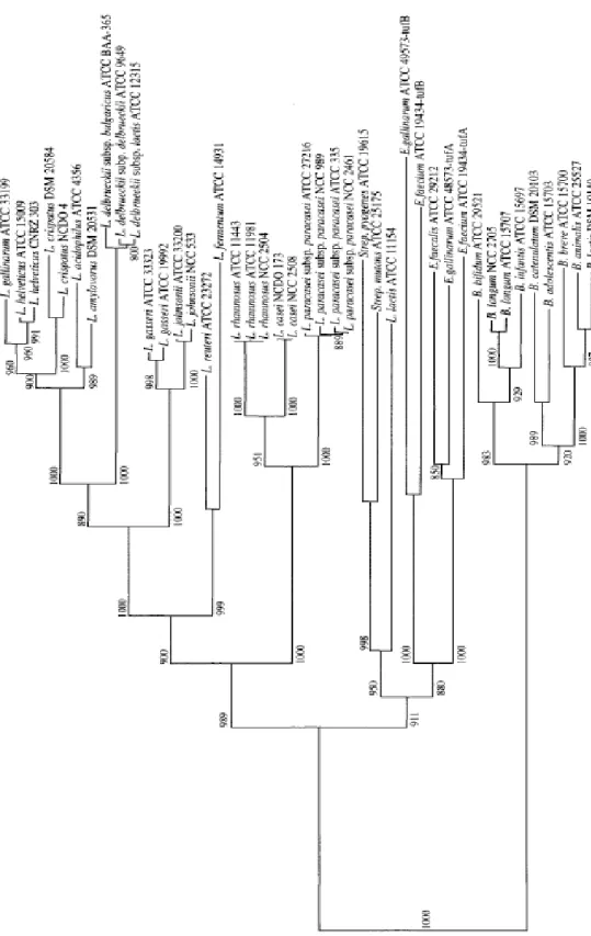
The phylogenetic structure of the Lactobacillaceae family considers
Lactococcus lactis and Streptococcus thermophilus as group limits.
The first phylogenetic analysis of lactobacilli was carried out by Collins et al. in 1991, on a small number of species known at that time: they proposed to divide the genus Lactobacillus into 3 groups:
L. delbrueckii group, L. casei-Pediococcus group and Leuconostoc
group, which contained some lactobacillus.
In 1995, Schleifer and Ludwig confirmed Collins evidences and they changed L. delbrueckii group with group name L. acidophilus. In addition, these authors noted that the group L. casei-Pediococcus could be divided into four sub clusters. In the first group the percentage of homology of 16S rDNA varies from 90.8% to 99.3%. It includes L. delbrueckii (G+ C = 50%) with the three subspecies (L. delbrueckii subsp. lactis, which includes the two old species L.
29
delbrueckii subsp. bulgaricus) and species in the group identified
by Collins, such as L. acidophilus (G+ C = 34-37%), L. amylovorus (G+ C = 40-41%), L. crispatus (G+ C = 33-35%), L. gallinarum (G+ C = 36-37%), L. gasseri (G+ C = 33-35%) and L. johnsonii (G+ C = 35-38%).
The group Lactobacillus casei-Pediococcus is the largest and heterogeneous, where the percentage of homology of 16S rDNA varies from 90.3% to 99%. It includes 37 species of Lactobacillus and 5 species of Pediococcus. Finally, Leuconostoc group: includes species assigned to the new genus Weissella, Oenococcus oeni and heterofermentative lactobacilli.
The recent description of a large number of species and the consequent re-examination of the phylogenesis splits these groups into smaller and more flexible groups. This strategy of "grouping" has been adopted by Hammes and Hertel (2003) and by Dellaglio Felis in 2005 (81) (Table 2-1).
30
Table 2-1 Phylogenetic grouping
Group Hammes and Hertel (2003) Dellaglio and Felis (2005) Felis and Dellaglio
L. delbrueckii group (delb) L. acetotolerans, L. acidophilus, L. amylolyticus, L. amylophilus, L. amylovorus, L. crispatus, L. delbrueckii, L. fornicalis, L. gallinarum, L. gasseri, L. hamsteri, L. helveticus, L. iners, L. intestinalis, L. jensenii, L. johnsonii, L. kefiranofaciens, L. kefirgranum, L. psittaci L. acetotolerans, L. acidophilus, L. amylolyticus, L. amylophilus, L. amylovorus, L. crispatus, L. delbrueckii, L. fornicalis, L.gallinarum, L. gasseri, L. hamsteri, L. helveticus, L. iners, L. intestinalis, L. jensenii, L. johnsonii, L. kalixensis, L. kefiranofaciens, L. kefirgranum L. kitasatonis, L. psittaci, L. suntoryeus, L. ultunensis L. acetotolerans, L. acidophilus, L. amylolyticus, L. amylophilus, L. amylotrophicus, L. amylovorus, L. crispatus, L. delbrueckii, L. fornicalis, L. gallinarum, L. gasseri, L. hamsteri, L. helveticus, L. iners, L. intestinalis, L. jensenii, L. johnsonii, L. kalixensis, L. kefiranofaciens, L. kitasatonis, L. psittaci, L. sobrius, L. ultunensis L. salivarius group L. acidipiscis, L. agilis, L. algidus, L. animalis, L. aviarius, L. cypricasei, L.
equi, L. mali, L. murinus, L. nagelii, L. ruminis, L. salivarius L. acidipiscis, L. agilis, L. algidus, L. animalis, L. aviarius, L. cypricasei, L. equi, L.
mali, L. murinus, L. nagelii, L. ruminis, L. saerimneri, L. salivarius, L. satsumensis L. acidipiscis, L. agilis, L. algidus*, L. animalis, L. apodemi, L. aviarius, L. equi, L. mali, L. murinus, L. nageli, L. ruminis, L. saerimneri, L. salivarius, L. satsumensis, L. vini L. reuteri group (reu) L. coleohominis, L. durianis, L. fermentum, L. frumenti, L. ingluviei, L. mucosae, L. oris, L. panis, L. pontis, L. reuteri, L. suebicus, L. thermotolerans, L. vaccinostercus, L. vaginalis L. antri, L. coleohominis, L. fermentum, L. frumenti, L. gastricus, L. ingluviei, L.
mucosae, L. oris, L. panis, L. pontis, L. reuteri, L. thermotolerans, L. vaginalis (L. reuteri group-a) associated with L. durianis, L. vaccinostercus, L. suebicus, L. rossii (L. reuteri group-b)
L. antri, L. coleohominis, L. fermentum,
L. frumenti, L. gastricus, L. ingluviei, L.
mucosae, L. oris, L. panis, L. pontis, L. reuteri, L. secaliphilus, L. vaginalis L. buchneri group (buch) L. buchneri, L. diolivorans, L. ferintoshensis, L. fructivorans, L. hilgardii, L. homohiochii, L. kefiri, L. kunkeei, L. lindneri, L. parabuchneri, L. parakefiri, L. sanfranciscensis L. buchneri, L. diolivorans, L. ferintoshensis, L. hilgardii, L. kefiri, L. parabuchneri, L. parakefiri (L. buchneri group-a) associated with L. fructivorans, L. homohiochii, L. lindneri, L. sanfranciscensis (L. buchneri group-b) L. buchneri, L. diolivorans, L. farraginis, L. hilgardii, L. kefiri, L. parabuchneri, L. parafarraginis, L. parakefiri associated with L. acidifarinae, L. namurensis, L. spicheri, and L. zymae (which form a robust group) L. alimentarius-L. farciminis group (al-far) / / L. alimentarius, L. farciminis, L. kimchii, L. mindensis, L. nantensis, L. paralimentarius, L. tucceti, L. versmoldensis L. casei group (cas) L. casei, L. manihotivorans, L. pantheris, L. paracasei, L. rhamnosus, L. sharpeae, L. zeae L. casei, L. paracasei, L. rhamnosus, L.
zeae (L. casei group-a) L. manihotivorans, L. pantheris, L. sharpeae (L. casei group-b)
appear as distinct clusters, not robustly
associated with each other
L. casei, L. paracasei, L. rhamnosus, L. Zeae L. sakei group (sakei) L. curvatus, L. fuchuensis, L. graminis, L. sakei L. curvatus, L. fuchuensis, L. graminis, L. sakei L. curvatus, L. fuchuensis, L. graminis, L. Sakei
31 L. fructivorans group (fru) / / L. fructivorans, L. homohiochii, L. lindneri, L. sanfranciscensis L. coryniformis group (cor) / / L. bifermentans, L. coryniformis, L. rennini, not robustly associated with L. composti L. plantarum group (plan) L. alimentarius, L. arizonensis, L. collinoides, L. farciminis, L. kimchii, L. malefermentans, L. mindensis, L. paralimentarius, L. paraplantarum, L. pentosus, L. plantarum, L. versmoldensis L. arizonensis, L. collinoides, L. paraplantarum, L. pentosus, L. plantarum (L. plantarum group-a) associated with L. alimentarius, L. farciminis, L. kimchii, L. mindensis, L. paralimentarius, L. versmoldensis (L. plantarum group-b) the affiliation of L. collinoides was poorly supported L. plantarum, L. paraplantarum, L. pentosus L. perolens group (per) / / L. perolens, L. harbinensis, L. paracollinoides L. brevis group (bre) / L. acidifarinae, L. brevis, L. hammesii, L. spicheri, L. zymae L. brevis, L. hammesii, L. parabrevis Pediococcus dextrinicus group (Pdex) P. dextrinicus, L. concavus, L. oligofermentans (the latter sometimes poorly supported)
Pediococcus
Not reported 1 single cluster (not including P. dextrinicus)
2 clusters, not associated: the first
comprises P. cellicola, P. damnosus P.
parvulus, P. inopinatus, while the second
includes P. acidilactici, P. claussenii, P.
pentosaceus and P. stilesii Couples (couple) L. rossiae-L. siliginis (1) L. vaccinostercus-L. suebicus (2) L. manihotivorans-L. collinoides (3) Single species (ss) L. bifermentans, L. brevis, L. coryniformis and L. perolens
L. algidus, L. kunkeei, L. malefermentans, L. paracollinoides, L. perolens, Paralactobacillus selangorensis L. kunkeei, L. malefermentans, L. pantheris, L. sharpeae, Paralactobacillus selangorensis Table 2-1 Continue
The greatest discrepancy in the taxonomy of the genus
Lactobacillus is due to the lack of ability to correlate between the
32 historical division of the genus Lactobacillus based on the type fermentation was excellently revisited by Pot et al. (1994) (87), who pointed out that the terms "homofermentative", "heterofermentative", "obliged homofermentative, "facultative heterofermentative " and " bliged heterofermentative" can have different meanings according to the authors, creating misunderstandings. The most recently accepted definition is given by Hammes and Vogel (1995) as described in the paragraph on the metabolism of lactic acid bacteria (Table 2-2).
33
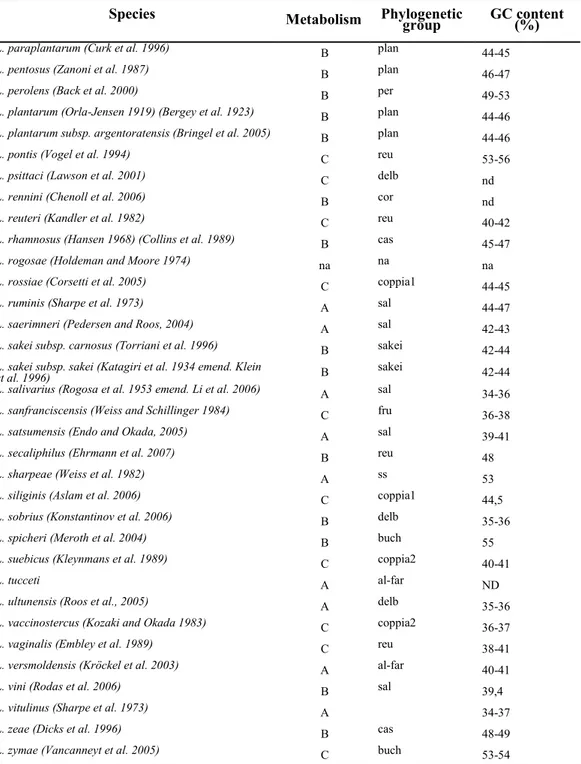
Table 2-2 Taxonomic characteristics of the species belonging to the genus Lactobacillus Species Metabolism Phylogenetic group GC content (%)
L. fuchuensis (Sakala et al. 2002) B sakei 41–42
L. gallinarum (Fujisawa et al. 1992) A delb 36-37
L. gasseri (Lauer and Kandler 1980) A delb 33-35
L. gastricus (Roos et al., 2005) C reu 41-42
L. graminis (Beck et al. 1989) B sakei 41-43
L. hammesii (Valcheva et al. 2005) B bre nd
L. hamsteri (Mitsuoka and Fujisawa 1988) B delb 33-35
L. harbinensis (Miyamoto et al. 2006) B per 53-54
L. helveticus (Orla-Jensen 1919) (Bergey et al. 1925) A delb 38-40
L. hilgardii (Douglas and Cruess 1936) C buch 39-41
L. homohiochii (Kitahara et al. 1957) B fru 35-38
L. iners (Falsen et al. 1999) A delb 34-35
L. ingluviei (Baele et al. 2003) C reu 49-50
L. intestinalis (ex Hemme 1974) (Fujisawa et al. 1990) B delb 33-35
L. jensenii (Gasser et al. 1970) B delb 35-37
L. johnsonii (Fujisawa et al. 1992) A delb 33-35
L. kalixensis (Roos et al., 2005) A delb 35-36
L. kefiranofaciens subsp. kefiranofaciens (Fujisawa et al. 1988) A delb 34-38
L. kefiranofaciens subsp. kefirgranum (Takizawa et al. 1994)
(Vancanneyt et al. 2004) A delb 34-38 L. kefiri (Kandler and Kunath 1983) C buch 41-42
L. kimchii (Yoon et al. 2000) B al-far 35
L. kitasatonis (Mukai et al. 2003) B delb 37-40
L. kunkeei (Edwards et al. 1998) C ss nd
L. lindneri (Back et al. 1997) C fru 35
L. malefermentans (Farrow et al. 1989) C ss 41-42
L. mali (Carr and Davies 1970, emend. Kaneuchi et al., 1998) A sal 32-34
L. manihotivorans (Morlon-Guyot et al. 1998 ) A coppia3 48-49
L. mindensis (Ehrmann et al. 2003) A al-far 37-38
L. mucosae (Roos et al. 2000) C reu 46-47
L. murinus (Hemme et al. 1982) B sal 43-44
L. nagelii (Edwards et al. 2000) A sal nd
L. namurensis (Scheirlinck et al. 2007) C buch 52
L. nantensis (Valcheva et al. 2006) B al-far 38,6
L. oligofermentans (Koort et al. 2005) C Pdex 35,3-39,9
L. oris (Farrow and Collins 1988) C reu 49-51
L. panis (Wiese et al. 1996) C reu 49-51
L. pantheris (Liu and Dong 2002) A ss 52-53
L. parabrevis (Vancanneyt et al. 2006) C bre 49
L. parabuchneri (Farrow et al. 1989) C buch 44
L. paracasei subsp. paracasei (Collins et al. 1989) B cas 45-47
L. paracasei subsp. tolerans (Collins et al. 1989) B cas 45-47
L. paracollinoides (Suzuki et al. 2004) C per 44-45
L. parafarraginis (Endo and Okada 2007) B buch 40
L. parakefiri (Takizawa et al. 1994) C buch 41-42
L. paralimentarius (Cai et al. 1999) B al-far 37-38
A: Obliged homofermentative; B: Facoltative heterofermentative; C: Facoltative heterofermentative; nd: not determinated; na: not classificated
34
Species Metabolism Phylogenetic
group GC content (%)
L. paraplantarum (Curk et al. 1996) B plan 44-45
L. pentosus (Zanoni et al. 1987) B plan 46-47
L. perolens (Back et al. 2000) B per 49-53
L. plantarum (Orla-Jensen 1919) (Bergey et al. 1923) B plan 44-46
L. plantarum subsp. argentoratensis (Bringel et al. 2005) B plan 44-46
L. pontis (Vogel et al. 1994) C reu 53-56
L. psittaci (Lawson et al. 2001) C delb nd
L. rennini (Chenoll et al. 2006) B cor nd
L. reuteri (Kandler et al. 1982) C reu 40-42
L. rhamnosus (Hansen 1968) (Collins et al. 1989) B cas 45-47
L. rogosae (Holdeman and Moore 1974) na na na
L. rossiae (Corsetti et al. 2005) C coppia1 44-45
L. ruminis (Sharpe et al. 1973) A sal 44-47
L. saerimneri (Pedersen and Roos, 2004) A sal 42-43
L. sakei subsp. carnosus (Torriani et al. 1996) B sakei 42-44
L. sakei subsp. sakei (Katagiri et al. 1934 emend. Klein
et al. 1996) B sakei 42-44 L. salivarius (Rogosa et al. 1953 emend. Li et al. 2006) A sal 34-36
L. sanfranciscensis (Weiss and Schillinger 1984) C fru 36-38
L. satsumensis (Endo and Okada, 2005) A sal 39-41
L. secaliphilus (Ehrmann et al. 2007) B reu 48
L. sharpeae (Weiss et al. 1982) A ss 53
L. siliginis (Aslam et al. 2006) C coppia1 44,5
L. sobrius (Konstantinov et al. 2006) B delb 35-36
L. spicheri (Meroth et al. 2004) B buch 55
L. suebicus (Kleynmans et al. 1989) C coppia2 40-41
L. tucceti A al-far ND
L. ultunensis (Roos et al., 2005) A delb 35-36
L. vaccinostercus (Kozaki and Okada 1983) C coppia2 36-37
L. vaginalis (Embley et al. 1989) C reu 38-41
L. versmoldensis (Kröckel et al. 2003) A al-far 40-41
L. vini (Rodas et al. 2006) B sal 39,4
L. vitulinus (Sharpe et al. 1973) A 34-37 L. zeae (Dicks et al. 1996) B cas 48-49
L. zymae (Vancanneyt et al. 2005) C buch 53-54
Table 2-2 Continue
The phylogenetic structure of lactobacilli is extremely complicated and from the data recorded in the literature it is evident that the taxonomy of the genus is still ambiguous for certain species and for this reason subjected to periodic reinterpretations.
35
2.2 Comparative genomic analysis
Phylogenetic distance between species is now highlighted by different methods of comparative analysis, which can help to explore the characteristics of Lactobacillus species whose genome has been sequenced.
L. plantarum has a relatively large number of proteins involved in
the sugars and lipids metabolism and amino acids transport, these became relevant when compared to L.johnsonii, which, however, can only use an extracellular proteinase to degrade peptides. This could explain and justify their different ecological niches;
L.johnsonii, in fact is only found in the GI tract rich in proteins. The
most "flexible" L. plantarum is able to colonize other environments such as plants, which require the ability to ferment a wider range of sugars.
There is only slight evidence of the correlation between the two genomes. Dot-plot comparisons and genomic alignments show little homology, have been found only 28 genes‟ clusters in common, with 6 genes in the same order. In addition, these clusters are also present in Bacillus subtilis, Enterococcus faecalis Listeria
36 correlation between L. plantarum and L. johnsonii is only minimally higher than that exhibited with other Gram-positive bacteria (88)
Although L. gasseri and L. acidophilus genomes group were compared. Many similarities have been identified between the members of the L. acidophilus group , which includes L.
acidophilus, L. gasseri and L. plantarum. The genes‟ disposition is
highly conserved and especially in the genomes of the two latter species mentioned and with the exception of a chromosomal reverse in the replication terminal site. Synteny studies have also revealed a gene region probably related with production of exopolysaccharides cell surface. This cluster is conserved among the three species and may have important properties in dairy products fermented by these microorganisms (89).
37
3 Molecular methods of identification
Traditionally, lactobacilli, and in general all the species belonging to the LAB (Lactic Acid Bacteria), have been identified on the basis of phenotypic characteristics, such as cell morphology, type of sugar fermentation, different growing temperatures, protein patterns of cell wall or the entire cell (90). Unfortunately, these phenotypic/biochemical typings are not completely accurate. Their limits are: lack of reproducibility, ambiguity of some techniques (often resulting from flexibility of bacterial growth), lack of reproducibility on a large scale and lack of discriminatory power. Another disadvantage of phenotypic analysis is represented by the fact that information carried by the entire genome is not always expressed, for example the expression of a gene may be related to environmental conditions (90).
Genotypic techniques are recognized as very important for identification purposes. Benefits arising from their use are high discriminatory power and their universal applicability. Strains strongly correlated with similar phenotypic characteristics, can be effectively identified by molecular techniques, mainly based on the PCR, such as RFLP (Restriction Fragment Length Polymorphism),
38
ARDRA (Amplified Ribosomal Restriction Fragment Analysis), AFLP (Amplified Fragment Length Polymorphism), RAPD (Randomly Amplified Polymorphic DNA), PFGE (Pulse-Field Gel
Electrophoresis), ribotyping, DGGE (Denaturing Gradient Gel
Electrophoresis) and TGGE (Terminal Gradient Gel
Electrophoresis), etc..
RFLP was the first method of molecular typing used. Profiles in bands that result from enzymatic cuts. Subsequent separation into DNA fragments, obtained by electrophoretic run, is known as DNA fingerprinting. Due to the high specificity of endonuclease and the stability of the chromosomal DNA, the profiles obtained after complete digestion of the DNA, are reproducible. A general criticism of this method is the complexity of the profile of bands. However, many scholars believe that the proper choice of enzymes and the use of specific conditions make the RFLP technique relatively fast and easily accomplished (90).
ARDRA (90)(91) is essentially a restriction polymorphisms analysis of amplified gene rRNA encoding. The effectiveness of the method in distinguishing between species or between the subspecies depends on the choice of restriction enzymes and the length of the amplicon. Since some bacterial species have high
39 homology of the sequence of rDNA genes, it is difficult to select endonucleases capable of producing distinct patterns of restriction for species closely related. The method generates highly reproducible restriction patterns reproducible in different laboratories.
AFLP (92) combines the power of RFLP technique to the flexibility of PCR-based methods. The genomic DNA is digested with two restriction enzymes, one with a low cut-off frequency, the second with high-frequency cutting. Nucleotide adapters are ligated to the double stranded DNA fragments using the binding sites of primers for PCR amplification. The use of complementary primers to adapters and sequence of the cleavage sites allow amplification patterns strain-specific to be obtained (93). AFLP technique is mainly used in clinical trials also it had effective application for typing strains belonging to the L. acidophilus and L. johnsonii (94)(95).
In RAPD, also known as AP-PCR (arbitrarily Primed PCR), random sequences oligonucleotides with a length of 10bp are used in PCR at low stringency. The profile that emerges is of an array of gene amplicons "anonymous." Generally this method is used to discriminate the species and sometimes different strains of the same
40 species. It has been widely used for typing lactobacilli and bifidobacteria from various habitats (96)(97). For example, Tynkkynen et al. (98), have distinct 12 genotypes between 24 belonging to the L. casei group, many of which are of human origin.
The method is simple to perform and rapid, but the reproducibility of the results is poor, therefore optimization and standardization of the technique is required. The use of different thermal cyclers, different DNA polymerases in different concentrations, methods of preparation and DNA extraction, concentrations of the primers and magnesium chloride, can cause variations in the RAPD patterns and, they are not comparable if carried out in different laboratories. PFGE technique allows the separation of large DNA fragments, in a continuous redirecting electrical field. Entire genome is digested with endonucleases (which rarely cut) and resulting macro fragments are separated by PFGE.
PFGE protocols have been designed for both lactobacilli and for bifidobacteria, and it shows a greater discriminating power if compared to other molecular typing methods, such as ribotyping. (90) (98). Due to the intense work involved, PFGE is not a feasible technique for large-scale typing of isolates.