CASE REPORT
CASE REPORTS
ABSTRACT
Introduction: the appearance of the lumen-apposing metal stent (LAMS) has meant an authentic revolution. To date, the results are promising but it is necessary to note the technical incidents and LAMS-related complications. Case report: an EUS-transmural guided drainage using a HotAXIOS was planned for a 36-year-old man with oral intolerance due to a voluminous walled-off necrosis. The distal flange was left in the collection, but a total distal malposition occurred during the proximal flange delivery, despite correct apposition with visualization of the black mark. A rescue technique was performed inserting a sec-ond LAMS over-the-guidewire salvaging the initial failed transmural drainage.
Discussion: This case is a reminder that in similar scenarios, extreme tension of the echoendoscope can cause a mal-function of the AXIOS stent delivery system, and lead to a total distal malposition. This “LAMS-in-LAMS” technique is feasible, effective, and a very helpful rescue technique in cases of dislodged LAMS.
Key words: Lumen apposing metal stent. Transmural drain-age. Endoscopic ultrasound. Stent in stent.
INTRODUCTION
The appearance of the lumen-apposing metal stent (LAMS) has resulted in a revolution in the interventional endoscopic ultrasound (EUS) field. The results to date are promising, although the technical incidents and LAMS-related compli-cations should be noted. We present the case of a
36-year-old male with oral intolerance due to a duodenal obstruc-tion caused by a voluminous walled-off necrosis (WON), secondary to a severe acute pancreatitis. EUS-transmural guided drainage was decided upon by a multidisciplinary committee.
CASE REPORT
Interventional procedureThe choice of LAMS in this case was due to the duodenal location (a greater risk of technical incidents during the de-ployment step), significant dimensions of pancreatic fluid collection (80 x 200 mm in diameters) and the nature of the lesion (WON). Severe inflammatory duodenal stenosis impeded the advance of the linear echoendoscope (GF-UCT180, Olympus) to the second duodenal portion and involved excessive flexion of the tip portion. First, a 0.035-inch (MET-35-480, Cook) guidewire was inserted into the collection through a 19-G needle (ECHO-19, Cook). Then a Hot AXIOS™ Stent (Hot AXIOS, 10 x 10 mm, BostonSC) was used to facilitate the transduodenal drainage. The dis-tal flange was left in the collection. However, a todis-tal disdis-tal malposition occurred during step #4 (proximal flange de-livery) despite a correct apposition (step #3) by endoso-nography and endoscopic control, with visualization of the black mark.
Rescue technique
As the guidewire was inserted in the cavity, it was possi-ble to perform a rescue technique by inserting a second stent over-the-guidewire. Another LAMS with a wider
di-A novel rescue maneuver for a distal dislodged lumen-apposing metal stent
(LAMS): “LAMS-in-LAMS” technique
Maria Curieses-Luengo
1,2, Claudia F. Consiglieri
1, Manuel Puga and Joan B. Gornals
1,31Endoscopy Unit. Department of Digestive Diseases. Hospital Universitari de Bellvitge. IDIBELL. Barcelona, Spain. 2Endoscopy Unit. Department
of Digestive Diseases. Hospital Río Carrión. Palencia, Spain. 3Health Sciences. Universitat Oberta de Catalunya
Curieses-Luengo M, Consiglieri CF, Puga M, Gornals JB. A novel rescue ma-neuver for a distal dislodged lumen-apposing metal stent (LAMS): “LAMS-in-LAMS” technique. Rev Esp Enferm Dig 2019;111(3):243-245.
DOI: 10.17235/reed.2019.5924/2018
Received: 25/09/2018 · Accepted: 27/10/2018
Correspondence: Joan B. Gornals. Endoscopy Unit. Department of Digestive Diseases. Hospital Universitari de
Bellvitge-IDIBELL (Bellvitge Biomedical Research Institute). Feixa Llarga s/n. 08907 L’Hospitalet de Llobregat, Barcelona. Spain. e-mail: [email protected]
Author´s contributions : Maria Curieses: acquisition of data, analysis and interpretation of data and drafting of the article. Claudia F. Consiglieri: analysis and interpretation of data and critical revision of the article for important intellectual content. Manuel Puga: analysis and interpretation of data and critical revision of the article for important intel-lectual content. Joan B. Gornals: study concept and design, acquisition of data, analysis and interpretation of the data and critical revision of the article for important intellectual content.
1130-0108/2019/111/3/243-245 • REVISTA ESPAÑOLA DE ENFERMEDADES DIGESTIVAS © Copyright 2019. SEPD y © ARÁN EDICIONES, S.L.
REV ESP ENFERM DIG 2019:111(3):243-245
M. Curieses-Luengo et al.
REV ESP ENFERM DIG 2019:111(3):243-245
DOI: 10.17235/reed.2019.5924/2018
244
ameter (Hot AXIOS™ Stent, 10 x 15 mm) was used to drag the displaced LAMS during its apposition. It was correctly deployed, salvaging the initial failed transmural drainage. An oral GI contrast was performed one day later and the contrast leak at the area of the duodenal ostomy was not observed, thus confi rming the technical success of the res-cue technique. Therefore, cleaning sessions of the cavity were started with the use of a standard video gastroscope. The access to the interior of the voluminous cavity through the stable ostomy was performed without technical or clin-ical incidents. This facilitated the aspiration of the retained contrast fl uid and accumulated detritus.
Figure 1 details an ex-vivo “LAMS-in-LAMS” image; a rescue stent (Hot AXIOS™ stent 10 x 15 mm, green line) is located inside the mal-positioned stent (Hot AXIOS™ stent 10 x 10 mm, red line). The large size of the pancre-atic fl uid collection can be seen on the radiological image (Fig. 2A), with part of the retained contrast and both LAMS stents within the ostomy tract. A fl uoroscopy image (Fig. 2B) shows the integrity of the “LAMS-in-LAMS” technique, which allowed the advance of a video gastroscope through the ostomy into the cavity and the aspiration and cleaning of the cavity.
DISCUSSION
This case is a reminder that, in similar scenarios, extreme tension of the echoendoscope can cause a malfunction of the AXIOS™ stent delivery system and lead to a total distal malposition of the LAMS (1-3) (Table 1). In this particular case, and despite the dimensions of the collection, an initial EUS-guided puncture was performed for the placement of a guidewire within the cavity, prior to the use of the LAMS. The main reason was the sensation of instability and ten-sion of the echoendoscope position in the duodenal bulb. The technical intention was to offer stability during the different LAMS steps, minimizing the risk of technical in-cidents and, in case of a failed technique, offer a potential rescue technique over-the-guidewire.
The selection of a second LAMS with larger fl anges than the fi rst LAMS ensured a perfect fi t for both LAMS stents. This strategy was fi rst tested (ex-vivo simulation) using two LAMS of 10 x 10 mm and 10 x 15 mm in diameter in the in-Fig. 1. Ex-vivo image of the lumen-apposing metal stent
(LAMS); “LAMS-in-LAMS” technique. Red line: the fi rst displaced LAMS. Green line: the second LAMS.
Fig. 2. Radiological image showing the size and silhouette
of the pancreatic fl uid collection containing contrast inside and both LAMS stents located within the ostomy tract. B. Fluoroscopic image of the “LAMS-in-LAMS” technique, which salvaged the initial failed transmural drainage. The integrity of the ostomy allows the advance of an upper endoscope for cleaning the cavity.
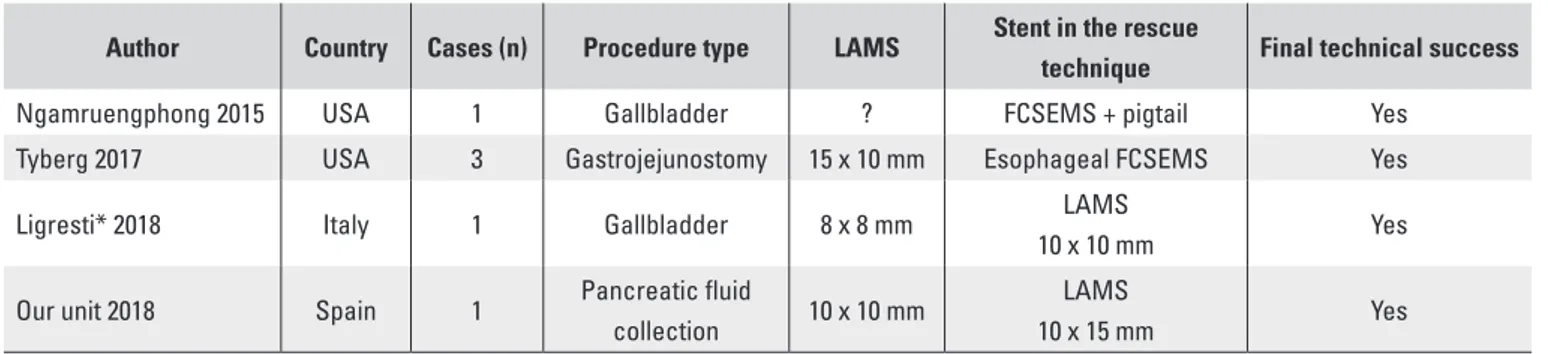
Table 1. Literature review of total distal dislodged lumen-apposing metal stent and the respective endoscopic rescue
technique using stents
Author Country Cases (n) Procedure type LAMS Stent in the rescue
technique Final technical success
Ngamruengphong 2015 USA 1 Gallbladder ? FCSEMS + pigtail Yes
Tyberg 2017 USA 3 Gastrojejunostomy 15 x 10 mm Esophageal FCSEMS Yes
Ligresti* 2018 Italy 1 Gallbladder 8 x 8 mm LAMS
10 x 10 mm Yes
Our unit 2018 Spain 1 Pancreatic fl uid
collection 10 x 10 mm
LAMS
10 x 15 mm Yes
LAMS: lumen-apposing metal stent; FCSEMS: fully covered metal stent. *Case of a buried LAMS stent, with the distal fl ange inside the gallbladder. “LAMS-in-LAMS” technique was applied one month after of the initial procedure.
A novel rescue maneuver for a distal dislodged lumen-apposing metal stent (LAMS): “LAMS-in-LAMS” technique
REV ESP ENFERM DIG 2019:111(3):243-245
DOI: 10.17235/reed.2019.5924/2018
245
terventional room. It confirmed a longer body of the second LAMS due the compression of the first LAMS.
CONCLUSION
To date, this novel technique has only been reported in a buried LAMS case, one month after a EUS-guided gall-bladder drainage (4). To the best of our knowledge, this is the first case describing this “LAMS-in-LAMS” technique, which salvaged the failed EUS-guided transmural drainage of a pancreatic collection during the initial interventional procedure. In our opinion, this “LAMS-in-LAMS” technique is feasible, effective and a helpful rescue technique for cas-es of dislodged LAMS. We think that the intentional use of a second LAMS (same design) will fit better than other stent designs (i.e., tubular design).
REFERENCES
1. Ngamruengphong S, Kumbhari V, Tieu AH, et al. EUS-guided rescue of early dislodgement of a lumen apposing stent. Gastrointest Endosc 2015;82:1124. DOI: 10.1016/j.gie.2015.06.009
2. Tyberg A, Pérez-Miranda M, Sánchez-Ocaña R, et al. Endoscopic ultra-sound-guided gastrojejunostomy with a lumen-apposing metal stent: a multicenter, international experience. Endoscopic Int Open 2016;4:276-8. DOI: 10.1055/s-0042-101789
3. Tyberg A, Zerbo S, Barthet M, et al. A novel technique for salvaging a dislodged lumen-apposing metal stent during creation of an endoscopic gastrojejunostomy. Gastrointest Endosc 2017;49:1007-8.
4. Ligresti D, Cipolletta F, Amata M, et al. Buried lumen-apposing metal stent (LAMS) following endoscopic ultrasound-guided gallbladder drainage: the LAMS-in-LAMS rescue treatment. Endoscopy 2018;50:822-3. DOI: 10.1055/a-0624-2050