Analysis and coordination of different
mechanisms controlling tube elongation during
the development of the embryonic system
in Drosophila melanogaster
Ivette Olivares Castiñeira
Aquesta tesi doctoral està subjecta a la llicència Reconeixement- NoComercial –
CompartirIgual 4.0. Espanya de Creative Commons.
Esta tesis doctoral está sujeta a la licencia Reconocimiento - NoComercial – CompartirIgual
4.0. España de Creative Commons.
This doctoral thesis is licensed under the Creative Commons
Departamento de Genética, Microbiología y Estadística Programa de Doctorado de Genética (HDK0S) Facultad de Biología Universidad de Barcelona
Analysis and coordination of different mechanisms controlling
tube elongation during the development of the embryonic
tracheal system in Drosophila melanogaster
Memoria presentada por Ivette Olivares Castiñeira Para optar al grado de DOCTOR por la Universidad de Barcelona Tesis dirigida por Marta Llimargas Casanova En el Instituto de Biología Molecular de Barcelona (IBMB)- Consejo superior de investigaciones Científicas (CSIC) Marta Llimargas Casanova Pedro Martínez Serra Directora Tutor Ivette Olivares Castiñeira Autora
ACKNOWLEDGEMENTS
M’agradaria donar les gràcies a totes aquelles persones que de més i de menys han contribuït durant aquests 4 anys, sense la seva ajuda i suport aquest difícil camí no hauria estat el mateix.
Primer de tot vull donar les gràcies a la meva directora, la Marta Llimargas, per donar-me l’oportunitat d’endinsar-me en aquest món de la investigació, tan complicat i feixuc però alhora tan fascinant. Agrair-te tot el que he après tan a nivell de treball com de persona i gràcies per fer-me créixer personalment i professionalment. Agradecer también a la segunda persona que más importante ha sido para mi durante esta etapa, a ti Anni. Sin tu incondicional ayuda nada hubiera sido lo mismo. Tanto a nivel laboral, en el que me has ayudado muchísimo, como personal hemos pasado muy buenos momentos. Nuestras risas infinitas por tonterías (zapatos de pelo), nuestra adición a comprar cosas por internet (bolsos, zapatos, relojes, pulseras…), ponernos de acuerdo con la ropa casi cada día, el apoyo mutuo y la complicidad con la que venir cada día al trabajo se ha hecho mucho más llevadero. También agradecer a dos personas que siempre han estado ahí para ayudarme, Nico y Esther. A ti Nico por tu gran ayuda con los CRISPR y en todo lo molecular en general. Sin ti nada saldría en el lab, siempre tienes alguna solución para todo y gracias por animar siempre el laboratorio. A ti Esther por ayudarme siempre con cualquier problema en el lab, por el apoyo que siempre me has dado, por las risas y por nuestras salidas fuera del laboratorio, roma, cursas, perritos. Echaré de menos al equipo Running y nuestros super desayunos.
Agrair al meu company de taula durant aquests quatre anys, Guillem. Gràcies per les bones converses, pels riures, per compartir els mateixos maldecaps de la tesi i per ser un excel·lent company de tesi, bona sort en la nova etapa. A Ettore por ser la super nueva incorporación. Aunque no vayamos a compartir muchos años, al menos los que sí han sido para ver que eres muy paciente y trabajador y en los momentos de desespero también me has dado tu apoyo. Mucha suerte en tu tesis, seguro que te irá genial.
Muchas gracias también a todas esas personas que encontré al principio y que por desgracia ya no están. Pili, fuiste como una madre para todos nosotros, siempre preocupándote por todos y también quiero agradecerte las risas en el lab y todo el apoyo que tuve por tu parte. A mis chicas, Arzu y Lorena, aunque estemos lejos nunca olvidaré nuestras noches en la rosa negra, nuestras risas, mojitos y momentos en los que me sentí muy contenta de haberos conocido. Espero que en algún momento nos volvamos a ver. A todos aquellos que ya no están pero que también formaron parte de estos años, Guille, Pablo, Paula, Oscar y Gonzalo por las risas y buenos momentos en la flyroom, Kyra, Delia y Ale por ser los italianos más divertidos y por pasar muy buenos momentos en el lab, a la Marina, per ser una super estudiant i pels bons moments i riures que vàrem passar, i la Neus, la millor tècnic que podíem haver tingut. Va ser un plaer poder coincidir amb tu. Agradecer a todos los miembros del laboratorio de Jordi Casanova y Marta, al Jordi pels llargs labmeetings preguntant y debatint, al Marc per la seva ajuda en els nostres projectes, a Juanjo por siempre ayudarme en cualquier duda que he tenido en los experimentos y por
las risas en la flyroom y a Sofia por su incondicional ayuda, a Sara, Xavi, Bea, Fridi, Sandra y a Yolanda porque sin ti el lab sería un caos. A las chicas de Gerardo, Marta per ajudar-me també amb els CRISPRs i pels riures al café, a la Núria per compartir riures, roba, viatges i bons moments, a Laura, Sara y Aikaterini. I després de tots vosaltres, vull agrair a la meva família que sempre m’han donat força i un somriure en els moments difícils. Als meus pares, Eduard i Núria, per estar sempre al meu costat i donar-me el suport i el millor consell. Sense vosaltres no hauria estat el mateix però sé que sempre estareu al meu costat. També a la meva avia que sé que estarà orgullosa de mi. A los padres de Ruben, Juan y Palmira, por todo el apoyo recibido y por darme las fuerzas necesarias para no decaer nunca. A la Manoli, Josep, Anna i Núria per preocupar-vos sempre per mi i preguntar com portes la tesi?
A tots els meus amics de Roda i del Palcam, per tots els anys que fa que ens coneixem i gràcies per donar-me sempre l’empenta i el suport, per estar al meu costat i per continuar sent amics després de tants anys. Por último, y una de las personas más importantes de mi vida, agradecer a Ruben el apoyo recibido cada día, los ánimos incesantes en los momentos más difíciles y agobiantes de la tesis, por empujarme a realizar todo aquello que quiero y darme ese poder y fuerza para realizarlo. Gracias infinitas porque sin ti todo hubiera sido más difícil y gracias por tu amor incondicional.
Per a Eduard, Núria i Ruben….
INDEX I
INDEX
1. INTRODUCTION ... 1 1.1 Drosophila tracheal system as a model to study tubulogenesis ... 1 1.2 Overview of the embryonic tracheal development ... 1 1.3 The genetic program controlling tracheal development ... 3 1.4 Mature branching pattern ... 3 1.5 Tracheal tube architecture ... 4 1.6 Cellular organization of tracheal system ... 7 1.7 Tracheal tube maturation: tube elongation and expansion ... 8 1.7.1 Mechanisms that regulate tube growth ... 9 1.7.2 Circumferential enlargement ... 11 1.7.3 Axial elongation ... 12 1.7.3.1 Modification of the aECM ... 12 1.7.3.2 Crb plays a key role in tube size control ... 14 1.7.3.3 Src kinases control axial elongation ... 16 1.8 Epidermal growth factor receptor ... 17 1.8.1 EGFR activity in the tracheal system ... 18 1.8.2 EGFR signalling pathway ... 19 1.8.3 EGFR ligands ... 20 1.8.4 Nuclear factors ... 21 1.9 Endocytic routes ... 23 1.9.1 The Retromer complex ... 25 1.9.2 The retromer complex and apical polarity trafficking ... 26 1.9.3 Trafficking of luminal cargoes ... 28 1.9.4 Rab GTPases ... 28 2. OBJECTIVES ... 33 3. MATERIAL AND METHODS ... 37 3.1 Drosophila strains ... 37 3.2 Immunohistochemistry ... 39 3.3 Antibodies ... 40 3.4 Image acquisition and processing ... 41 3.5 Morphometric analysis ... 41 3.5.1 Tube size quantifications ... 41 3.5.2 Cell junctions length ... 42 3.5.3 Crb intensity levels ... 42 3.5.4 Crb subcellular accumulation ... 42 3.5.5 Crb anisotropy accumulation ... 42 3.5.6 Crb Serp colocalization ... 43 3.5.7 Analysis of cargoes in endosome ... 43 3.5.8 Cell number ... 43 3.6 FRAP assay ... 43 3.7 Time-lapse imaging ... 44 3.7.1 Vesicle dynamics ... 44 3.7.2 Crb apical subcellular accumulation ... 44 3.8 Quantifications and statistics ... 44 4. RESULTS ... 47 4.1 EGFR requirement in tracheal tube size ... 47INDEX II 4.1.1 The modulation of EGFR activity controls tube length ... 47 4.1.2 Cell number and cell shape ... 49 4.2 EGFR activation in tube size control ... 50 4.2.1 The role of Spi in the control of DT elongation ... 50 4.2.2 The activity of Vein and keren in tracheal formation ... 52 4.3 The requirement of the RAS-MAPK pathway in tube size control ... 54 4.3.1 EGFR controls tube size by the RAS-MAPK pathway ... 54 4.3.2 The nuclear requirement of EGFR signalling in tube size ... 55 4.4 EGFR mechanism of tube size regulation ... 58 4.4.1 EGFR controls Serp accumulation in the aECM ... 58 4.4.2 EGFR controls Crb accumulation in the DT ... 60 4.5 Crb apical subcellular accumulation ... 62 4.5.1 Crb subcellular accumulation during tracheal formation ... 64 4.5.2 Crb subcellular accumulation depends on its endocytosis ... 66 4.6 Crb apical localisation and recycling pathways ... 68 4.6.1 The role of Rab4 in Crb recycling ... 68 4.6.2 The role of RE-Rab11 in Crb recycling ... 71 4.7 Serp and Crb localise in common sorting endosomes ... 73 4.7.1 Serp and Crb recycling mutually affect each other ... 75 4.8 EGFR is required for the proper organisation of Serp-Crb endosomes ... 78 4.9 EGFR affects WASH complex localisation ... 79 4.10 Crb anisotropic accumulation in DT cell junctions ... 83 4.10.1 Crb anisotropically accumulation in the control ... 83 4.10.2 Crb anisotropic accumulation is decreased in the Src42ADN mutant embryos ... 86 4.11 Constitutively activation of Src42A affects Crb junctional accumulation and Serp luminal deposition ... 89 5. DISCUSSION ... 95 5.1 EGFR restricts tube elongation in the trachea ... 95 5.2 A role for EGFR in endocytic traffic regulation ... 97 5.3 The regulation of Crb localisation in the trachea ... 98 5.4 The regulation of Crb anisotropic accumulation in DT cell junctions ... 101 5.5 A possible connection between Src42A activity and intracellular trafficking ... 102 6. CONCLUSIONS ... 105 7. BIBLIOGRAPHY ... 109 8. APPENDICES ... 125 8.1 List of abbreviations ... 125 8.2 Movie legends ... 128 8.3 Summary ... 129 8.4 Resumen en castellano ... 131 8.5 Published papers ... 132
1. INTRODUCTION
1. INTRODUCTION 1
1. INTRODUCTION
1.1
Drosophila tracheal system as a model to study
tubulogenesis
A major goal of developmental biology is to understand organ formation. The branched tubular network is one of the most common structural designs used in organ formation, and it is present in organs such as the lung, kidney and the vascular system. These organs carry out essential functions such as the transport of gases or body fluids. The morphogenesis of branched tubular structures is known as tubulogenesis.
Drosophila melanogaster has been proven to be an excellent model for identifying the
underlying molecules and mechanisms of many aspects for development and disease. As with all multicellular organisms, Drosophila contains a variety of tubular organs such as the salivary gland, the tracheal system (respiratory organ) and the Malpighian tubules (the excretory system). Since the mechanisms of tube formation are conserved from worms to humans (Andrew and Ewald 2010) and it is difficult to analyse these structures in mammalian systems, due to their complexity, the Drosophila tracheal system has emerged as a good model to study tubulogenesis. The tracheal system of Drosophila, a highly branched tubular network, is a vey well-described system for understanding the molecular and cellular underpinnings of tube formation. Furthermore, the huge amount of genetic tools and cellular approaches helps to understand in detail the formation of branched structures.
1.2
Overview of the embryonic tracheal development
The tracheal system is established during embryonic development when a group of epithelial cells are determined to become part of the tracheal system as ten ectodermal placodes (or clusters) on each side of the stage (stg) 9 embryo (Manning and Krasnow, 1993, Affolter and Caussinus 2008, Maruyama and Andrew 2012, Schottenfeld et al. 2010, Uv et al. 2003, Loganathan et al. 2016). Subsequently, at stg 10 of embryogenesis, these cells on either side of the embryo invaginate in an ordered choreography to form the internalised tracheal sacs through an apical constriction mechanism. This structure generates the luminal cavity, which is subsequently expanded and remodelled during the branching process. Later, at stg 11, the around 40 tracheal cells of each placode undergo the final round of mitotic division resulting in 80 cells per tracheal segment (metamere). Thereafter, subsets of cells within each sac begin to migrate towards specific directions in a stereotypical manner to form the distinctive branches visible from stg 12 onwards. At stg 13-14, through cell rearrangements, and cell shape changes, tracheal cells undergo a distinct sequential programme of branch sprouting, directed branch outgrowth and branch fusion. These buds elongate to form branches of distinct cellular architecture, ranging from multicellular tubes to fine branches. From stg 14 onwards, an interconnection of the
1. INTRODUCTION
2
metameric, branched units at distinct fusion points occurs. Meanwhile, terminal tracheal cells at the branch tips extend the luminal space into individual cell extensions, which adopt tree-like structures and reach every cell. The air enters the trachea through specialized openings, the spiracles, and at the tip of the terminal branches, gas is exchanged with the surrounding tissue once the tracheal system becomes physiologically active at the end of the embryogenesis (Affolter and Caussinus 2008) (Fig.1). Figure 1. Development of embryonic Drosophila tracheal system. There are ten tracheal placodes at each
side of the embryo at stage 10. Tracheal cells undergo apical constriction and starts to invaginate to form internalized tracheal sacs at stage 11. From stage 12, subsets of cells bud towards different directions giving rise to the different branches that will continue extending as development proceeds. From stage 14, the tracheal cells will mediate branch fusion with contralateral or adjacent branches, forming a continuous tubular tracheal network until the end of embryogenesis (From Atlas of Drosophila Development by Volker Hartenstein).
1. INTRODUCTION 3
1.3
The genetic program controlling tracheal
development
The genetic program and the signalling pathways underlying tracheal development have been extensively studied for the last years. Tracheal cell specification requires a combination of transcription factors, including trachealess (trh), ventral veinless (vvl) and
knirps/knirps-related (Affolter and Shilo 2000). Once the tracheal placodes are formed, the
cells invaginate by apical constriction to form internalized tracheal sacs (Isaac and Andrew 1996, Llimargas and Casanova 1999), in a process that requires the expression of Trh (Isaac and Andrew 1996, Wilk et al. 1996). In Trh mutants, tracheal primordia fail to accumulate apical actin and do not constrict, remaining at the surface of the embryo. Therefore, the invagination of the tracheal pits is associated with an accumulation of actin at the cell surfaces and both invagination and actin accumulation are dependent on trh activity (Isaac and Andrew 1996, Llimargas and Casanova 1999). Trh controls invagination partly through the transcriptional activation of rhomboid (rho) (Boube et al. 2000, Zelzer and Shilo 2000), which encodes a protein essential for the processing of the Epidermal Growth Factor ligand (EGF ligand) (Lee et al. 2001). However, in rho mutants tracheal invagination is only partially affected, indicating that additional pathways may function downstream of Trh to control internalization. In this respect, a role for the apical determinant Crumbs (Crb) in tracheal invagination was proposed (Letizia et al. 2011).
Upon invagination, the migration of specific tracheal branches along distinct trajectories requires the Fibroblast Growth Factor (FGF)/ Breathless (Btl) signalling pathway (Sutherland et al. 1996). Subsequent branch sprouting and outgrowth occurs without cell division as cells migrate towards neighbouring cells or towards tissues that express sources of the Btl ligand Branchless (Bnl) and attractant signal of the Fibroblast Growth Factor Receptor (Fgfr). Therefore, Bnl/Fgf secretion controls the migratory behaviour and the direction of tracheal cell movement (Sutherland et al. 1996). Cells at the tip of the tracheal branches, called tip cells, appear to be highly dynamic and send out filopodia and lamelopodia in response to Btl/Fgfr signalling, thereby regulating cytoskeleton dynamics (Okenve-Ramos and Llimargas 2014a, b, Ribeiro et al. 2002). In the absence of Fgfr signalling, cells remain in the sac-like shape, and filopodia and lamelopodia are not seen (Ribeiro et al. 2002), demonstrating that Bnl/Fgf signalling regulates both motility and the directionality of tracheal cell movement in the embryo. The Fgf signalling leads to phosphorylation and activation of the Ras/Mitogen-Activated Protein Kinase (MAPK) cascade, which in turn regulates the expression of several target genes to control cell behaviour (Affolter and Caussinus 2008).
1.4
Mature branching pattern
The tracheal system is a complex structure that consists of interconnected metameric units of different-sized tubes that extend over the entire embryo until the end of the embryogenesis. Different branches can be distinguished in the mature embryonic tracheal1. INTRODUCTION 4 system (Fig.2A,B,C). The dorsal trunk (DT) extends along the anterior-posterior axis of the embryo and forms the major tracheal tube. The dorsal branches, (DBs) extend towards the dorsal midline. DBs of each side fuse with their contralateral ones at the dorsal midline. The transverse connective (TC) sprouts from the DT and extends ventrally. The visceral branches (VBs) sprout from the TC and extend internally towards the gut. A lateral trunk (LT) forms parallel to the DT. The ganglionic branches (GBs) extend from the TC and reach the ventral nerve cord. Finally, the group of cells that remain near the site of the invagination form the spiracular branches (SBs) (Manning and Krasnow, 1993).
Figure 2. Structure of a tracheal metamere. (A, B) Lateral view of the embryonic tracheal system of Drosophila. Abbreviations: Anterior and posterior spiracle (AS, PS). (C) A schematic representation of a single
tracheal metamere. The main branches are indicated: the dorsal trunk (DT); dorsal branch (DB); transverse connective (TC), visceral branch (VB); lateral trunk (LT); ganglionic branch (GB); spiracular branch (SB). Adapted from (Uv et al. 2003).
1.5
Tracheal tube architecture
Each tracheal branch within a metamere has fixed cell numbers (Samakovlis et al. 1996) and characteristic tubular dimensions (Beitel and Krasnow 2000). We can classify the tracheal branches into four major types according to their size and cellular architecture (Uv et al. 2003): Type I tubes, such as the DT (the major branch of the trachea) and the majority of TC (the stalk connecting all of the tracheal branches within each metamere), are multicellular tubes. In these tubes, several cells are connected by intercellular junctions and surround an extracellular central lumen.
The type II tubes, such as the LT, GB, DB and VB, are narrower and multicellular. These tubes comprise a linear arrangement of single cells whose apical surfaces surround an extracellular lumen. Cells connect with themselves through autocellular junctions and with their neighbours through intercellular junctions. The main function of type I and II tubes are to transport gases.
1. INTRODUCTION
5
Type III tubes interconnect type I and type II tubes of the adjacent or contralateral tracheal metameres to mediate luminal branch fusion and form a continuous network. They are composed of tubular protrusions of single cells attached to each other, resulting in seamless tubes without intracellular junctions. Meanwhile, the two fusion cells connect themselves by intercellular junctions and form an intracellular lumen.
Type IV tubes, such as the lateral ganglionic (LG) branches, are highly branched intracellular cytoplasmic extensions that form in terminal cells at the tips of the tubes. Their main function is to contact target tissues for gas exchange (table1, Fig.3A,B,C).
Table1. Types of tracheal tubes. Abbreviations used: DT: Dorsal trunk, TC: Transverse connective, LT:
Lateral trunk, GB: Ganglionic branch, DB: Dorsal branch, VB: Visceral branch and LG: Lateral ganglionic. Adapted from (Zuo et al. 2013). Type Cellular make-up surrounding the central lumen Tracheal tubes Connections of cells surrounding the lumen Lumen I Multicellular DT, TC Intercellular junction Extracellular
II Multicellular LT, GB, DB, VB Autocellular
junction Extracellular III Tubular protrusions of single cells Connect type I and II (fusion cells) Seamless, no intracellular junction Intracellular IV Intracellular cytoplasmic extension LG (terminal cells) Seamless, no intracellular junction Intracellular
1. INTRODUCTION
6
Figure 3. Types of tracheal tubes. (A) Scheme representation shows the types of tubes that form in the Drosophila trachea, indicating which type of tube is found in each branch. The lumen is shown in pink, with
the apical membrane in dark pink. The basal surface is dark blue and the cytoplasm is lighter blue. Adapted from (Kerman et al. 2006, Tonning et al. 2005). (B) Four distinct cellular types of tubes. Abbreviations used: AJ: Adherens junctions, SJ: Septate junctions. Adapted from (Uv et al. 2003). (C) Schematic overview of the different patterns of tubes. Intracellular tubes are shown in blue, autocellular tubes in green and intercellular in red. Abbreviations used: DT: Dorsal trunk, TC: Transverse connective, LT: Lateral trunk, GB: Ganglionic branch, DB: Dorsal branch, VB: Visceral branch, LG: Lateral ganglionic and SB: Spiracular branch. Adapted from (Ribeiro et al. 2004).
1. INTRODUCTION 7
1.6
Cellular organization of tracheal system
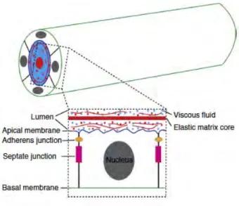
The Drosophila trachea is a ramifying network of epithelial tubes. The tracheal cells have features characteristic of typical epithelial such as apicobasal polarity, maintenance of tissue integrity via cell-cell and cell-extracellular matrix adhesions, a basement membrane and the capacity for directed secretion. The development of the tracheal system involves dramatic tissue remodelling, including cell rearrangements and cell shape changes. Remarkably, during remodelling cells that form the tracheal tubes remain attached to one another maintaining their epithelial integrity and polarity. A single layer of polarized epithelial cells composed of apical and basal membrane domains forms the embryonic tracheal tube. The apical membrane of the tracheal cells faces the luminal cavity filled with an apical extracellular matrix (aECM)1 whereas, the basolateral membrane is in contactwith neighbouring cell or either basal membrane (Fig.4).
Figure 4. Schematic representation of tracheal epithelial cells. Top: epithelial cells surrounding the
tracheal luminal space by its apical side. Bottom: single epithelial cell facing the apical membrane to the lumen. Inside the lumen, aECM is formed by a viscous fluid and an elastic matrix core. From (Dong and Hayashi 2015).
Epithelial cells contact each other through junctional structures that serve to link the cells and provide structural support, prevent the diffusion of membrane proteins and lipids, and provide barrier functions. Apical and basal regulators are associated with distinct membrane domains:
Adherens junctions (AJ) contain E-cadherin (E-Cadh) and Armadillo (βCatenin) form a cell-cell adhesive belt around the apical membrane, known as the Zonula adherens (ZA) that segregate the apical and basolateral membrane domains. Just apical to the ZA lies the
1 Apical Extracellular Matrix (aECM): It is an extracellular scaffolding matrix that provides mechanical support for
tissue assembly and organ shape. It is mainly constituted of secreted transmembrane proteins and polysaccharides, fibrous structural proteins, fluids and signalling molecules, forming an organized meshwork closely associated with the cell membrane (Labouesse, 2012). aECM is present both at the basal and apical surfaces of epithelia.
1. INTRODUCTION 8 Subapical region (SAR), also described as marginal zone. The SAR is defined by the presence of Crb and Par complexes, which have an important role in establishing epithelial polarity (Knust and Bossinger 2002). The Septate Junctions (SJs), equivalent to the tight junctions in vertebrates, are found below the AJs at the basolateral membrane. The SJ functions as the diffusion barrier that controls the exchange of solutes between epithelial cells. The SJ is composed of several protein complexes. (Laprise and Tepass 2011, Swanson and Beitel 2006, Wu et al. 2004). The most apical part of the plasma membrane forms an apical free membrane or region (AFR) and in the tracheal cells it faces directly the extracellular space contacting the tube lumen (Fig.5A). Figure 5. Organization of the Drosophila epithelial cells. (A) Scheme of Drosophila epithelial cell. The tracheal cells are epithelial cells with polarized plasma membrane associated with two membrane domains: Apical and basolateral membrane. The apico-basal polarity region of these cells is maintained through interactions between apical polarity regulators and basolateral polarity regulators. Adapted from (Tepass 2012).
1.7
Tracheal tube maturation: tube elongation and
expansion
After a morphogenetic phase in which the branches form by branching morphogenesis, tracheal branches undergo a process of maturation (Tsarouhas et al. 2007). This branch maturation process includes the acquisition of the correct size concerning length and diameter, unique to each type of branch (Ghabrial A. et al. 2003) and required for proper tube function. The DT is one of the most extensively studied branches in the context of tube size control and geometry regulation (Beitel and Krasnow 2000).
1. INTRODUCTION 9
1.7.1
Mechanisms that regulate tube growth
The tracheal tubes have to be constructed with an optimal shape for the efficient circulation of gas. In Drosophila, the growth of embryonic tracheal tubules proceeds in two dimensions, by axial elongation and diameter expansion. Maintaining a constant tube diameter and fitting axial length, which are controlled by distinct genetic programs and cellular mechanisms (Beitel and Krasnow 2000), are two key requirements for proper tube function (Fig.6A,B,C). Figure 6. Schematic drawings of DT elongation and expansion. (A, B, C) Scheme representing the DT axial extension (tube elongation) along the long axis and the diameter expansion (tube dilatation, circumferential) increasing the circumference of the lumen cavity. Luminal space is labelled in green. Cell outline and nucleus are drawn in black line and filled circle, respectively. Adapted from (Hayashi and Dong 2017).
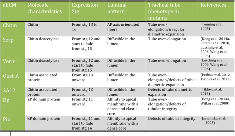
The aECM, which fills the entire tube lumen, is essential for the generation of these two growth forces. The aECM is formed by various proteins and polysaccharides secreted from the tracheal cells (Devine et al. 2005, Tonning et al. 2005). The components of the aECM include: chitin, chitin deacetylation domain proteins (serpentine/Serp and vermiform/Verm), chitin associated proteins (2A12/Gasp and Obstructor-A/Obst-A), and zona pellucida (ZP) domain proteins (Dumpy/Dp and Piopio/Pio) (Table2, Fig.7). These components span a wide range of molecular sizes and are distributed in unique patterns, suggesting that aECM is a complex of various structural elements.
1. INTRODUCTION
10
Table 2. Apical ECM components. Abbreviations used: ZP: Zona pellucida, AP: Anterior-Posterior. From
(Dong and Hayashi 2015)
aECM Molecule
characteristics Expression Stg Luminal pattern Tracheal tube phenotype in mutants
References Chitin Chitin From stg 13 to
16 AP axis orientated fibers Tube over-elongation/irregular diametric expansion
(Tonning et al. 2005)
Serp Chitin deacetylase From stg 12 and start to fade from stg 15 Diffusible in the lumen Tube over-elongation (Dong et al. 2014a, Forster et al. 2010, Luschnig et al. 2006, Wang et al. 2006)
Verm Chitin deacetylase From stg 12 and start to fade from stg 15 Diffusible in the lumen Tube over-elongation (Luschnig et al. 2006, Wang et al. 2006) Obst-A Chitin associated
protein From stg 13 onward Diffusible in the lumen Tube over-elongation/defects of tube diametric expansion
(Petkau et al. 2012, Tiklova et al. 2013)
2A12 Chitin associated
protein From stg 13 onward Diffusible in the lumen Defects of tube diametric expansion
(Tiklova et al. 2013)
Dp ZP domain protein From stg 11
onward Affinity to apical membrane with a dense and elastic core Tube over-elongation/defects of tubular integrity (Dong et al. 2014a, Wilkin et al. 2000)
Pio ZP domain protein From stg 11 and start to fade from stg 14 Affinity to apical membrane with a dense core Defects of tubular integrity (Jazwinska et al. 2003)
Figure 7. Model for the tracheal tube size control in Drosophila. The expansion of the diameter of the
tracheal tubes from narrow to longer takes place during the embryogenesis by the transient chitin fibrils matrix (red) that is also required for the normal organization of the apical βH spectrin cytoskeleton (blue). Septate junctions mediate the Verm and Serp secretion, which is required for the subsequent modification of chitin matrix, to prevent enlarges of the tracheal cells and tubes. From (Swanson and Beitel 2006).
1. INTRODUCTION
11
1.7.2
Circumferential enlargement
The deposition of the aECM in the luminal space is associated with the onset of a diametrical expansion of the tube (Forster et al. 2010). This diametrical expansion is carried out in a defined time interval between embryonic stages 14-15.
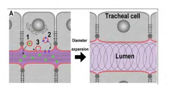
Different luminal proteins present in the tracheal cell cytoplasm are secreted into the lumen by different vesicle trafficking COPI (Coat complex protein I) (Armbruster and Luschnig 2012, Jayaram et al. 2008) and COPII-mediated complexes (Coat complex protein II) (Norum et al. 2010) (COPI: γCOP, σCOP; COPII: sec23, sec24, sar1, sec13) and by the Diaphanous-dependent secretory pathway (Massarwa et al. 2009). All these secretory pathways are required for the deposition of aECM components and the addition of apical membrane, leading to expansion of the intraluminal space (Fig.8).
Furthermore, the chitin fibrils are deposited along the tube lumen and coordinate uniform tube expansion. In the absence of chitin synthesis, the lumen is expanded in an irregular manner causing tube dilations (Tonning et al. 2005). Thus, the chitin cable coordinates the behaviour of the tracheal cells that surround it, and stabilizes the epithelium during diametric expansion (Schottenfeld et al. 2010).
Figure 8. Model with the secretion roles implicated in tracheal tube expansion. (A) Secretory activity
mediates tracheal lumen expansion by (1) soluble secreted proteins in the lumen, (2) membrane proteins delivered to the apical cell surface and (3) membrane material released into the lumen cavity required for the proper diameter expansion. From (Forster et al. 2010). Moreover, the chitin fibrils inside the lumen send signal to the epithelial cells to organize the luminal diameter (Swanson and Beitel 2006).
1. INTRODUCTION 12
1.7.3
Axial elongation
Following diametrical expansion the tube grows axially until the end of the tracheal development. Studies using tube elongation defective mutants (Beitel and Krasnow 2000, Ghabrial A. S. et al. 2011) have shown that axial growth requires Septate junctions (Llimargas et al. 2004, Paul et al. 2003, Wang et al. 2006, Wu et al. 2004), the subapical protein complex (Dong et al. 2014a, Laprise et al. 2010), planar cell polarity (Chung et al. 2009) and the maintenance of chitin deacetylases and Src kinase levels (Forster and Luschnig 2012, Luschnig et al. 2006, Nelson et al. 2012, Wang et al. 2006) Disruption of all these processes, with the exception of maintenance of Src kinase levels, leads to elongated tubes. However, the exact molecular mechanisms that cause tube over-elongations remain uncharacterized.
1.7.3.1 Modification of the aECM
The proper organization and modification of the aECM is required to restrict tube over-elongation. When the remodelling of the chitin polymer is compromised due to a loss of function of the secreted chitin deacetylases (Serp amb Verm) (Luschnig et al. 2006, Wang et al. 2006), the tracheal tubules exceed their normal length and show convoluted patterning. Therefore, such over-elongated tube phenotype suggests that an axial elongation force is present in the tracheal cells and that aECM restricts the excess elongation.
Chitin undergoes a deacetylation reaction resulting in the exposure of an amino group that converts insoluble chitin into a biologically compatible form with a rigid structure, the chitosan (Kim, 2011). The modification of the chitin cable is essential to restrict tube over-elongation. Serp and Verm, secreted proteins which contain both chitin binding and deacetylase domains, are putative chitin deacetylases present in the tracheal lumen and their function is required to restrict tracheal tube length (Fig.9E,F) (Luschnig et al. 2006, Wang et al. 2006). The lack of chitin modification by the absence of Serp and Verm leads to an over-elongated and convoluted DT at stages 15-16 of tracheal development, while there is little or no effect in the diameter of the DT (Fig.9A-D, F). This suggests that tube length is controlled separately from diameter by modulation of physical properties of the chitin ECM (Luschnig et al. 2006) (Fig.9F).
1. INTRODUCTION
13
Figure 9. Serpentine and Vermiform, chitin deacetylases. Adapted from (Luschnig et al. 2006).
(A-D) Stage 16 embryos showing genotypes immunostained with a fluorescent chitin binding protein
labelling the tracheal lumen. (B,C) Single Serp and Verm mutant display a slightly convoluted DT compared to the control (A). (D) In double mutants Serp-Verm, display a strong phenotype of elongated and tortuous DT. The diameter is not affected in the mutant conditions. (E) Scheme showing the protein domains of Serp and Verm: Sp, N-terminal signal peptide; CBD, Chitin Binding Domain; LDLa, LDL receptor type A ligand binding motif; and CDA, Chitin deacetylase domain. (F) Summary of the different roles of chitin regulating diameter versus tube length. In the top panels is shown
the DT expansion of wild-type embryos. Middle panels show the requirements for serp and verm chitin modification in the regulation of tube length. Lower panels show the requirement of the presence of chitin for tube diameter regulation and tube stability. In chitin modification mutants the chitin cylinder and the lumen tube are longer and convoluted, whereas in chitin synthesis mutants, the chitin cylinder is absent and tubes are not dilated properly.
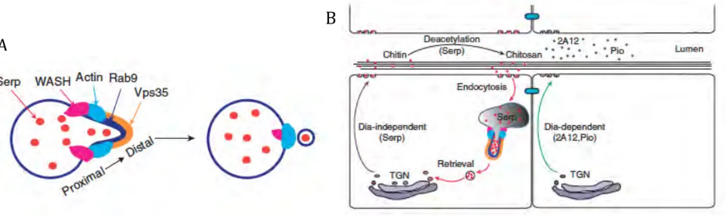
Serp and Verm are first detected at early stage 13 embryos in the cytoplasm of tracheal cells. Later on, the two proteins accumulate in the lumen and at the apical membrane. Maintenance of Serp in the lumen requires recycling through the endocytic pathway, through a Rab9-mediated retrograde2 trafficking process (Dong et al. 2013); see
below in chapter 1.9.3. In addition, Serp is also transcytosed3 from the fat body to the
tracheal lumen being retained there through chitin association (Dong et al. 2014b). These mechanisms ensure the optimal accumulation of Serp in the lumen, facilitating the remodelling of the aECM to balance the continuous elongation of tracheal tubes. The transcytosis and retrograde pathways are specific for Serp protein and do not act on other luminal proteins such as Verm or Pio (Dong et al. 2014b, Dong et al. 2013).
2 Retrograde trafficking pathway: transport of cargoes from the endosomes to the trans-Golgi network (TGN) (Dong et al. 2013). 3 Transcytosis: a type of transcellular transport in which the cargoes are transported across the interior of the cell. It is a key process to facilitate protein translocation across epithelial barriers (Dong et al. 2014b) F E
1. INTRODUCTION 14
1.7.3.2 Crb plays a key role in tube size control
The Crb transmembrane protein, a component of the SAR, is a key regulator of apical polarity in Drosophila epithelial cells (Tepass 2012, Tepass and Knust 1990, Tepass et al. 1990). Crb contains one large extracellular domain, which is composed of 29 epidermal growth factor (EGF)-like repeats and four laminin-A globular-domain-like repeats. The cytoplasmic domain, called the intra domain, consists of two highly conserved regions, the C-terminal PSD-95/Disc-large/ZO-1 (PDZ)-binding motif ERLI, and a 4.1/ezrin/radixin/moesin (FERM)-binding domain. The intra domain is responsible for the interaction with different proteins, like Stardust, Patj and Lin-7, with which it forms a complex, and also connects with the apical βH-spectrin (βH), the actin cytoskeleton and basolateral polarity complexes (Bulgakova and Knust 2009) (Fig.10A,B). Loss of Crb causes severe defects in epithelial polarity and AJ integrity (Bulgakova and Knust 2009, Tepass et al. 2001). Figure 10. Drosophila Crb protein complex and Crb protein domains. (A) Scheme of the core proteins of Drosophila Crb complex. Crb complex is formed by four components: Crb, Stardust (Sdt), PATJ and Lin-7. From (Bulgakova and Knust 2009). (B) Molecular interaction of Crb protein domains. The cytoplasmic tail of Crb protein has two conservedmotifs: a FERM domain binding site (FDB) and a PDZ-domain binding site (PDB) that interact with several partners in order to regulate different processes such as the formation, structure and maintenance of the apical domain of epithelial cells to establish the apico-basal polarity, epithelial cell shape, and the regulation of cell proliferation and survival. From (Tepass 2012).
During tracheal tube elongation, Crb overexpression causes overexpansion of the apical domain and this leads to an increased DT length (Dong et al. 2014a, Laprise et al. 2010, Laprise et al. 2006) (Fig.11). High levels of Crb are detected during the invagination of the tracheal placodes, being reduced as tracheal development proceeds (Letizia et al. A
1. INTRODUCTION
15
2011) suggesting that Crb accumulation in the apical membrane has to be controlled in order to allow correct DT length.
At late stages, Crb plays a key role in controlling the length and diameter of the DT through negative regulatory interactions with the FERM proteins Yurt (Yrt) and Coracle (Cora). Crb regulates the apical membrane size at the same time that controls tracheal tube length. Apical and basolateral epithelial polarity proteins interact with each other in order to regulate the tracheal tube length independently of the aECM requirement (Laprise et al. 2010) (Fig.11).
Figure 11. Model of interactions between Crb with Yrt and Cora related with the tube length regulation. The apical polarity protein Crb is controlling epithelial tube size through the interaction with Yrt
and Cora, two SJ-associated polarity proteins. From (Laprise et al. 2010).
In shrub4 mutants, Crb is lost from the apical membrane and forms huge cytoplasmic
aggregates at late stages. It has been proposed that the loss of shrub, an ESCRT-III component, causes defective Crb turnover, leading to Crb mislocalization and misregulation, which induces apical membrane overgrowth. In shrub mutants, the DT is over-elongated and bent in a sinusoidal pattern with no changes in tube diameter (Dong et al. 2014a) (Fig.12).
4 Shrub: ESCRT III-mutant. Endosomal sorting complex required for transport (ESCRT) is cytoplasmic machinery
required for membrane deformation, internalization and scission (Wollert et. 2009). In Drosophila the Shrub gene encodes Vps32, a subunit of ESCRT III, which regulates endocytotic sorting of membrane-associated proteins leading to lysosomal degradation (Hori et al. 2011).
1. INTRODUCTION 16 Figure 12. Scheme showing Shrub’s control of apical membrane biosynthesis through the regulation of Crb activity. The left panel shows the WT conditions where the balance between Rab11 and the Retromer – mediated recycling and degradation pathways controls Crb activity. In the right panel, the loss of Shrub causes Crb to accumulate in enlarged endosomes promoting a Crb overactivation, which induces the synthesis of the apical membrane, resulting in apical membrane overgrowth. From (Dong et al. 2014a).
1.7.3.3 Src kinases control axial elongation
Src-family kinases have a role in the control of tube size in mammalian epithelial and endothelial tubular systems. Moreover, Src proteins associate with the cytoskeleton and adherens junctions (Thomas and Brugge 1997). It has been shown that in Drosophila, Src42A also plays a key role in tracheal tube length control (Forster and Luschnig 2012, Nelson et al. 2012). The downregulation of Src42A produces a reduction of tube length, while the overexpression leads to elongated tubes. The results indicated that Src42A is required to increase DT length and that its activity is required autonomously in the trachea for DT elongation (Fig.13A,B). It was proposed that Src42A acts together with Daam, a Diaphanous-related formin (Aspenstrom et al. 2006), in controlling DT apical growth.dDaam mutants display a short and thicker DT length, similar to Src42A mutants, suggesting
that dDaam and Src42A control together directional apical surface growth (Nelson et al. 2012). A fine analysis at the cellular level indicated that in Src42A mutants the cell’s apical surface is significantly reduced and the surface axial extension is biased toward a more circumferential axis, compared to control. This revealed a clear effect of Src42A in polarised cell shape changes. In addition, it was proposed that Src42A affects E-Cadh recycling at AJs (Forster and Luschnig 2012).
In summary, Src42A regulates tube length by an anisotropic mechanical force that impinges on expansion along the longitudinal axis (anterior-posterior axis). This is achieved by regulating AJs remodelling (Forster and Luschnig 2012) and by polymerization of actin through the formin DAAM leading to axial apical expansion (Nelson et al. 2012).
1. INTRODUCTION 17 Figure 13. Model for the regulation of Src in Drosophila tracheal tube expansion. From (Ochoa-Espinosa et al. 2012)
(A) DT elongation stars at mid-stage of the embryogenesis. In the absence of Src42A, DT expansion is
disrupted and display short DT phenotype.
(B) Src initiates the elongation of tubes by the regulation of cell rearrangement and anisotropic growth of the
apical surface, whereas the aECM and SJ proteins are required to prevent an overelongation of the tubes. Src42A mediates the cell changes by the induction of adherens junction turnover. At the same time, Src42A promotes the axial elongation of apical surfaces united with dDaam/formin. The circumferential expansion is carried out in a Src42A-independent manner.
1.8
Epidermal growth factor receptor
The Epidermal Growth Factor Receptor (EGFR), a Tyrosine Kinase Receptor (RTK), triggers one of the principal conserved signalling pathway operating in development and homeostasis. During development, EGFR signalling controls important aspects such as migration, differentiation, proliferation, cell growth and survival. In addition, a miss-regulation of EGFR activity can lead to cancer and metastasis. Given its relevance, it is crucial to identify the whole plethora of EGFR activities and the downstream molecular machinery that leads to the different cellular outcomes.
1. INTRODUCTION 18
1.8.1
EGFR activity in the tracheal system
Roles for EGFR during tracheal development have already been proposed. EGFR is required for tracheal invagination and to maintain the epithelial integrity.
EGFR signalling controls the initiation of apical constriction, cell rearrangements and cell migration allowing invagination. In the absence of EGFR signalling, the cells of tracheal placodes remain at the surface of the embryo and consequently an abnormal tracheal tree is formed. This is consistent with an EGF-dependent activation of MAPK cascade in the tracheal placode at stg 10 (Llimargas and Casanova 1999). Additionally, EGFR controls the activity of the RhoGTPase Rho1 during tracheal invagination by regulating the RhoGTPase-Activating Protein (RhoGAP), encoded by crossveinless-c (cv-c), that affects the actin-myosin apical distribution. Hence, the EGFR pathway regulates cytoskeleton organization through the regulation of cv-c and Rho1, which, in turn, modify the apical actin required for the correct invagination and proper tracheal development (Fig.14A) (Brodu and Casanova 2006). Figure 14. Model for the regulation of tracheal cells invagination. (A) Trh induces and organizes tracheal invagination triggering EGFR signalling. EGFR activity is responsible for the proper tracheal invagination. Sal, a partner of tracheal invagination, is expressed in the dorsal half of the placodes. Its role is, at least in part, to downregulate EGFR signalling activity. Once EGFR is activated, cv-c controls the actin organization. Proper actin assembly, display a well-localized actin rings in the apical region, which leads to a proper apical cell constriction From (Brodu and Casanova 2006).
EGFR pathway has also been to maintain tracheal branch epithelial integrity through activation of the canonical MAPK pathway, but independently of the nuclear transcription factor Pointed. Downregulation of the EGFR pathway results in loss of tube integrity, whereas upregulation leads to increased tissue stiffness. This regulation of tissue integrity correlates with differences in the accumulation of E-cadh at cell-cell junctions (Cela and Llimargas 2006). A
1. INTRODUCTION 19
1.8.2
EGFR signalling pathway
The EGFR is one of the most versatile RTK known and it has been extensively studied. EGFR functions in a wide range of cellular processes such as cell fate determination, cell proliferation, cell migration and apoptosis. Several positive and negative regulators modulate EGFR activity (Adamson 1990, Dominguez et al. 1998, Schweitzer and Shilo 1997, Sibilia et al. 1998).
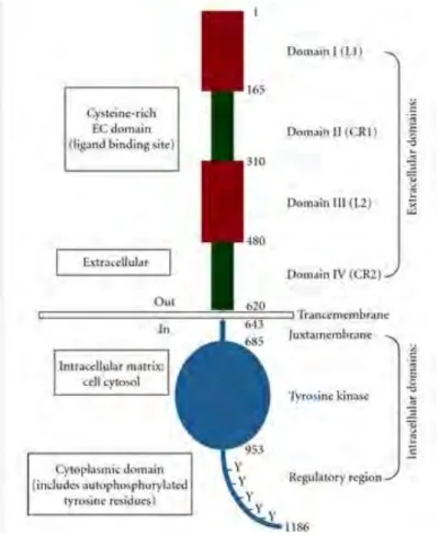
EGFR is a transmembrane protein containing an extracellular ligand-binding domain, a single membrane-spanning domain, and a cytoplasmic protein tyrosine kinase domain (Fig.15). At the plasma membrane, the un-stimulated EGFR is present as a monomer. Upon binding of an extracellular ligand, the receptor undergoes dimerization resulting in trans-autophosphorylation of its cytoplasmic domain. The ligand-binding domain is identifiable in the glycosylated extracellular domain by the presence of conserved cysteine-rich clusters. The cytoplasmic domain contains a juxtamembrane region required for signal attenuation by protein kinase C (PKC), and is followed by the tyrosine kinase domain (Src homologous domain 1-SH1-). The latter is the most conserved region and mediates auto-phosphorylation of the carboxy-terminal tail on six tyrosine residues, located in the non-catalytic tail. The phosphorylation of these residues allows recruitment of various adapter proteins with a Src homology domain 2 (SH2) or phosphotyrosine binding domain (PTB). In addition, EGFR can be phosphorylated by other kinases such as PKC in order to regulate its distribution at the membrane. The carboxy-terminal tail contains motifs for receptor internalization and degradation. After ligand binding, EGFR is endocytosed rapidly for signal attenuation.
In Drosophila, there is a single gene encoding EGFR, called DER (Bogdan and Klambt 2001). DER signalling is used broadly and throughout different contexts such as in development and growth (Schweitzer and Shilo 1997, Wasserman and Freeman 1997).
1. INTRODUCTION 20
Figure 15. Overview of the EGFR domains. EGFR is a monomer composed of 1186 amino acids. Its
extracellular domain is characterized by two cysteine-rich motifs where the ligand binds. Then is followed by a transmembrane domain. The intracellular domain is composed of a juxtamembrane domain, a tyrosine kinase domain and a regulatory region domain (Flynn et al. 2009).
1.8.3
EGFR ligands
In Drosophila there are four active EGFR ligands: Spitz (Spi), Vein (Vn), Gurken (Grk) and Keren (Krn) (Fig.16,1). The main activating ligand is Spi, a membrane-tethered EGF ligand that resembles the mammalian ligand transforming growth factor -α (TGF-α) (Freeman 1994, Rutledge et al. 1992, Schweitzer et al. 1995a, Tio and Moses 1997, Tio et al. 1994). The full-length Spi is unable to transduce the signal and must be processed into an active, soluble form. The precursor of Spi is transported from the endoplasmic reticulum to the Golgi compartment by Star (a transmembrane protein) in order to be processed (Kolodkin et al. 1994). Once Spi is in the Golgi compartment it is cleaved at its transmembrane domain by Rhomboid-1 (Rho1), an intramembrane serine protease (Urban et al. 2001).
There are two other membrane-tethered EGFR ligands in Drosophila: Grk and Krn. Grk is restricted to oogenesis, being expressed in the oocyte and signaling to the EGFR expressing follicle cells leading to egg polarization (Gonzalez-Reyes et al. 1995, Neuman-Silberberg and Schupbach 1993). It has been suggested that Grk, like Spi, requires post-translational activation in order to signal (Ghiglione et al. 2002). Rho2, a member of the
1. INTRODUCTION
21
rhomboid family, is expressed in the oocyte and may carry out Grk processing (Guichard et al. 2000). Krn is highly similar in structure to Spi and was identified originally by the
Drosophila Genome Project. The processing of Krn is regulated in an analogous manner to Spi, by Star and Rho proteins (Reich and Shilo 2002, Urban et al. 2002). The last activating ligand is Vn, which resembles the vertebrate Neuroregulin. Unlike the other ligands, Vn is a secreted ligand, which possesses an inherently weaker activation capacity and is used in tissues where low levels of EGFR activation are required (Schnepp et al. 1996, Schnepp et al. 1998). Vn is required for muscle attachment cell fate (Yarnitzky et al. 1997) and also induces several distal cell fates in the leg (Campbell 2002, Galindo et al. 2002). Argos (Aos) is a secreted ligand that has a single EGF-like domain with an expanded B-loop. It was initially assumed that Aos binds to EGFR through its EGF-like domain, functioning as a competitive inhibitor (Freeman et al. 1992, Jin et al. 2000, Schweitzer et al. 1995b). However, it was later found that Aos is associated predominantly with Spi and prevents Spi binding, EGFR dimerization and consequently EGFR activation (Klein et al. 2004). Figure 16. EGFR ligands. (1) Activating and inhibitory Drosophila epidermal growth factor receptor (EGFR, DER) ligands. Four activating ligands and one inhibitory ligands of EGFR have been described. Spi, Krn and Grk are produced as transmembrane precursors that have to be cleaved (pink arrow) to generate the active ligand and to be secreted. Vn is generated as a secreted protein. Aos is produced as a secreted protein and binds to Spi protein to prevent EGFR signalling. From (Shilo 2003).
1.8.4
Nuclear factors
EGFR signals through the MAPK pathway, one of the most conserved signalling cascades in multicellular organisms (Chen R. E. and Thorner 2007). Upon EGFR dimerization, the cytoplasmic domain leads to a trans-phosphorylation of the tyrosine residues contained in the tyrosine kinase domain (Arkhipov et al. 2013). This recruits
1. INTRODUCTION
22
different adaptor proteins; in particular, the cytoplasmic growth factor receptor bound protein 2 (Grb2). Subsequently, Grb2 recruits the Guanine exchange factor Son Of Sevenless (SOS) that activates the membrane associated Ras. Ras, then, initiates a cascade of phosphorylation of the three kinases: Raf, MAPKK (MEK) and MAPK (ERK or Rolled in
Drosophila) (Futran et al. 2013, Sopko and Perrimon 2013, Wortzel and Seger 2011)
(Fig.17). The activity of MAPK (ERK) is negatively regulated by MAPK phosphatase 3 (MKP3), which dephosphorylates the critical threonine and tyrosine residues of MAPK (Kim et al. 2004).
Figure 17. EGFR canonical signalling pathway. Ligands bind to EGFR on the cell surface, which upon
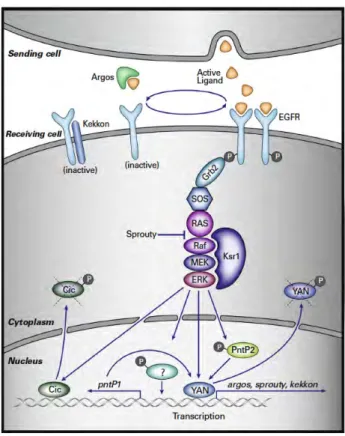
dimerization trigger its canonical signalling pathway involving Sos/Ras/Raf/MEK/MAPK pathway. Ksr1 (Kinase suppressor of Ras), a scaffold protein, regulates the Raf/MEK/MAPK activity increasing the efficiency of signalling. The main transcriptional output of the signalling pathway is mediated by the ETS protein Pointed (Pnt). The isoform PntP2 is activated mainly by MAPK phosphorylation, while the second isoform, PntP1, is constitutively active and its transcriptionally regulated by MAPK. In addition, Yan a constitutive repressor protein that competes for Pnt-binding sites, is removed from the nucleus and degraded upon phosphorylation by MAPK. Capicua (Cic), an HMG-box protein, is another repressor that is removed from the nucleus and degraded by the MAPK phosphorylation. Additional downregulation of the pathway is mediated by Sprouty and Kekkon, which attenuate the signalling maintaining the signalling pathway in check. From
(Shilo 2014).
MAPK has a wide variety of substrates both nuclear and cytoplasmic (Fig.17). The cytoplasmic substrates are non-transcriptional factors that affect other cell biological processes such as cell migration (Gabay et al. 1997, Sutherland et al. 1996), although not much is known about these substrates. The role of the nuclear substrates is better known. Two principal nuclear transcriptional substrates have been described to transduce the
1. INTRODUCTION
23
MAPK canonical signal: the ETS-domain protein Pointed (Pnt), which is one of the major activators of transcription downstream of RTKs, and the transcriptional repressor Yan. Pnt is produced in two forms. PntP2 is directly activated by MAPK phosphorylation upon EGFR activation. In contrast, PntP1 is a constitutively active form but requires MAPK transcriptional induction (Brunner et al. 1994, Gabay et al. 1996, Klambt 1993, O'Neill et al. 1994, Scholz et al. 1993).
MAPK also regulates nuclear transcription through the phosphorylation and inactivation of the transcriptional repressors like Yan/Anterior open (Aop) and Capicua (Cic). Yan is an ETS-domain protein that lacks the transcription activation domain and blocks the binding of Pnt. Upon MAPK phosphorylation, Yan is exported to the cytoplasm and degraded (Lai and Rubin 1992, Rebay and Rubin 1995, Tootle et al. 2003). Cic is an HMG-box protein whose phosphorylation by MAPK leads to its inactivation and nuclear export (Astigarraga et al. 2007).
1.9
Endocytic routes
Endocytic recycling is critical for many basic processes like signalling, polarity, cell adhesion, migration, cytokinesis or nutrient uptake. Cells internalise cargoes such as extracellular material, ligands, lipids or membrane proteins and return them back to the plasma membrane or to the extracellular space, ensuring their correct localisation and function, thereby coordinating the endocytic recycling with the endocytic uptake. Following endocytosis from the plasma membrane, internalized cargoes first reach the early sorting endosome. The sorting endosome acts as the main platform by which endocytic cargoes are sorted and further transported to their final destination through different specific pathways. Cargoes targeted to degradation will be transported from the sorting endosome to the late endosome (LE), which is also known as the multivesicular body5 (MVBs), and
then to the lysosome. Alternatively, cargoes can be recycled back to membrane or the extracellular space. In these cases, cargoes can either undergo a retrograde transport to the TransGolgi Network (TGN), which allows access to the secretory pathway or they can recycle to the plasma membrane from the sorting endosome using a direct route or an indirect one through the recycling endosome (RE) (Grant and Donaldson 2009, Hsu et al. 2012). Different Rabs proteins (see also chapter 1.9.4) are associated with and control each of these trafficking steps. For instance, Rab5 mediates internalisation to early endosomes (EE), Rab4 is associated with a short-loop recycling pathway back to the plasma membrane, Rab9 is associated with a retrieval pathway from the endosomes to the TGN and Rab11 is associated with the recycling to the cell surface through RE (Bhuin and Roy 2014, Wandinger-Ness and Zerial 2014) (Fig.18).
Cargo retrieval to the TGN or to the membrane usually relies on the presence of sorting signals in the cargoes that are recognized by specific coat complexes. This interaction facilitates the partition of cargoes into different discrete domains in the sorting
5 Multivesicular Body: an endocytic intermediate organelle in the lysosomal degradative pathway that
1. INTRODUCTION
24
endosomes. The subsequent tubulations, which eventually detach from these endosomes, become trafficking vesicles delivered to distinct intracellular routes. One of these coat complexes is the retromer complex (Burd and Cullen 2014, Gallon and Cullen 2015, Seaman et al. 2013, Wang and Bellen 2015). Figure 18. Overview of endosomal sorting pathways. Once proteins are internalized, they are sorted into
the early endosomes, which are characterized by the presence of the Rab GTPase Rab5. Afterwards, endosomes maturate and switch between Rab5 and Rab7, which mediate the conversion of these endosomes to late endosomes. Also, in many cases, exist an intermediate organelle called MVB that mediates the cargo transfer to the lysosomes. Proteins can also be directly recycled back from EE to the plasma membrane by Rab4 GTPase. Recycling of other proteins to the cell surface occurs through RE, marked by Rab11. Rab9 carries out cargo delivered from the endosomes to the TGN. From (Rajendran et al. 2010).
1. INTRODUCTION
25
1.9.1
The Retromer complex
The retromer complex is a conserved protein complex originally identified in yeast. The retromer regulates endosomal sorting and trafficking by rescuing cargoes from degradation and mediates two trafficking pathways: the retrograde transport from the endosomes through the TGN, and a direct delivery from endosomes to the plasma membrane (Burd and Cullen 2014, Gallon and Cullen 2015, Seaman et al. 2013, Wang and Bellen 2015). The retromer is a key component of the endosomal protein sorting machinery and operates by recognising specific membrane proteins or cargoes that are concentrated into discrete domains of the sorting endosome membrane to transport them to their appropriate destination. Moreover, the retromer complex regulates the subcellular localization of proteins and prevents its lysosomal degradation (Seaman 2012).
The retromer complex contains a Vps26-Vps29-Vps35 trimer (termed the cargo-selective complex, CSC) (Burd and Cullen 2014), which forms a stable structure that is absolutely required for retromer activity (Arighi et al. 2004, Seaman 2004). The CSC is regarded as the core of the retromer complex.
The retromer complex associates with different proteins, like sorting nexins (Snx). The association of the CSC with Snx proteins determines cargo selection and the trafficking route, i.e to the TGN or to the plasma membrane (Burd and Cullen 2014, Gallon and Cullen 2015, Seaman et al. 2013, Wang and Bellen 2015). Moreover, the small GTPase Rab7a and the Snx3, which associate with the Vps35 subunit, have been implicated in mediating the proper recruitment of the CSC to the membrane (Harterink et al. 2011b, Rojas et al. 2008, Seaman 2012, Seaman et al. 2009). In addition, the retromer associates with the WASH6
complex, which promotes the formation of branched actin networks on endosomes and generates discrete actin patches that facilitates cargo sorting and stabilization of the retromer tubule formation and scission (Seaman et al. 2013) (Fig.19,1,2).
6 WASH: (Wiskott-Aldrich Syndrome Protein and Scar Homolog): actin nucleation-promoting factors that regulate the actin nucleating properties of the Arp2/3 complex (Campellone and Welch 2010). WASH associates with a regulatory complex composed of FAM21 (family sequence similarity 21), SWIP, strumpellin and CCDC53 (coiled-coil domain containing protein 53) (Gomez and Billadeau 2009, Derivery et al., 2009).
1. INTRODUCTION
26
Figure 19. Scheme showing the retromer complex pathways during regulation of the endosomal protein sorting trafficking. In this scheme is depicted a general overview of the two retromer recycling
pathways. At the bottom (2) is shown an example of retromer retrieval pathway from EE to TGN. The CSC is recruited to the endosomal membrane mediated by Snx3 and Rab7a. The CSC recruits WASH complex to mediate the production of actin patches on the surface of the endosomes. Above (1) is shown an example of a direct pathway from EE to cell surface. Once the CSC is recruited to the endosome membrane, together with Snx27 and the WASH complex. It regulates the cargo specific route from the endosomes to cell surface. From (Seaman 2012).
In summary, the core complex acts as a recruiting hub in endosomal sorting and trafficking, which coordinates the cargo uptake/enrichment and actin polimerization to achieve cargo sorting into recycling pathways rather than lysosomal degradation. The Snx and other retromer-associated proteins, in combination with the CSC, provide functional diversity and spatiotemporal regulation of retromer-mediated trafficking pathways (Burd and Cullen 2014, Mukadam and Seaman 2015).
1.9.2
The retromer complex and apical polarity trafficking
The retromer complex regulates several biological processes, such as lysosome maturation (Arighi et al. 2004, Seaman 2004), polymeric IgA transcytosis (Verges et al. 2004), Wingless (Wnt) secretion (Belenkaya et al. 2008, Franch-Marro et al. 2008, Harterink et al. 2011a, Port et al. 2008), apoptotic cell clearance (Chen D. et al. 2010) and the efflux of the phytohormone auxin (Jaillais et al. 2007). However, how the retromer regulates apical-basal polarity in epithelial cells remains unknown.
1
1. INTRODUCTION
27
Recent studies have suggested a link between the retromer complex and Crb recycling. In the follicle epithelium, during both oogenesis and embryogenesis, loss of Vps35 disrupts the epithelial cell layering, and apical markers such as Crb and Patj are lost from the membrane (Pocha et al. 2011, Zhou et al. 2011). Moreover, overexpression of Crb rescues both cuticle loss and polarity defects in Vps35 mutant embryos (Zhou et al. 2011), suggesting a role for the retromer in Crb recycling and prevention of its lysosomal degradation (Fig.20). Additionally, wing imaginal discs mutant for Scribble (Scrib), a regulator of basolateral membrane identity, show defective retromer-dependent sorting of Crb (de Vreede et al. 2014). Thus, the retromer complex is a hub that coordinates and sorts different polarity proteins to the correct domains at the plasma membrane. This key role of the retromer in recycling apical proteins determines the establishment of epithelial cell polarity. Figure 20. Trafficking of Crb in Wild type and Retromer-deficient conditions. In Wild type, Crb is located
in the apical domain (purple) and absent from the basolateral domain (blue). After Crb endocytosis, it is retrieved from endosomes to the apical membrane by the Retromer, although, the specific pathways by which Crb is recycled to the apical domain remain unclear. Presumptive recycling pathways are: First, transport of endocytosed Crb through the TGN to the apical domain. Second, direct or indirect traffic, by Rab11 recycling endosomes. Loss of Retromer activity, leads to a reduction of Crb retrieval from the endosomes, so that Crb is delivered to the lysosomes to be degraded. This results in a strong reduction of Crb levels and severe defects in the epithelial polarity. From (Pocha and Wassmer 2011). Crb recycling has also been shown to require other alternative pathways. For instance, Rab11 regulates Crb trafficking in the Drosophila embryonic ectoderm (Roeth et al. 2009), and the Exocyst 84 (Exo84) has a role in the apical localization of Crb (Blankenship et al. 2007).