DOCTORAL SCHOOL IN BIOLOGY
Biology Applied to Human Health
XXVII DOCTORAL PROGRAM
Prostate specific antigen (PSA): from the catalytic activity to the
clinical value
L’antigene prostatico specifico (PSA): dall’attività catalitica
all’uso clinico
Luigi Tomao
Supervisor: Prof. Paolo Ascenzi
Coordinator: Prof. Paolo Visca
Table of contents
Chapter 1 – Introduction and aims 6
1.1 – The prostate cancer (PCa) 6
1.1.1 – Anatomy of the prostate 6 1.1.2 – Epidemiology of PCa 7 1.1.3 – Pathogenic mechanisms of PCa 8 1.1.4 – Laboratory medicine 12
1.2 – The prostate specific antigen (PSA) 14 1.2.1 – Enzymology of the PSA 14 1.2.2 – Clinical use of the PSA 15
1.3 – The new PSA-related biomarkers 18
1.3.1 – The prostate cancer gene 3 (PCA3) 18
1.3.2 – The [-2]proPSA 21
1.3.3 – The Galectin 3 22
1.4 – PCa treatment: the radical prostatectomy (RP) 23
1.5 - Aims of the thesis 25
References 26
Papers introduction 38
References 42
Papers 44
Characterization of the prostate-specific antigen (PSA)
catalytic mechanism: a pre-steady-state and steady-state study 45 PCA3 in prostate cancer and tumor aggressiveness detection
on 407 high-risk patients: a National Cancer Institute experience 56 Diagnostic and prognostic value of serum [-2]proPSA and
galectin-3 related indices in prostate cancer: a retrospective study 79 Perioperative changes in pro and anticoagulant factors in
prostate cancer patients undergoing laparoscopic and robotic
radical prostatectomy with different anaesthetic techniques 105 The impact of different anaesthetic and surgery protocols
in peri-operative variations of VEGF and vWf in patients
Concluding remarks 142
References 146
Chapter 1
Introduction and aims
1.1 – The prostate cancer
1.1.1. Anatomy of the prostate
The prostate, whose name derives from the greek proseitos (i.e. "set before", in relation to its position relative to the bladder), is an exocrine gland that surrounds the urethra. In adults prostate weighs about 20-25 g and histologically consists of glandular alveoli surrounded by a fibro-muscular matrix. The main function of the prostate is represented by the production of seminal liquid (Fig. 1).
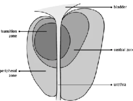
Based on its pathophysiological and embryological characteristics, the prostate is divided into four zones (Fig. 2):
The transition zone: it constitutes the 5% of the gland surrounding the urethra;
The central zone: it accounts for about the 2% of the gland and it is placed under the proximal urethra;
The peripheral zone: it comprises the 70-75% of the gland surrounding the central area and extending to the apex of the gland; The fibromuscular stroma: it is place before the other zones. Most of the prostate cancers (PCa) originate in the peripheral zone, therefore, about the 70% of them are classified as adenocarcinoma [Schulz et al., 2003]. Less common is the possibility that neoplastic transformations could occur in the medial portion or in the transition zone of the gland (20%) that are typical sites of the benign prostatic hyperplasia (BPH). The central area, which constitutes the bulk of the prostate, rarely represent a tumor site (5%), but more often it is invaded by large tumors originated from the neighboring portions [Hising et al., 2006].
1.1.2. Epidemiology of the prostate cancer
PCa is the most common male cancer in Western populations and, after lung cancer and colon-rectum cancer, is the third leading cause of cancer related death [Siegel et al., 2013]. In the USA the highest incidence is found, with 217,730 new cases diagnosed in 2013, on a male population of 150 million people (0.14%) and 32,050 deaths (mortality = 0.025%) [Siegel et al., 2013]. In Europe, however, the incidence of PCa is similar, with 382,000 new diagnosed cases in 2012 on a male population of 360 million people (0.10%) and 87,400 deaths (mortality = 0.024%) [Ferlay et al., 2013]. On the other side, in some countries of Southeast Asia, from two to
ten times lower incidence rates were reported [Sankaranarayanan et al., 2011].
Over the last years, in many Western industrialized countries, an increase in PCa incidence occurred. This may reflect the introduction in the clinical practice of the determination of prostate specific antigen (PSA), in the form of opportunistic screening, with the consequent diagnosis of a higher number of asymptomatic or preclinical PCa forms [Croswell et al., 2011]. The use of this diagnostic tool, however, did not affect the mortality rate for this pathology [Jemal et al., 2010], probably because the majority of PCa identified by the PSA test is not intended to clinically manifest in the course of life even withot the screening [Guidelines AIOM 2009]. Tumors that exhibit this clinical course are called "latent cancers" and they are well documented also by post mortem autopsies. These analyses showed an incidence of PCa of 10-30% in men between 50 and 60 years old and of 50-70% in subjects between 70 and 80 years old [Haas et al., 2008].
1.1.3. Pathogenic mechanisms of PCa
As for the majority of solid tumors, the etiology of PCa is multifactorial as a result of a complex interaction between genetic factors (responsible for the familiar and racial incidence) and environmental factors (related to diet and lifestyle). This disease is steadily increasing and age is one of the most relevant risk factors; the PCa occurrence, in fact, is rare in men under 50 years old, but it increases dramatically after 65 years old, while the higher number is diagnosed between 70 and 74 years old [Carlsson et al., 2014]. Another factor that seems to be important in PCa development is the influence of male sex hormones. Since the prostate is an androgen-dependent gland it develops and maintains its tropism due to testosterone levels. However, there are no definitive data concerning the role of circulating androgens in PCa occurrence [Ismail et al., 2011]. On the other side, environment, lifestyle and diet are well documented risk factors for PCa. A particular work conducted on Asian immigrants moving to the USA, showed that the incidence of PCa increases in men starting from the second generation, thus emphasizing the importance of environmental factors in the development of this disease [Shimizu et al., 1991]. In other studies it was reported that the consumption of red meat in association with smoking, intake of alcohol and obesity, could play a significant role in higher the PCa risk [Meyerhardt et al., 2010]. The consumption of vegetables, however, seems to be important as a protective factor. The low incidence of this pathology in the Asian populations may therefore be related to the low consumption of red meat and the high consumption of vegetables, whose nutritional principles could play a protective role [Desgrandchamps et al., 2010].
From the molecular point of view, many are the mechanisms underlying the PCa onset and progression. In particular, it has been proposed as a recurring
or chronic inflammation may play a pivotal role in the neoplastic transformation [De Marzo et al., 2004]. During the inflammatory response, in fact, cells of the immune system synthesize numerous oxidizing agents capable to induce genetic damages to the resident epithelial cells [Sciarra et al., 2008]. One of the most interesting aspects, in that sense, was the finding of genetic alterations, characterizing the beginning stages of PCa, even in cells affected by inflammatory atrophic processes [Vecchione et al., 2007]. Moreover, at an inflammation level, epithelial cells often show signs of oxidative stress, such as increasing expression level of the glutathione-S-transferase (GSTP1).
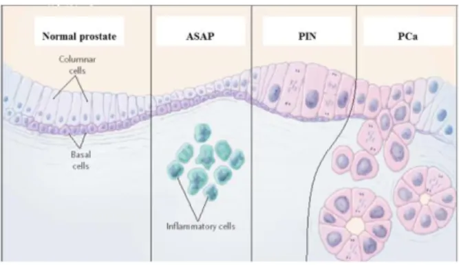
On the other side, histological examination revealed the occurrence of specific lesions of the prostatic glandular tissue, defined as prostatic intraepithelial neoplasia (PIN). Such injuries are attributable to histopathological changes of low grade (LGPIN) or high grade (HGPIN) and, from many authors, they are considered direct precursors of PCa [Dickinson, 2010]. PIN lesions are frequently found in the peripheral zone of the prostate (where most of the PCa originate); the prostatic epithelial cells in those sites showed often the same chromosomal alterations detected in cancer cells. Moreover, cells of the PIN lesions show cell wall alterations similar to those observed in tumor cells; the thickening of the epithelium basal layer can be also be observed (Fig. 5). Changes in gene expression are also showed by the pre-neoplastic cells, as documented by the reduction of the cadherins and cytoskeleton components levels [Nelson et al., 2003].
PIN lesions, on the other side, differ from PCa lesions for the presence of an intact basement membrane that does not allow the invasion of the glandular stroma. Furthermore, these lesions do not produce high levels of PSA and, therefore, they can be detected only by biopsy [Dickinson, 2010].
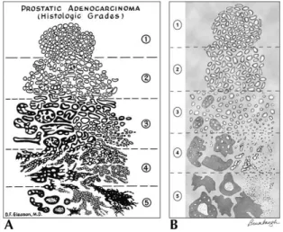
The international reference system used for classifying histologically a PCa is called Gleason system. This system consider the glandular differentiation
degree and the infiltration degree. Figure 4 shows the 5 pattern of increasing aggressiveness considered in the Gleason system. These patterns are classified as follows:
Gleason 1: Tumor composed by well defined, tight, uniform, single and not
confluent glandular nodules.
Gleason 2: Tumor with a minimal extension of to the neoplastic glands
toward the tumor lesion periphery. This lesion is localized in the context of normal tissue.
Gleason 3: Tumor invading the normal tissue; glands show considerable
variability in shape and size.
Gleason 4: Glands with obvious neoplastic confluent alterations.
Sometimes there are cribriform glands with irregular edges.
Gleason 5: Tumor without glandular differentiation, it is characterized by
stretches of anaplastic cells and necrotic areas.
In the Gleason system the main framework (predominant) and the secondary framework (less represented) are considered and to both, a score between 1 and 5, is assigned. 1 indicates the most differentiated and 5 the less differentiated and most aggressive pattern. If a tumor shows a single histological framework, to the primary and secondary pattern the same score is assigned. The two scores are then combined in order to generate the so called Gleason score (Gs), whose value fluctuates from 2 (1 + 1) to 10 (5 + 5), that represent the highest degree of malignancy [Guidelines AIOM 2009].
The diagnosis of PCa is exclusively made by biopsy. The importance of the ultrasound-guided prostate biopsy is due not only to obtain a definitive diagnosis, but also some useful information for guiding the therapeutic strategy [Eichler et al., 2006]. This examination, however, is not diriment in case of a negative report; in fact the 10-30% of patients with a negative biopsy may have a PCa diagnosis in further biopsies [Djavan et al., 2005]. The detection rate for a PCa biopsy depends not only on the sampling technique, but also on the criteria used to perform eventual further biopsies [Eichler et al., 2006]. Moreover, a negative biopsy is usually associated with a risk reduction of finding a high degree PCa in a subsequent biopsy [Borden et al., 2007]. To date, in Italy, it is recommended to repeat a PCa biopsy only one or more of the following indications are satisfied [Guidelines AIOM 2009]:
• Inadequacy of the first biopsy (under 6 sampling, absence of prostate glands, too small fragments);
• Previous histological diagnosis of uncertain or suspicious pre-neoplastic lesions, such as HGPIN or atypical small acinar proliferation (ASAP);
• Progressively increasing PSA serum levels, or changes in digital rectal examination (DRE) results.
During a prostate biopsy is not uncommon to experience adverse events such as pain, hematuria, hematospermia and rectal bleeding; more serious adverse events, such as infections (1.8%) or considerable bleeding (0.6%), are instead infrequent. Complications related to the prostate biopsy brought the researchers to study new strategies to facilitate the PCa diagnosis in order to avoid this invasive clinical practice when it is not firmly recommended [Eichler et al., 2006].
The correct identification of the tumor differentiation state is important to determine the best therapeutic strategy and to obtain prognostic information. Despite considerable advances in imaging technology, it is not yet possible to get these information through such diagnostic tools. In fact, especially for the early stages, PCa can be diagnosed only with a biopsy [Verma et al., 2011].
In the TNM classification the local extension (T), the commitment of the lymph nodes (N) and the presence of distant metastasis (M) are considered. The study of pathological material analyzed after the radical prostatectomy (RP) provides information in the tumor stage definition according to the TNM system that involves the following indications [Schröder et al., 1992]:
Primary tumor (T)
pT2 Organ confined
pT2a Unilateral, involving one-half of 1 lobe or less
pT2b Unilateral, involving more than one-half of 1 lobe but not both lobes
pT2c Bilateral disease pT3 Extraprostatic extension
pT3a Extraprostatic extension or microscopic invasion of the bladder neck
pT3b Seminal vesicle invasion
pT4 Invasion of the bladder and rectum
Regional lymph nodes (N)
pN0 No positive regional nodes pN1 Metastases in regional nodes(s)
Distant metastasis (M)
M0 No distant metastasis M1 Distant metastasis
M1a Non-regional lymph nodes(s) M1b Bone(s)
M1c Other site(s) with or without bone disease
1.1.4. Laboratory medicine
Since it is not expected, at least in the short term, to reach a reduction in PCa incidence through an effective primary prevention, there is no doubt that secondary prevention remains the only available tool to influence the evolution of this disease and reduce, consequently, the PCa-related mortality. Therefore, the best way to obtain an early detection appears to be an individual or population opportunistic screening. The screening test that seems to be the more appropriate for this purpose, considering the cost, convenience, and diagnostic accuracy, is the PSA [Guidelines AIOM 2009]. On the other side, the role of PSA screening in reducing PCa-related mortality is still controversial. In fact, conflicting results have emerged from several observational studies [Crawford et al., 2011; Sciarra et al., 2011]; Therefore, the recommendations for PSA screening differ between the various organizations and scientific societies.
Although the PSA serum test increased the early diagnosis of PCa, a major disadvantage of this marker is its low specificity, which brings, every year, to the execution of a high percentage of negative biopsies (60-75%), especially in patients with PSA levels between 4 and 10 ng/ml, the so called gray zone [Hessels et al., 2009]. The PSA low specificity is due to the fact that its increase in serum is not an event that closely reflects the presence of a PCa, but it can also be found in patients with BPH and prostatitis. Consequently, although the normal cutoff value for PSA is 4 ng/ml, the probability to have a PCa exists even below this threshold, as well as values higher than 4 ng/ml do not necessarily indicate a PCa. Therefore, the strategy to perform a biopsy whenever serum PSA levels increase exposes
the male population to undergo a biopsy that is often useless and linked to several complications [Nogueira et al., 2010 ].
A great effort is therefore constantly turned to the research of new biomarkers, in order to improve the PCa diagnosis and/or the ability to detect the asymptomatic and most aggressive forms. Among the new identified biomarkers, the prostate cancer gene 3 (PCA3) seemed to have good diagnostic potential, giving conflicting results as concerns its prognostic value [Hessels et al., 2009].
The PCA3 gene (also known as DD3 or DD3PCA3) is located on chromosome 9 and is transcribed into a non-coding mRNA which is overexpressed in tumor cells, with a level from 60 to 100 times higher compared to normal cells [Nogueira et al., 2010]. Numerous studies have demonstrated the clinical utility of the PCA3 assay [Ankerst et al., 2008; Deras et al., 2008; Kirby et al., 2009; De la Taille et al., 2011], stressing that this tests could be useful in the following cases [Schilling et al., 2010]: • Males with a high PSA serum levels who underwent one or more
negative biopsies.
• Males with a normal PSA serum levels and a family history of PCa. • Males with high PSA serum levels and a concomitant disease of the
urinary tract.
Some preliminary studies also suggest the utility of the PCA3 assay in discriminating tumors of different aggressiveness [Haese et al., 2008; Nakanishi et al., 2008], even if the most promising are those in which it emerges how the PCA3 test is able to predict a prostate biopsy outcome after a previous negative biopsy [Marks et al., 2007] [Haese et al., 2008]. These studies also contributed to investigate another open question concerning PCA3 test, that is its optimal cutoff. Most of the published data indicated that a threshold of 35 (dimensionless, see paragraph 1.3.1) represents a point in which a better balance between sensitivity and specificity can be found for PCa diagnosis [Kouriefs et al., 2009].
The role of the laboratory medicine is therefore to validate such new biomarkers with the help of well conducted and independent prospective studies, in order to clarify the effective usefulness of their introduction in the clinical practice, in order to do not commit the same errors made with the old biomarkers. In this light, two more markers are currently under investigation in PCa early diagnosis: a particular truncated isoform of the PSA pro-enzyme, the [-2]proPSA (p2PSA) and an adhesion molecule that seems to be involved in PCa progression, the Galectin 3 (Gal3). Immunohistochemical studies showed that p2PSA is the most abundant form of truncated proPSA in tumor tissues [Mikolajczyk et al., 2000] and several studies were able to demonstrate the utility of the serum quantification of this biomarker in patients with serum PSA in the grey zone candidate to a further biopsy after at least previous negative biopsy [Mikolajczyk et al., 2003; Catalona et al., 2003; Sokoll et al., 2003; Sokoll
et al., 2008; Le et al., 2010; Catalona et al., 2011; Guazzoni et al., 2012; Lazzeri et al., 2012]. On the other side Gal3 expression has been reported to vary between healthy and tumor conditions [Takenaka et al., 2004; Balan et al., 2010; Newlaczyl et al., 2011; Wang et al., 2013]. A recent study demonstrated that the expression levels of Gal3 decrease in prostate tumor tissue when compared with normal tissue [Araújo-Filho et al., 2013], while another research found that, in patients with metastatic PCa, Gal3 serum levels were significantly higher than those observed in normal patients, opening, for the first time, to an hypothetical application of this marker in PCa diagnosis [Balan et al., 2013].
In this scenario it is clear that, to date, the possibility to early detect a PCa has increased, since beside the old diagnostic factors, such as the PSA serum levels, the DRE and the diagnostic imaging techniques, some interesting and innovative tests can be used giving a valid support.
1.2 – The prostate specific antigen (PSA)
1.2.1 – Enzymology of the PSA
The prostate specific antigen is a 30 kDa serine protease belonging to the kallikrein family and it’s also known as the kallikrein related peptidase 3 (KLK3). PSA is produced almost exclusively by the prostate glandular cells and it is secreted as part of the seminal fluid in order to keep the semen fluidity after ejaculation.
Like all other members of the kallikrein family, PSA is synthesized in an inactive form as a zymogen which is composed of a pre-peptide (also known as signal peptide) and a pro-peptide (which maintains the enzyme in the latent form). Inside the epithelial cell, the 17 amino acid pre-sequence is first cleaved off by signal peptidases. Afterwards, in the extracellular environment, the additional 7 amino acid pro-sequence is removed by human kallikrein 2 (hK2) [Williams et al., 2007]. PSA shows a conserved position of the Asp102 / His57 / Ser195 catalytic triad [Watt et l., 1986], however, unlike most of kallikreins, which display a trypsin-like proteolytic specificity (i.e., they cleave on the carboxyl side of a positively charged amino acid residue, namely Arg and Lys), PSA shows instead a chymotrypsin-like substrate specificity (i.e., it cleaves on the carboxyl side of a hydrophobic amino acid residue, namely Tyr, Phe, Trp, and Leu). In addition, PSA is the only member of the kallikrein family that catalyzes the cleavage of substrates displaying the Gln residue at the P1 position [LeBeau et al., 2009].
PCa can increase the amount of PSA released into the bloodstream, even though serum PSA is kept inactive in a variety of different forms. As a matter of fact, serum PSA falls into two general categories: the free PSA
(fPSA), which includes all the unbound zymogen forms, and the complexed PSA, where also active forms are kept latent through the binding of serum protease inhibitors. Notably, PSA present in the extracellular fluid, surrounding prostate epithelial cells, has been reported to be enzymatically active, suggesting that its proteolytic activity plays a role in the PCa physiopathology [Denmeade et al., 2001].
The most important physiological substrates for PSA have been proposed to be semenogelin I (SgI) and semenogelin II (SgII). These proteins are synthesized and secreted by the seminal vesicles in spermatic fluid and are involved in the formation of a gel matrix that wraps around ejaculated spermatozoa, preventing their functionalization (mainly via inhibition of reactive oxygen species) [Malm et al., 2000]. The gel matrix breaks down under the PSA enzymatic action, facilitating the spermatozoa movements [Suzuki et al., 2007]. PSA cleaves preferentially the Tyr-Glu peptide bonds and generates multiple soluble fragments of SgI and SgII [Peter et al., 1998] that seem to be the main antibacterial components in human seminal plasma [Edström et al., 2008]. These findings, together with the ability of PSA to process a number of growth regulatory proteins that are important in cancer growth and survival (such as Insulin-like growth factor binding protein, Parathyroid hormone-related protein, latent Transforming growth factor-beta 2 as well as extracellular matrix components, like fibronectin and laminin) [Cohen et al., 1992; Iwamura et al., 1996; Lilja et al., 2000; Dallas et al., 2005], suggest that PSA can facilitate tumor growth and metastasis dissemination [Williams et al., 2007; Webber et al., 1995; Ishii et al., 2004]. On the other hand, PSA has been reported to slow down blood vessel formation, thus playing likely an important role in slowing the growth of prostate cancer [Mattsson et al., 2008]. PSA is synthesized to high levels by normal and malignant prostate epithelial cells and, under pathological conditions, it is abundantly secreted in the extracellular compartments. For this reason, it is the main biomarker currently used for early diagnosis of prostate cancer. Therefore, serum levels of PSA are also useful to detect eventual recurrent forms and to follow up treatment response in not operable and metastatic tumors [Ilic et al., 2013]. As a whole, although PSA is currently used as a PCa biomarker, its role in the PCa pathobiology remains obscure [Williams et al., 2007].
1.2.2 – Clinical use of the PSA
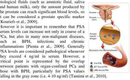
The PSA can be found in the circulation in both free form (fPSA) and conjugated to inhibitors, such as α-1-antichymotrypsin (ACT) and the α-2-macroglobulin (αMG) (Fig. 5). The immunoassays commonly used in todays clinical practice are able to quantify both the fPSA fraction and the one linked to the ACT (tPSA), while they can not measure the PSA linked to the αMG [Shariat et al., 2011]. Although the PSA can be found in other
biological fluids (such as amniotic fluid, saliva and human milk), only the amount produced by the prostate can reach significant blood levels, so it can be considered a prostate specific marker [Kouriefs et al., 2009].
However it is important to remember that PSA serum levels can increase not only in course of a PCa, but also in many non-malignant diseases, such as BPH, infections and chronic inflammations [Pienta et al., 2009]. Generally PSA levels are considered pathological whenever they exceed 4 ng/ml in serum. However, a critical point is represented by the overlap between patients with organ-confined PCa and those with BPH, particularly for PSA values
falling in the gray zone (i.e. 4-10 ng/ml) [Tamimi et al., 2010].
On the other side, it is important to observe that the 25-30% of patients with PCa show PSA values between 2.5 and 4 ng/ml [Hessels et al., 2009]. However, changing the PSA threshold is very risky, in fact, reducing the cutoff to 1.1 ng/ml, the 83.4% of PCa would be diagnosed, but the false positives would be the 61%. Conversely, with a threshold of 3.1 ng/ml, the test sensitivity and specificity would be 32% and 87%, respectively, while using a cutoff of 2.1 ng/ml they would be 53% and 73%, respectively. Today a threshold of 4 ng/ml should therefore be considered a conventional cutoff, characterized by a low predictive value, both negative and positive, no longer suitable for the decision to undergo a biopsy or not [Nogueira et al., 2010].
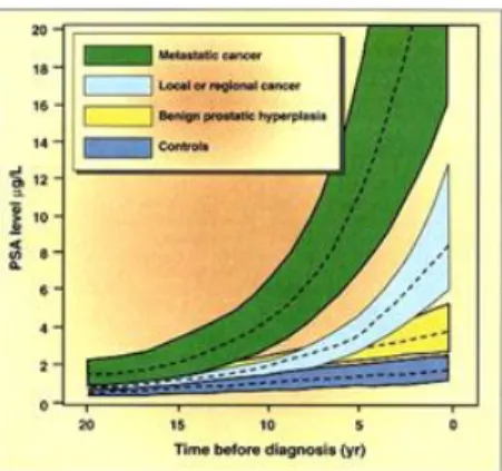
Attempting to improve the specificity of PSA test for the early diagnosis of PCa, some PSA-related parameters were used. The PSA velocity (Fig. 6): it is an index of the increasing rate of PSA over time and is obtained measuring the quantitative annual variation of PSA. This parameter is used to monitor patients with PSA levels in the gray zone. It was observed that in PCa the increase in PSA levels generally exceeds 0.75 ng/ml per year, or it undergoes an annual increase of 20% compared to baseline value. This parameter therefore represent an important diagnostic approach, but it requires careful standardization protocols before a possible routine use. To adopt this criterion, in fact, repeated PSA testing are necessary, for a minimum period of twelve months and preferably for several years. The inability to provide answers of clinical relevance in a short time is, therefore, the limit of this approach [Roobol et al., 2004].
The PSA ratio: it is a mathematical index calculated as fPSA/tPSA and it is also known as percentage of fPSA (%fPSA). For unknown reasons, patients with PCa tend to have a reduced amount of circulating fPSA compared to patients with a benign prostatic disease [Hoffman et al., 2000]. It was demonstrated that the PSA ratio reduces the number of unnecessary biopsies in subjects with tPSA between 4 and 10 ng/ml, but the optimal cutoff, even in this case, is not unanimously agreed [Pepe et al., 2010].
The introduction of PSA serum test in the clinical practice was an important step in the history of oncology; in fact before this test the two/third of the PCa were diagnosed only after metastasization. PSA mass screening improved early diagnosis of PCa, permitting more effective therapeutic interventions [Makarov et al., 2006].
In 2001, the American Cancer Society guidelines, suggested that men after 50 years old and with a normal risk of PCa should carry out an annual PSA and digital rectal testing, anticipating this timing in high-risk subjects. However in order to classify a screening procedure as acceptable it is necessary that its effectiveness, in terms of mortality reduction and cost/benefit, is confirmed by prospective and randomized studies. A large scale clinical trial questioning the real usefulness of PSA screening was conducted in Europe and produced, in 2009, some interesting data concerning the PSA impact on PCa-related mortality. The European Randomized Study of Screening for Prostate Cancer (ERSPC) started in the early 90s and enrolled, in seven European countries, a total of 182,000 individuals from 50 to 74 years old. This men underwent a PSA serum test every four years on average. After a mean follow-up of 9 years, the cumulative incidence of PCa was 8.2% for the PSA screened group and 4.8% in the control group, with a PCa-related mortality rate, between the first and the second group, of 0.80 (p = 0.04). The difference in the absolute death risk, instead, was found to be of 0.71 deaths per 1000 men, indicating
that 1408 people should be screened with PSA to prevent one case of PCa-related death. Authors concluded that beside a reduction of cancer-PCa-related mortality of about 20%, the PSA screening is associated to a high percentage of false positives [Schröder et al., 2009 ].
The potential benefits resulting from a screening program based on the determination of the serum PSA levels are, therefore, still unsure and not supported by clear evidence. The remarkable early diagnosis, the high number of false positives and the latent PCa treatment are, in fact, important negative effects of the PSA screening. Moreover this aspect should be taken together to the inability of this marker to discriminate between patients with aggressive PCa forms from those that are not intended to clinically manifest [Guidelines AIOM 2009].
1.3 – The new PSA-related biomarkers
1.3.1 – The prostate cancer gene 3 (PCA3)
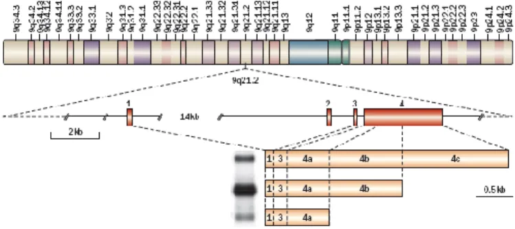
In 1999, a gene specifically expressed in prostate cells was identified, using the differential display analysis, a technique that compares the expression profiles of mRNAs in the tumor tissue to the adjacent normal tissue [Bussemakers et al., 1999]. Using Northern blot analysis, the DD3 (differential display clone 3) was found to be significantly overexpressed in tumor tissue compared to normal tissue from the same patients. In particular, the median expression of this mRNA resulted to be 34 times higher in cancer cells than in normal cells [Hessels et al. , 2003]. According to the current nomenclature of the human genome, the gene was then renamed PCA3 (prostate cancer gene 3), in order to highlight its close relationship with PCa. Using a RT-PCR, it was shown that the PCA3 is a gene specifically expressed in the prostate resulting silent in the other human tissues, although, to date, it is not clear the role of this mRNA in prostate epithelial cells [Day et al., 2011]. PCA3 is a 25 kb gene located on chromosome 9q21-22 and it is composed by four exons. The molecular characterization of the PCA3 transcript revealed that alternative polyadenylation in three different positions of exon 4 could generate different transcripts. Furthermore, an alternative splicing event, may give a transcript in which exon 2 is totally deleted. The transcript that is found more frequently in prostate cells, however, contains exons 1, 3, 4a and 4b (Fig. 7).
The absence of an Open Reading Frame (ORF) and the presence of stop codons which interrupt the protein structure, indicate that the PCA3 does not encode for a specific protein and that its transcript is not then translated [Bussemakers et al., 1999]. Even if the role of this gene is still unknown, it was proposed that its transcript could be implicated in gene expression or splicing regulation [Hessels et al., 2009]. Recently, it was demonstrated that the PCA3 gene is incorporated in the intron 6 of a second gene, BMCC1, implicated in the control of normal cells transformation into cancer cells [Clarke et al., 2009].
The association between PCA3 increased expression levels and PCa highlighted the potential of its mRNA as oncologic marker [Deras et al., 2008]. In 2006 a commercial kit, approved by the Food and Drug Administration (FDA), was produced in order to quantify the number of PCA3-mRNA copies in urine samples. This test is based on the technology of the transcription-mediated amplification (TMA) and it was called PROGENSA PCA3 assay (Gen-Probe Inc., San Diego, CA, USA). This test allows the quantification of the number of PSA-mRNA copies too, in order to obtained the PCA3 score calculated as PCA3-mRNA copies per ml/ PSA-mRNA copies per ml x 1000 [Groskopf et al., 2006]. The number of mRNA-PSA copies is an index of the amount of nuclear material derived from prostate cells in the urine sample, so the PCA3 score gives the expression of the PCA3 gene corrected for the amount of prostate cells in the sample, estimated through the evaluation of the mRNA-PSA copies. The cutoff for this test was set at 35, a value that seemed to give the balance in terms of sensitivity and specificity [Kouriefs et al., 2009]. To date, many studies have been performed and most of them showed how the PCA3 test represented a useful tool to predict PCa, but questions about the optimal cutoff and the ability of PCA3 to predict tumor aggressiveness still remain highly controversial [Day et al., 2011; Luo et al., 2014].
Several studies suggested that the threshold of 35 proposed by Gen-Probe Inc., using the PROGENSA PCA3 assay, could be modified, getting lower
or even higher, in a way that is probably dependent on the population features. In this respect, the cutoff value of 20 seems to increase the PCA3 test sensitivity without affecting the specificity [Hessels et al., 2003; Van Gils et al., 2008; Haese et al., 2008; Bollito et al., 2012; Filella et al., 2013; Gittelman et al., 2013]. Some studies also demonstrated that PCA3 is effective only after the first negative biopsy, but a recently published meta-analysis showed that PCA3 can be used for repeat biopsy to improve accuracy of PCa detection, since a large number of unnecessary biopsies can be avoided by using a PCA3 score cutoff of 20 [Fall et al., 2007; Luo et al., 2014].
The second debated aspect in which scientists focused in the last period concerns the possible association between the PCA3 score and the tumor stage. The PCA3 score is strongly associated to the fraction of cancer cells in the urine sample as a result of the DRE. In this view, larger and more aggressive tumors could release more easily a wider number of neoplastic cells respect to smaller and less aggressive PCa forms, producing higher values of PCA3 score [Hessels et al., 2010]. Many authors attempted to validate this hypothesis by evaluating the association between PCA3 score and tumor volume, measured after radical prostatectomy (RP), and other clinical and pathological PCa features, often reporting conflicting results [Van Gils et al, 2008]. From this point of view it is well known that subjects with organ-confined PCa and Gs ≥ 7 have a worst prognosis than those with Gs ≤ 6, even following RP or radiation therapy [Heidenreich et al., 2011; Albertsen et al., 2011; van den Bergh et al., 2014]. To recognize a low grade from a more aggressive PCa is therefore essential for therapeutic purposes, but currently the only way to discriminate patients with low or high grade PCa is to perform a biopsy. The possibility of using the PCA3 test as a prognostic marker is desirable, but the possibility to evaluate tumor aggressiveness by the PCA3 test is openly debated [Auprich et al., 2011; Haese et al., 2008; Filella et al., 2013; van Poppel et al., 2012; Hessels et al., 2010; Durand et al., 2012; Liss et al., 2011; Auprich et al., 2011; Nakanishi et al., 2008]. Indeed, the wide range of results obtained in previous studies may be due to different experimental conditions and may reflect the selected cohort features. In fact, the use of urine sediments or whole urine samples, collected before or without a previous DRE, can give rise to different results that are not often comparable in judging the prognostic value capabilities of the PCA3 test. On the other hand, the characteristics of the screened population could be important too. In fact, the choice to enroll only patients with a certain risk for PCa, or depending on the number of previous biopsies, can drive data towards an easier or less easy association between the result of the PCA3 test and the tumor aggressiveness.
1.3.2 – The [-2]proPSA
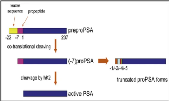
PSA is normally secreted from the prostatic epithelial cells as proPSA, an inactive proenzyme containing 244 amino acids. Once released into the prostate lumen, the 7-amino acid peptide is eliminated extracellularly by human kallikrein enzymes hK-2 and hK-4, becoming the active or mature form of PSA with 237 amino acids. The forms with some part of the peptide yet bound to them remain as proPSA (Fig. 8) [Mikolajczyk et al., 2001].
In proPSA, the smaller the part bound to the peptide in the leader region, the more difficult it is to activate. This makes the isoform of proPSA containing 2 residues in the leader region (the [-2]proPSA) the most stable component of proPSA in the serum. The [-2]proPSA (p2PSA) is produced much more in the periphery of the prostate, particularly under neoplastic conditions, so, although other proPSA isoforms maybe present in significant levels in serum samples, p2PSA appears to be more consistently correlated with PCa [Mikolajczyk et al., 2000]. In men with PSA levels between 6.0 and 24.0 ng/ml, the p2PSA fraction was found to be significantly higher in men with PCa and some prospective studies demonstrated that p2PSA better discriminated between PCa and benign disease compared to PSA and PSA ratio [Mikolajczyk et al., 2002].
Further studies evidenced that, among men with PSA levels between 2 and 10 ng/ml, a combination of p2PSA and fPSA, the so called percentage of p2PSA (%p2PSA = p2PSA/fPSA) was more cancer-specific than PSA and PSA ratio [Catalona et al., 2003]. Moreover, subsequent studies demonstrated a correlation between p2PSA levels and clinically significant cancer, including more advanced pathologic stage, higher tumor volume, and higher tumor grade [Catalona et al., 2004]. In addition, more recently Beckman Coulter Inc. has also developed a mathematical formula combining tPSA, fPSA and p2PSA: the Beckman Coulter prostate health index (PHI = (p2PSA/fPSA)× tPSA1/2). This mathematical regression model was approved by the FDA in June 2012 and provided a better overall result for PCa discrimination in the tPSA range of 2-10 ng/ml [Le et al., 2010;
Guazzoni et al., 2011; Romero Otero et al., 2014]. In this light, the use of p2PSA, either incorporated in %p2PSA or PHI, provides superior discrimination between PCa and benign disease in men with tPSA levels of 2.5 to 10 ng/ml and negative DRE; however, confirmatory validation studies are needed to determine the optimal incorporation of this marker into clinical practice, as well as to definitively assess its ability in the identification of the most aggressive PCa forms.
1.3.3 – The Galectin 3
Galectin 3 (Gal3) is one of the proteins which can be cleaved by PSA. It is a unique chimera-type member of the galectin family, which contains a small N-terminal part, collagen-like sequence, and carbohydrate-binding domain similar to other galectins. Gal3 is the only member of the galectin family that can form oligomers through intermolecular interactions involving the collagen-like sequence [Hirabayashi et al., 1998; Barondes et al., 1994; Barondes et al., 1994]. The collagen-like sequence, rich in proline, tyrosine, and glycine residues contributes to self-aggregation [Lepur et al., 2012]. Until today, the three-dimensional structure of intact Gal3is unknown. However, the X-ray crystal structure of its carbohydrate recognition domain (CRD) was resolved, showing high similarity to the structure of CRD domains of other galectins [Seetharaman et al., 1998]. The unfolded structure of collagen-like sequence, which probably exhibits random-coil conformation, opens this sequence to different post-translational modifications, such as phosphorylation and cleavage by proteases, which in turn change the ability of Gal3 to create oligomers and change the localization in the cell.
Gal3 is mainly a cytosolic protein that often can be found in the nucleus and is secreted outside of the cell despite the fact that it lacks the classical leader signals at the N-terminal [Strik et al., 2001; Davidson et al., 2002; Yu et al., 2002]. Lack of Gal3 in knockout mice is associated with reduced mast cell function, reduced accumulation of asthma-associated leukocytes in airway inflammation and reduced peritoneal inflammatory responses. Endogenous Gal3 has also been shown to play a role in phagocytosis by macrophages and can mediate cytokine production by mast cells when functioning intracellularly [Cummings et al., 2009]. The fact that galectin-3 knockout mice do not show more drastic phenotypic changes leads to the assumption that other galectins can take over the role of Gal3. Most adult tissues without Gal3 do not show pathological changes; however, its role is more obvious in inflammatory responses, cell proliferation, motility, and apoptosis [Dumic et al., 2006].
This protein can be found in a wide variety of tissues as well as in blood. Experimental data available today demonstrate an association between Gal3 levels (in terms of up-regulation as well as down-regulation) and numerous
pathological conditions such as heart failure, infection with microorganisms, diabetes, and tumor progression [Chen et al., 2005; Takenaka et al., 2004; Shekhar et al., 2004; Puglisi et al., 2004; Califice et al., 2004; Balan et al., 2010; Newlaczyl et al., 2011]. Outside of the cell Gal3 is involved with a variety of extracellular functions such as cell adhesion, migration, invasion, angiogenesis, immune functions, apoptosis, and endocytosis [Ochieng et al., 2004; Nangia-Makker et al., 2008]. Experimental and clinical data demonstrate a correlation between galectin expression and tumor progression and metastasis, and therefore, galectins have the potential to serve as reliable tumor markers [Balan et al., 2010]. The expression of Gal3 in PCa is controversial. Previously published work demonstrated that expression of Gal3 was significantly decreased compared with normal and pre-malignant tissue [Araújo-Filho et al., 2013]. However, another study demonstrated an increased cleavage of Gal3 during the progression of PCa. This data implicate Gal3 in PCa progression and suggest that this protein may serve as both a diagnostic marker and a therapeutic target for future disease treatments [Wang et al., 2009]. To confirm this hypothesis, a recent work showed a significant increase of Gal3 serum levels in patients with metastatic PCa compared to normal patients [Balan et al., 2013]. It is therefore now essential to understand whether this marker can also discriminate PCa patients (even at an organ-confined tumor stage) and patients with benign prostatic pathologies, as well as it could work as a standalone marker or only in combination with the PSA, as the other previously screened biomarkers, resulting then only complementary and not substitutive of the PSA test.
1.4 – PCa treatment: the radical prostatectomy (RP)
Although some controversies remain over ideal diagnostic and treatment strategies for PCa, complete removal of the prostate remains the gold standard in the surgical management of localized disease. Hugh Hampton Young first described the perineal prostatectomy over 100 years ago in 1905 [Young, 1905]. Subsequently, the first retropubic radical prostatectomy (RRP) was performed by Millin in 1947 [Millin, 1947]. Anatomic studies in the 1970s and early 1980s led to improved appreciation of periprostatic features (dorsal venous complex, endopelvic fascia, autonomic innervation, and striated sphincter) to decrease morbidity of surgery and improve overall outcomes [Walsh, 1998; Bianco et al., 2005]. More recently, in 1997, Schuessler et al. described the first laparoscopic radical prostatectomy (LRP) reporting the feasibility of technique despite its association with long operative times [Schuessler et al., 1997]. Since that time, numerous European and US centers continued to improve and refine
technical aspects of the laparoscopic approach [Guillonneau et al., 1999; Touijer et al., 2005]. Several robotic systems were introduced around the turn of the century. The da Vinci system (Intuitive Surgical Inc, CA, USA) was first introduced in 1999. Following a merger with Computer Motion Inc. (AESOP and ZEUS systems) in 2003, Intuitive Surgical has become the sole producer of robotic surgical devices [Yates, et al., 2011]. After initially embarking into cardiothoracic surgery, the da Vinci robot found popularity within the urological community. From the initial descriptions of robot assisted laoaroscopic prostatectomy (RALP) in 2000 [Abbou et al., 2001; Binder et al., 2001], it has become widely adopted by urologists. By 2008, roughly 80% of RPs in the United States were performed robotically [Freire et al., 2010]. RALP has continued to evolve rapidly since that time with contributions including procedural step by steps, technical modifications, and outcomes data from various surgeons throughout the literature.
The surgical resection of a tumor in the absence of distal lymph node metastases is generally resolutive. However, some neoplasia, including PCa, are able to generate metastases after a few years, even if they are not detected at the moment of the surgery [Roato et al., 2008; Gupta et al., 2010; Pietras et al., 2010, Meyer et al., 2010; Roberts et al., 2013]. Accordingly, some investigations have suggested that a number of factors in the perioperative period could promote metastasization. These include the surgery approach and its associated stress response, the anaesthetic regimen, the acute pain and the administration of opioid analgesics, all of which could induce the liberation of angiogenic factors [Condon et al., 2004; Lee et al., 2009; Gottschalk et al., 2010; Mao et al., 2013]. One of the most affective factors seemed to be the anaesthetic regimen and this opened a wide debate regarding the use of a regional anaesthesia (RA) in place of the classic general anaesthesia (GA). In particular, RA, aside from reducing the amount of intra-operatively required GA and postoperative opioid consumption, has been consistently shown to attenuate the neuroendocrine response to surgery and, therefore, peri-operative immunosuppression [Melamed et al., 2003; Snyder et al., 2010; Mao et al., 2013]. Recent retrospective analyses indicate that RA for breast and prostate cancer surgery is associated with a markedly reduced risk of tumor recurrence and metastasization compared to systemic opioid administration [Exadaktylos et al., 2006; Biki et al., 2008]. This finding strengthens the hypothesis that paravertebral RA might reduce the incidence of metastases and recurrences compared to GA. Deepening these issues could be very important to understand if some anaesthetic regimens (e.g. GA and RA) or RP technique (e.g. LRP and RALP) could promote angiogenesis in vivo and if the administration of anti-angiogenic agents could be helpful in preventing the pro-metastasization events derived from the peri-operative manipulations.
1.5 - Aims of the thesis
Prostate cancer is the most common cancer in men of the Western countries [Siegel et al., 2013], but the molecular mechanisms of prostate cells neoplastic transformation are still unclear [Koul et al., 2010]. Despite the high incidence rate, an early diagnosis followed by surgery, radiotherapy or chemotherapy could be resolutive for this pathology [Roberts et al., 2013]. In this context, researches focused on the PCa pathogenic mechanisms, the evaluation of new PCa-related markers and less invasive PCa surgical methods, hoping to reach a positive impact on PCa-related recurrence and mortality [Kirby, 2014]. Early detection of PCa is currently based on the trans-rectal ultrasound scan, the digital rectal examination and the evaluation of the PSA serum levels [Romero et al., 2014]. PSA is a serine proteases that was found to be able to cleave a number of growth regulatory proteins that are important in cancer growth and survival, so it was related to tumor growth and metastasis dissemination [Ishii et al., 2004]. PSA serum level, on the other side, has been used for several decades as the only PCa-related marker, but in the last years clear limits for this test were demonstrated [Schröder et al., 2009; Crawford et al., 2011]. New PCa-related biomarkers have been intensively scrutinized over the last decade, but despite the new findings and the disclosure of some positive diagnostic performances, they are currently not uniformly accepted in clinical practice [Romero et al., 2014]. Even if the PSA serum test is no longer considered a helpful tool for PCa early diagnosis it is still a fundamental marker of PCa recurrence after surgical prostate removal [Carthon et al., 2013]. At the same time, a debate on whether some surgery approaches or anaesthetic drugs in PCa patient management could increase the risk of tumor spreading and metastasization was recently opened [Lee et al., 2009; Mao et al., 2013].
In light of these considerations, the present thesis aims to approach the PCa pathology from different points of view, trying to i) reveal new insights for the catalytic mechanism of PSA, ii) evaluate the PSA clinical utility as a diagnostic test, as well as the diagnostic and prognostic potential of new PSA-related biomarkers such as PCA3, p2PSA and Gal3, iii) to investigate changes in activation markers of the haemostatic system, endothelium and angiogenesis in patients with PCa undergoing different laparoscopic radical prostatectomy protocol and intra-operative anaesthetic regimens.
References
Abbou CC, Hoznek A, Salomon L, Olsson LE, Lobontiu A, Saint F, Cicco A, Antiphon P, Chopin D. Laparoscopic radical prostatectomy with a remote controlled robot. (2001). Journal of Urology, 165(6):1964–6. Albertsen PC, Moore DF, Shih W, Lin Y, Li H, Lu-Yao GL. Impact of
comorbidity on survival among men with localized prostate cancer. (2011). J Clin Oncol, 29(10):1335-41.
Ankerst DP, Groskopf J, Day JR, Blase A, Rittenhouse H, Pollock BH, Tangen C, Parekh D, Leach RJ, Thompson I. Predicting prostate cancer risk through incorporation of prostate cancer gene 3. (2008). J Urol, 180(4):1303-8.
Arújo-Filho JL, Melo-Junior MR, Carneiro Beltrão EI, Amorim de Lima LR, Lins Antunes CB, Bezerra de Carvalho L. Immunochemiluminescent detection of galectin-3 in tumoral tissue from prostate. (2013). Int J Clin Exp Pathol, 6(9):1861-7.
Auprich M, Bjartell A, Chun FK, de la Taille A, Freedland SJ, Haese A, Schalken J, Stenzl A, Tombal B, van der Poel H. Contemporary role of prostate cancer antigen 3 in the management of prostate cancer. (2011). Eur Urol, 60(5):1045-54.
Auprich M, Chun FK, Ward JF, Pummer K, Babaian R, Augustin H, Luger F, Gutschi S, Budäus L, Fisch M, Huland H, Graefen M, Haese A. Critical assessment of preoperative urinary prostate cancer antigen 3 on the accuracy of prostate cancer staging. (2011). Eur Urol, 59(1):96-105.
Balan V, Nangia-Makker P, Raz A. Galectins as Cancer Biomarkers. (2010). Cancers, 2(2):592-610.
Balan V, Wang Y, Nangia-Makker P, Kho D, Bajaj M, Smith D, Heilbrun L, Raz A, Heath E. Galectin-3: a possible complementary marker to the PSA blood test. (2013). Oncotarget, 4(4):542-9.
Barondes SH, Castronovo V, Cooper DN, Cummings RD, Drickamer K, Feizi T, Gitt MA, Hirabayashi J, Hughes C, Kasai K. Galectins: a family of animal beta-galactoside-binding lectins. (1994). Cell, 76(4):597-8.
Barondes SH, Cooper DN, Gitt MA, Leffler H. Galectins. Structure and function of a large family of animal lectins. (1994). J Biol Chem, 269(33):20807-10.
Bianco FJ, Scardino PT, Eastham JA. Radical prostatectomy: long-term cancer control and recovery of sexual and urinary function (‘trifecta’). (2005). Urology, 66(5):83–94.
Biki B, Mascha E, Moriarty DC, Fitzpatrick JM, Sessler DI, Buggy DJ. Anesthetic technique for radical prostatectomy surgery affects cancer
recurrence: a retrospective analysis. (2008). Anesthesiology, 109:180-7.
Binder J, Kramer W. Robotically-assisted laparoscopic radical prostatectomy. (2001). BJU International, 87(4):408–10.
Bollito E, De Luca S, Cicilano M, Passera R, Grande S, Maccagnano C, Cappia S, Milillo A, Montorsi F, Scarpa RM, Papotti M, Randone DF. Prostate cancer gene 3 urine assay cutoff in diagnosis of prostate cancer: a validation study on an Italian patient population undergoing first and repeat biopsy. (2012). Anal Quant Cytol Histol, 34(2):96-104. Borden LS Jr, Wright JL, Kim J, Latchamsetty K, Porter CR. An abnormal digital rectal examination is an independent predictor of Gleason > or =7 prostate cancer in men undergoing initial prostate biopsy: a prospective study of 790 men. (2007). BJU Int, 99(3):559-63.
Bussemakers MJ, van Bokhoven A, Verhaegh GW, Smit FP, Karthaus HF, Schalken JA, Debruyne FM, Ru N, Isaacs WB. DD3: a new prostate-specific gene, highly overexpressed in prostate cancer. (1999). Cancer Res, 59(23):5975-9.
Califice S, Castronovo V, Van Den Brule F. Galectin-3 and cancer. (2004). Int J Oncol, 25(4):983-92.
Carlsson S, Assel M, Sjoberg D, Ulmert D, Hugosson J, Lilja H, Vickers A. Influence of blood prostate specific antigen levels at age 60 on benefits and harms of cancer screening: population based cohort study. (2014). BMJ Int, 348:g2296.
Carter HB, Ferrucci L, Kettermann A, Landis P, Wright EJ, Epstein JI, Trock BJ, Metter EJ. Detection of life-threatening prostate cancer with prostate-specific antigen velocity during a window of curability. (2006). J Natl Cancer Inst, 98(21):1521-7
Carthon BC, Marcus DM, Herrel LA, Jani AB, Rossi PJ, Canter DJ. Therapeutic options for a rising PSA after radical prostatectomy. (2013). Can J Urol, 20(3):6748-55.
Catalona WJ, Bartsch G, Rittenhouse HG, Evans CL, Linton HJ, Amirkhan A, Horninger W, Klocker H, Mikolajczyk SD. Serum pro prostate specific antigen improves cancer detection compared to free and complexed prostate specific antigen in men with prostate specific antigen 2 to 4 ng/ml. (2003). J Urol, 170(6):2181-5.
Catalona WJ, Bartsch G, Rittenhouse HG, Evans CL, Linton HJ, Horninger W, Klocker H, Mikolaiczyk SD. Serum pro-prostate specific antigen preferentially detects aggressive prostate cancers in men with 2 to 4 ng/ml prostate specific antigen. (2004). J Urol, 171(6):2239-44.
Catalona WJ, Partin AW, Sanda MG, Wei JY, Klee GG, Bangma CH, Slawin KM, Marks LS, Loeb S, Broyles DL, Shin SS, Cruz AB, Chan DW, Sokoll LJ, Roberts WL, van Schaik RH, Mizrahi IA. A multicenter study of [-2]pro-prostate specific antigen combined with
prostate specific antigen and free prostate specific antigen for prostate cancer detection in the 2.0 to 10.0 ng/ml prostate specific antigen range. (2011). J Urol, 185(5):1650–5.
Chen HY, Liu FT, Yang RY. Roles of galectin-3 in immune responses. (2005). Arch Immunol Ther Exp (Warsz), 53(6):497-504.
Clarke RA, Zhao Z, Guo AY, Roper K, Teng L, Fang ZM, Samaratunga H, Lavin MF, Gardiner RA. New genomic structure for prostate cancer specific gene PCA3 within BMCC1: implications for prostate cancer detection and progression. (2009). PLoS One, 4(3):4995.
Cohen P, Graves HC, Peehl DM, Kamarei M, Giudice LC, Rosenfeld RG. Prostate-specific antigen (PSA) is an insulin-like growth factor binding protein-3 protease found in seminal plasma. (1992). J Clin Endocrinol Metab, 75:1046-53.
Condon ET, Wang JH, Redmond HP. Surgical injury induces the mobilization of endothelial progenitor cells. (2004). Surgery, 135(6):657-61.
Crawford ED, Grubb R 3rd, Black A, Andriole GL Jr, Chen MH, Izmirlian G, Berg CD, D'Amico AV. Comorbidity and mortality results from a randomized prostate cancer screening trial. (2011). J Clin Oncol, 29(4):355-61.
Croswell JM, Kramer BS, Crawford ED. Screening for prostate cancer with PSA testing: current status and future directions. (2011). Oncology, 25(6):452-60.
Cummings RD, Liu FT. Galectins. In: Varki A, Cummings RD, Esko JD, Freeze HH, Stanley P, Bertozzi CR, Hart GW and Etzler ME. (2009). Essentials of Glycobiology, chapter 33.
Dallas SL, Zhao S, Cramer SD, Chen Z, Peehl DM, Bonewald LF. Preferential production of latent transforming growth factor beta-2 by primary prostatic epithelial cells and its activation by prostate-specific antigen. (2005). J Cell Physiol, 202(2):361-70.
Davidson PJ, Davis MJ, Patterson RJ, Ripoche MA, Poirier F, Wang JL. Shuttling of galectin-3 between the nucleus and cytoplasm. (2002). Glycobiology, 12(5):329-337.
Day JR, Jost M, Reynolds MA, Groskopf J, Rittenhouse H. PCA3: from basic molecular science to the clinical lab. (2011). Cancer Lett, 301(1):1-6.
De la Taille A, Irani J, Graefen M, Chun F, de Reijke T, Kil P, Gontero P, Mottaz A, Haese A. Clinical evaluation of the PCA3 assay in guiding initial biopsy decisions. (2011). J Urol, 185(6):2119-25.
De Marzo AM, DeWeese TL, Platz EA, Meeker AK, Nakayama M, Epstein JI, Isaacs WB, Nelson WG. Pathological and molecular mechanisms of prostate carcinogenesis: implications for diagnosis, detection, prevention, and treatment. (2004). J Cell Biochem, 91(3):459-77.
Denmeade SR, Sokoll LJ, Chan DW, Khan SR, Isaacs JT. Concentration of enzymatically active prostate-specific antigen (PSA) in the extracellular fluid of primary human prostate cancers and human prostate cancer xenograft models. (2001). Prostate, 48(1):1-6.
Deras IL, Aubin SM, Blase A, Day JR, Koo S, Partin AW, Ellis WJ, Marks LS, Fradet Y, Rittenhouse H, Groskopf J. PCA3: a molecular urine assay for predicting prostate biopsy outcome. (2008). J Urol, 179(4):1587-92.
Desgrandchamps F, Bastien L. Nutrition, dietary supplements and prostate cancer. (2010). Prog Urol, 20(8):560-5.
Dickinson SI. Premalignant and malignant prostate lesions: pathologic review. (2010). Cancer Control, 17(4):214-22.
Djavan B, Milani S, Remzi M. Prostate biopsy: who, how and when. An update. (2005). Can J Urol, 12(1):44-8.
Dumic J, Dabelic S, Flögel M. Galectin-3: an openended story. (2006). Biochim Biophys Acta, 1760(4):616-635.
Durand X, Xylinas E, Radulescu C, Haus-Cheymol R, Moutereau S, Ploussard G, Forgues A, Robert G, Vacherot F, Loric S, Allory Y, Ruffion A, de la Taille A. The value of urinary prostate cancer gene 3 (PCA3) scores in predicting pathological features at radical prostatectomy. (2012). BJU Int, 110(1):43-9.
Edström AM, Malm J, Frohm B, Martellini JA, Giwercman A, Morgelin M, Cole AM, Sorensen OE. The major bactericidal activity of human seminal plasma is zinc-dependent and derived from fragmentation of the semenogelins. (2008). J Immunol, 181(5):3413-21.
Eichler K, Hempel S, Wilby J, Myers L, Bachmann LM, Kleijnen J. Diagnostic value of systematic biopsy methods in the investigation of prostate cancer: a systematic review. (2006). J Urol, 175(5):1605-12. Exadaktylos AK, Buggy DJ, Moriarty DC, Mascha E, Sessler DI. Can
anesthetic technique for primary breast cancer surgery affect recurrence or metastasis?. (2006). Anesthesiology, 105:660-4.
Fall K, Garmo H, Andrèn O, Bill-Axelson A, Adolfsson J, Adami HO, Johansson JE, Holmberg L. Scandinavian Prostate Cancer Group Study No. 4. Prostate-specific antigen levels as a predictor of lethal prostate cancer. (2007). J Natl Cancer Inst, 99(7):526-32.
Ferlay J, Steliarova-Foucher E, Lortet-Tieulent J, Rosso S, Coebergh JWW, Comber H, Forman D, Bray F. Cancer incidence and mortality patterns in Europe: Estimates for 40 countries in 2012. (2013). Eur J Cancer, 49:1374-1403.
Filella X, Foj L, Milà M, Augé JM, Molina R, Jiménez W. PCA3 in the detection and management of early prostate cancer. (2013). Tumour Biol, 34(3):1337-47.
Freire MP, Choi WW, Lei Y, Carvas F, Hu JC. Overcoming the learning curve for robotic-assisted laparoscopic radical prostatectomy. (2010). Urologic Clinics of North America, 37(1):37–47.
Gittelman MC, Hertzman B, Bailen J, Williams T, Koziol I, Henderson RJ, Efros M, Bidair M, Ward JF. PCA3 molecular urine test as a predictor of repeat prostate biopsy outcome in men with previous negative biopsies: a prospective multicenter clinical study. (2013). J Urol, 190(1):64-9.
Gottschalk A, Sharma S, Ford J, Durieux ME, Tiouririne M. Review article: the role of the perioperative period in recurrence after cancer surgery. (2010). Anesth Analg, 110:1636-43.
Groskopf J, Aubin SM, Deras IL, Blase A, Bodrug S, Clark C, Brentano S, Mathis J, Pham J, Meyer T, Cass M, Hodge P, Macairan ML, Marks LS, Rittenhouse H. APTIMA PCA3 molecular urine test: development of a method to aid in the diagnosis of prostate cancer. (2006). Clin Chem, 52(6):1089-95.
Guazzoni G, Lazzeri M, Nava L, Lughezzani G, Larcher A, Scattoni V, Gadda GM, Bini V, Cestari A, Buffi NM, Freschi M, Rigatti P, Montorsi F. Preoperative prostate-specific antigen isoform p2PSA and its derivatives, %p2PSA and prostate health index, predict pathologic outcomes in patients undergoing radical prostatectomy for prostate cancer. (2012). Eur Urol, 61(3):455–66.
Guazzoni G, Nava L, Lazzeri M, Scattoni V, Lunghezzani G, Maccagnano C, Dorigatti F, Ceriotti F, Pontillo M, Bini V, Freschi M, Montorsi F, Rigatti P. Prostate-specific antigen (PSA) isoform p2PSA significantly improves the prediction of prostate cancer at initial extended prostate biopsies in patients with total PSA between 2.0 and 10 ng/ml: results of a prospective study in a clinical setting. (2011). Eur Urol, 60(2):214–22.
Guidelines AIOM 2009. The prostate cancer.
Guillonneau B, Cathelineau X, Barret E, Rozet F, Vallancien G. Laparoscopic radical prostatectomy: technical and early oncological assessment of 40 operations. (1999). European Urology, 36(1):14–20. Gupta SC, Kim JH, Prasad S, Aggarwal BB. Regulation of survival,
proliferation, invasion, angiogenesis, and metastasis of tumor cells through modulation of inflammatory pathways by nutraceuticals. (2010). Cancer and Metastasis Reviews, 29(3):405–34.
Haas GP, Delongchamps N, Brawley OW, Wang CY, de la Roza G. The worldwide epidemiology of prostate cancer: perspectives from autopsy studies. (2008). Can J Urol, 15(1):3866-71.
Haese A, de la Taille A, van Poppel H, Marberger M, Stenzl A, Mulders PF, Huland H, Abbou CC, Remzi M, Tinzl M, Feyerabend S, Stillebroer AB, van Gils MP, Schalken JA. Clinical utility of the PCA3 urine
assay in European men scheduled for repeat biopsy. (2008). Eur Urol, 54(5):1081-8.
Heidenreich A, Bellmunt J, Bolla M, Joniau S, Mason M, Matveev V, Mottet N, Schmid HP, van der Kwast T, Wiegel T, Zattoni F. European Association of Urology: EAU guidelines on prostate cancer. Part 1: screening, diagnosis, and treatment of clinically localised disease. (2011). Eur Urol, 59(1):61-71.
Helpap B, Egevad L. Correlation of modified Gleason grading with pT stage of prostatic carcinoma after radical prostatectomy. (2008). Anal Quant Cytol Histol, 30(1):1-7.
Hessels D, Klein Gunnewiek JMT, van Oort I, Karthaus HFM, van Leenders GJL, van Balken B, Kiemeney LA, Witjes JA, Schalken JA. DD3(PCA3)-based molecular urine analysis for the diagnosis of prostate cancer. (2003). Eur Urol, 44(1):8-16.
Hessels D, Schalken JA. The use of PCA3 in the diagnosis of prostate cancer. (2009). Nat Rev Urol, 6(5):255-61.
Hessels D, van Gils MP, van Hooij O, Jannink SA, Witjes JA, Verhaegh GW, Schalken JA. Predictive value of PCA3 in urinary sediments in determining clinico-pathological characteristics of prostate cancer. (2010). Prostate, 70(1):10-6.
Hirabayashi J, Kasai KI. Evolution of animal lectins. (1998). Prog Mol Subcell Biol, 19:45-88.
Hising AW, Chokkalingam AP. Prostate cancer epidemiology. (2006). Front Biosci, 11:1388-413.
Hoffman RM, Clanon DL, Littenberg B, Frank JJ, Peirce JC. Using the free-to-total prostate-specific antigen ratio to detect prostate cancer in men with nonspecific elevations of prostate-specific antigen levels. (2000). J Gen Intern Med, 15(10):739-48
Ilic D, Neuberger MM, Djulbegovic M, Dahm P. Screening for prostate cancer. (2013). Cochrane systematic review, 1:CD004720.
Ishii K, Otsuka T, Iguchi K, Usui S, Yamamoto H, Sugimura Y, Yoshikawa K, Hayward SW, Hirano K. Evidence that the prostate-specific antigen (PSA)/Zn2+ axis may play a role in human prostate cancer cell invasion. (2004). Cancer Lett, 207(1):79-87.
Ismail M, Ferroni M, Gomella LG. Androgen suppression strategies for prostate cancer: is there an ideal approach?. (2011). Curr Urol Rep, 12(3):188-96.
Iwamura M, Hellman J, Cockett AT, Lilja H, Gershagen S. Alteration of the hormonal bioactivity of parathyroid hormone-related protein (PTHrP) as a result of limited proteolysis by prostate-specific antigen. (1996). Urology, 48(2):317-325.
Jansen FH, Roobol M, Jenster G, Schröder FH, Bangma CH. Screening for prostate cancer in 2008 II: the importance of molecular subforms of
prostate-specific antigen and tissue kallikreins. (2009). Eur Urol, 55(3):563-74.
Jemal A, Siegel R, Xu J, Ward E. Cancer statistics, 2010. (2010). CA Cancer J Clin, 60(5):277-300.
Kirby R. Optimising the management of early prostate cancer. (2014). Practitioner, 258(1770):15-8.
Kirby RS, Fitzpatrick JM, Irani J. Prostate cancer diagnosis in the new millennium: strengths and weaknesses of prostate-specific antigen and the discovery and clinical evaluation of prostate cancer gene 3 (PCA3). (2009). BJU Int, 103(4):441-5.
Koul HK, Kumar B, Koul S, Deb AA, Hwa JS, Maroni P, van Bokhoven A, Lucia MS, Kim FJ, Meacham RB. The role of inflammation and infection in prostate cancer: Importance in prevention, diagnosis and treatment. (2010). Drugs Today, 46(12):929-43.
Kouriefs C, Sahoyl M, Grange P, Muir G. Prostate specific antigen through the years. (2009). Arch Ital Urol Androl, 81(4):195-8.
Lazzeri M, Briganti A, Scattoni V, Lunghezzani G, Larcher A, Gadda gm, Lista G, Cestari A, Buffi N, Bini V, Freschi M, Rigatti P, Montorsi F, Guazzoni G. Serum index test %[-2]proPSA and Prostate Health Index are more accurate than prostate specific antigen and %fPSA in predicting a positive repeat prostate biopsy. (2012). J Urol, 188(4):1137–43.
Le BV, Griffin CR, Loeb S, Carvalhal GF, Kan D, Baumann NA, Catalona WJ. [-2]Proenzyme prostate specific antigen is more accurate than total and free prostate specific antigen in differentiating prostate cancer from benign disease in a prospective prostate cancer screening study. (2010). J Urol, 183(4):1355-9
LeBeau AM, Singh P, Isaacs JT, Denmeade SR. Prostate-specific antigen is a "chymotrypsin-like" serine protease with unique P1 substrate specificity. (2009). Biochemistry, 48:3490-6.
Lee JW, Shahzad MM, Lin YG, Armaiz-Pena G, Mangala LS, Han HD, Kim HS, Nam EJ, Jennings NB, Halder J, Nick AM, Stone RL, Lu C, Lutgendorf SK, Cole SW, Lokshin AE, Sood AK. Surgical stress promotes tumor growth in ovarian carcinoma. (2009). Clin Cancer Res, 15(8):2695-702.
Lepur A, Salomonsson E, Nilsson UJ, Leffler H. Ligand induced galectin-3 protein self-association. (2012). J Biol Chem, 287(26):21751-6. Lilja H, Piironen TP, Rittenhouse HG, Mikolajczyk SD, Slawin KM.
Comprehensive Textbook of Genitourinary Oncology. (2000). Lippincott Williams and Wilkins, Philadelphia, 638-50.
Liss MA, Santos R, Osann K, Lau A, Ahlering TE, Ornstein DK. PCA3 molecular urine assay for prostate cancer: association with pathologic features and impact of collection protocols. (2011). World J Urol, 29(5):683-8.