Università degli Studi di Ferrara
DOTTORATO DI RICERCA IN
FARMACOLOGIA E ONCOLOGIA MOLECOLARE
CICLO XXIII
COORDINATORE Prof. Antonio Cuneo
PHARMACOLOGICAL AND NEUROBIOLOGICAL
STUDIES ON NEUROPEPTIDE S
AND ITS RECEPTOR
Settore Scientifico Disciplinare BIO/14
Dottorando Tutore
Dott. Ruzza Chiara Dott. Calò Girolamo
_______________________________ _____________________________
(firma) (firma)
List of abbreviations 3
Abstract 5
1. INTRODUCTION 7
1.1. Orphan G-protein coupled receptors and reverse pharmacology 7
1.2. Neuropeptide S 11 1.3. NPS receptor 13 1.4. Biological actions of NPS 16 1.4.1. In vitro studies 16 1.4.2. In vivo studies 18 1.5. NPSR ligands 28 1.5.1. NPSR peptide ligands 28
1.5.2. NPSR non peptide ligands 31
1.6. NPSR knockout mice 33
1.7. Aims 35
2. MATERIALS AND METHODS 36
2.1. Drugs and reagents 36
2.2. Buffer composition 36
2.3. In vitro studies 37
2.3.1. Calcium mobilization assay 37
2.4. Ex vivo studies 40
2.4.1. DNA extraction and PCR 40
2.5. In vivo studies 41
2.5.1. Animals 41
2.5.2. Locomotor activity 44
2.5.3. Righting Reflex 44
2.5.4. Elevated plus maze 45
2.5.5. Stress induced hyperthermia 45
2.5.6. Open field 46
2.5.7. Novel object recognition 46
2
2.5.9. Formalin test 47
2.6. Data analysis and terminology 48
3. RESULTS AND DISCUSSION 50
3.1. In vitro pharmacological characterization of NPS analogues modified 50
in position 2, 3 and 4 3.2. Biological activity of NPS analogues modified in position 5 59
3.3. In vivo neuropeptide S actions in mice 64
3.4. In vitro and in vivo pharmacological characterization of peptide NPSR 77
antagonists 3.5. In vitro and in vivo pharmacological characterization of the non peptide 92
NPSR antagonist SHA 68 3.6. Behavioural characterization of NPSR knock out mice 101
4. GENERAL CONCLUSIONS 119
List of abbreviations
5-HT serotonin
aa amino acid
Aib amino isobutyric acid
ANOVA analysis of variance
BSA bovine serum albumin
cAMP cyclic adenosine monophosphate
CHO chinese hamster ovary
CNS central nervous system
CRF corticotropin release factor
DNA desoxyribonucleic acid
dNTP deoxribonucleotide triphosphates
EPM elevated plus maze
FIU fluorescence intensity units
FLIPR fluorometric imaging plate reader
FST forced swimming test
GABA γ-aminobutyric acid
GPCR G-protein coupled receptor
GPR154 G-protein coupled receptor 154
GPRA G-protein coupled receptor for asthma susceptibility
GTP guanosin 5’-triphosphate
HBSS Hanks’ Balanced Salt Solution
HEK human embryonic kidney
HEPES 2-[4-(2-Hydroxyethyl)-1-piperazinyl]ethanesulfonic acid
hNPSR human neuropeptide S receptor
HTS high throughput screening
i.c.v. intracerebroventricular(ly)
i.p. intraperitoneal(ly)
IL-1β interleukin-1 beta
4
IP3 inositol 1,4,5-trisphosphate
IUPHAR International Union of Pharmacology
LA locomotor activity
LC locus ceruleus
mNPSR mouse neuropeptide S receptor
mRNA messenger ribonucleic acid
NA noradrenaline
NOR novel object recognition
NPS neuropeptide S
NPSR receptor neuropeptide S receptor
NPSR(+/+), NPSR(-/-) NPSR receptor wild type and knockout
OF open field
OX orexin
PCR polymerase chain reaction
PLC phospholipase C
ppNPS neuropeptide S peptide precursor
RR righting reflex
RT-PCR reverse transcriptase polymerase chain reaction
s.e.m. standard error of the mean
SAR structure-activity relationship
SHA 68 (9R/S)-3-oxo-
1,1-diphenyl-tetrahydro-oxazolo[3,4-a]pyrazine-7-carboxylic acid 4-fluoro-benzylamide
SIH stress induced hyperthermia
SNP single nucleotide polymorphism
TNF-α tumor necrosis factor α
Abstract
Neuropeptide S (NPS) is the last neuropeptide identified via reverse pharmacology techniques. NPS selectively binds and activates a previously orphan GPCR, now named NPSR,
producing intracellular Ca2+ mobilization and stimulation of cAMP levels. Biological functions
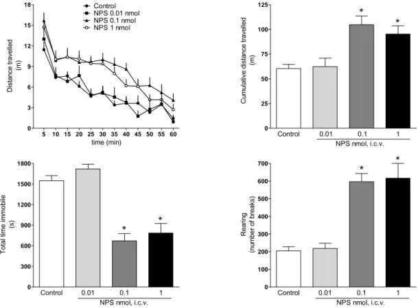
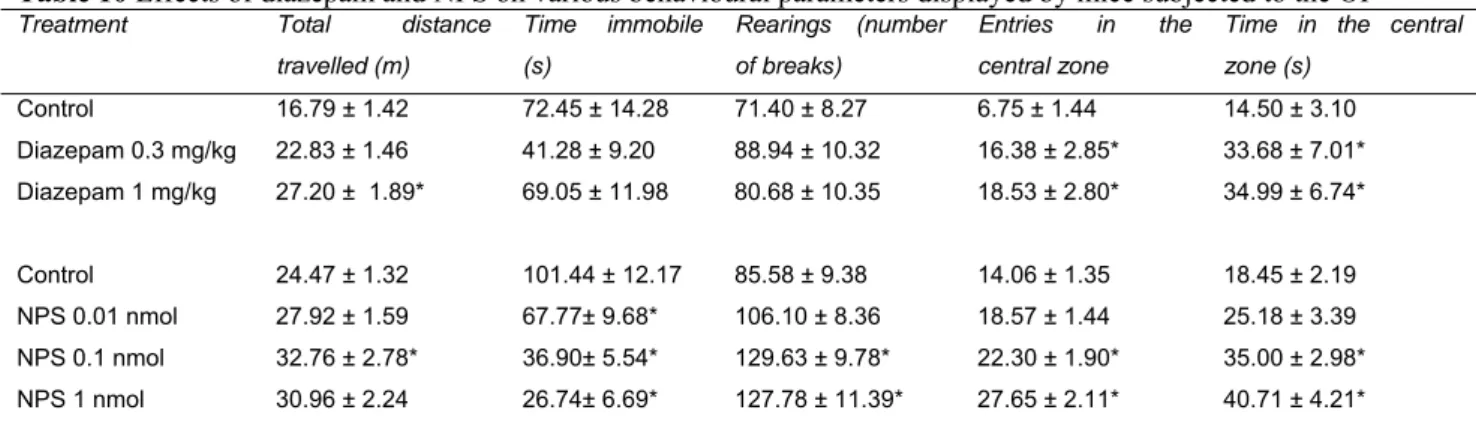
modulated by the NPS/NPSR system include anxiety, arousal, locomotion, food intake, learning and memory, pain and drug addiction. In our laboratories we provided further evidence that NPS injected supraspinally in mice acts as a stimulatory anxiolytic. In fact, in the mouse righting reflex (RR) test, NPS (0.01- 1 nmol, i.c.v.) was able to reduce in a dose dependent manner the percent of animals losing the RR in response to diazepam (15mg/kg, i.p.) and their sleep time. Furthermore, NPS in the same range of doses caused a significant increase in locomotor activity (LA) in mice. These effects were associated with a clear anxiolytic-like action elicited by NPS in the mouse elevated plus maze (EPM) test, open field (OF) test and stress-induced hyperthermia (SIH) assay. Thus NPS evokes an unique pattern of behavioural effects: stimulation associated with anxiolysis. To deeply investigate the biological roles played by the NPS/NPSR system the development of pharmacological (i.e. selective NPSR ligands, particularly antagonists) and genetic (i.e. receptor knockout animals) tools are needed. In collaboration with the medicinal chemistry group of the University of Ferrara, we performed a series of classical structure-activity (SAR) studies on NPS sequence. Specifically, NPS positions 2, 3, 4 and 5 were investigated in details, since they were demonstrated to be crucial for NPS bioactivity. Studies focussed on NPS position 5 led to the identification of the first generation of NPSR peptide antagonists. In vitro, in HEK293 cells stably
expressing the mouse NPSR, [D-Cys(tBu)5]NPS up to 100 μM did not stimulate Ca2+ mobilization
but was able to counteract in a competitive manner the stimulatory action of NPS (pA2: 6.44). In
vivo, in the RR test, [D-Cys(tBu)5]NPS at 10 nmol was inactive per se but dose dependently
antagonized the arousal-promoting action of NPS 0.1 nmol. [D-Val5]NPS acted in vitro as a pure
NPSR antagonist, with a pKB of 6.54 in inhibition experiments. In vivo, in LA test, [D-Val5]NPS at 10 nmol completely blocked the stimulatory effect evoked by NPS. In a further medicinal chemistry study, the potent NPSR antagonist [tBu-D-Gly5]NPS was identified. In vitro, [tBu-D-Gly5]NPS did
not stimulate calcium mobilization but blocked the stimulant action of NPS with a pKB of 7.06. In
vivo, in RR assay, [tBu-D-Gly5]NPS (0.1-10 nmol, i.c.v.) was inactive per se but dose dependently
antagonized the arousal-promoting action of NPS 0.1 nmol. Similarly in the LA assay [t
6
evoked by 0.1 nmol NPS in a dose dependent manner. SHA 68 has been previously identified as the first non peptide NPSR antagonist. In our laboratories we further assessed the pharmacological profile of SHA 68 in vitro and in vivo. In vitro SHA 68 was inactive per se up to 10 μM while it
antagonized NPS-stimulated Ca2+ mobilization in a competitive manner showing a pA2 value of
8.06. In vivo, in the mouse RR assay, SHA68 50 mg/kg i.p. fully prevented the arousal promoting action of NPS 0.1 nmol. In LA experiments, SHA 68 50 mg/kg i.p. was able to partially counteract the stimulant effects elicited by NPS 0.1 nmol. Instead, the anxiolytic-like effects of NPS 0.1 nmol in mouse OF test were slightly reduced by SHA 68. Collectively these data demonstrated the exclusive involvement of NPSR in the arousal promoting and locomotor stimulant effects of NPS. Finally, we backcrossed on the CD-1 strain the NPSR knockout mice originally generated on the 129Sv/Ev genetic background. A first phenotype analysis revealed no locomotor differences between NPSR(+/+) and NPSR(-/-) mice, with the exception of rearing behaviour that was reduced in knockout animals. Furthermore, the behaviour of NPSR(+/+) and NPSR(-/-) mice in the EPM, OF and SIH tests is superimposable. Similarly no differences were detected in the novel object recognition, forced swimming, RR and formalin assays. However, the stimulant actions of 1 nmol NPS in RR and in LA test could be detected in NPSR(+/+) but not in NPSR(-/-) mice. Collectively these data demonstrated that endogenous NPS/NPSR system does not play a role in the control of locomotion, anxiety, depression and memory, at least under the present experimental conditions. These results demonstrated that the NPS stimulant effects are selectively due to NPSR activation, corroborating the findings obtained with NPSR antagonists. In conclusion, the research activity performed during the PhD program led to the identification of the first generation of NPSR peptide antagonists. The use of these research tools in parallel with knockout studies generated converging evidence demonstrating that the biological effects of NPS are selectively due to the selective activation of NPSR.
introduction
1. INTRODUCTION
1.1 Orphan G-protein coupled receptors and the reverse pharmacology approach
G-protein coupled receptors (GPCRs) are one of the largest family of proteins acting as modulators of intercellular interactions and regulating biological functions in the human body, in particularly in the central nervous system (CNS). There are numerous GPCRs in living organisms, but the function of many of them is still unknown. The human genome encompasses ~ 800 GPCRs, of which more than half are olfactory and/or taste GPCRs. GPCRs are targets of most of the primary messengers including neurotransmitters, all neuropeptides, glycoprotein hormones, lipid mediators and other small molecules; thus they have considerable pharmaceutical interest. Drugs acting at GPCRs are used to treat numerous disorders. More than 30% of the approximately 500 clinically used drugs, are modulators of GPCRs function, representing around 9% of global pharmaceutical sales; this makes GPCRs the most successful of any target class in terms of drug discovery 1.
367 transmitter GPCRs have been identified within the human genome, the majority of these GPCRs have been identified on the basis of their sequence similarities, either by homology cloning or by bioinformatics analyses. Some of these receptors luck their endogenous ligand and they are defined ‘orphans’. Currently the orphan GPCRs are ~ 140.
The first step in the characterization of an orphan GPCRs is the search of the activating ligand. As the genomes of most studied model organisms have now been sequenced, the process of discovery of GPCRs-ligand pairs has been reversed. Until recently, neuropeptides have been traditionally identified either on the basis of their chemical characteristics 2 or of their effects in particular assay systems 3. Although highly successful, these approaches had reached a stand still by the mid 80’s.
Through DNA recombination techniques, it is now possible to transfect the sequence of an orphan receptor of which the function is not yet known, into an appropriate cellular expression system. This leads to the use of orphan receptors as baits to isolate their natural ligands from fractions of tissue extracts in high-throughput screening (HTS). This approach has been named “reverse pharmacology”. The expression system provides the necessary trafficking and G-protein-signalling machinery to enable the successful identification of the activating ligand. By exposing the transfected cell to a tissue extract containing the natural ligand of the orphan receptor, a change
introduction
8
in intracellular second messengers will be induced and will serve as a parameter to monitor orphan receptor ligand purification. Despite the logic of the theory, the process is not simple, since the chemical nature of the ligand and the type of the second messenger response that it will generate, are unknown. However, structural features in an orphan GPCR will determine its relationship to known receptors and will help in evaluating the nature of the receptor’s ligand and its cellular action. Indeed, an orphan receptor which is related, even to a low degree, to a particular receptor family has a higher probability of sharing a ligand of the same physical nature and a coupling to similar G proteins. Notably this strategy has already led to several significant discoveries. The orphan receptor strategy was first proven to be successful with the discovery of the neuropeptide
nociceptin/orphanin FQ as the endogenous ligand of the orphan GPCR opioid receptor-like 1 4-5.
Figure 1 The orphan receptor strategy (Civelli et al.,TRENDS in Neurosciences Vol.24 No.4 April 2001). The orphan
receptor strategy was developed to identify the natural ligands of orphan G-protein-coupled receptors (GPCRs) with the aim of discovering novel transmitters (defined in the main text). This strategy involves: (1) expression of the cloned orphan GPCR in an heterologous cell line; (2) exposure of this transfected cell line to a tissue extract that is expected to contain the natural ligand; (3) recording of the change in second messenger response elicited by activation of the orphan GPCR; (4) fractionation of the tissue extract and isolation of a surrogate, the active component; (5) determination of the chemical structure of the active component and (6) chemical synthesis of the active component and demonstration that it exhibits identical activity to that of the purified ligand.
introduction This first successful example of orphan receptor strategy was followed by the identification of other novel bioactive peptides such as: hypocretins and orexins, prolactin-releasing peptide, apelin, ghrelin, melanin-concentrating hormone, urotensin II, neuromedin U, metastin, neuropeptide B, neuropeptide W. The last success of reverse pharmacology was in 2004 the identification of neuropeptide s, the subject of this thesis, as the endogenous ligand of the previously orphan GPCR
GPR154, now named NPSR. Each of these discoveries was a landmark in its field 6. The success in
GPCRs deorphanization led to the approach being used by pharmaceutical industry 7, which had
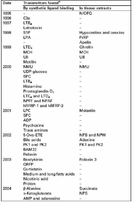
mastered the HTS of thousands of ligands. This led to thousands of potential transmitters and unexpected ligands (also of non-peptide nature) being tested on dozens of orphan GPCRs and a revival of the reverse pharmacology approach. The novel information emerging from reverse pharmacology discoveries substantially increased our knowledge on the diverse physiological functions modulated by peptidergic systems and most likely will open novel avenues for treating several diseases including food intake and sleep disorders, pain, anxiety and depression in the near future. Table 1 summarizes the transmitters of peptidic and non-peptidic nature identified as ligands of orphan GPCRs 6.
introduction
introduction
1.2 Neuropeptide S
The identification of NPS as the endogenous ligand of the previous orphan GPCR GPR154
was first reported in the patent literature in 2002 8. The patent reported the sequence of GPR154
(GenBank accession numbers BD183774, BD183814, BD183773) and the isolation of NPS as its endogenous ligand, but nothing is provided about the biological functions and the pharmacological characteristics of this new system. Then, in 2004, an elegant study by Xu et al. 9 described for the first time some functional features of the NPS/GPR154 system. Following the description by Xu et
al. 9, in the present work the GPR154 receptor will be indicated as NPSR. However this latter
abbreviation has to be considered provisional since it is not in line with IUPHAR recommendations for nomenclature of receptors (the receptor name should not include the letter R as an abbreviation for receptor 10).
The primary sequence of NPS in humans is SFRNGVGTGMKKTSFQRAKS. The amino acid at the N-terminus of the peptide is serine (S) in all animal species and this was the reason of
naming the peptide NPS 9. The NPS sequence is highly conserved among vertebrates with few
variations located in the centre and C-terminus of the peptide 11 (figure 2). The N-terminal sequence Ser1-Phe2-Arg3-Asn4-Gly5-Val6-Gly7 is identical in all species, thus suggesting that this may represent the bioactive core of this peptide 11. Curiously, the peptide is absent in fish genomes (e.g., zebrafish and fugu) and is also not found in amphibian or reptile DNA sequences, indicating that this transmitter arose relatively late during vertebrate evolution 11. As other neuropeptides, NPS is cleaved from a larger precursor protein. NPS peptide precursor (ppNPS) is a typical neuropeptide precursor containing a hydrophobic signal peptide at the beginning of its sequence and a pair of basic residues (Lys-Arg) before the mature NPS sequence 12.
introduction
Figure 2 Alignment of NPS peptide structures from various species. Sequences were deduced from GenBank finished
and unfinished genome sequences. Amino acids different from human NPS are highlighted in red 13.
The regional distribution of NPS in rat has been described in two publications by Xu et al.
9,14, using quantitative real time polymerase chain reaction (RT-PCR) and in situ hybridization
techniques. Quantitative RT-PCR showed that NPS mRNA are expressed in various tissues and the
highest levels were found in brain, thyroid, salivary, and mammary glands 9. Xu and colleague
focused their attention to the distribution of NPS and its receptor in the CNS. In situ hybridization experiments demonstrated that in rat brain ppNPS mRNA is expressed discretely in a few areas, with strongest expression in the locus (LC) coeruleus area, principle sensory trigeminal nucleus, and lateral parabrachial nucleus. Moderate expression was also found in a few scattered cells of the
dorsomedial hypothalamic nucleus and the amygdala 9. In particular, in the LC area NPS-expressing
neurons defines a previously unrecognized population of cells located between the noradrenergic
LC and the Barrington’s nucleus 9. Double label in situ hybridization studies demonstrated that
most of the NPS expressing cells in the LC area are glutamatergic neurons, few are cholinergic,
while none produce GABA 14. In the principle sensory trigeminal nucleus many of the NPS
expressing neurons use glutamate as a neurotransmitter 14. Finally, in the lateral parabrachial
nucleus NPS positive cells co-express CRF 14. NPS seems to be co-expressed with excitatory
neurotransmitters and on this basis it has been proposed that NPS may provide additional excitatory input to the postsynaptic target of these excitatory neurons.
introduction
1.3 NPS receptor
NPSR is also know as vasopressin receptor-related receptor (VRR1) 15 or G protein coupled
receptor for asthma susceptibility (GPRA) 16. NPSR is a typical GPCR, but it shows moderate
homology to other members of the GPCR family, the closest relatives being the vasopressin (i.e., V1a and V2) receptors. Human and mouse NPSR were studied in heterologous expression systems,
showing that NPS both stimulates intracellular calcium levels and cAMP accumulation with EC50
values in the low nanomolar range 9,17. This indicates that the NPSR can signal via both Gq and Gs protein to increase cellular excitability.
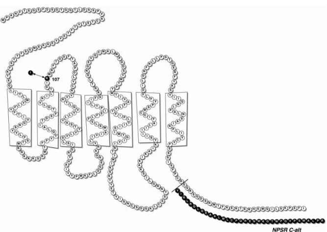
Multiple single-nucleotide polymorphisms (SNP) and several splice variants have been identified in the human NPSR (hNPSR) gene that is located on chromosome 7p14-15 (figure 3). On
note Laitinen et al. 16 described different polymorphisms in the hNPSR gene linked to an increased
susceptibility for asthma and potentially other forms of allergy that are characterized by high IgE
serum levels in Finnish and Canadian asthma patients 16. In particular the study described two
different C-terminal splicing variant of hNPSR, named “GPRA isoform A” (GenBank accession
number NP_997055) and “GPRA isoform B” (Gen-Bank accession number NP_997056) 16. The
“GPRA isoform A” corresponds to the NPSR receptor extensively studied by Xu 17. The most
interesting of SNPs described for hNPSR gene is an Asn-Ile exchange at position 107 of the mature
hNPSR protein (SNP591694 A>T; refSNP ID: rs324981) 17. This receptor polymorphism seems to
have functional implications since the hNPSRIle107 receptor displayed similar binding affinity but
higher NPS potency (by approx. 10-fold) than hNPSRAsn107 17. This result has been replicated in
different laboratories, using different assays (intracellular calcium and cAMP accumulation) as well
as different cell types (HEK293 and CHO) 17-18. Instead, the C-terminal splice variant of NPSR did
not appear to cause measurable differences in the pharmacological profile of the receptor 17-18. It is worthy of mention that the rat and mouse NPSR contain Ile at position 107. Different
epidemiological studies reported a gender-specific association of the hNPSRAsn107Ile
polymorphism with panic disorder, sleep behaviour, bowel disease and asthma susceptibility 16,19-21, however, the pathophysiological implications of NPSR polymorphisms are far from being fully understood.
introduction
Figure 3 Schematic diagram of the hNPSR protein showing the presumed location of the N107I polymorphism and the
sequence of the alternatively spliced carboxyl-terminal tail in NPSR C-alt 17.
Quantitative RT-PCR and in situ hybridization experiments reported that the NPSR mRNA
is widely distributed in the brain 9,14. High levels of expression are found in areas involved in
olfactory processing, including the anterior olfactory nucleus, the endopiriform nucleus, and the piriform cortex. NPSR mRNA is also highly expressed in the amygdaloid complex, in the thalamus and in the preoptic region. Furthermore, multiple hypothalamic nuclei, including the dorsomedial and the ventromedial hypothalamic nucleus and the posterior arcuate nucleus, express high levels of NPSR mRNA. In addition, NPSR mRNA is strongly expressed in major output and input regions of hippocampus, including the parahippocampal regions, the lateral entorhinal cortex, and the retrosplenial agranular cortex. These data suggested that the NPS system may play a key role in modulating a variety of physiological functions, especially arousal, anxiety, learning and memory, and energy balance. NPSR transcripts were also found in peripheral tissues, including thyroid,
salivary, and mammary glands, which might indicate additional endocrine functions 9. Recently,
Leonard and colleagues generated and validated a NPSR-specific antibody to determine the distribution of the NPSR protein in the rat brain 22. The localization of the NPSR receptor identified by the use of the antibody was consistent with the mRNA distribution identified by Xu et al. using
in situ hybridization techniques 14. NPSR protein was in fact detected in the medial amygdala,
introduction regions. Additionally, NPSR protein was localized in the pyramidal cell layer of the ventral hippocampus, the medial habenula, and was widely distributed in the cortex 22.
introduction
1.4 Biological actions of NPS
1.4.1 In vitro studies
Most of the in vitro studies were performed with cells expressing the recombinant NPSR receptor. In these studies NPS was able to increase both intracellular calcium and cAMP levels,
displaying similar high potency (EC50 in the low nanomolar range) at the rat, mouse and human
NPSR. As mentioned above, NPS displayed 10 fold higher potency at the hNPSRIle107 isoform than
hNPSRAsn107.
Only limited information is available on the in vitro effects of NPS in cells/tissues expressing the native NPSR. In our laboratories we assessed the effects of NPS in a series of cell lines measuring intracellular calcium mobilization and in several animal tissues taken from the gastrointestinal, genitourinary, and respiratory system measuring myotropic effects; however, we were not able to find any NPS sensitive preparation. The only peripheral cell types reported to be
sensitive to NPS are macrophages and lymphocytes 23-24. Specifically, macrophages responds to
NPS with reduced adhesion and increased phagocytosis, chemotaxis and production of the
pro-inflammatory cytokines IL-1β, IL-6 and TNF-α 23-24; moreover NPS stimulates lymphocytes
proliferation 24. These findings together with those coming from genetic studies, which provide
evidence for association of NPSR gene polymorphism with chronic inflammatory diseases of the respiratory 16 and gastrointestinal 19 systems, indicate that the NPS/NPSR system may have a role in modulating innate immunity and chronic inflammatory diseases of epithelial barrier organs.
As far as CNS preparations are concerned, NPS has been reported to modulate synaptic
activity in mouse brain slices 25-26 and neurotransmitter release in mouse synaptosomes 27. In
particular, two complimentary studies performed in mouse coronal brain slices reported distinct
actions of NPS in the amygdala. Meis et al. 26 reported in response to NPS an increase in
glutamatergic synaptic transmission onto basolateral amygdala GABAergic interneurons; this effect was sensitive to tetrodotoxin suggesting dependence from action potential propagation. The endopiriform nucleus was identified as the site of action of NPS. This NPS sensitive circuit might be responsible for the inhibitory effect of the peptide on the expression of contextual fear 26. Using
the same preparation, Jungling et al. 25 demonstrated that NPS increases glutamatergic transmission
to intercalated GABAergic neurons in the amygdala via presynaptic NPS receptors onto connected principal neurons. This electrophysiological effect of NPS may likely be important for both the anxiolytic-like action of the peptide and for its ability to facilitate extinction of aversive
introduction
memories25. The electrophysiological actions of NPS modulating the activity of the circuit
endopiriform cortex, lateral, basolateral, and central amygdala has been nicely summarized by Pape
et al. 28. Recently, electrophysiological studies demonstrated that NPS depolarizes and thereby
excites ventromedial hypothalamic neurons 29. This depolarization is postsynaptic and involves R-
and T-type calcium channels and non selective cation channels 29. The excitatory effect on
ventromedial hypothalamic neurons can represent the cellular process by which NPS participates in
the regulation of food intake and energy homeostasis 29. Furthermore, NPS has been reported to
behave as an extremely potent (pM range of concentrations) inhibitor of the release of 5-HT and NA from mouse frontal cortex synaptosomes. In parallel experiments NPS did not modify the release of GABA and glutamate and weakly reduced, only at high concentrations, dopamine and
acetylcholine release 27. However, no evidence was provided for the involvement of the NPSR in
these neurochemical actions of NPS. Based on the reported cellular actions of NPS (i.e. increase in intracellular calcium concentrations and cAMP), the ability of NPS to inhibit neurotransmitter release should be considered unexpected. However, the existence of excitatory G protein coupled receptor mediating inhibitory effects on neurotransmitter release has been repeatedly reported (for detailed discussion see Raiteri et al. 27). There is convincing evidence in the literature that elevation of 5-HT and NA levels is associated with anxiety like behaviours; therefore, the inhibition of 5-HT and NA release elicited by NPS may represent at least one of the mechanisms by which NPS promotes its anxiolytic-like effects. The results obtained by Raiteri et al. with mouse frontal cortex
synaptosomes 27 are in contrast with the recent study performed by Si and colleagues 30 using in
vivo microdialysis in freely moving rats. Si et al. 30 shown in fact that NPS administration increases
release of dopamine in the medial prefrontal cortex but does not change 5-HT levels in this brain area. This stimulatory effect of NPS on dopamine release in the medial prefrontal cortex might be
functionally connected to the effects of NPS on fear extinction and anxiety 30. Furthermore, NPS
given in the ventral tegmental area was reported to increase dopamine levels in the nucleus
accumbens shell in rats, activating the mesolimbic dopaminergic pathway 31. NPS induced
introduction
1.4.2 In vivo studies
Locomotor activity
In the pivotal study by Xu et al. 9 it has been reported that the supraspinal administration of NPS in mice stimulates locomotor activity (LA). This effect is evoked by low doses (0.1 nmol) of peptide, lasts for about 1 hr, and is similar in naïve mice and animals habituated to the test chamber. These findings were later confirmed in several studies both in mice 32-34 and rats 35, suggesting that the hyperlocomotor action of NPS is a robust phenomenon among experimental conditions and animal species. The involvement of NPSR in the locomotor stimulatory effect of NPS has been
demonstrated with the use of the NPSR antagonist SHA 68 33 that was able to counteract the
stimulatory effect of NPS being per se inactive. These results were corroborate by the use of the NPSR knockout (NPSR(-/-)) mice. NPS was in fact able to increase animals LA in NPSR wild type (NPSR(+/+)) but not in NPSR(-/-) mice and no differences were recorded between NPSR(+/+) and
NPSR(-/-) mice 36-38. All these findings demonstrate that this peptide action is due to NPSR
activation and suggest that the endogenous NPS system does not exert a tonic control on animal locomotor behaviour. It was demonstrated that a brain area important for NPS hyperlocomotor
action is the ventral tegmental area 31. Specifically, NPS seems activate the mesolimbic
dopaminergic system, that origins from this area and projects in the nucleus accumbens. In fact the NPS stimulant effects were completely block by the administration in the shell of the nucleus accumbens of sulpiride, a dopamine D2 –like receptor antagonist 31. This finding indicates a role of the dopaminergic pathway in the neurochemical mechanisms responsible for NPS induced
hyperlocomotion. Furthermore, a study by Boeck et al. 39 demonstrated that the pharmacological
blockade of adenosine A2A receptors by caffeine and the selective A2A antagonist ZM241385
significantly attenuate NPS stimulant effects. Thus the endogenous adenosine system seems play a
role in mediating this NPS function. Also corticotrophin releasing factor signalling via CRF1
receptors seems to be involved in the hyperlocomotor action of NPS. In fact this effect of NPS is blocked by the selective CRF1 antagonist antalarmin and is not evident in CRF1(-/-) mice 34. The microinjection of NPS into the paraventricular nucleus 35 or amygdale 25 mimicked the effects of the peptide given i.c.v. on food intake and anxiety states, respectively, but did not stimulate locomotion. Thus it is likely that these brain areas are not important for the locomotor stimulant effect of NPS.
introduction
Arousal and sleep
Electroencephalografic studies in rats demonstrated that the i.c.v. injection of NPS reduced all stages of sleep promoting wakefulness 9. Similar to the locomotor stimulant action in mice, this effect of NPS lasted for about 1 h and can be evoked using low doses of peptide (0.1-1 nmol). The arousal promoting action of NPS has been also demonstrated using the righting reflex assay. In fact NPS dose dependently (0.1 – 10 nmol) reduced the sleep time of rat treated with ketamine (100
mg/kg) and thiopental (45 mg/kg) 40. The NPSR antagonist [D-Cys(tBu)5]NPS at the dose of 20
nmol was able to completely prevent the arousal promoting action of NPS 1 nmol giving evidence
that the arousal promoting action of NPS is due to selective NPSR activation 40. Moreover,
[D-Cys(tBu)5]NPS per se prolonged both ketamine and thiopental anesthesia duration, suggesting that
the NPS/NPSR system tonically controls the anesthesia state 40. These findings parallel the
peptidergic arousal promoting system of the orexins. In fact, orexin receptor antagonists prolonged
barbiturate sleeping time in rats 41 and emergence from general anesthesia 42. Recent studies
performed with NPSR(-/-) mice suggested a role played by NPSergic pathways in the modulation of circadian rhythm. In fact NPSR(-/-) mice displayed reduced late peak wheel running (an index
of activity of the internal clock) 36 and reduced LA during the dark phase 37 compared to
NPSR(+/+) mice. Clearly further studies performed both with NPSR ligands and NPSR(-/-) mice are needed to investigate the role of the NPS/NPSR system in the regulation of wakefulness and sleep physiology and pathology. As far as the brain areas possibly relevant for the arousal promoting effect of NPS are concerned, NPSR mRNA has been reported to be expressed in several structures known to play a major role in the regulation of arousal including the thalamus, hypothalamus, ventral tuberomammilary nucleus, substantia nigra and ventral tegmental area, and
the pontine reticular nucleus 14. In particular the thalamic midline nuclei which integrate the
arousal circuit reticular formation-thalamus-cortex express high levels of NPSR mRNA. Microinjection and electrophysiological studies are now needed to establish the role of the above mentioned brain structures in the arousal promoting action of NPS. Interestingly enough, a recent genetic epidemiology study performed on 749 subjects found a clear association between the
NPSRAsn107Ile polymorphism and mean bedtime delay 20. While these findings require replication
in other samples, they provide evidence for a role of the NPS/NPSR system in regulating sleep physiology in humans.
introduction
Anxiety and mood
The studies by Xu et al. 9,14 demonstrated a strong expression of NPSR mRNA in different
brain areas related to stress, including the amygdala, bed nucleus of the stria terminalis, hypothalamus, raphe nucleus and ventral tegmental area. This NPSR distribution suggests that the NPS system might influence emotional behaviours such as stress and anxiety responses. The pivotal
study by Xu et al. 9 reported that the supraspinal administration of NPS in the 0.01 – 1 nmol range
of doses evokes a clear anxiolytic-like effects in mice subjected to a battery of validated assays including the elevated plus maze (EPM), the light-dark box, and the open field (OF) 9. These initial observations were later confirmed and extended to other assays such as the four-plate test and
elevated zero maze 34,43. These tests are based on the natural aversion of rodents for open or
unfamiliar spaces and anxiolytic drugs increase exploration of these exposed areas. It should be considered that these assays are sensitive to the confounding effects of drugs, like NPS, that stimulate locomotor activity since in these tests anxiety levels are measured as inhibited behaviours. However NPS was reported to be able to reduce in a dose dependent manner the number of marbles
buried in the marble burying test 9,34, a model which is not biased by locomotion since anxiety
levels are measured as an active behaviour. Furthermore Leonard and colleagues 43 demonstrated
that anxiolytic-like effects can be measured in response to NPS in mice in the stress induced hyperthermia (SIH) test. These results provide evidence that the anxiolytic-like action of NPS is a genuine effect. Subsequently the anxiolytic-like effects of NPS were confirmed and extended to a different species, the rat, and a different assay, the defensive burying test 44. It was demonstrated that NPS (0.1 – 10 nmol) reduces cumulative burying behaviour in a dose dependent manner without modifying other parameters including latency to contact the probe, burying behaviour
latency, number of shocks received or immobility/freezing duration 44. Again, since the main
parameter predictive of anxiolysis in this assay is the inhibition of an active defensive behaviour, motor activity can be considered a minor bias in the outcome of the defensive burying test 45.
The NPS effects were also investigated in the Pavlovian fear conditioning test 25, an
established model for fear and emotional memory. In this paradigm, an aversive stimulus (unconditioned stimulus, i.e. short electric foot shock) is associated with a neutral stimulus (conditioned stimulus). After the training (pairing of unconditioned stimulus and conditioned stimulus), associative fear memory is tested by presentation of conditioned stimulus alone and fearful responses can be recorded reliably by measuring ‘‘freezing’’ behaviour. Repeated presentation of the conditioned stimulus alone without the aversive stimulus leads to a gradual decline in fear responses, which indicates extinction of fear memory. It was demonstrated that NPS
introduction given into the amygdala produces a significant decrease of fear behaviour and facilitates the extinction of conditioned fear responses 25. Next, Fendt et al. 46 reported that the intra amygdala
administration of NPS completely block the expression of the fear-potentiated startle 46. Thus the
NPS/NPSR system seems to have a role in controlling the mechanisms of conditioned fear, in particular eliciting fear reducing effects.
The amygdala can be the brain area crucial for NPS anxiolytic action. In fact, the intra
amygdala injection of NPS promoted clear anxiolytic effects in the mouse EPM and OF assays 25,
mimicking the effects of the peptide given i.c.v 9. Furthermore the NPS effects on fear expression
were detected giving it into the amygdala 25,46. Another brain region important for the NPS effects
on anxiety and fear seems to be the medial prefrontal cortex, where NPS induces an increase in dopamine levels, probably through the activation of the dopaminergic neurons originated in the ventral tegmental area 30.
The endogenous NPS/NPSR system might tonically control anxiety levels since the intra amygdala injection of the NPSR antagonist SHA 68 produces robust anxiogenic effects in mice
subjected to the open field 25. Furthermore a study by Duangdao and colleagues 36 reported that
NPSR(-/-) mice display an anxious phenotype compared to their wild type littermates in the
light-dark box, EPM and OF. On the contrary Zhu et al. 38 and Fendt et al. 37 observed no differences
between the behaviour of NPSR(+/+) and NPSR(-/-) mice in the OF, EPM and elevated zero maze tests, suggesting that the endogenous NPSergic pathway does not tonically control anxiety levels. The hypothesis that the NPS/NPSR system does not play a role in modulating anxiety is corroborated by the absence of anxiogenic effects per se of the NPSR antagonist SHA 68 in the
EPM and defensive baring assays in rat 47. These indications need however to be confirmed in
future studies investigating the effects of different chemically unrelated NPSR receptor selective antagonists as well as performing systematic investigations of the phenotype of NPSR(-/-) and
possibly ppNPS(-/-) 48 mice in different assays predictive of anxiety states. Interestingly NPS
failed to elicit anxiolytic-like actions in OF and elevated zero maze assays in mice lacking the
NPSR protein 38, demonstrating that the NPS anxiolytic activity is selectively due to the NPSR
activation. This is also demonstrated by the fact that SHA 68 was able to counteract the NPS effects in the EPM and defensive baring assays in rats 47.
On note different epidemiological studies reported an association between NPSR SNPs and
susceptibility to panic disorders. In particular the NPSR isoform NPSRAsn107 was found
underrepresented in patients with panic disorder and the NPSRIle107 isoform was found associated
with an over interpretation of fear reaction and a tendency to catastrophizing 21,49-51. This
introduction and seems inconsistent with findings in rodent models, where NPS have been shown to exert a dose dependent anxiolytic effect. Thus the role plays by the NPS/NPSR system in the control of panic is still unclear and further studies are needed to address this issue.
Interestingly enough, the effect of NPS on emotional behaviour seems to be restricted to anxiety and fear since the peptide was found inactive in tests such as the tail suspension 43 or the forced swimming test (FST) (Rizzi et al., unpublished results) that are sensitive to the antidepressant- like effects of drugs. However preliminary results indicate that NPS may alter both anxiety- and depression-like behaviours in a rat genetic model of depression. In fact, in flinders sensitive rats NPS decreased depression-like behaviour in the forced swimming test and anxiety-related behaviour on the EPM in a dose-dependent manner (0.05 – 1 nmol, i.c.v.). In contrast, NPS did not alter the behaviour of flinders resistant animals 52. A recent study performed with NPSR(-/-) mice reported that male mice lacking the NPSR receptor display increased depression-like behaviour in the FST. However Duangdao et al. reported no differences in the behavior of NPSR
deficient mice compared to wild type mice in the FST 36. Thus the role of the NPS/NPSR system in
mood regulation is not well understood.
In conclusion, the initial findings by Xu et al. 9 were replicated and extended in different laboratories confirming that NPS promotes in the same range of doses stimulation of locomotor activity and arousal associated to a genuine anxiolytic-like action. Thus, the proposal of NPS as an
activating anxiolytic 9, is confirmed after six years of research activities. This rather unique
behavioral profile challenges the common idea that anxiolytics are also sedative (i.e. benzodiazepines) or that stimulants are also anxiogenic (i.e. caffeine, cocaine, and amphetamines). The only substance which shares this behavioral profile with NPS is nicotine which increases
arousal and wakefulness and produces, at least in smokers, anxiolysis and antistress effects 53.
Interestingly enough, chronic nicotine treatment in rats increases both NPS and NPSR expression in
the brainstem and NPSR in the hypothalamus 54. Thus nicotine might produce some of its effects
via regulation of the endogenous NPS/NPSR system.
introduction
Drug addiction
The unique pattern of behavioral effects elicited by NPS (arousal promoting action associated with anxiolysis) together with the expression of NPSR in brain areas involved in the rewording effects of drugs has prompted the investigation of NPS effects on drug addiction.
In conditioned place preference studies in mice, NPS neither induced place preference nor aversion. However, NPS blocked the acquisition of conditioned place preference to morphine. Moreover, the expression of conditioned place preference induced by morphine was also inhibited
by NPS. These results revealed the involvement of NPS in rewarding activities of morphine 55.
The effects of NPS were also evaluated on ethanol drinking in alcohol-preferring and non
preferring rats 56. NPS given i.c.v. reduced ethanol intake in alcohol-preferring, but not in non
preferring rats. The peptide neither altered anxiety-like behavior in the EPM test nor modified general motor activity. However, there was an increase in the amount of time spent in the center of the activity cage following infusions of 0.6 nmol of NPS in preferring, but not in non preferring rats, indicating anxiolytic actions of the peptide. Thus, this study suggests a role for NPS in the modulation of ethanol drinking and possibly anxiety-like behavior in rats selectively bred for high alcohol drinking 56.
Recently, two complimentary studies investigated the role of NPS in drug seeking behavior
34,57. Cocaine-seeking behavior was evaluated in mice by Paneda et al. 34. It was demonstrated that
i.c.v. NPS reinstates extinguished cocaine-seeking behavior in a dose-dependent manner.At the
highest dose tested i.e. 0.45 nmol, NPS increased active lever pressingin the absence of cocaine to
levels that were equivalent to those observed during self-administration. This action of NPS
involved corticotropin-releasing factor receptor signaling via CRF1receptor since CRF1(-/-) mice
did not respond to the cocaine reinstatement effects of NPS and the CRF1 antagonist antalarmin
blocked the increase in active lever responding in response to NPS 34. Ethanol seeking behavior
was investigated in rats by Cannella et al. 57. In self-administration experiments, the stable
response rates observed for ethanol reinforcement were not modified by i.c.v NPS (1.0 and 2.0 nmol). In reinstatement experiments, ethanol-associated cues induced robust rates of ethanol seeking. NPS i.c.v. injection resulted in a significant increase of ethanol seeking elicited by ethanol-associated cues. Site-specific NPS injection (0.1 and 0.5 nmol) into the lateral hypothalamus also reinstated extinguished responding to ethanol. This effect was selectively
blocked by pre-treatment with the OX1 receptor antagonist SB-334867 which did not modify
introduction
ethanol-seeking elicited by environmental conditioning factors 57. Furthermore NPSR mRNA
expression was found enhanced in rat with a history of ethanol addiction in different brain areas, suggesting an involvement of the NPS/NPSR system in controlling mechanisms of ethanol abuse
58. Finally, a very recent study performed by Kallupi et al. 59 confirmed that NPS increases
conditioned reinstatement of cocaine-seeking in rats through the activation of orexin immunoreactive neurons in the lateral hypothalamus. Interestingly the administration of the two
chemically distinct NPS receptor antagonists SHA 68 and [D-Cys(tBu)5]NPS potently and
selectively prevented cue-induced cocaine seeking, suggesting that the endogenous NPS may have
a role in the pathophysiology of drug relapse 59. Based on these findings, the NPSR receptor
represents an important and unique target for the treatment of drug craving and the prevention of relapses in addicted patients.
Food intake
Several studies demonstrated that NPS is able to reduce food intake acting as an anorexigenic signal in the brain. The first evidence for this NPS action was provided by Beck et al.
60 who demonstrated that the i.c.v. injection of NPS in the 0.4 – 4 nmol range strongly inhibited
chow intake in overnight fasted rats with effects of longer duration with the highest dose. In the same study similar inhibitory effects were observed for the spontaneous intake of a palatable diet
60. Smith and colleagues 35 later demonstrated that the injection of low NPS doses in the
paraventricular nucleus of the hypothalamus produces robust anorexigenic effects. The observations in rats were confirmed in chicks where NPS inhibits food intake both injected i.c.v.
and in the paraventricular nucleus or lateral hypothalamus 61. In a follow up study 62 the same
authors demonstrated that chickens lines selected for low or high body weight are differently sensitive to NPS. These data indicate that NPS may differentially affect appetite-related processes in hypo- and hyperphagic individuals. Subsequently, Fedeli et al. 63 further demonstrated that NPS elicits a marked inhibition of palatable food consumption in rats and that the paraventricular nucleus of the hypothalamus represents an important site of action for this NPS effect 63. Moreover
the NPSR antagonists [D-Cys(tBu)5]NPS was able to completely counteract the NPS action on
palatable food intake being per se inactive 63. These results demonstrate that the NPSR protein is
involved in the anorectic action of NPS and suggest that the NPS/NPSR system does not exert a tonic control on this biological function. This hypothesis were then confirmed in two parallel studies by Peng et al. 64 using the NPSR antagonists [D-Val5]NPS and Cifani et al. 65 using the
introduction
[Aib5]NPS and [Ala3]NPS. [D-Val5]NPS was in fact able to block the NPS anorectic action being
per se inactive in fasted mice 64, moreover [D-Cys(tBu)5]NPS and [tBu-D-Gly5]NPS counteracted
the palatable food intake inhibition induced by NPS and they did not affect palatable food intake
alone 65. [Aib5]NPS, like NPS, reduced food intake, instead [Ala3]NPS blocked the NPS action
being per se inactive 65. All these results corroborate the evidence that NPS elicits inhibition of food consumption through selective NPSR activation and that the NPSergic pathway does not have a role in the tonic regulation of feeding behaviour. Obviously studies performed with NPSR(-/-) mice are needed to further confirm these findings.
Interestingly, the NPS effects on food consumption were not modified by the CRF receptor antagonists CRF 9–41 63 and NBI-27914 64, indicating that palatable food intake inhibition by NPS is independent from the activation of the CRF system.
While the above mentioned studies converge indicating that NPS reduce food intake, the
report by Niimi et al. 66 provided opposite findings showing that the i.c.v. injection of NPS (1
nmol) in rats slightly but significantly stimulated feeding. In the same study it has been shown that centrally administered NPS increased Fos-like immunoreactivity in orexin-expressing neurons of the lateral hypothalamic area. Thus the orexin system may take part to the actions of NPS on food intake.
Learning and memory
NPSR mRNA is expressed significantly in the major input and output regions of hippocampal formation, which are critical in the modulation of learning and memory. The first study aimed to investigate the effects of NPS on spatial learning and memory was performed by Han et al. 67 using the Morris water maze task. It was demonstrated that NPS (1 nmol) given i.c.v. before training trials facilitates spatial memory acquisition, in fact NPS treated mice spent significant more time in the target quandrant of the maze 67. Furthermore NPS was able to mitigate
the spatial memory impairment caused by the N-methyl-D-aspartate receptor antagonist MK801 67
and by rapid eye movement sleep deprivation 68. This first study by Han and colleagues 67 did not provide any indication regarding the role exerts by the endogenous NPS in regulating the mechanisms of spatial learning and memory. However a subsequent study performed with NPSR(-/-) mice reported no differences between the performance of NPSR(+/+) and NPSR(-NPSR(-/-) mice in
the Morris water maze test, suggesting that endogenous NPS may not modulate spatial learning
introduction inhibitory avoidance test NPS (1 nmol) was able to enhance emotionally aversive long-term
memory until 96 hours after post training session 69. Similarly, NPS increased the non-aversive
memory in the novel object recognition (NOR) task until one week after the training trial 69. Of
note, NPS was effective when given after the training session, instead pre-training or pre-retrieval NPS administrations failed to enhance memory retention, suggesting that NPS may act during the
consolidation phase 69. Interestingly the NPSR antagonist SHA 68 completely counteracted the
NPS effect in the inhibitory avoidance paradigm demonstrating that the mechanism by which NPS
facilitate emotionally aversive memory is the selective activation of the NPSR protein 69.
Furthermore NPSR deficient mice shown impaired performance in the inhibitory aversive memory, novel object recognition, and novel place or context recognition, suggesting that activity of the
endogenous NPS system is required for memory formation 69. The central noradrenergic system
seems to be involved in the NPS-induced memory enhancement, since the block of the
noradrenergic receptors by propranolol attenuated this NPS effect 69. In summary NPS system can
have a significant role in the facilitation of various type of memory, such us spatial, emotional aversive and non aversive memory.
Pain transmission
NPSR mRNA is highly expressed in the periaqueductal gray 14, which is an important
component of descending analgesic system 70. Using the tail withdrawal and hot-plate test Li et al.
71 demonstrated that NPS (0.01–1 nmol, i.c.v.) caused a dose-dependent antinociceptive effect. The
first findings by Li et al. were then confirmed by Peng and colleagues 72, using the formalin test. This is a very useful model of clinical pain in which the first phase seems to be caused by direct activation of peripheral nociceptors, while the second phase is considered to be dependent on an inflammatory reaction caused by peripheral and central sensitization. NPS injected i.c.v. significantly reduced both the first and the second phase of nociceptive behaviour 72. Interestingly, the antinociceptive effects elicited by NPS in the tail withdrawal and hot-plate test were
completely prevented by the NPSR peptide antagonist [D-Cys(tBu)5]NPS 71, while the NPSR
antagonist [D-Val5]NPS was able to block the NPS actions in the formalin assay 72. The NPSR
antagonists given alone did not modify nociceptive transmission 71-72. These results reveal that
NPS may produce antinociceptive effects through NPSR activation and suggest that the endogenous NPS pathway does not control pain transmission under these experimental condition. Naloxone did not affect NPS antinociceptive activity in all the assays, indicating that opioid systems are not involved in this action 71-72. The antinociception induced by NPS can be due to the
introduction activation of neurons in periaqueductal gray, as suggested by the increased c-Fos expression in this brain area measured in response to NPS administration 72.
Colonic transit
It has also been reported that NPS inhibited, in a dose-dependent manner (0.001–1 nmol), distal colonic transit (measured as fecal pellet output and bead expulsion time) after i.c.v. but not
i.p. administration 73. The effect evoked by NPS 0.01 nmol was sensitive to the NPSR antagonist
[D-Val5]NPS (0.1 and 1 nmol, i.c.v.), demonstrating the involvement of NPSR in this biological
action of NPS 73. Of note, two different studies reported that NPS, in HEK293cells trasfected with the NPSR receptor, increases the expression of several peptide involved in the control of physiological motor and sensory functions in the gastrointestinal tract, such as somatostatin,
cholecystokinin, vasoactive intestinal peptide and peptide YY 74-75. Obviously further study are
needed to verify this NPS action in native intestinal enteroendocrine cells.
In conclusion there is convincing evidence in the literature that the NPS/NPSR system regulates multiple biological functions including locomotor activity, arousal and wakefulness, fear expression and anxiety, food intake and gastrointestinal functions, drug addiction, memory processes, and pain transmission.
introduction
1.5 NPSR ligands
1.5.1 NPSR peptide ligands
Soon after the identification of the NPS/NPSR system, different research groups started medicinal chemistry programs directed to identify the NPS bioactive sequence and its crucial residues involved in NPSR recognition. To this aim classical Ala- and D-scans together with N- and
C-terminal truncation studies were performed 17-18,76. The systematic replacement of the NPS amino
acid sequence with Ala residues can give information on the contribution of each amino acid side chain for NPS bioactivity. Nineteen Ala-substituted NPS analogues were synthesized and evaluated in a calcium mobilization assay using HEK293 cells expressing the hNPSR receptor by Roth et al. 76. Results of these studies indicated that positions 2, 3, 4, and 7 are crucial for NPS bioactivity. In
particular, the replacement of Phe2 with Ala generated a completely inactive NPS analogue while
[Ala4]NPS and [Ala7]NPS displayed a drastic loss of potency. Interestingly, [Ala3]NPS bound to the NPSR receptor with about 10-fold reduced potency compared to NPS and was able to elicit
maximal effects corresponding to 50% of those elicited by the natural peptide. Thus, [Ala3]NPS
behaves as a NPSR partial agonist. Ala substitution in the other NPS positions was fully tolerated
and did not significantly modified either the potency or the efficacy of the [Alax]NPS analogs.
Similar studies provided converging evidence demonstrating that the sequence Phe2-Arg3-Asn4 is
critical for NPS biological activity 18. This study also confirmed the important role played by Gly7 for NPSR recognition. D-scan investigation was applied to collect information on the contribution of the single amino acid chirality for NPS biological activity. Confirming Ala-scan results, these studies revealed that the NPS portion Phe2-Arg3-Asn4 is of great importance for NPSR binding and activation 76. In fact, replacement of these residues with their enantiomers consistently produced an
important loss of peptide potency. Inversion of chirality of Val6 generated a low potency NPS
derivative. D-amino acid substitution of all the other chiral positions did not modify either the potency or the efficacy of the [D-Xaax]NPS analogues 76. N- and C-terminal truncation studies
17-18,76 consistently identified in the N-terminal portion of NPS the crucial amino acid sequence needed
for NPSR binding and activation. In particular, the deletion of Ser produced a moderate decrease in
peptide potency while further deletion of Phe2 generated an inactive analog 18,76. In contrast,
systematic deletion of up to ten residues from the C-terminal part of NPS did not produce major
changes in peptide biological activity 17-18,76. Further, C-terminal shortening of the NPS sequence
introduction reporting NPS(1–6) as a high potency NPSR agonist. The NPS(1–10) fragment was further investigated in vivo in locomotor activity experiments performed in mice. While NPS elicited a dose-dependent stimulatory effect in the range 0.01–1 nmol, NPS(1–10) was found inactive up to
10 nmol 76. These results indicated that the NPS(11–20) sequence seems to be important for
maintaining in vivo biological activity. Collectively, the data obtained with these first SAR studies demonstrated that: (i) the most important residues for NPSR recognition are Phe2-Arg3-Asn4; (ii) the sequence Val6-Gly7 is also important for NPSR bioactivity; (iii) the C-terminal 11–20 sequence of NPS is not required for in vitro activity while it is necessary for its in vivo biological effects. Important suggestions for the design of novel peptide ligands can be obtained investigating the conformation of a given peptide in different environments. NPS conformational investigations performed by NMR experiments indicated that NPS presents a completely disordered conformation
in water 77. However, several NH-NH cross peaks were observed by Bernier et al. 18 in the NPS
region 5–13; this lead these authors to hypothesize the presence of a nascent helix in this region, which, during the NPSR binding process, may favour the formation of a stable α helix structure. On the other hand, disordered NPS conformations were observed in different NMR solvent mixtures that may favour helicity 77. This NPS behaviour has been ascribed, at least in part, to the presence in position 5, 7, and 9 of the flexible amino acid residue Gly. In order to force NPS to adopt a stable a helix structure, residues 7, 9, and 13 were replaced with Ala. NMR analyses confirmed that
[Ala7,9,13]NPS adopts a very stable helix spanning all the peptide sequence but this peptide was
completely inactive at NPSR 77. Single amino acid substitutions indicated that only the replacement
of Gly7 with Ala or Aib (2-amino-2-methylpropionic acid) is able to induce a significant helical
structure, whereas [Ala9]NPS and [Ala13]NPS showed a limited degree of helicity 77. Interestingly,
[Ala9]NPS and [Ala13]NPS were almost as active as NPS at NPSR while [Ala7]NPS and [Aib7]NPS
were found inactive. These results indicate that a helical conformation centred around position 7 is not compatible with NPS biological activity. Different results were obtained with substitution of
Gly5 with L- and D-Ala. In fact, both substitutions generated peptide derivatives that showed only
slightly reduced potency compared to NPS 77. This suggests that an a helix conformation (favoured
by L-Ala but not by D-Ala) is, at least in this portion of the NPS sequence, not important for NPSR
binding. Interestingly, [D-Ala5]NPS behaves as a partial agonist at NPSR with efficacy
corresponding to half of that of NPS. The replacement of Gly5 with the achiral α helix promoting
residue Aib did not produce a significant increase in peptide helicity and generated an NPS analogue with reduced potency and, more importantly, with a statistically significant reduction in
(L-introduction this causes a reduction in potency. In addition, this reduction of conformational freedom may favour partial agonist ([D-Ala5]NPS, [Aib5]NPS), or full agonist ([L-Ala5]NPS) pharmacological activities.
Collectively, this conformation-activity study together with SAR investigations
demonstrated that: (i) the sequence Gly5-Val6-Gly7 may represent a flexible peptide region
important for inducing and/or stabilizing NPS bioactive conformations; (ii) an α helix conformation around position 7 is not compatible with NPS biological activity; (iii) modifications of Gly5 may be critical for the design of NPSR ligand with reduced efficacy.
introduction
1.5.2 NPSR non peptide ligands
The first example of non peptide molecules able to interact with the NPSR was reported in
the patent literature by Takeda researchers 78. These compounds are characterized by a
3-oxo-tetrahydro-oxazolo[3,4-a]pyrazine scaffold mainly substituted in position 1 and 7. Among this series of compounds, SHA 68 (figure 3) i.e. the racemic mixture (9R/S)-3-oxo- 1,1-diphenyl-tetrahydro-oxazolo[3,4-a]pyrazine-7-carboxylic acid 4-fluoro-benzylamide (reported as example 25 in Fukatzu et al. 78) has been selected by Okamura et al. 33 for in vitro and in vivo pharmacological
characterization. In radioligand ([125I][Tyr10]NPS) binding experiments SHA 68 displayed high
affinity (pKi 7.3) for human NPSR. In cells expressing the hNPSR SHA 68 was inactive per se
while antagonizing the stimulatory effects of NPS on calcium mobilization in a competitive manner.
Similar high pA2 values were obtained at hNPSR Ile107 (7.6) and Asn107 (7.8) receptor isoforms.
SHA 68 appears to be selective for NPSR since it did not affect signalling at 14 NPSR unrelated
G-protein coupled receptors 33. Pharmacokinetic analysis demonstrated that SHA 68 reaches
pharmacologically relevant levels in plasma and brain after i.p. administration. Furthermore, peripheral administration of SHA 68 in mice (50 mg/kg i.p.) was able to antagonize NPS-induced
stimulation of locomotor activity 33. In a separate study SHA 68 given into the amygdala exerted
functionally opposing responses compared to NPS 25. Thus, available data demonstrated that SHA
68 behaves as a potent and selective NPSR pure antagonist. SAR studies performed on position 7 of SHA 68 indicate that a urea functionality is required for potent NPSR antagonist activity while alkylation of the urea nitrogen or replacement with carbon or oxygen generated less potent derivatives. In addition, compounds with a-methyl substitution or elongated alkyl chains had reduced potency, indicating a limited tolerance for position 7 substituents. The only chemical modification tolerated in this position was the elimination of the fluorine atom in the para position of the phenyl ring; this generates a molecule (SHA 66), which displays similar potency to the parent compound 33,79.
introduction
Figure 4 Chemical structure of the non peptide NPSR antagonist SHA 68
Subsequently, Merck researchers, using HTS techniques, identified and developed two new
structural classes of potent NPSR antagonists, characterized by a quinoline 80 and a tricyclic
imidazole 81 scaffold. From the first series of compounds the NPSR antagonist NPSR-QA1 has been
identified. This compound showed potent binding to the rat NPSR receptor in in vitro binding
displacement studies with an IC50 of 1.3 nM, furthermore it provided high occupancy of NPSR in
the brain after i.p. dosing 80. From the tricyclic imidazole series the compound NPSR-PI1 was
identified, with an IC50 of 43 nM in FLIPR experiments. It exhibited significant plasma free
fractions after i.p. administration, enabling in vivo central nervous system exposure 81. Thus NPSR-QA1 and NPSR-PI1 are potent, highly selective and CNS penetrant NPSR antagonist and they represent a useful pharmacologic tool for further study of the effects of central NPSR antagonism in vivo. Up to now no data are reported on the effectiveness of these NPSR antagonists in vivo.
Recently GlaxoSmithKline researchers published a new series of NPSR antagonists, with 5-phenyl-2-[2-(1-piperidinylcarbonyl)phenyl]-2,3-dihydro-1H-pyrrolo[1,2-c]imidazol-1-one structure
introduction
1.6 NPSR knockout mice
NPSR(-/-) mice were generated on 129S6/SvEv strain in Taconic laboratories as described
in Allen et al. 83. The inactivation of the NPSR protein was obtained by replacing the majority of
exon 4 with the neomycin cassette. This deletion is predicted to remove the third transmembrane
spanning domain and regions of the intracellular loop 2 83. Allen and colleagues investigated the
functional significance of NPSR in the regulation of lung physiology and pathology.Despite the
loss of functional NPSR, no differences were detected in the development of asthma and allergic lung pathology in NPSR(-/-) mice. Neither qualitative nor quantitative differences in the cellular infiltrate, nor the development of airway hyperresponsiveness in response to methacholine,
distinguished the mutant mice from their littermate controls 83. Thus, no evidence for abnormal
respiratory or immunological functions were recorded in mice lacking NPSR. These first findings
by Allen and colleagues were very recently confirmed in a study performed by Zhu et al. 84 using
NPSR deficient mice in which the exon 2 was deleted. No differences in the lung inflammation and respiration in response to ovalbumine or A. fumigatus exposure were measured between NPSR(+/+) and NPSR(-/-) mice, suggesting that endogenous NPS is not directly involved in the development of experimental asthma 84.
In the last few years the behavioural phenotype of NPSR(-/-) mice was deeply investigated
in different laboratories, using NPSR(-/-) mice on a 129S6/SvEv 36 or C57BL/6 genetic background
37-38,50. The first of these studies was performed by Duangdao et al. 36. They reported that NPSR(-/-)
mice display lower late peak wheel running activity during the subjective evening at the end of the dark phase, suggesting that a functional NPS system is required for expression or maintenance of arousal. This hypothesis was corroborate by the subsequent results obtained by Fendt and
colleagues 37, that recorded a decreased LA during the dark phase in mice lacking the NPSR
protein.
Interestingly, NPSR(-/-) mice were found by Duangdao et al. 36 to display increased levels
of anxiety-like behaviours in the OF, EPM and light dark box tests, providing evidence for a substantial role of the endogenous NPS system in regulating anxiety levels. On the contrary, no
differences were observed in the OF and in the elevated zero maze assays by Zhu and colleagues 38
and in the OF and EPM tests by Fendt et al.37. Importantly, NPS failed to have anxiolytic effects in
the elevated zero maze and OF tests in mice lacking the NPSR receptor 38. Thus it was
introduction No differences were recorded between NPSR(+/+) and NPSR(-/-) mice by Duangdao et al.
36 in the FST, indicating that NPS/NPSR system is not involved in regulating depressive-like
behaviour, instead Zhu et al. 38 found an increased depressive phenotype in male NPSR(-/-) mice in
the FST but not in the tail suspension test. Furthermore NPSR(-/-) mice are reported to be less reactive in the acoustic startle response test by Zhu et al. and Fendt et al. 37-38, but no differences were recorded by Duangdao et al. 36 in this paradigm.
No differences were reported between NPSR(+/+) and NPSR(-/-) mice in terms of
spontaneous LA 36-38, furthermore the sensitivity of NPSR(-/-) mice to the stimulant action of
metamphetamine 38 and cocaine 37 was superimposable to that of NPSR(+/+) mice. These results
clearly indicate that the endogenous NPS does not control mice locomotion and is not involved in the mechanisms by which cocaine and metamphetamine elicit stimulation. All the research groups demonstrated that NPS is able to increase mice locomotion in NPSR(+/+) but not in NPSR(-/-)
mice, thus the NPS stimulant effects are selectively due to the NPSR activation 36-38. NPSR(-/-)
appear to have improved motor coordination in the accelerating rotarod test when compared to NPSR(+/+) mice, instead body weight and body length appeared equal between both groups of mice
36.
A very recent study by Okamura et al. 69 shown that mice lacking the functional NPSR
receptor display deficits in inhibitory avoidance memory, NOR and novel place or context recognition, suggesting that activity of the endogenous NPS system is required for memory formation. Instead the performance of NPSR(-/-) mice in the Morris water maze test was similar to NPSR(+/+) mice, indicating that the NPS/NPSR system is not involved in spatial learning and
memory mechanisms 38.
Finally, the plasma corticosterone levels after a stressful event (FST or immobilization) were
found superimposable in NPSR(+/+) and NPSR(-/-) mice 38,50, instead after 1 hour of
immobilization the gene expression levels of some anxiety and stress-related genes was found altered in NPSR(-/-) mice. In particular these mice presented an upregulation of JunB and interleukin 1 β in the cortex and hypothalamus and a downregulation of the neurotrophic factor neurotrophin 3 in cortex and striatum 50.


![Figure 12 Concentration-response curves to NPS and [Bip 2 ]NPS obtained in the same plates of HEK293](https://thumb-eu.123doks.com/thumbv2/123dokorg/4735572.46277/55.892.260.633.252.528/figure-concentration-response-curves-nps-bip-obtained-plates.webp)
![Table 8 Effects of NPS and [X 5 ]NPS analogues residues in HEK293](https://thumb-eu.123doks.com/thumbv2/123dokorg/4735572.46277/64.892.86.758.294.800/table-effects-nps-x-nps-analogues-residues-hek.webp)