Determinants of Procedural Pain Intensity in the
Intensive Care Unit
The Europain
® Study
Kathleen A. Puntillo1, Adeline Max2, Jean-Francois Timsit3, Lucile Vignoud4, Gerald Chanques5,6, Gemma Robleda7, Ferran Roche-Campo7, Jordi Mancebo7, Jigeeshu V. Divatia8, Marcio Soares9,
Daniela C. Ionescu10, Ioana M. Grintescu11, Irena L. Vasiliu11, Salvatore Maurizio Maggiore12, Katerina Rusinova13, Radoslaw Owczuk14, Ingrid Egerod15, Elizabeth D. E. Papathanassoglou16, Maria Kyranou17, Gavin M. Joynt18, Gast ´on Burghi19, Ross C. Freebairn20, Kwok M. Ho21, Anne Kaarlola22, Rik T. Gerritsen23, Jozef Kesecioglu24, Miroslav M. S. Sulaj25, Michelle Norrenberg26, Dominique D. Benoit27, Myriam S. G. Seha28, Akram Hennein29, Fernando J. Periera30, Julie S. Benbenishty31, Fekri Abroug32, Andrew Aquilina33, J ´ulia R. C. Monte34,
Youzhong An35, and Elie Azoulay2
1
Department of Physiological Nursing, University of California, San Francisco, San Francisco, California;2Medical Intensive Care Unit, University of Paris-Diderot, Saint Louis Hospital, Paris, France;3Medical Intensive Care Unit, H ˆopital A. Michallon, Grenoble,
France;4Institut Albert Bonniot, INSERM U823, Grenoble, France;5D ´epartement d’Anesth ´esie-R ´eanimation, H ˆopital Saint Eloi, France;6Unit ´e U1046 de l’Institut National de la Sant ´e et de la Recherche, University of Montpellier, Montpellier, France;
7
Servei de Medicina Intensiva, Hospital de Sant Pau, Barcelona, Spain;8Anaesthesia, Critical Care and Pain, Tata Memorial Hospital, Mumbai, India;9D’Or Institute for Research Education, Postgraduate Program, Instituto Nacional de C ˆancer, Rio de Janeiro, Brazil;10Department of Anesthesia and Intensive Care I, Iuliu Hatieganu University of Medicine and Pharmacy, Cluj-Napoca, Romania;11Anesthesia and Intensive Care Department, Clinical Emergency Hospital, Bucharest, Romania;12Department of Anesthesiology and Intensive Care, Policlinico A. Gemelli, Universit `a Cattolica del Sacro Cuore, Rome,
Italy;13Department of Anaesthesiology and Intensive Care, General University Hospital, First Faculty of Medicine of Charles University, Prague, Czech Republic;14Department of Anaesthesiology and Intensive Therapy, Medical University of Gdansk,
Gdansk, Poland;15Trauma Centre, Copenhagen University Hospital Rigshospitalet, Copenhagen, Denmark;16Department of Nursing, Cyprus University of Technology, Nicosia, Cyprus;17Nursing Department, Papageorgiou Hospital, Thessaloniki, Greece;18The Chinese University of Hong Kong, Department of Anaesthesia and Intensive Care, Prince of Wales Hospital, Shatin, Hong Kong;19Intensive Care Unit, Hospital Maciel, Santorio Americano and Sanatorio Hospital in Montevideo, Montevideo, Uruguay;20Intensive Care Services, Hawke’s Bay Hospital, Hastings, New Zealand;21Department of Intensive Care Medicine and School of Population Health, Royal Perth Hospital and University of Western Australia, Perth, Australia;22Department of Surgery, Helsinki University Central Hospital, Helsinki, Finland;23Department of Intensive Care, Medical Centre Leeuwarden,
Netherlands;24Department of Intensive Care Medicine, University Medical Center, Utrecht, Netherlands;25Clinic of Anesthesiology and Intensive Medicine, Jessenius Faculty of Medicine in Martin-Comenius University in Bratislava, University Hospital Martin, Martin, Slovakia;26Intensive Care Unit Department, Erasme Hospital, Belgium;27Department of Intensive Care, Erasme University Hospital, Universit ´e Libre de Bruxelles, Brussels, Belgium;28ICU Maennedorf, Spital Maennedorf, Maennedorf, Switzerland;29ICU Department, Khoula Hospital, Muscat, Sultanate of Oman;30Intensive Care Unit of Clinica Las Americas, Medellin, Columbia;31ICU, Neurosurgery, Medical ICU, Hadassah Hebrew University Hospital, Jerusalem, Israel;32Intensive Care Unit, CHU F.Bourguiba, Monastir, Tunisia;33Department of Anaesthesia and Intensive Care, Mater Dei Hospital, Msida, Malta;34Servic¸o Cuidados Intensivos, Hospital Santo Ant ´onio, Centro Hospitalar do Porto, Porto, Portugal; and35Department of Critical Care Medicine, Peking University People’s Hospital, Beijing, China
( Received in original form June 27, 2013; accepted in final form November 4, 2013 )
Supported by a grant from the European Society of Intensive Care Medicine/European Critical Care Research Network Award (ECCRN Established Investigator Award, 2009) and by an academic grant from AP-HP, H ˆopital Saint-Louis, Paris, France.
Author Contributions: K.A.P., E.A., A.M., study concept and design. K.A.P., A.M., E.A., training materials. K.A.P., E.A., J.-F.T., L.V., data analysis and interpretation. K.A.P., E.A., J.-F.T., L.V., manuscript preparation and drafting. K.A.P., E.A., J.-F.T., L.V., statistical methods, statistical data analysis. A.M., G.C., G.R., J.M., F.R.-C., J.V.D., M.S., D.C.I., I.M.G., I.L.V., S.M.M., K.R., R.O., I.E., E.D.E.P., M.K., G.M.J., G.B., R.C.F., K.M.H., A.K., R.T.G., J.K., M.M.S.S., M.N., D.D.B., M.S.G.S., A.H., F.J.P., J.S.B., F.A., A.A., J.R.C.M., Y.A., acquisition of the data and manuscript critique and review. All authors approved the manuscript submitted.
Correspondence and requests for reprints should be addressed to Kathleen A. Puntillo, R.N., Ph.D., 2 Koret Way, Box 0610, Department of Physiological Nursing, University of California, San Francisco, San Francisco, CA 94143-0610. E-mail: [email protected]
This article has an online supplement, which is accessible from this issue’s table of contents at www.atsjournals.org
Am J Respir Crit Care Med Vol 189, Iss 1, pp 39–47, Jan 1, 2014 Copyright© 2014 by the American Thoracic Society
Originally Published in Press as DOI: 10.1164/rccm.201306-1174OC on November 21, 2013 Internet address: www.atsjournals.org
Abstract
Rationale:Intensive care unit (ICU) patients undergo several diagnostic and therapeutic procedures every day. The prevalence, intensity, and risk factors of pain related to these procedures are not well known.
Objectives:To assess self-reported procedural pain intensity versus baseline pain, examine pain intensity differences across procedures, and identify risk factors for procedural pain intensity.
Methods:Prospective, cross-sectional, multicenter, multinational study of pain intensity associated with 12 procedures. Data were obtained from 3,851 patients who underwent 4,812 procedures in 192 ICUs in 28 countries.
Measurements and Main Results:Pain intensity on a 0–10numeric
rating scale increased significantly from baseline pain during all procedures (P, 0.001). Chest tube removal, wound drain removal, and arterial line insertion were the three most painful procedures, with median pain scores of 5 (3–7), 4.5 (2–7), and 4 (2–6), respectively. By multivariate analysis, risk factors independently associated with greater procedural pain intensity were the specific procedure; opioid
administration specifically for the procedure; preprocedural pain intensity; preprocedural pain distress; intensity of the worst pain on the same day, before the procedure; and procedure not performed by a nurse. A significant ICU effect was observed, with no visible effect of country because of its absorption by the ICU effect. Some of the risk factors became nonsignificant when each procedure was examined separately.
Conclusions:Knowledge of risk factors for greater procedural pain intensity identified in this study may help clinicians select
interventions that are needed to minimize procedural pain. Clinical trial registered with www.clinicaltrials.gov (NCT 01070082).
Keywords:symptom assessment; analgesia; prevention
At a Glance Commentary
Scientific Knowledge on the Subject: Most intensive care unit patients undergo several procedures during their stay. No comprehensive data are available on the prevalence, intensity, and risk factors of procedural pain in intensive care unit patients.
What This Study Adds to the Field: All 12 procedures studied significantly increased pain intensity from baseline. Risk factors for higher procedural pain intensity were the specific procedure; higher preprocedural pain intensity and distress; higher“worst pain” intensity before the procedure; and opioid administration specifically to control procedural pain. Knowledge of pain intensities associated with specific procedures and of risk factors for higher pain intensity may help clinicians select interventions that are needed to minimize procedural pain.
Pain management has been identified as a fundamental human right (1). Yet, despite significant advances in pain control after the gate control theory of pain was introduced in 1965 (2), patients continue to experience pain. Effective control of acute pain can improve clinical outcomes (3), providing an impetus to identifying and treating various causes of acute pain.
Pain in intensive care unit (ICU) patients has received considerable attention in the last 20 years. Unrelieved pain has long been identified as one of the greatest concerns for ICU patients (4), can cause insufficient sleep (5), and is one of the main sources of psychological stress for ICU patients (6–12). Pain associated with stress can persist after hospital discharge (11, 13), adding to a long-term psychological burden on patients.
Pain is a multidimensional
phenomenon with physical and emotional components. Pain intensity reflects the physical component of pain and pain distress the emotional component (14). Procedural pain is defined here as pain
associated with nonsurgical procedures, such as chest tube removal (CTR) or wound care. Although the adverse effects of procedural pain have not received sufficient research attention, it stands to reason that procedural pain, as a type of acute pain (15, 16), can be a threat to tissue integrity, initiating a series of psychological, physiologic, and inflammatory stress responses. Indeed, procedural pain in ICU patients has been identified as a negative physiologic (17) and psychological stressor (18).
The Thunder Project II reported in 2001 is the largest study to date of procedural pain in ICU patients and provides major insight into the magnitude, intensity, and behavioral indices of procedural pain (18, 19). However, the findings may not be current, because ICU clinicians have directed increasing attention to the physical and emotional comfort of patients in recent years (20–22). Our objective was to assess the prevalence, intensity, and determinants of procedural pain in adult ICU patients. We believed
that doing so could establish the
significance of its impact on ICU patients, thus providing a framework for future efforts toward identifying short- and long-term adverse consequences and preventive strategies. Thus, we conducted
a prospective cross-sectional study of procedural pain in a large number of ICUs in 28 countries.
Methods
Study Design and Population
A prospective, cross-sectional, multicenter, multinational design was used to assess the characteristics and determinants of pain associated with 12 procedures commonly performed in ICUs. The study, named Europain®, received support from the European Society of Intensive Care Medicine (ESICM). ESICM ethics section members and other ICU researchers who had participated in previous international studies (e.g., Conflicus [23]) were invited to participate as national coordinators (NCs),
and additional NCs were recruited by the core study investigators (E.A., A.M., and K.A.P.). Local ICU coordinators were recruited by NCs, via announcements on the ESICM webpage, and at 2010 critical care conferences in Brussels, Belgium and Barcelona, Spain. One or more physicians or nurses working in the study ICUs in each of the countries volunteered to be ICU coordinators.
Patients were eligible if they were 18 years of age or older, able to speak English or the primary language of the country where they were admitted, met institutional review board (IRB) requirements, and were to undergo at least one of the study procedures as part of their standard care. Exclusion criteria
were marked clinical instability, treatment with neuromuscular blockers, any condition associated with altered pain perception (e.g., Guillain-Barr´e syndrome), any condition likely to interfere with behavioral assessments of pain (e.g., decerebrate posturing), and/or a definitive or probable diagnosis of delirium by the ICU clinician.
Ethics and Consent
Ethics committee approval for the study was obtained at the study coordinating center in Paris and at the home institution of the principal investigator (K.A.P.). IRB approval that met local legislation criteria, including patient consent requirements, was
mandatory for study participation in all
ICUs. Failure to obtain this approval (n = 9 countries) or withdrawal after IRB approval (n = 2 countries) left 28 of an original 39 countries remaining in the study (Figure 1).
Data Collection
The main study investigators (E.A., A.M., and K.A.P.) developed the data collection packet in the English language. The packet was sent to NCs in Israel, Dutch-speaking Belgium, and Greece for feedback, which was used to clarify the packet contents. The packet was then translated into 12 different languages by bilingual professionals in various countries and back-translated by 12 other bilingual translators according to the Brislin model for instrument translation for cross-cultural
research (24, 25). English language was used in the remaining countries.
To ensure standardization of data collection, the study investigators presented the project to NCs and ICU coordinators attending the 2009 annual ESICM meeting. Feedback from the NCs was used to improve the data collection protocol. The data collection packet was then emailed to the NCs, who were asked about questions or concerns. In addition, a blog was created for training and communication purposes (http://europain.chu-stlouis.fr). Finally, the main study investigators were available to answer questions from NCs about data collection methods.
Measures
Pain intensity was measured using a 0–10 numeric rating scale (NRS), with higher numbers indicating greater pain intensity. This measure is widely used to assess pain intensity and has construct (26, 27) and concurrent validity (26, 28). Before the procedure, patients were asked,“what number would you give the worst pain you have had today, using this scale, where 0 = no pain and 10 = worst possible pain”? Before the procedure, they were also asked the following question:“How intense is your pain right now, on this scale, where 0 means no pain and 10 the worst possible pain”? Immediately after the procedure, patients were asked,“how intense was your pain during the procedure, on this scale where 0 = no pain and 10 = worst possible pain”? Pain distress was measured using a 0–10 NRS, with higher numbers indicating greater distress. Before the procedure, patients were asked the following question:“How distressful (or bothersome) is your pain right now, on this scale, where 0 means no distress and 10 means severe distress”? Immediately after the procedure, patients were asked,“how distressful [or bothersome] was your pain during the procedure, on this scale where 0 = no distress and 10 = very distressing”? Pain behaviors were also recorded before and during the procedure (data not shown).
Protocol
The ICU coordinators selected the procedures to be studied in their ICUs from a predefined list on the basis of their usual practices in their own ICUs (see Table E1 in the online supplement for the list of procedures and definitions). Patients could be enrolled for one procedure or for two
procedures performed on the same day or two consecutive days, but not at the same time. Table E2 outlines the data collection protocol. Two data collectors were present for each procedure; the person performing the procedure could not be a data collector. To allow an assessment of interrater reliability, the two data collectors observed the patient and procedure separately. Pain intensity and distress were assessed before and immediately after the procedure, with the latter being measures of procedural pain.
Data were collected between April 1, 2011 and January 1, 2012. The ICU coordinators sent all completed data collection packets to the study center in Paris, where the data were entered into a database. One study coordinator entered the data and another audited the entries. In addition, a dedicated research assistant audited 8% of the entire database. Finally, when the data were received by the statistics center in Grenoble, France, an SAS program (SAS Institute, Cary, NC) was built to detect inconsistencies in thefinal database, which were then resolved by returning to the original raw data. The database was locked in January 2012.
Statistical Analyses
Results are expressed as numbers (%) for categorical variables and medians
(interquartile range for continuous variables, unless stated otherwise). The primary outcome measure for this report was procedural pain intensity on a 0–10 NRS and was studied using negative binomial regression in a hierarchical model with ICU and country as random effects. A
multivariate model adjusting for potential confounding factors was built. Variables associated with P values less than 0.20 by univariate analysis were entered into the multivariate model and kept if the P value was less than 0.05. Adjusted relative risks and 95% confidence intervals were calculated for each parameter estimate. P values less than 0.05 were considered significant. Analyses were computed using the SAS 9.3 software package (SAS Institute).
To avoid creating an additional level in thefinal hierarchical model, a single observation per procedure was used. For NRS values, this observation was the mean of the values recorded by the two data collectors. For binary variables, when data from one observer were missing, the data from the other observer were used.
Concordance between the two observers for pain scores was excellent (kappa.0.90).
Results
A total of 192 ICUs in 28 countries participated in the study. Of the 5,107 procedures in 4,080 ICU patients observed for the study, 4,812 procedures (94%) in 3,851 patients were evaluable (Figure 1). The number of procedures per country ranged from 15 in China to 864 in France (see Figure E1 for the list of countries and number of patients enrolled per country and Table E3 for characteristics of ICUs). The number of procedures on patients unable to report pain intensity (n = 1,303) or pain distress (n = 1,340) before the procedure, or pain intensity (n = 1,290) or pain distress (n = 1,322) immediately after the procedure, were not taken into account in comparisons.
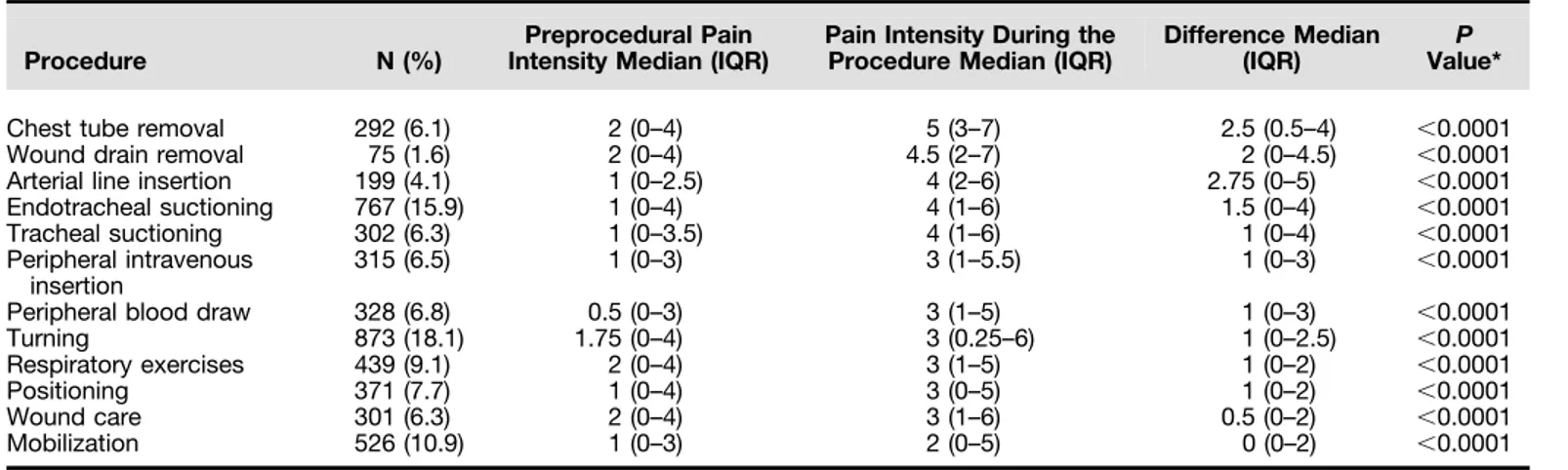
Table 1 lists the main patient characteristics. Among the 3,851 patients, 1,392 (37.4%) received mechanical ventilation during the procedure. Of all patients, 2,467 (65.1%) were able to speak or otherwise communicate. Median Sequential Organ Failure Assessment (SOFA) score at admission was 3 (2–6) and median Richmond Agitation Sedation Score on the procedure day was 0 (21 to 0). The ICU mortality rate was 10%. The most common procedure was turning (n = 873) and the least common was wound drain removal (n = 75) (Table 2). In general, patients reported mild preprocedural pain intensity (i.e., NRS scores of 1–4) (29), and experienced a significant increase in pain intensity during the procedure (P, 0.001) for all procedures.
Pain intensity varied significantly across procedures (Table 2). CTR, wound drain removal, and arterial line insertion were the three most painful procedures, with median procedural pain scores of 5 (3–7), 4.5 (2–7), and 4 (2–6), respectively. Mobilization was the least painful procedure, with a median procedural pain score of 2 (0–5).
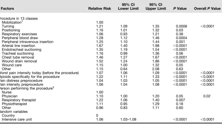
Univariate analyses identified several risk factors for higher procedural pain intensity, when random effects of country and ICU were accounted for (see Table E4). By multivariate analysis, all factors in the model were independently associated with higher procedural pain intensity. Factors associated with greater pain intensity were
the specific procedure, use of opioids specifically for the procedure, higher preprocedural pain intensity, higher preprocedural pain distress, higher intensity of worst pain on the day of the procedure, and procedure not performed by a nurse (Table 3). A significant ICU effect was found, with no detectable country effect because of absorption by the ICU effect.
Table E5 presents the results of multivariate analyses for each separate procedure. The person performing the procedure became nonsignificant for each procedure, and other potential risk factors became nonsignificant, dependent on the procedure.
Discussion
To our knowledge, this is the largest multinational study documenting the prevalence, intensity, and risk factors of procedural pain intensity experienced by adult ICU patients. Most patients had mild pain (NRS scores, 1–4) (29) before the procedure, indicating an improvement over a previous report of baseline resting pain in a smaller sample of ICU patients (30). Nevertheless, all procedures induced a significant increase in pain, although no procedure caused severe pain (mean NRS score = 7–10) (29). For the three most painful procedures (CTR, wound drain removal, and arterial line insertion) pain intensity more than doubled during the procedure compared with the
preprocedural level. Procedural pain was not significantly associated with patient-related variables (e.g., age, sex, or comorbid conditions, such as anxiety and chronic pain). However, several risk factors for higher procedural pain were identified.
The most painful procedure was CTR. This higher degree of pain is in keeping with previous reports (31–33), although CTR pain intensity was lower in our study. Previously, CTR was associated with a mean NRS pain intensity score of 7.7 (31). Opioids have proved effective in
minimizing pain associated with CTR (34, 35), as has ketorolac, a nonsteroidal antiinflammatory agent. In fact, when equianalgesic doses of morphine or ketorolac were administered at time to peak effect during CTR, both were effective in minimizing pain (36). Because CTR is usually a scheduled procedure, pain
Table 1: Patient Characteristics (N = 3,851)
N (%) or Median (IQR) During study enrollment
Age in years (n = 3,793) 62 (50 to 73)
Sex
Male 2,325 (60.8)
Female 1,498 (39.2)
Country of birth: same as country of ICU admission
No 212 (5.9)
Yes 3,401 (94.1)
Native speaker of the primary language of the country of admission
No 126 (3.5)
Yes 3,491 (96.5)
Speaks the primary language of this country
No 12 (0.3)
Yes 3,758 (99.7)
Able to speak or otherwise communicate
No 1,324 (34.9)
Yes 2,467 (65.1)
Tracheostomy or endotracheal tube
No 2,207 (58.6)
Yes 1,558 (41.4)
SOFA score (n = 3,377) 3 (2 to 6)
RASS (n = 3,758) 0 (21 to 0)
Invasive mechanical ventilation
No 2,331 (62.6)
Yes 1,392 (37.4)
Comorbidities before hospital admission Diabetes No 2,812 (76) Yes 887 (24) Heart disease No 1,906 (51.2) Yes 1,817 (48.8)
Chronic lung disease
No 2,861 (78)
Yes 807 (22)
Alcohol abuse
No 3,033 (88)
Yes 412 (12)
Chronic opioid use (i.e., regular use for.3 mo)
No 3,426 (96.9)
Yes 110 (3.1)
Chronic pain (i.e., for.3 mo)
No 3,245 (92.4)
Yes 268 (7.6)
Neuropathic pain
No 3,428 (97.2)
Yes 98 (2.8)
Anxiety before hospital admission
No 2,888 (86.4)
Yes 456 (13.6)
Depression before hospital admission
No 2,926 (88.2)
Yes 392 (11.8)
Died in the ICU
No 3,186 (89.8)
Yes 362 (10.2)
Definition of abbreviations: ICU = intensive care unit; IQR = interquartile range; RASS = Richmond Agitation Sedation Score; SOFA = Sequential Organ Failure Assessment.
prevention by opioid administration or nonopioid agents, such as ketorolac, should be possible in most patients. Pain caused by wound drain removal, the second most painful procedure, has been effectively reduced by preventive local lidocaine injection (37). Severe pain during arterial line insertion has been reported by adults in wards (38) and ICUs (39), and arterial line insertion was the most frequently reported unpleasant experience by 48% of 100 ICU patients (4). Our patients reported less intense pain during this procedure. Reasons for thisfinding of lower pain intensity are unknown.
We categorized procedures involving moving the patients as turning, respiratory exercises, positioning, or mobilization. We found that each caused mild pain (NRS score, 3–4) (29). These findings are encouraging, especially considering the increasing use of early mobilization in ICU patients. Although early mobilization has been reported to decrease deconditioning, improve functional status, and decrease ICU and hospital stay lengths (40, 41), pain resulting from early mobilization has received little attention. Recently, however, a quality-improvement project was conducted to reduce severe pain and stress-related events while moving ICU patients during bathing, massage, sheet change, and repositioning (35). The incidence of severe pain and serious adverse events during patient movement decreased significantly after a change in analgesic ordering practice patterns occurred across the quality improvement project. Although patients in
our study reported only mild pain with mobilization, thisfinding may not apply to all patients and all ICUs. Therefore, pain must be assessed objectively in each patient because lower pain can improve functional status, such as greater mobility (16).
We identified several risk factors for higher procedural pain intensity, including the specific procedure. Compared with mobilization, the risk of increased pain intensity was 20–67% greater with turning, arterial line insertion, peripheral blood draw, intravenous line insertion, endotracheal tube suctioning, CTR, and wound drain removal. Among these procedures, endotracheal tube suctioning is the only one likely to be performed on an emergency basis. When a procedure is not emergent, analgesics can be used for pain prevention. Using nonpharmacologic approaches, such as talking to the patient in a soothing manner, providing information about what is being done, and having family members present, may also provide support to the patient during procedures (42). However, the strength of existing research on nonpharmacologic approaches for procedural pain is limited (16).
When considering all procedures together, there were several risk factors for increased procedural pain intensity: use of opioids specifically for the procedure, higher preprocedural pain intensity, higher preprocedural pain distress, higher intensity of worst pain on the day of the procedure, and procedure not performed by a nurse. Higher pain intensity and higher pain distress before the procedure were
associated with a high risk of increased pain during the procedure. In addition, patients who reported“worst pain” during the day of the procedure also experienced higher procedural pain. Thesefindings make it all the more important that baseline and preprocedural pain be assessed. However, Payen and colleagues (43) found that pain assessments before ICU procedures were performed only 35% of the time. Validated and reliable self-assessment (44) and pain behavior (45, 46) tools are available for use in patients with and without communication capabilities, respectively. Because procedural pain seems to be affected by baseline pain, further research efforts are needed to validate the
effectiveness of a standardized preprocedural pain assessment and an algorithmic approach to administration of a preprocedure analgesic according to pain assessmentfindings. The preprocedural pain assessment should include that of the patient’s current pain intensity, pain distress, and the degree of“worst pain” that day before the procedure. This research step could prove the clinical utility of preprocedural pain assessment and a preemptive analgesic intervention on prevention of procedural pain.
An additional risk factor for higher procedural pain intensity was opioid administration specifically for the
procedure. That is, the patients who received opioids reported increased procedural pain intensity, suggesting the following
possibilities: (1) the amount of opioid received may not have been sufficient for
Table 2: Differences in Pain Intensity from before the Procedure to during the Procedure
Procedure N (%)
Preprocedural Pain Intensity Median (IQR)
Pain Intensity During the Procedure Median (IQR)
Difference Median
(IQR) Value*P
Chest tube removal 292 (6.1) 2 (0–4) 5 (3–7) 2.5 (0.5–4) ,0.0001
Wound drain removal 75 (1.6) 2 (0–4) 4.5 (2–7) 2 (0–4.5) ,0.0001
Arterial line insertion 199 (4.1) 1 (0–2.5) 4 (2–6) 2.75 (0–5) ,0.0001
Endotracheal suctioning 767 (15.9) 1 (0–4) 4 (1–6) 1.5 (0–4) ,0.0001
Tracheal suctioning 302 (6.3) 1 (0–3.5) 4 (1–6) 1 (0–4) ,0.0001
Peripheral intravenous insertion
315 (6.5) 1 (0–3) 3 (1–5.5) 1 (0–3) ,0.0001
Peripheral blood draw 328 (6.8) 0.5 (0–3) 3 (1–5) 1 (0–3) ,0.0001
Turning 873 (18.1) 1.75 (0–4) 3 (0.25–6) 1 (0–2.5) ,0.0001
Respiratory exercises 439 (9.1) 2 (0–4) 3 (1–5) 1 (0–2) ,0.0001
Positioning 371 (7.7) 1 (0–4) 3 (0–5) 1 (0–2) ,0.0001
Wound care 301 (6.3) 2 (0–4) 3 (1–6) 0.5 (0–2) ,0.0001
Mobilization 526 (10.9) 1 (0–3) 2 (0–5) 0 (0–2) ,0.0001
Definition of abbreviation: IQR = interquartile range. Pain intensity was scored on a 0–10 numerical rating scale. *Wilcoxon signed rank sum test.
the procedure, (2) opioids may have not been timed to peak effect in relation to time of the procedure, (3) opioids may have been given to patients who experienced more pain during previous procedures, (4) opioids may be a surrogate marker of more “sensitive” patients, and (5) opioids may have been used more often during the more painful procedures. These possibilities deserve future consideration. Nevertheless, in a previous study, a higher proportion of patients reporting pain before a procedure received an opioid for the procedure compared with patients reporting no pain (47). However, only 17–50% of patients received preemptive opioids in that study (47). Given the evidence that many procedures are painful and that many patients are already in a painful state, increased attention to preprocedural pain assessment and sufficient preventive analgesic therapy is in order. This can be done while
taking into consideration potential adverse effects of analgesic therapies and instituting measures to avoid them.
The influence of these risk factors did not depend on level of acuity because conducting the analyses on two groups (those below the median SOFA score of 3.5 and those equal to or above the median SOFA score) elicited the same results. However, the influence of these risk factors on increasing procedural pain intensity did vary according to each procedure. When considering procedures with samples greater than 400 (i.e., turning, endotracheal tube suctioning, mobilization, and respiratory exercises), the following observations can be made. Higher pain intensity and/or higher pain distress before the procedure continued to be associated with a high risk of increased pain during those specific procedures. In addition, patients undergoing those procedures who reported“worst pain” during the day of the
procedure also experienced higher procedural pain.
In the model examining all procedures together, pain intensity was less when the procedure was performed by a nurse than when it was performed by a physician, respiratory therapist, or physiotherapist when considering all procedures together. However, the effect of the person performing the procedure became nonsignificant when examining each procedure separately. The suggestion that procedural pain can be influenced by the person performing the procedure is intriguing and deserves consideration in future studies of procedural pain.
The prospective design and rigorous standardization of data collection are strong points of our study. However, several limitations must be acknowledged. First, neither the countries nor the ICUs were selected at random. However, the number of
Table 3: Effect of the Procedures on Pain Intensity, as Reported on a 0–10 Numeric Rating Scale When Adjusted on the Other Cofounders in a Multivariate Hierarchical Binomial Model (N = 2,769)*
Factors Relative Risk
95% CI Lower Limit
95% CI
Upper Limit P Value OverallP Value Procedure in 13 classes
Mobilization† 1.00
Turning 1.21 1.09 1.35 0.0006 ,0.0001
Positioning 1.16 1.01 1.32 0.03
Respiratory exercises 1.06 0.93 1.21 0.38
Peripheral blood draw 1.28 1.12 1.46 0.0004
Peripheral intravenous insertion 1.25 1.10 1.44 0.001
Arterial line insertion 1.67 1.40 1.98 ,0.0001
Endotracheal suctioning 1.35 1.19 1.54 ,0.0001
Tracheal suctioning 1.16 0.99 1.36 0.06
Chest tube removal 1.46 1.27 1.67 ,0.0001
Wound drain removal 1.52 1.24 1.86 ,0.0001
Wound care 1.15 1.00 1.32 0.05
Other 1.15 0.64 2.08 0.63
Worst pain intensity today (before the procedure) 1.07 1.06 1.09 ,0.0001 ,0.0001
Opioids specifically for the procedure 1.22 1.11 1.33 ,0.0001 ,0.0001
Pain distress preprocedure 1.04 1.03 1.05 ,0.0001 ,0.0001
Pain intensity preprocedure 1.06 1.04 1.08 ,0.0001 ,0.0001
Person performing the procedure‡
Nurse 1.00 Physician 1.10 1.00 1.20 0.05 0.02 Respiratory therapist 1.22 1.06 1.40 0.007 Physiotherapist 1.11 0.95 1.29 0.18 Other 0.96 0.83 1.11 0.60 Random variables Country 0
Intensive care unit 1.06 1.03–1.08 ,0.0001 ,0.0001
Definition of abbreviation: CI = confidence interval.
Variables not retained in the final model because of their nonsignificant effects were age, depression, chronic opioid use, anxiety, Sequential Organ Failure Assessment score, Richmond Agitation Sedation Scale score, chronic pain, medication on the same day before the procedure, and opioid on the day of the procedure.
*Mixed effect including random effect of intensive care unit and country.
†
Mobilization was the reference.
participating countries and ICUs was larger than in any previous study of procedural pain. Second, patients were enrolled on the basis of convenience, and the 10% mortality rate suggests enrollment bias toward patients having less severe acute illnesses. Nevertheless, our sample represented a large proportion of patients undergoing ICU procedures, suggesting substantial generalizability of our findings. Third, the number of study patients varied considerably across countries. Fourth, pain intensity may have been underestimated because of patient sedation. However, the median Richmond Agitation Sedation Score was 1.07 (1.02–1.12). Fifth, delirium, an exclusion criterion, may have been present and undiagnosed in some patients. Finally, psychological aspects of procedural pain and the importance of attending to nonpharmacologic measures that might ameliorate the distress were not addressed in this report but will be addressed in a separate report.
Our study shows that ICU patients throughout the world often experience a twofold increase in pain from baseline during procedures. Importantly, patients
with higher pain intensity before a procedure and those given opioids for the procedure were at greater risk for increased procedural pain. Efforts to minimize procedural pain should include routine assessments of pain, because preprocedural pain intensity affected the risk of increased procedural pain. Patients receiving opioid infusions may still need additional preemptive analgesia before a procedure. Preventing or reducing procedural pain rather than waiting for patients to experience it is a superior, proactive approach to patient care (16). Dedicated pain assessment instruments, procedural pain-control protocols, and educational programs are effective for minimizing procedural pain (21) and should be used more widely in ICUs.
Conclusions
Procedural pain in ICUs is extremely common. CTR, wound drain removal, and arterial line insertion are the most painful procedures. These three procedures and several others can result in a twofold
increase in pain intensity from baseline. Yet, no procedure under study was associated with severe pain, suggesting that analgesic practices for procedural pain are improving. Nevertheless, identifying short- and long-term adverse consequences of procedural pain and determining the effectiveness of specific analgesic interventions in minimizing procedural pain, especially patients at highest risk, deserve research investigation.n
Author disclosures are available with the text of this article at www.atsjournals.org. Acknowledgment: The authors acknowledge Ms. Marine Chaize, Dr. Nancy Kentish-Barnes, Mr. Pierre Doucet, and Dr. Adeline Max for creating the study blog. They are grateful to Drs. Carmen Mabel Arroyo Novoa, Milagros Figueroa Ramos, and Elena Portecolone, and to several national coordinators, for the translations/back-translations of the data collection packet. They are indebted to A. Wolfe, M.D., for editing this manuscript. The authors thank the ICU patients who agreed to participate in this study, and all ICU coordinators who were responsible for the data collection in their units (see the online supplement for a full list of names and institutions).
References
1. Brennan F, Carr DB, Cousins M. Pain management: a fundamental human right. Anesth Analg 2007;105:205–221.
2. Melzack R, Wall PD. Pain mechanisms: a new theory. Science 1965;150: 971–979.
3. Usichenko TI, R ¨ottenbacher I, Kohlmann T, J ¨ulich A, Lange J, Mustea A, Engel G, Wendt M. Implementation of the quality management system improves postoperative pain treatment: a prospective pre-/post-interventional questionnaire study. Br J Anaesth 2013;110:87–95. 4. Turner JS, Briggs SJ, Springhorn HE, Potgieter PD. Patients’ recollection
of intensive care unit experience. Crit Care Med 1990;18:966–968. 5. Jones J, Hoggart B, Withey J, Donaghue K, Ellis BW. What the patients
say: a study of reactions to an intensive care unit. Intensive Care Med 1979;5:89–92.
6. Wilson VS. Identification of stressors related to patients’ psychologic responses to the surgical intensive care unit. Heart Lung 1987;16: 267–273.
7. Ballard KS. Identification of environmental stressors for patients in a surgical intensive care unit. Issues Ment Health Nurs 1981;3:89–108. 8. Cochran J, Ganong LH. A comparison of nurses’ and patients’ perceptions
of intensive care unit stressors. J Adv Nurs 1989;14:1038–1043. 9. Rotondi AJ, Chelluri L, Sirio C, Mendelsohn A, Schulz R, Belle S, Im K,
Donahoe M, Pinsky MR. Patients’ recollections of stressful experiences while receiving prolonged mechanical ventilation in an intensive care unit. Crit Care Med 2002;30:746–752.
10. van de Leur JP, van der Schans CP, Loef BG, Deelman BG, Geertzen JH, Zwaveling JH. Discomfort and factual recollection in intensive care unit patients. Crit Care 2004;8:R467–R473.
11. Schelling G, Stoll C, Haller M, Briegel J, Manert W, Hummel T, Lenhart A, Heyduck M, Polasek J, Meier M, et al. Health-related quality of life and posttraumatic stress disorder in survivors of the acute respiratory distress syndrome. Crit Care Med 1998;26: 651–659.
12. Schelling G, Richter M, Roozendaal B, Rothenh ¨ausler HB, Krauseneck T, Stoll C, Nollert G, Schmidt M, Kapfhammer HP. Exposure to high stress in the intensive care unit may have negative effects on health-related quality-of-life outcomes after cardiac surgery. Crit Care Med 2003;31:1971–1980.
13. Granja C, Gomes E, Amaro A, Ribeiro O, Jones C, Carneiro A, Costa-Pereira A; JMIP Study Group. Understanding posttraumatic stress disorder-related symptoms after critical care: the early illness amnesia hypothesis. Crit Care Med 2008;36:2801–2809.
14. Price DD, Harkins SW, Baker C. Sensory-affective relationships among different types of clinical and experimental pain. Pain 1987;28: 297–307.
15. Carr DB, Goudas LC. Acute pain. Lancet 1999;353:2051–2058. 16. Czarnecki ML, Turner HN, Collins PM, Doellman D, Wrona S,
Reynolds J. Procedural pain management: a position statement with clinical practice recommendations. Pain Manag Nurs 2011;12: 95–111.
17. G ´elinas C, Johnston C. Pain assessment in the critically ill ventilated adult: validation of the Critical-Care Pain Observation Tool and physiologic indicators. Clin J Pain 2007;23:497–505.
18. Puntillo KA, White C, Morris AB, Perdue ST, Stanik-Hutt J, Thompson CL, Wild LR. Patients’ perceptions and responses to procedural pain: results from Thunder Project II. Am J Crit Care 2001;10: 238–251.
19. Puntillo KA, Morris AB, Thompson CL, Stanik-Hutt J, White CA, Wild LR. Pain behaviors observed during six common procedures: results from Thunder Project II. Crit Care Med 2004;32:421–427.
20. Puntillo K, Pasero C, Li D, Mularski RA, Grap MJ, Erstad BL, Varkey B, Gilbert HC, Medina J, Sessler CN. Evaluation of pain in ICU patients. Chest 2009;135:1069–1074.
21. Chanques G, Jaber S, Barbotte E, Violet S, Sebbane M, Perrigault PF, Mann C, Lefrant JY, Eledjam JJ. Impact of systematic evaluation of pain and agitation in an intensive care unit. Crit Care Med 2006;34: 1691–1699.
22. Harvey MA. Palliative care makes intensive care units intensive care and intensive caring units. Crit Care Med 2011;39:1204–1205. 23. Azoulay E, Timsit JF, Sprung CL, Soares M, Rusinov ´a K, Lafabrie A,
Abizanda R, Svantesson M, Rubulotta F, Ricou B, et al.; Conflicus Study Investigators and for the Ethics Section of the European Society of Intensive Care Medicine. Prevalence and factors of intensive care unit conflicts: the Conflicus Study. Am J Respir Crit Care Med 2009;180:853–860.
24. Jones PS, Lee JW, Phillips LR, Zhang XE, Jaceldo KB. An adaptation of Brislin’s translation model for cross-cultural research. Nurs Res 2001;50:300–304.
25. Brislin RW. Back-translation for cross-cultural research. J Cross-Cultural Psych 1970;1:187–216.
26. Downie WW, Leatham PA, Rhind VM, Wright V, Branco JA, Anderson JA. Studies with pain rating scales. Ann Rheum Dis 1978;37:378–381. 27. Jensen MP, Karoly P, Braver S. The measurement of clinical pain
intensity: a comparison of six methods. Pain 1986;27:117–126. 28. Reading AE. A comparison of pain rating scales. J Psychosom Res
1980;24:119–124.
29. Serlin RC, Mendoza TR, Nakamura Y, Edwards KR, Cleeland CS. When is cancer pain mild, moderate or severe? Grading pain severity by its interference with function. Pain 1995;61:277–284.
30. Chanques G, Sebbane M, Barbotte E, Viel E, Eledjam JJ, Jaber S. A prospective study of pain at rest: incidence and characteristics of an unrecognized symptom in surgical and trauma versus medical intensive care unit patients. Anesthesiology 2007;107:858–860. 31. Puntillo KA. Dimensions of procedural pain and its analgesic management
in critically ill surgical patients. Am J Crit Care 1994;3:116–122. 32. Puntillo KA. Effects of interpleural bupivacaine on pleural chest tube removal
pain: a randomized controlled trial. Am J Crit Care 1996;5:102–108. 33. Carson MM, Barton DM, Morrison CC, Tribble CG. Managing pain
during mediastinal chest tube removal. Heart Lung 1994;23:500–505. 34. Friesner SA, Curry DM, Moddeman GR. Comparison of two
pain-management strategies during chest tube removal: relaxation exercise with opioids and opioids alone. Heart Lung 2006;35:269–276. 35. Joshi VS, Chauhan S, Kiran U, Bisoi AK, Kapoor PM. Comparison of
analgesic efficacy of fentanyl and sufentanil for chest tube removal after cardiac surgery. Ann Card Anaesth 2007;10:42–45.
36. Puntillo K, Ley SJ. Appropriately timed analgesics control pain due to chest tube removal. Am J Crit Care 2004;13:292–301, discussion 302, quiz 303–304.
37. Yiannakopoulos CKA, Kanellopoulos AD. Innoxious removal of suction drains. Orthopedics 2004;27:412–414.
38. Morrison RS, Ahronheim JC, Morrison GR, Darling E, Baskin SA, Morris J, Choi C, Meier DE. Pain and discomfort associated with common hospital procedures and experiences. J Pain Symptom Manage 1998;15:91–101.
39. Nelson JE, Meier DE, Oei EJ, Nierman DM, Senzel RS, Manfredi PL, Davis SM, Morrison RS. Self-reported symptom experience of critically ill cancer patients receiving intensive care. Crit Care Med 2001;29:277–282.
40. Needham DM, Korupolu R, Zanni JM, Pradhan P, Colantuoni E, Palmer JB, Brower RG, Fan E. Early physical medicine and rehabilitation for patients with acute respiratory failure: a quality improvement project. Arch Phys Med Rehabil 2010;91:536–542.
41. Schweickert WD, Kress JP. Implementing early mobilization interventions in mechanically ventilated patients in the ICU. Chest 2011;140:1612–1617.
42. Faigeles B, Howie-Esquivel J, Miaskowski C, Stanik-Hutt J, Thompson C, White C, Wild LR, Puntillo K. Predictors and use of nonpharmacologic interventions for procedural pain associated with turning among hospitalized adults. Pain Manag Nurs 2013;14: 85–93.
43. Payen JF, Chanques G, Mantz J, Hercule C, Auriant I, Leguillou JL, Binhas M, Genty C, Rolland C, Bosson JL. Current practices in sedation and analgesia for mechanically ventilated critically ill patients: a prospective multicenter patient-based study. Anesthesiology 2007;106:687–695, quiz 891–892.
44. Chanques G, Viel E, Constantin JM, Jung B, de Lattre S, Carr J, Ciss ´e M, Lefrant JY, Jaber S. The measurement of pain in intensive care unit: comparison of 5 self-report intensity scales. Pain 2010;151:711–721. 45. G ´elinas C, Fillion L, Puntillo KA, Viens C, Fortier M. Validation of the
critical-care pain observation tool in adult patients. Am J Crit Care 2006;15:420–427.
46. Payen JF, Bru O, Bosson JL, Lagrasta A, Novel E, Deschaux I, Lavagne P, Jacquot C. Assessing pain in critically ill sedated patients by using a behavioral pain scale. Crit Care Med 2001;29:2258–2263. 47. Puntillo KA, Wild LR, Morris AB, Stanik-Hutt J, Thompson CL, White C.
Practices and predictors of analgesic interventions for adults undergoing painful procedures. Am J Crit Care 2002;11:415–429, quiz 430–431.