An underestimated cause of chronic
cough: The Protracted Bacterial
Bronchitis
Paola Di Filippo, Alessandra Scaparrotta, Marianna Immacolata Petrosino, Marina Attanasi, Sabrina Di Pillo, Francesco Chiarelli, Angelika Mohn
Abstract:
Chronic cough in childhood is associated with a high morbidity and decreased quality of life. Protracted bacterial bronchitis (PBB) seems to be the second most common cause of chronic cough in children under 6 years of age. Its main clinical feature is represented by wet cough that worsens when changing posture and improves after the introduction of antibiotics. Currently, the mainstay of PBB treatment is a 2‑week therapy with a high dose of antibiotics, such as co‑amoxiclav, to eradicate the infection and restore epithelial integrity. It is very important to contemplate this disease in a child with chronic cough since the misdiagnosis of PBB could lead to complications such as bronchiectasis. Clinicians, however, often do not consider this disease in the differential diagnosis and, consequently, they are inclined to change the antibiotic therapy rather than to extend it or to add steroids. Data sources of this review include PubMed up to December 2016, using the search terms “child,” “chronic cough,” and “protracted bacterial bronchitis.”
Keywords:
Asthma, bronchiectasis, children, cough, protracted bacterial bronchitis
C
ough is the most common reasonwhy a patient asks a doctor for a consultation.[1]
Pediatric chronic cough in children younger than 15 years of age is defined as a daily cough that persists longer than 4 weeks.[2]
Chronic cough in childhood is associated with a high morbidity and decreased quality of life scores,[3] impacting sleep and school
attendance.[4]
In a recent study including 563 children with chronic cough, the most common final diagnoses were asthma (24.9%), asthma‑like symptoms (19%), protracted bacterial bronchitis (PBB) (11.9%), and upper airway cough syndrome (9.1%). The frequency of causes changes according to age groups: PBB was the second most common cause in
children under 6 years of age, whereas the psychogenic cough was the second common cause of cough in children over 6 years of age.[5]
PBB is frequently missed, misdiagnosed (e.g., as asthma), or inadequately treated,[6]
resulting in a persistence of symptoms and potential structural damage of the respiratory system (bronchiectasis).[7]
Despite the increased clinical recognition, the underlying mechanisms of PBB remain to be clarified.
Definition
Lower respiratory tract bacterial infections may manifest as the classic bacterial pneumonia or as PBB. This syndrome is a relatively new clinical entity, first described in a study conducted in Australia in 2006.[8] Address for
correspondence:
Dr. Paola Di Filippo, Department of Pediatrics, University of Chieti, Via dei Vestini 5, 66100 Chieti, Italy. E‑mail: paolina__@ hotmail.it Submission: 08‑01‑2017 Accepted: 26‑04‑2017 Department of Pediatrics, University of Chieti, 66100 Chieti, Italy
Review Article
Access this article online
Quick Response Code:
Website:
www.thoracicmedicine.org
DOI:
10.4103/atm.ATM_12_17
How to cite this article: Di Filippo P, Scaparrotta A,
Petrosino MI, Attanasi M, Di Pillo S, Chiarelli F, et al. An underestimated cause of chronic cough: The protracted bacterial bronchitis. Ann Thorac Med 2018;13:7‑13.
This is an open access article distributed under the terms of the Creative Commons Attribution‑NonCommercial‑ShareAlike 3.0 License, which allows others to remix, tweak, and build upon the work non‑commercially, as long as the author is credited and the new creations are licensed under the identical terms.
PBB is a protracted or persistent infection of conducting airways and it is clinically defined as the presence of chronic wet/moist cough for more than 4 weeks.[9] It
is a common cause of chronic wet cough in children, and it was only recently described in pediatric or even respiratory pediatric textbooks and incorporated in national and international cough guidelines.[10]
Epidemiology
The prevalence of chronic cough was estimated to be 8% in a study of 1165 Australian children and 566 Nigerian children,[11] although some studies reported higher
percentages, up to 10% in both adults and children.[4]
Chronic cough represents a source of great frustration for parents and doctors. It has consequences on the child’s sleep, school performance, and ability, and it is associated with recurrent doctor visits and parental stress and worry.[12] It commonly happens that children were
visited by a general practitioner more than 20 times for their cough before being referred to a specialist.[4] The
prevalence of PBB among children with chronic wet cough is still not clear, but many studies report a prevalence of about 40%,[6,13,14] and coexisting bronchiectasis in 9%.[14]
Some studies report a probable increase after variations of the antibiotic therapy and after Haemophilus influenzae type B and pneumococcal conjugate vaccines.[9] On the
other hand, it is still unclear whether the increasing number of diagnoses of PBB is due to a true increase in incidence or to a better recognition.[6]
Nearly 54.5% of children referred to a tertiary center with cough for at least 3 weeks received a primary diagnosis of asthma, but after further investigations that included a bronchoscopy, the primary diagnosis became PBB in 40% of cases.[6]
Although it can involve any period of life (including adulthood), the majority of children with PBB are <6 years old (mean or median age of 1.8–4.8 years).[14,15,16]
Moreover, PBB is more frequent in males and it often coexists with airway malacia.[6]
Etiopathogenesis
The most common bacteria implicated in PBB are represented by nontypeable Haemophilus influenzae (NTHi), Streptococcus pneumoniae, and, less commonly,
Moraxella catarrhalis.[6,13]
H. influenzae (range: 47%–81%) is the most common
bacterium reported,[17] and most H. influenzae are NTHi
strains, representing different genotypes.[9]
In some studies,[8,12] S. pneumoniae (24%–39%) was
the second most common organism detected in
bronchoalveolar lavage (BAL) cultures, but in others,[7,9]
M. catarrhalis (range: 19–43%) was more commonly
found.
In a recent Chinese study investigating 66 hospitalized infants under the age of 3 years with chronic wet cough, 75.8% were diagnosed with PBB. H. influenzae (47.4%) and S. pneumoniae (36.8%) were the most commonly identified pathogens.[18]
More than one organism is often identified in BAL samples of patients affected by PBB,[7‑9] but how this
influences clinical presentation or outcomes is still not clear. Viral pathogens identified in children with PBB include respiratory syncytial virus, parainfluenza virus, and human metapneumovirus, but the most common virus identified is human adenovirus (HAdV).[19]
Children with PBB are significantly more likely than no cough controls to have coinfection with HAdV and
H. influenzae.[13]
The occurrence of PBB is related to impaired mucociliary clearance, systemic immune function defects, airway anomalies, and the formation of bacterial biofilm in the airway.[20]
A biofilm is a matrix secreted by some bacteria that enhances the attachment, facilitates access to nutrients, and decreases antibiotic penetration, protecting the bacteria against antibiotics and making the bacteria difficult to eradicate with standard length courses of antibiotics.[21]
C h i l d r e n w i t h P B B u s u a l l y h a v e n o r m a l immunoglobulin levels, antibody responses (to tetanus and H. influenzae type B vaccines), lymphocyte subsets, and propensity to atopy (IgE and RAST). Many children, however, exhibit elevated NK‑cell levels, probably associated with the recent viral infection and associated to elevated virus detection rates in BAL specimens (particularly HAdV). Tracheobronchomalacia is common, but rates are similar to one of the control groups.[19]
Lower airways of children with PBB are characterized by bacterial infection and airway neutrophilia. This suggests that pulmonary innate immunity and neutrophil pathway mediators may play an important role in pathogenesis.[8,22] Several studies, moreover, found an
upregulation of the toll‑like receptors (TLRs) TLR2 and TLR4,[13] human b‑defensin‑2, and mannose‑binding
lectin levels in the BAL of children with PBB,[23]
suggesting an epithelial–neutrophil interaction actively engaged in PBB.
A recent study found an increased expression of neutrophil‑related mediators in PBB, including
interleukin‑1 (IL‑1) pathway members, neutrophil α‑defensins (defensins 1–3), and the chemokine receptor CXCR2. IL‑1b and related mediators were associated with BAL neutrophils, cough symptoms, and disease recurrence, providing an insight into PBB pathogenesis. IL‑1 pathway signaling was associated with PBB disease recurrence.[24]
Clinical Features
The clinical features of PBB are nighttime coughing, shortness of breath during physical exercise, “wheezing,” and exacerbations with upper respiratory tract infections.[6] These are nonspecific symptoms and may
also be asthma manifestations.
Some authors have highlighted that children with PBB do not have “wheeze” but a “rattle” (a rattling sound), which is reflective of airway secretions.[15]
A viral infection would exacerbate both asthma and PBB. In PBB, this is likely to be due to the release of planktonic forms of bacteria from biofilms, presumably in response to the associated inflammation which may help disseminate the organism as observed in cystic fibrosis.[25]
Systemic symptoms are generally absent, including sinus and ear disease.[4] The affected children typically look
healthy, their growth and development are normal, and they lack signs of the underlying chronic suppurative lung disease, such as digital clubbing, chest wall deformity, and adventitial auscultatory chest findings.[16,19]
If present, systemic symptoms are minimal, like tiredness or lake of energy, but the impact on the general welfare is relevant, because of disturbed sleep or chronic infection.[22]
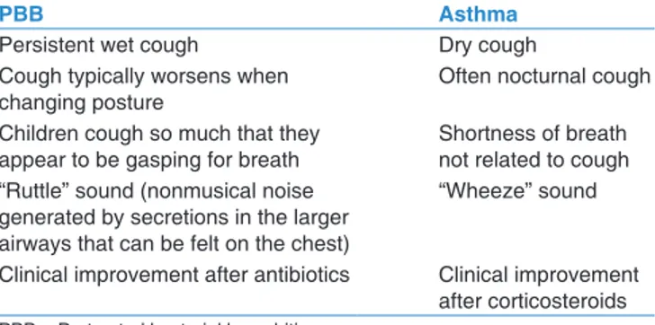
Nonspecific differences between asthma and PBB symptoms make the differential diagnosis not always easy [Table 1], and the addition of a spirometry test for children older than 6 years can help to differentiate. The
differences that may be helpful to distinguish between the two diseases are: (1) wet cough in PBB versus dry nocturnal cough of asthma; (2) cough typically worsens when changing posture, just after lying down in bed and as the first thing in the morning, but can be present all night long as in asthma; (3) children with PBB typically cough so much that they appear to be gasping for breath, while the shortness of breath observed in asthmatics is not directly related to coughing; (4) rattle sound (nonmusical noise generated by secretions in the larger airways that can be felt on the chest) versus the “wheeze” sound of asthmatics; (5) improvement after the introduction of antibiotics, while asthma improves after inhaling corticosteroids.[6]
The differential diagnosis with acute bronchitis is based on the resolution of the cough within 2–4 weeks in acute bronchitis. The major difficulties arise in cases of recurrent episodes of acute bronchitis, especially in winter, when respiratory virus circulation is very high.[26]
When parents report the failure of antibiotics, usually we tend to exclude the PBB. It is therefore important to ask more specific questions. In PBB, children often have an initial improvement in symptoms as long as they adhere to antibiotic therapy and then symptoms worse quickly when the antibiotic therapy is ceased.[6]
Diagnosis
The PBB diagnosis should be clinically suspected when typical features suggestive of a persistent infection of the conducting airways by pathogenic bacteria are present.[6]
The original diagnostic criteria for PBB include: (a) wet cough for more than 4 weeks; (b) identifiable lower airway bacterial infection on BAL culture (defined as bacterial growth ≥104 CFU/ml in BAL);[27] and (c) response to
antibiotics (amoxicillin/clavulanate) with resolution of cough within 2 weeks.[8] In clinical practice, however,
some of these diagnostic criteria are difficult to obtain in children due to the following reasons: it is impractical to perform bronchoscopy and BAL on every child with a chronic wet cough; children are often unable to expectorate a sufficient sputum for reliable culture;[16]
antibiotic usage within a month often results in a negative culture;[27] in a minority of children, the therapy
has to be extended to a period of maximum 4 weeks.[22]
More clinical diagnostic criteria have been recently established [Table 2], and the criterion “Documented lower airway infection (single bacterial species >104 CFU/ml in sputum or at BAL)’ has been replaced by: “Absence of symptoms or signs of other causes of wet or productive cough”[28] that includes the absence of
chest pain, history suggestive of inhaled foreign body,
Table 1: Differential diagnosis between protracted bacterial bronchitis and asthma
PBB Asthma
Persistent wet cough Dry cough Cough typically worsens when
changing posture Often nocturnal cough Children cough so much that they
appear to be gasping for breath Shortness of breath not related to cough “Ruttle” sound (nonmusical noise
generated by secretions in the larger airways that can be felt on the chest)
“Wheeze” sound
Clinical improvement after antibiotics Clinical improvement after corticosteroids PBB = Protracted bacterial bronchitis
dyspnea, exertional dyspnea, hemoptysis, failure to thrive, feeding difficulties (including choking/vomiting), cardiac or neurodevelopmental abnormalities, recurrent sino‑pulmonary infections, immunodeficiency, epidemiological risk factors for exposure to tuberculosis, respiratory distress signs, digital clubbing, chest wall deformity, auscultatory crackles, chest radiographic alterations (other than perihilar changes), and lung function abnormalities.[2,29]
Recently, Chang et al. have proposed four PBB definitions: (1) PPB‑micro includes the original criteria; (2) PPB‑clinical replaces the microbiological item with a clinical item; (3) PBB‑extended where, despite the presence of the diagnostic criteria of PBB‑clinical or PBB‑micro were present, only after 4 weeks of antibiotics; (4) Recurrent PBB: recurrent episodes (>3 per year) of PBB.[16]
The results of instrumental examinations show that chest X‑ray is often normal or may have only minor abnormalities, such as peribronchial wall thickening. These evidences are consistent with other conditions such as viral lower respiratory tract infections, poorly controlled asthma and recurrent minor aspirations.[6]
When hyperinflation is present, it should indicate asthma or the coexistence of asthma with PBB.[22]
Computed tomography (CT) scan should be done only after establishing a treatment failure by a suspicion of bronchiectasis.[22] When performed, spirometry is
normal.[30]
When a child can expectorate, a sputum specimen should be collected.[27] Cough swabs can be useful but have a
relatively low sensitivity.[15]
The gold standard method to sample the lower airways in young children is flexible bronchoscopy with BAL (FB‑BAL). This is safe and has a low rate of complications.[31] The timing of this examination should
be discussed with parents; in general, it is performed when there is a relapse after three courses of antibiotics, whereas some parents prefer to have a definitive diagnosis at the onset. Typically, we can find secretions and edematous collapsible bronchi that collapse during suctioning while undertaking a BAL.[6]
The guidelines of the European Respiratory Society recommend a triple aliquot sample from a single of the six lobes, including the lingula.[31] FB‑BAL with the
sampling of six lobes has previously demonstrated a heterogeneous distribution of bacteria within the lungs of children with cystic fibrosis.[32] Due to this
heterogeneity, the sampling is limited to one or two lobes, a number of organisms would have been missed with consequent false‑negative results.[7] In
a retrospective study,[7] which analyzed each of the
six lobes in fifty children with suspected PBB, it was demonstrated that positive cultures were obtained from 41/50 (82%) of FB‑BAL procedures versus 46% and 63% seen in previous studies in which only a single lobe was sampled.[33] The higher rate of positive cultures
seems to be related to the number of lobes sampled: sampling one lobe, the percentage of positive cultures was 66% (33/50), and sampling 2 lobes (one lobe and lingula), it was 70% (35/50).
If a triple aliquot of BAL is performed, it is recommended that the first aliquot is sent for microbiological culture because this is the more proximal sample, and the second and third aliquots are pooled for cytological and noncellular studies.[31]
As previously mentioned, elevated neutrophil counts in BAL cytology of patients with PBB were demonstrated.[8] In general, a positive BAL culture is
caused by infection and not by colonization. Assuming that all patients with PBB are symptomatic, no positive airway culture could be presumed to be due to colonization. In conclusion, cytology results are not included as a diagnostic criterion, and we do not think they would have provided any additional information in this particular clinical setting.[7]
Even if flexible bronchoscopy is a safe procedure with a very low incidence of major complications (about 2%), this test remains invasive and difficult to perform, especially in children. Furthermore, since the complications can be dangerous, a careful analysis of indications is needed.[34]
On the other hand, further studies are required to evaluate the most appropriate timing of performing bronchoscopy in children with PBB.
Table 2: Diagnostic criteria of protracted bacterial bronchitis
Original diagnostic criteria (PBB-micro) Modified diagnostic criteria (PBB‑clinical)
Chronic wet cough (>4 weeks) Chronic wet cough (>4 weeks) Documented lower airway infection (single bacterial
species >104 CFU/ml in sputum or at BAL)
No symptoms or signs of other causes of wet or productive cough Resolution of the cough after a 2‑week course of an
appropriate oral antibiotic (amoxicillin‑clavulanate) Resolution of the cough after a 2‑week course of an appropriate oral antibiotic (amoxicillin‑clavulanate) PBB = Protracted bacterial bronchitis, BAL = Bronchoalveolar lavage
The Natural Course of Protracted Bacterial
Bronchitis
If untreated, PBB in some children may progress into chronic suppurative lung disease and possibly bronchiectasis.[22]
It has been suggested that PBB, chronic suppurative lung disease and bronchiectasis represent a clinical continuum. The invasion by encapsulated bacteria results, indeed, in innate immune activation and a subsequent neutrophilic inflammation. In the absence of an adequate treatment, probably, the persistent inflammation causes impaired ciliary clearance, chronic mucus hypersecretion, further airway inflammation, and then bacterial stasis. This saturates the bactericidal ability of the immune system, promoting the chronic inflammation. A vicious cycle of infection‑inflammation ensues and probably pushes patients along the clinical spectrum of the BBP, to chronic suppurative lung disease and finally bronchiectasis.[20,35]
The apoptotic cells can undergo secondary necrosis, thus potentially releasing toxic cell contents and perpetuating airway inflammation. A recent study hypothesized that a reduced alveolar macrophage phagocytic host response to apoptotic cells or that NTHi may contribute to neutrophilic inflammation and NTHi colonization in both PBB and bronchiectasis, identifying similar mechanisms underlying both diseases. Whether these mechanisms also contribute to the progression of PBB to bronchiectasis remains unknown.[36]
Recurrent PBB and bronchiectasis in children share common characteristics, with a similar clinical presentation, such as the association with neutrophilic inflammation and the frequent isolation from the lower airways of potentially pathogenic microorganisms.[36]
Douros et al.[37] found a correlation between duration of
wet cough and abnormalities seen on CT scans. However, if recurrent PBB is antecedent to bronchiectasis, the development of the disease must be studied in a longitudinal cohort study.
In a recent prospective longitudinal cohort study of children with PBB,[38] two significant risk factors
for bronchiectasis were identified: recurrent (>3 episodes/year) PBB and the presence of H. influenzae infection in the lower airways. Multivariate logistic regression showed that recurrent PBB status and
H. influenzae infection were significantly associated
with bronchiectasis diagnosis (odds ratio [OR], 11.48; 95% confidence interval [CI], 2.33–56.50; P = 0.003 and OR, 7.60; 95% CI, 1.53–37.79; P = 0.013, respectively); the survival analysis, through Cox regression, concurred with logistic regression findings.
There is high‑quality evidence that when specific cough pointers (e.g., digital clubbing) are present in children with wet cough, further investigations (e.g., flexible bronchoscopy, chest CTs, and immunity tests) should be undertaken.[39]
Therapy
The mainstay of PBB treatment is an oral antibiotic, although the use of intravenous and nebulized antibiotics has been reported.[15] There is high‑quality evidence
that, in children aged ≤14 years, with chronic wet or productive cough, the use of appropriate antibiotics improves cough resolution.[39] The aim of the treatment
is to eradicate the infection and restore epithelial integrity.[6,14]
If therapy is started blindly, co‑amoxiclav is the most widely used first‑line treatment. As soon as results of the microbiological studies are available, the antibiotics can be chosen according to the isolated organism and its sensitivity.[12]
In a recent study, co‑amoxiclav resulted to be an appropriate antibiotic for most of the children under examination. After the results of bronchoscopy, however, a different antibiotic was used in 17/50 patients, and, in particular, in four children, amoxicillin rather than co‑amoxiclav was prescribed. The culture in two children revealed the presence of organisms with
in vitro resistance to co‑amoxiclav. Eleven children,
in whom a Staphylococcus aureus was isolated, were treated with flucloxacillin or clarithromycin rather than co‑amoxiclav.[7]
When co‑amoxiclav cannot be used, macrolides or cephalosporins are other options.[10]
As regards the duration of the antibiotic therapy, the 2008 British Thoracic Society cough guidelines[10] recommend
that all children with PBB should receive 4–6 weeks of antibiotics. This recommendation, however, was based on expert opinions as, at that time, no supportive high‑quality studies existed.
It was demonstrated that a 2‑week therapy with co‑amoxiclav can lead to resolution of the chronic wet cough and to a dramatic improvement in the child’s quality of life in a statistically significant number of children.[6,12]
Shorter courses of antibiotics tend to result in a partial resolution or a relapse of the cough after a few days of treatment, differing from community‑acquired pneumonia for which 5–7 days’ treatment is usually sufficient.[6]
Further studies are now needed to help identify whether the patients with PBB requiring a longer course of antibiotics are different (e.g., outcomes) from those responding to shorter courses.[16]
Donnelly et al. found that more than half (51%) of the analyzed cohort was completely symptom free after two courses of antibiotics, with only 13% requiring six or more courses or needing continuous prophylactic antibiotics for at least one winter.[15]
When the wet cough does not improve by 4 weeks of antibiotics, there is moderate quality evidence that children should be referred to a major center for further investigations to determine the potential presence of underlying lung or other diseases.[39]
It is not clear whether vaccines against NTHi are effective. The use of pneumococcal conjugate vaccines has not reduced the incidence of this condition.[6]
It is important to remember that asthma is the most common cause of significant respiratory symptoms in childhood, and we must guard against returning to mistreating asthma with antibiotics. Considering the dramatic impact of antibiotics on the lives of children with PBB, there is a risk of the overuse of antibiotics to treat asthma, misdiagnosing mild asthmatics as having PBB. It is also important to recognize that asthma per se predisposes to PBB, particularly if poorly controlled, and hence in a patient with definite asthma who has a persistent wet cough, antibiotics may have an important role; however, they should not be used to treat exacerbations of asthma mislabeled as “chest infections.”[6]
If PBB fails to respond or becomes recurrent, further investigations such as complete blood count, assessment of immunoglobulins, functional antibody response to vaccination, sweat test, and high‑resolution CT are required to rule out other etiologies such as cystic fibrosis, primary ciliary dyskinesia, immunodeficiencies, or dysfunctional swallowing and aspiration issues.[40] CT scan has an important role, because it has
been demonstrated that children with a chronic wet cough failing to resolve after 4 weeks of appropriate oral antibiotics have increased likelihood of developing bronchiectasis.[41]
Conclusion
In a child with chronic wet cough without symptoms and signs suggestive of another diagnosis, PBB should always be suspected, remembering that chronic cough is not always asthma, especially if there is an improvement of symptoms under antibiotic therapy. Further research
is needed to better establish clinical criteria that can help clinicians to obtain PBB diagnosis, because the actual proposed criteria for PBB are nonspecific and may lead to significant overdiagnosis and antibiotic overuse that may cause antibiotic resistance.
It is important to remember that PBB is characterized by a prolonged duration of wet cough and that antibiotics should not be prescribed for acute exacerbations of asthma.
Financial support and sponsorship
Nil.
Conflicts of interest
There are no conflicts of interest.
References
1. Irwin RS. Introduction to the diagnosis and management of cough: ACCP evidence‑based clinical practice guidelines. Chest 2006;129 1 Suppl: 25S‑7S.
2. Chang AB, Glomb WB. Guidelines for evaluating chronic cough in pediatrics: ACCP evidence‑based clinical practice guidelines. Chest 2006;129 1 Suppl: 260S‑83S.
3. Newcombe PA, Sheffield JK, Chang AB. Minimally important change in a Parent‑Proxy Quality‑of‑Life questionnaire for pediatric chronic cough. Chest 2011;139:576‑80.
4. Marchant JM, Newcombe PA, Juniper EF, Sheffield JK, Stathis SL, Chang AB. What is the burden of chronic cough for families? Chest 2008;134:303‑9.
5. Gedik AH, Cakir E, Torun E, Demir AD, Kucukkoc M, Erenberk U,
et al. Evaluation of 563 children with chronic cough accompanied
by a new clinical algorithm. Ital J Pediatr 2015;41:73.
6. Craven V, Everard ML. Protracted bacterial bronchitis: Reinventing an old disease. Arch Dis Child 2013;98:72‑6. 7. Narang R, Bakewell K, Peach J, Clayton S, Samuels M, Alexander J,
et al. Bacterial distribution in the lungs of children with protracted
bacterial bronchitis. PLoS One 2014;9:e108523.
8. Marchant JM, Masters IB, Taylor SM, Cox NC, Seymour GJ, Chang AB. Evaluation and outcome of young children with chronic cough. Chest 2006;129:1132‑41.
9. Priftis KN, Litt D, Manglani S, Anthracopoulos MB, Thickett K, Tzanakaki G, et al. Bacterial bronchitis caused by Streptococcus
pneumoniae and nontypeable Haemophilus influenzae in children:
The impact of vaccination. Chest 2013;143:152‑7.
10. Shields MD, Bush A, Everard ML, McKenzie S, Primhak R; British Thoracic Society Cough Guideline Group. BTS guidelines: Recommendations for the assessment and management of cough in children. Thorax 2008;63 Suppl 3:iii1‑15.
11. Faniran AO, Peat JK, Woolcock AJ. Measuring persistent cough in children in epidemiological studies: Development of a questionnaire and assessment of prevalence in two countries. Chest 1999;115:434‑9.
12. Marchant J, Masters IB, Champion A, Petsky H, Chang AB. Randomised controlled trial of amoxycillin clavulanate in children with chronic wet cough. Thorax 2012;67:689‑93.
13. Marchant JM, Gibson PG, Grissell TV, Timmins NL, Masters IB, Chang AB. Prospective assessment of protracted bacterial bronchitis: Airway inflammation and innate immune activation. Pediatr Pulmonol 2008;43:1092‑9.
14. Chang AB, Robertson CF, Van Asperen PP, Glasgow NJ, Mellis CM, Masters IB, et al. A multicenter study on chronic
cough in children: Burden and etiologies based on a standardized management pathway. Chest 2012;142:943‑50.
15. Donnelly D, Critchlow A, Everard ML. Outcomes in children treated for persistent bacterial bronchitis. Thorax 2007;62:80‑4. 16. Chang AB, Gibson PG, Marchant JM, Upham JW, Masters IB,
Redding GR, et al. Protracted bacterial bronchitis: The last decade and the road ahead. Pediatr Pulmonol 2016;51:225‑42.
17. Kompare M, Weinberger M. Protracted bacterial bronchitis in young children: Association with airway malacia. J Pediatr 2012;160:88‑92.
18. Wang Y, Hao C, Chi F, Yu X, Sun H, Huang L, et al. Clinical characteristics of protracted bacterial bronchitis in Chinese infants. Sci Rep 2015;5:13731.
19. Wurzel DF, Marchant JM, Yerkovich ST, Upham JW, Mackay IM, Masters IB, et al. Prospective characterization of protracted bacterial bronchitis in children. Chest 2014;145:1271‑8.
20. Paul SP, Hilliard T. The importance of recognizing protracted bacterial bronchitis in children. Indian J Pediatr 2014;81:1‑3. 21. Stewart PS. Mechanisms of antibiotic resistance in bacterial
biofilms. Int J Med Microbiol 2002;292:107‑13.
22. Chang AB, Redding GJ, Everard ML. Chronic wet cough: Protracted bronchitis, chronic suppurative lung disease and bronchiectasis. Pediatr Pulmonol 2008;43:519‑31.
23. Chang AB, Yerkovich ST, Gibson PG, Anderson‑James S, Petsky HL, Carroll ML, et al. Pulmonary innate immunity in children with protracted bacterial bronchitis. J Pediatr 2012;161:621‑5.e1.
24. Baines KJ, Upham JW, Yerkovich ST, Chang AB, Marchant JM, Carroll M, et al. Mediators of neutrophil function in children with protracted bacterial bronchitis. Chest 2014;146:1013‑20.
25. Chattoraj SS, Ganesan S, Jones AM, Helm JM, Comstock AT, Bright‑Thomas R, et al. Rhinovirus infection liberates planktonic bacteria from biofilm and increases chemokine responses in cystic fibrosis airway epithelial cells. Thorax 2011;66:333‑9.
26. Thompson M, Vodicka TA, Blair PS, Buckley DI, Heneghan C, Hay AD; TARGET Programme Team. Duration of symptoms of respiratory tract infections in children: Systematic review. BMJ 2013;347:f7027.
27. De Schutter I, De Wachter E, Crokaert F, Verhaegen J, Soetens O, Piérard D, et al. Microbiology of bronchoalveolar lavage fluid in children with acute nonresponding or recurrent community‑acquired pneumonia: Identification of nontypeable
Haemophilus influenzae as a major pathogen. Clin Infect Dis
2011;52:1437‑44.
28. Chang AB, Landau LI, Van Asperen PP, Glasgow NJ,
Robertson CF, Marchant JM, et al. Cough in children: Definitions and clinical evaluation. Med J Aust 2006;184:398‑403.
29. Chang AB. Cough, cough receptors, and asthma in children. Pediatr Pulmonol 1999;28:59‑70.
30. Chang AB, Van Asperen PP, Glasgow N, Robertson CF, Mellis CM, Masters IB, et al. Children with chronic cough: When is watchful waiting appropriate? Development of likelihood ratios for assessing children with chronic cough. Chest 2015;147:745‑53.
31. de Blic J, Midulla F, Barbato A, Clement A, Dab I, Eber E,
et al. Bronchoalveolar lavage in children. ERS Task Force on
bronchoalveolar lavage in children. European Respiratory Society. Eur Respir J 2000;15:217‑31.
32. Gilchrist FJ, Salamat S, Clayton S, Peach J, Alexander J, Lenney W. Bronchoalveolar lavage in children with cystic fibrosis: How many lobes should be sampled? Arch Dis Child 2011;96:215‑7. 33. Zgherea D, Pagala S, Mendiratta M, Marcus MG, Shelov SP,
Kazachkov M. Bronchoscopic findings in children with chronic wet cough. Pediatrics 2012;129:e364‑9.
34. de Blic J, Marchac V, Scheinmann P. Complications of flexible bronchoscopy in children: Prospective study of 1,328 procedures. Eur Respir J 2002;20:1271‑6.
35. Al Subie H, Fitzgerald DA. Non‑cystic fibrosis bronchiectasis. J Paediatr Child Health 2012;48:382‑8.
36. Hodge S, Upham JW, Pizzutto S, Petsky HL, Yerkovich S, Baines KJ, et al. Is alveolar macrophage phagocytic dysfunction in children with protracted bacterial bronchitis a forerunner to bronchiectasis? Chest 2016;149:508‑15.
37. Douros K, Alexopoulou E, Nicopoulou A, Anthracopoulos MB, Fretzayas A, Yiallouros P, et al. Bronchoscopic and high‑resolution CT scan findings in children with chronic wet cough. Chest 2011;140:317‑23.
38. Wurzel DF, Marchant JM, Yerkovich ST, Upham JW, Petsky HL, Smith‑Vaughan H, et al. Protracted bacterial bronchitis in children: Natural history and risk factors for bronchiectasis. Chest 2016;150:1101‑8.
39. Chang AB, Oppenheimer JJ, Weinberger M, Rubin BK, Irwin RS. Children with chronic wet or productive cough – Treatment and investigations: A systematic review. Chest 2016;149:120‑42. 40. Bidiwala A, Krilov LR, Pirzada M, Patel SJ. Pro‑con debate:
Protracted bacterial bronchitis as a cause of chronic cough in children. Pediatr Ann 2015;44:329‑36.
41. Goyal V, Grimwood K, Marchant J, Masters IB, Chang AB. Does failed chronic wet cough response to antibiotics predict bronchiectasis? Arch Dis Child 2014;99:522‑5.