Oral mucosal lesions and risk of all-cause and
cardiovascular mortality in people treated
with long-term haemodialysis: The ORAL-D
multinational cohort study
Marinella Ruospo1, Suetonia C. Palmer2, Giusi Graziano1, Patrizia Natale1,3,
Valeria Saglimbene1,4, Massimo Petruzzi3, Michele De Benedittis3, Jonathan C. Craig5,
David W. JohnsonID6,7, Pauline Ford6, Marcello Tonelli8, Eduardo Celia1, Ruben Gelfman1,
Miguel R. Leal1, Marietta To¨ ro¨ k1, Paul Stroumza1, Luc Frantzen1, Anna
Bednarek-Skublewska1,9, Jan Dulawa1,10, Domingo del Castillo1, Staffan Scho¨ n1, Amparo G. Bernat1, Jo¨ rgen HegbrantID1, Charlotta Wollheim1, Letizia Gargano1, Giovanni F.
M. StrippoliID1,3,4*, on behalf of the ORAL-D Investigators
1 Diaverum Medical Scientific Office, Lund, Sweden, 2 University of Otago, Christchurch, New Zealand, 3 University of Bari, Bari, Italy, 4 University of Sydney, Sydney, Australia, 5 Flinders University, Adelaide,
Australia, 6 University of Queensland, Brisbane, Australia, 7 Translational Research Institute, Brisbane, Australia, 8 University of Calgary, Calgary, Canada, 9 Medical University of Lublin, Lublin, Poland,
10 Medical University of Silesia, Silesia, Poland
¶ Membership of the ORAL-D study is provided in the Acknowledgments. *[email protected]
Abstract
Background
Chronic kidney disease is a risk factor for oral diseases, which may be associated with pre-mature death. We evaluated the risk of all-cause and cardiovascular mortality associated with oral mucosal lesions in adults with kidney failure treated with long-term haemodialysis.
Methods
Oral mucosal lesions (herpes, ulceration, neoformation, white lesion, red lesion, oral candi-diasis, geographical tongue, petechial lesions, and fissured tongue) were evaluated within the Oral Diseases in Haemodialysis (ORAL-D) study, a multinational cohort study of 4726 haemodialysis adults. We conducted cox regression analyses adjusted for demographic and clinical variables to evaluate the association with all-cause and cardiovascular mortality.
Results
Overall, 4205 adults (mean age 61.6±15.6 years) underwent oral mucosal examination with 40% affected by at least one lesion. The prevalence of oral lesions was (in order of fre-quency): oral herpes 0.5%, mucosal ulceration 1.7%, neoformation 2.0%, white lesion 3.5%, red lesion 4.0%, oral candidiasis 4.6%, geographical tongue 4.9%, petechial lesions 7.9%, and fissured tongue 10.7%. During median follow-up of 3.5 years, 2114 patients died (1013 due to cardiovascular disease). No association was observed between any individual
a1111111111 a1111111111 a1111111111 a1111111111 a1111111111 OPEN ACCESS
Citation: Ruospo M, Palmer SC, Graziano G, Natale P, Saglimbene V, Petruzzi M, et al. (2019) Oral mucosal lesions and risk of all-cause and cardiovascular mortality in people treated with long-term haemodialysis: The ORAL-D multinational cohort study. PLoS ONE 14(6): e0218684.https://doi.org/10.1371/journal. pone.0218684
Editor: Micah Chan, University of Wisconsin, UNITED STATES
Received: March 15, 2019 Accepted: June 6, 2019 Published: June 21, 2019
Copyright:© 2019 Ruospo et al. This is an open access article distributed under the terms of the Creative Commons Attribution License, which permits unrestricted use, distribution, and reproduction in any medium, provided the original author and source are credited.
Data Availability Statement: All relevant data are within the manuscript and its Supporting Information files.
Funding: MR, PN, VS, EC, RG, MRL, MT, PS, AB-S, JD, LF, JNF, DC, SAB-S, AGB, JH, CW, LG are employees of Diaverum. Diaverum Renal Services provided support in the form of salaries for authors MR, PN, VS, EC, RG, MRL, MT, PS, AB-S, JD, LF, JNF, DC, SS, AGB, JH, CW, LG, but did not have
oral lesion and all-cause or cardiovascular mortality when adjusted for comorbidities, except for oral candidiasis, which was associated with all-cause mortality (adjusted hazard ratio 1.37, 95% CI 1.00 to 1.86) and cardiovascular mortality (adjusted hazard ratio 1.64, 95% CI 1.09 to 2.46).
Conclusion
Oral mucosal lesions are prevalent in haemodialysis patients. Oral candidiasis appears to be a risk factor for death due to cardiovascular diseases.
Introduction
Patients with chronic kidney disease (CKD) may experience oral mucosal diseases more fre-quently than the general adult population [1].
A systematic review of observational data has shown a prevalence of 8.6% for mucosal ulceration, 2.5% for oral herpetic lesions, and 19.6% for oral candidiasis among people with kidney failure [1]. Recent studies have demonstrated that oral mucosal lesions are frequent in patients with CKD, including those receiving dialysis. Oral candidiasis is one of the most com-mon forms of mucosal disease in this population [2][3]. However, knowledge about the preva-lence and prognostic implications of oral mucosal lesions in the setting of dialysis is
constrained by the existence of relatively few studies. Oral mucosal disease may reduce health-related quality of life and is increased among patients with long term conditions [4]. To date, the relationship of oral mucosal lesions with clinical outcomes, such as mortality, for people with CKD has not been investigated [5].
The Oral Diseases in Haemodialysis study (ORAL-D) was a prospective multinational cohort study involving 4205 participants treated with dialysis. ORAL-D was designed to evalu-ate the prevalence and severity of oral disease in adults treevalu-ated with haemodialysis and the associations of oral disease and dental practices with all-cause and cardiovascular mortality [6]. Previous ORAL-D analyses have evaluated prevalence and mortality outcomes associated with dental disease and periodontitis [7–9]. The aim of this study was to evaluate the associa-tion of oral mucosal lesions with all-cause and cardiovascular mortality.
Materials and methods
The reporting of this analysis follows the Strengthening the Reporting of Observational studies in Epidemiology (STROBE) guidelines (S1 Item) [10].
Design and setting
We evaluated the point prevalence of oral mucosal lesions and their association with mortality in hemodialysis patients, as a substudy of ORAL-D, the methods of which have been reported previously [6–9]. In brief, the ORAL-D study involved a standardized oral and dental examina-tion among 4726 unselected patients with kidney failure treated with long term hemodialysis in seven countries in Europe and South America. All participants provided written and informed consent before participation.
We received ethics approval for the ORAL Diseases in Hemodialysis (ORAL-D) study from the following responsible local Human Research Ethics Committees: Comitè de Protection des Personnes Sud-Medierranèe II reference number 2010-A01125-34 (France), Komisja
any additional role in the study design, data collection and analysis, decision to publish, or preparation of the manuscript. The specific roles of these authors are articulated in the ‘author contributions’ section. Diaverum Renal Services provided funding to assist with the cost of oral examination visits in countries where these required specific funding. Funding from Diaverum Renal Services was also applied to cover overhead costs for study coordinators in each contributing country, material printing and distribution and procurement of standardized examination kits for all patient assessments.
Competing interests: MR, PN, VS, EC, RG, MRL, MT, PS, AB-S, JD, LF, JNF, DC, SS, AGB, JH, CW, LG are employees of Diaverum. GS holds a consultancy with Diaverum Renal Services. JH and GS received unrestricted funding from Diaverum Renal Services, a provider of renal services and LCO (Le Cliniche Odontoiatriche, Italy). Funding was used to assist with the cost of oral examination visits in countries where these required specific funding. Funding was also applied to cover overhead costs for study coordinators in each contributing country, material printing and distribution and procurement of standardized examination kits for all patient assessments. This does not alter our adherence to PLOS ONE policies on sharing data and materials.
Bioetyczna, Slaskiego Uniwersytetu Medycznego W Katowicach reference number KNW/ 0022/KB/204/10 (Poland), CE da Diaverum Portugal reference number 01/2010 (Portugal), Comite Etico de Investigacion Clinica (CEIC) de la Fundaction Puigvert reference number T2011/01 and Agencia Valenciana de Salud, Departament de Salut Valencia reference number 38/11 (Spain), and Szegedi Tudomanyegyetem, Szent-Gyorgyi albert klinikai kozpont, and Regionalis human orvosbiologiai kutatasetikai bizottsaga reference number 168/2009 (Hun-gary). Ethics approval was not required for this type of study in Italy or Argentina. The study was performed in accordance with the Declaration of Helsinki.
Study population
Consecutive adults who were aged 18 years or older and treated with long-term hemodialysis for any duration were included from a convenience sample of clinics operated by a single dial-ysis provider in Europe (France, Hungary, Italy, Poland, Portugal, and Spain) and South America (Argentina). We enrolled participants between July 2010 and February 2012. Patients were not enrolled if they had cognitive impairment sufficient to preclude consent or if they preferred not to participate.
Data sources
Demographic, clinical, laboratory and dialysis-related data were obtained from a centralized clinical database linked to particpants using a unique identification code. These data included age, sex, country, education level, marital and occupational status, alcohol intake, smoking his-tory, physical activity, housing, family income, body mass index, comorbidities (including car-diovascular disease and diabetes), medication, serum parameters (including hemoglobin, phosphorus, parathyroid hormone, calcium, and albumin levels), and dialysis characteristics. Cause-specific mortality was ascertained and entered by treating clinicians on a monthly basis.
Exposures
The exposure of interest was any oral mucosal lesion. All participants underwent a standard-ized oral examination before the start of a dialysis treatment by a trained dentist, according to a systematic approach recommended by the World Health Organization (WHO) [11]. Exam-ining dentists participated in audio-teleconferences to discuss the study protocol and oral examination, as detailed previously [6]. The dentists recorded the presence of herpes, ulcera-tion, neoformaulcera-tion, white lesion, red lesion, oral candidiasis, geographical tongue, petechial lesions and fissured tongue based on their clinical appearance. They were subsequently classi-fied based on their infectious/traumatic nature. No tissue biopsies were taken. Patients with identified lesions requiring specific therapy were referred to a practitioner for diagnosis and treatment.
Outcomes
The outcomes of interest were all-cause mortality and death due to cardiovascular causes. Car-diovascular death was defined as a sudden death or death due to acute myocardial infarction, pericarditis, atherosclerotic heart disease, cardiomyopathy, cardiac arrhythmia, cardiac arrest, valvular heart disease, pulmonary edema, congestive cardiac failure, cerebrovascular accident including intracranial haemorrhage, ischemic brain damage including anoxic encephalopathy, or mesenteric infarction or ischemia of the bowel. The cause of death was adjudicated by the usual attending physician who was not aware of the results of the oral examination.
Statistical analysis
Baseline demographic, socioeconomic, clinical and dialysis characteristics were evaluated as frequencies (%), mean and standard deviation (SD), or medians with interquartile range (IQR). The prevalence of each mucosal lesion was calculated.
The point prevalence of oral mucosal lesions was assessed. Participant characteristics asso-ciated with the presence of mucosal lesions were evaluated using logistic regression. We then used a multivariable logistic model to include those covariates that were statistically significant in univariate models or were clinically relevant. Estimates were expressed as odds ratios (OR) and adjusted odds ratios (adjOR) together with their respective 95% confidence intervals (CI).
To estimate the association between mucosal lesions and all-cause and cardiovascular mor-tality, we used a Cox proportional hazard regression model adjusted for country, age, sex, edu-cation, smoking history, myocardial infarction, diabetes, hemoglobin, serum albumin, serum phosphorus, time on dialysis,body mass index, and any mucosal lesions (ulceration, white lesion, red lesion, neoformation, petechial lesions, geographical tongue, fissured tongue, oral candidiasis, and herpes). These covariates were included in the final model on the basis of clin-ical relevance or the statistclin-ical significance in univariate Cox analyses and were formally tested for collinearity. Backward elimination was then used for variable selection: we started by fitting a full model with all the variables listed above, then removing at each step the variable with the smallest contribution to the model and continuing until all remaining variables were statisti-cally significant. The proportional hazards assumption was confirmed by fitting the interaction of selected covariates with time and graphically by plotting the log-negative-log of Kaplan-Meier estimator of the survival function.
Estimated mortality risks were expressed as hazard ratios (HR) and adjusted hazard ratios (adjHR) together with their respective 95% confidence intervals (CI). In addition, we used a random-effects Cox model fitted using shared frailty to account for clustering of mortality risk within countries. The Fine and Gray model was used to calculate the cumulative incidence function for cardiovascular mortality accounting for the competing risk of all-cause mortality. We repeated analyses restricted to participants with complete data for all variables. Statistical analyses were performed using SAS, version 9.4 (SAS Institute Inc).
Results
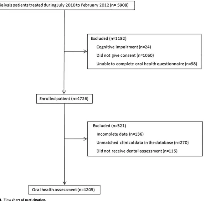
Overall, 4205 patients of the 4726 participants in ORAL-D (89%) underwent an examination for oral mucosal lesions between July 2010 and February 2012 and were included in the analy-sis (Fig 1andTable 1). The mean age was 61.6± 15.6 years and 58% of participants were men. Overall, 1029 (33.4%) participants had never smoked.
Prevalence
Forty percent of participants were affected by the presence of at least one oral lesion. In ascending order of frequency, 21 (0.5%) participants had oral herpes, 70 (1.7%) had mucosal ulceration, 85 had neoformation (2.0%), 147 (3.5%) white lesion, and 169 (4.0%) red lesion. Oral candidiasis was observed in 192 (4.6%) participants, geographical tongue in 207 (4.9%) participants, petechial lesions in 331 (7.9%), and fissured tongue in 450 (10.7%) participants.
In analyses adjusted for demographic and clinical characteristics, there was no statistically significant association between participant demographic and clinical variables and oral muco-sal lesions (S1andS2Tables). The prevalence of oral mucosal lesions (especially geographical tongue, fissured tongue, and candidiasis) varied significantly by country. Oral mucosal lesions tended to be more common among patients treated in Italy, Poland and Spain (S1 Table).
Mortality
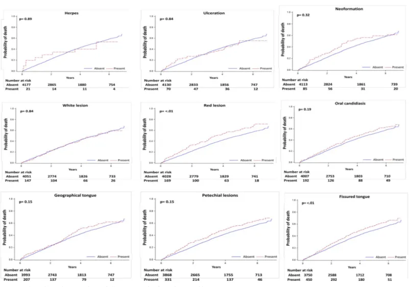
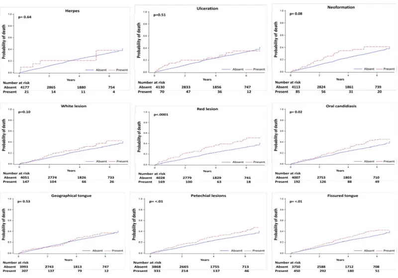
During a median follow-up of 3.5 (range, 1.6–5.8) years, 2114 (50%) deaths occurred of which 1013 (24%) were due to cardiovascular causes. As shown in Figs2and3, red lesion, fissured tongue, and petechial lesions were associated with all-cause mortality in unadjusted analyses, while red lesion, fissured tongue, petechial lesions, and oral candidiasis were associated with cardiovascular death.
Following multivariable adjustment, there was no association between individual oral mucosal lesions and all-cause and cardiovascular death, except for oral candidiasis (Table 2), which was statistically significantly associated with all-cause (adjusted hazard ratio 1.37, 95% CI 1.00–1.86) and cardiovascular mortality (adjusted hazard ratio 1.64, 95% CI 1.09–2.46).
Sensitivity and subgroup analyses
Comparable results were observed when the cumulative incidence of cardiovascular death was calculated after modelling other causes of death as competing risks (S2 Table). When the
Fig 1. Flow chart of participation.
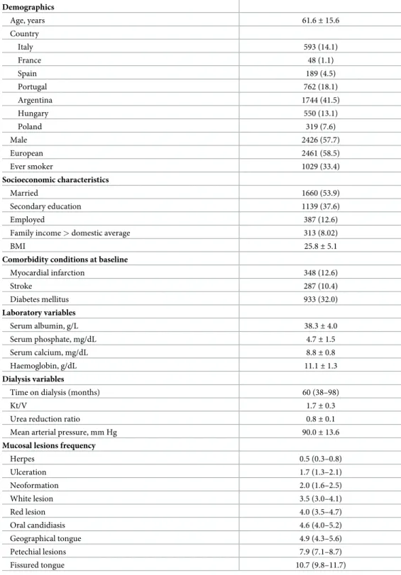
Table 1. Characteristics of study participants. Demographics Age, years 61.6± 15.6 Country Italy 593 (14.1) France 48 (1.1) Spain 189 (4.5) Portugal 762 (18.1) Argentina 1744 (41.5) Hungary 550 (13.1) Poland 319 (7.6) Male 2426 (57.7) European 2461 (58.5) Ever smoker 1029 (33.4) Socioeconomic characteristics Married 1660 (53.9) Secondary education 1139 (37.6) Employed 387 (12.6)
Family income > domestic average 313 (8.02)
BMI 25.8± 5.1
Comorbidity conditions at baseline
Myocardial infarction 348 (12.6) Stroke 287 (10.4) Diabetes mellitus 933 (32.0) Laboratory variables Serum albumin, g/L 38.3± 4.0 Serum phosphate, mg/dL 4.7± 1.5 Serum calcium, mg/dL 8.8± 0.8 Haemoglobin, g/dL 11.1± 1.3 Dialysis variables
Time on dialysis (months) 60 (38–98)
Kt/V 1.7± 0.3
Urea reduction ratio 0.8± 0.1
Mean arterial pressure, mm Hg 90.0± 13.6
Mucosal lesions frequency
Herpes 0.5 (0.3–0.8) Ulceration 1.7 (1.3–2.1) Neoformation 2.0 (1.6–2.5) White lesion 3.5 (3.0–4.1) Red lesion 4.0 (3.5–4.7) Oral candidiasis 4.6 (4.0–5.2) Geographical tongue 4.9 (4.3–5.6) Petechial lesions 7.9 (7.1–8.7) Fissured tongue 10.7 (9.8–11.7)
Results are expressed as number (%) for categorical variables and as mean± SD or median (25th, 75thcentile) for
continuous variables. Frequencies are expressed as proportion with 95% confidence interval.
analyses were conducted using shared frailty models to account for heterogeneity in survival between countries, the results were substantively similar (S3 Table).
Discussion
In the ORAL-D, a multinational prospective cohort study in haemodialysis patients, oral mucosal lesions affected 40% of patients. Oral candidiasis was associated with increased all-cause and cardiovascular mortality after controlling for clinical and demographic factors. There was no evidence of increased all-cause or cardiovascular mortality associated with other oral mucosal lesions.
The finding of oral candidiasis in 4.6% of patients with kidney failure is similar to the find-ing of oral candidiasis in 7% of patients with Human Immunodeficiency Virus (HIV) infec-tion, predominantly among patients with more advanced immunosuppression and higher viral load [12]. The similar prevalence of oral candida in HIV and kidney failure suggests a potential association with the lower immune function found in both conditions [13]. Notably, the point prevalence of oral ulceration, herpes, red and white lesions, and tongue changes were several-fold higher in the present cohort than observed in a general population characterized in the Third National Health and Nutrition Examination Survey from 1988–1994, suggesting
Fig 2. Unadjusted cumulative incidence of all-cause mortality for each mucosal lesion. https://doi.org/10.1371/journal.pone.0218684.g002
Fig 3. Unadjusted cumulative incidence of cardiovascular mortality for each mucosal lesion. https://doi.org/10.1371/journal.pone.0218684.g003
Table 2. All-cause and cardiovascular mortality associated with oral mucosal lesions.
Death (N, %) Unadjusted HR (95% CI) Adjusted HR (95% CI)�
Lesion (N patients) All-cause Cardiovascular All-cause Cardiovascular All-cause Cardiovascular
Herpes (21) 10 (48) 6 (29) 0.96 (0.52–1.79) 1.21 (0.54–2.70) 1.48 (0.54–4.03) 1.10 (0.26–4.58) Ulceration (70) 35 (50) 20 (29) 0.97 (0.69–1.35) 1.16 (0.74–1.81) 1.09 (0.65–1.84) 1.17 (0.55–2.51) Neoformation (85) 47 (55) 27 (32) 1.16 (0.87–1.55) 1.40 (0.96–2.05) 1.55 (0.94–2.55) 1.69 (0.80–3.59) White lesion (147) 74 (50) 46 (31) 0.98 (0.78–1.23) 1.28 (0.96–1.73) 0.90 (0.60–1.35) 0.93 (0.52–1.63) Red lesion (169) 100 (59) 58 (34) 1.39 (1.13–1.69) 1.69 (1.29–2.20) 0.98 (0.69–1.38) 1.07 (0.67–1.71) Oral candidiasis (192) 111 (58) 62 (32) 1.14 (0.94–1.38) 1.34 (1.04–1.74) 1.37 (1.00–1.86) 1.64 (1.09–2.46) Geographical tongue (207) 107 (52) 49 (24) 1.16 (0.95–1.40) 1.10 (0.82–1.46) 0.86 (0.62–1.18) 0.93 (0.57–1.54) Petechial lesions (331) 190 (57) 98 (30) 1.24 (1.07–1.44) 1.34 (1.09–1.65) 1.12 (0.87–1.44) 1.01 (0.68–1.51) Fissured tongue (450) 251 (56) 129 (29) 1.22 (1.07–1.39) 1.32 (1.10–1.59) 1.16 (0.93–1.45) 1.15 (0.84–1.59)
�The multivariable model was adjusted for country, age, sex, education, smoking history, myocardial infarction, diabetes, haemoglobin, serum albumin, serum phosphorus, time on dialysis, body mass index and oral mucosal lesions. HR: hazard ratio; CI: confidence interval.
mucosal changes may be more common with kidney failure [14]. This observation is consis-tent with our finding of a higher prevalence of dental conditions in end-stage kidney disease [7–9].
The observation that oral candidiasis is associated with mortality is consistent with prior studies showing an elevated risk for both all-cause mortality and cardiovascular mortality in patients with kidney failure who have other oral conditions, including tooth loss and dental caries. Oral candidiasis is an opportunistic infection of the mouth and is associated with ste-roid or antibiotic therapy, intercurrent comorbidity, diabetes, denture wearing and low sali-vary flow. Many of these features are present among people with kidney failure and are independently associated with poorer outcomes.
It is unclear in the present study whether oral candidiasis is causal in pathways for mortality or a confounding factor indicative of other comorbidities that represent the true aetiology of worse clinical outcomes, or an epiphenomenon occurring simultaneously with kidney failure but not necessarily caused by the condition. No association was found between any oral muco-sal lesion and any socio-demographic factor. This finding is consistent with observations in an Australian National Survey of Adult Oral Health showing no relationship between oral muco-sal lesions and social or clinical factors and may explain the lack of an association of non-can-didiasis lesions with mortality in this dialysis population [15]. Alternative explanations for a lack of statistical association between oral mucosal lesions with mortality is the relatively low prevalence of many oral mucosal lesions, which may have led to imprecision in the estimated mortality risk. Alternatively, the absence of a statistical association may reflect a difference in the pathobiology of lesions and the predominant causes of mortality in the presence of kidney failure.
While these observations suggest there is limited association of oral mucosal lesions with mortality outcomes in dialysis patients, this finding does not preclude other negative conse-quences of oral mucosal lesions for patients with kidney failure. Notably, this study indicates that approximately 5–10% of dialysis patients experience clinical mucosal lesions at any given time. Based on existing evidence, it is probable that oral lesions have a negative impact on oral-specific and general quality of life for dialysis patients [16]. Whether oral lesions lead to important functional limitations such as discomfort while eating, physical pain, taste, and speech articulation in dialysis patients warrants investigation.
Our finding of an association between oral candidiasis and increased mortality in hemodi-alysis patients suggests that prevention or treatment of oral mucosal lesions might improve clinical outcomes in this population. However, this hypothesis needs to be tested in a suffi-ciently powered randomized controlled trial along with demonstrated feasibility of screening and treatment for oral candidiasis in hemodialysis patients.
The strengths of this study include a large multinational sample size, inclusion of represen-tative patients in typical dialysis clinics, and inclusion of routine data collection for potential confounding factors such as financial status and education. Our study has potential limita-tions. First, due to the observational study design, this analysis cannot evaluate any causal impact of oral mucosal lesions on mortality risk. Although there was adjustment for an exten-sive number of potential confounders, the risk of residual confounding is still possible. Second, we evaluated all-cause mortality and cardiovascular mortality as key outcomes. While there was no evidence of an association for these outcomes, we did not evaluate linkages between oral mucosal lesions and other patient-important factors, including pain, health-related quality of life, nutrition, and oral function. Third, despite a higher prevalence of oral mucosal lesions than has been reported in the general population, the relatively few participants who had oral mucosal lesions may have reduced the power in the analyses to detect significant associations between mucosal lesions and mortality. Fourth, oral candidiasis and herpetic lesions were
assessed clinically and did not include microbiological testing or evaluation of type (e.g. pseu-domembranous or erythematous candidiasis), leading to the possibility of measurement error. Fifth, we observed different prevalence rates of oral mucosal lesions across the different coun-tries contributing data. While the dental examination was conducted using a standardized approach, differences in prevalence may have represented variation in diagnostic practices in each country.
In conclusion, the prevalence of oral mucosal lesions may be higher in dialysis patients than in the general population, although such lesions appear unrelated to measured sociodemo-graphic and clinical characteristics. Oral candidiasis was present in 5% of this dialysis popula-tion and was associated with all-cause and cardiovascular mortality.
Supporting information
S1 Item. STROBE statement—Checklist of items that should be included in reports of observational studies.
(PDF)
S1 Table. Association of clinical and demographic variables with mucosal lesions.
(PDF)
S2 Table. Association of mucosal lesions with cardiovascular mortality accounting for all-cause mortality as competing risk.
(PDF)
S3 Table. Association of mucosal lesions with all-cause and cardiovascular disease using a Cox proportional hazards model fitted with shared frailty to account for within-country clustering of mortality risks.
(PDF)
Acknowledgments
We thank all participants for involvement in the study.
Each author contributed important intellectual content during manuscript drafting or revi-sion, approved the final version of the manuscript, and agreed to be accountable for all aspects of the work.
Participating centres, facilitators and organizing committee
The ORAL-D Investigators are as follows.Argentina: S. Raña, M. Serrano, S. Claros, M. Arias, L.
Petracci, M. Arana, P. De Rosa, A. Gutierrez, M. Simon, V. Vergara, M. Tosi, M. Cernadas, I. Vilamajo´, D. Gravac, M. Paulo´n, L. Penayo, G. Carrizo, M. Ghiani, G. Perez, O. Da Cruz, D. Galarce, M. Gravielle, E. Vescovo, R. Paparone, C. Mato Mira, E. Mojico, O. Hermida, D. Florio, M. Yucoswky, W. Labonia, D. Rubio, G. Di Napoli, A. Fernandez, H. Altman, J. Rodriguez, S. Serrano, G. Valle, M. Lobos, V. Acosta, G. Corpacci, M. Jofre, L. Gianoni, G. Chiesura, M. Cap-devila, J. Montenegro, J. Bequi, J. Dayer, A. Go´mez, C. Caldero´n, E. Abrego, C. Cechı´n, J. Garcı´a, J. Corral, M. Natiello, A. Coronel, M. Muñiz, V. Muñiz, A. Bonelli, F. Sanchez, S. Maestre, S. Oli-vera, M. Camargo, V. Avalos, E. Geandet, M. Canteli, A. Escobar, E. Sena, S. Tirado, A. Peñalba, G. Neme, M. Cisneros, R. Oliszewski, V. Nascar, M. Daud, S. Mansilla, A. Paredes A´ lvarez, L. Gamı´n, M. Arijo´n, M. Coombes, M. Zapata; France: C. Boriceanu, S. Frantzen-Trendel; Hun-gary: K. Albert, I. Csaszar, E. Kiss, D. Kosa, A. Orosz, J. Redl, L. Kovacs, E. Varga, M. Szabo, K.
Magyar, G. Kriza, E. Zajko, A. Bereczki, J. Csikos, A. Kuti, A. Mike, K. Steiner, E. Nemeth, K. Tolnai, A. Toth, J. Vinczene, Sz. Szummer, E. Tanyi, R. Toth, M. Szilvia;Italy: N. Dambrosio, G.
Paparella, M. Sambati, C. Donatelli, F. Pedone, V.A. Cagnazzo, R. Antinoro, F. Torsello, C. Saturno, G. Giannoccaro, S. Maldera, E. Boccia, M. Mantuano, R. Di Toro Mammarella, M. Meconizzi, P.F. Steri, C. Riccardi, A. Flammini, L. Moscardelli, M. Murgo, N. San Filippo, S. Pagano, G. Marino, G. Montalto, S. Cantarella, B. Salamone, G. Randazzo, D. Rallo, A. Manis-calco, M. Fici, A. Lupo, P. Pellegrino, R. Fichera, A.D’Angelo, N. Falsitta;Poland: E.
Bochenska-Nowacka, A. Jaroszynski, J. Drabik, M. Birecka, D. Daniewska, M. Drobisz, K. Doskocz, G. Wyr-wicz;Portugal: L. Inchaustegui, C. Outerelo, D. Sousa Mendes, A. Mendes, J. Lopes, J. Barbas, C.
Madeira, A. Fortes, R. Vizinho, A. Cortesão, E. Almeida; Spain: A. Bernat, B. De la Torre, A. Lopez, J. Martı´n, G. Cuesta, R.M. Rodriguez, F. Ros, M. Garcia, E. Orero, E. Ros.
The ORAL-D dentists are as follows:Argentina: G. Bava (Buenos Aires), A. Bora (Caleta
Olivia), H. Gorena (Co´rdoba), T. Caldero´n (Mendoza), R. Dupuy, N. Alonso (Tucuma´n), V. Siciliano (Comodoro Rivadavia);France: S. Frantzen-Trendel; Hungary: K. Nagy, O¨ . Bajusz, I.
Pinke, G. Decsi, L. Gyergyoi, Zs. Jobba, Zs. Zalai, A´ . Zsedenyi, G. Kiss, M. Pinter, M. Keresz-turi (Faculty of Dentistry University of Szeged);Italy: M. Petruzzi (Bari), M. De Benedittis
(Bari); Poland: J. Szkutnik (Lublin), J. Sieczkarek (Lublin);Portugal: A. Capelo (Lisboa), A.
Caetano, K. MacGregor, M. Santos, S. Silva Pinheiro, L. Martins, D. Leitão, C. Izidoro (inde-pendent dentists);Spain: M. Garcia Gallart (independente dentist), C. Mendieta (Barcelona).
Author Contributions
Conceptualization: Marinella Ruospo, Suetonia C. Palmer, Michele De Benedittis, Jonathan
C. Craig, Giovanni F. M. Strippoli.
Data curation: Marinella Ruospo, Patrizia Natale, Valeria Saglimbene, Massimo Petruzzi,
Michele De Benedittis, Eduardo Celia, Ruben Gelfman, Miguel R. Leal, Marietta To¨ro¨k, Paul Stroumza, Luc Frantzen, Anna Bednarek-Skublewska, Jan Dulawa, Domingo del Cas-tillo, Staffan Scho¨n, Amparo G. Bernat, Letizia Gargano.
Formal analysis: Marinella Ruospo, Suetonia C. Palmer, Giusi Graziano, Giovanni F. M.
Strippoli.
Funding acquisition: Jo¨rgen Hegbrant, Giovanni F. M. Strippoli.
Investigation: Marinella Ruospo, Suetonia C. Palmer, Giusi Graziano, Patrizia Natale, Valeria
Saglimbene, Massimo Petruzzi, Michele De Benedittis, Jo¨rgen Hegbrant, Giovanni F. M. Strippoli.
Methodology: Jonathan C. Craig, Giovanni F. M. Strippoli.
Project administration: Jonathan C. Craig, Giovanni F. M. Strippoli. Resources: Jo¨rgen Hegbrant, Giovanni F. M. Strippoli.
Software: Marinella Ruospo, Suetonia C. Palmer, Giusi Graziano. Supervision: Jonathan C. Craig, Giovanni F. M. Strippoli.
Validation: Marinella Ruospo, Suetonia C. Palmer, Giusi Graziano, Patrizia Natale, Valeria
Saglimbene, Massimo Petruzzi, Michele De Benedittis, Jonathan C. Craig, David W. John-son, Marcello Tonelli, Jo¨rgen Hegbrant, Letizia Gargano, Giovanni F. M. Strippoli.
Visualization: Marinella Ruospo, Suetonia C. Palmer, Giusi Graziano, Patrizia Natale, Valeria
Saglimbene, Massimo Petruzzi, Michele De Benedittis, Jonathan C. Craig, David W. John-son, Pauline Ford, Marcello Tonelli, Jo¨rgen Hegbrant, Letizia Gargano, Giovanni F. M. Strippoli.
Writing – review & editing: Marinella Ruospo, Suetonia C. Palmer, Giusi Graziano, Patrizia
Natale, Valeria Saglimbene, Massimo Petruzzi, Michele De Benedittis, Jonathan C. Craig, David W. Johnson, Pauline Ford, Marcello Tonelli, Eduardo Celia, Ruben Gelfman, Miguel R. Leal, Marietta To¨ro¨k, Paul Stroumza, Luc Frantzen, Anna Bednarek-Skublewska, Jan Dulawa, Domingo del Castillo, Staffan Scho¨n, Amparo G. Bernat, Jo¨rgen Hegbrant, Char-lotta Wollheim, Letizia Gargano, Giovanni F. M. Strippoli.
References
1. Ruospo M, Palmer SC, Craig JC, Gentile G, Johnson DW, Ford PJ, et al. Prevalence and severity of oral disease in adults with chronic kidney disease: a systematic review of observational studies. Nephrol Dial Transplant 2014; 29(2):364–75. Epub 2013/10/02.https://doi.org/10.1093/ndt/gft401PMID: 24081863.
2. Gautam NR GN, Rao TH, Koganti R, Agarwal R, Alamanda M. Effect of end-stage renal disease on oral health in patients undergoing renal dialysis: A cross-sectional study. J INt Soc Prev Community Dent 2014; 4(3):164–9.https://doi.org/10.4103/2231-0762.142006PMID:25374834
3. Oyetola EO, Owotade FJ, Agbelusi GA, Fatusi OA, Sanusi AA. Oral findings in chronic kidney disease: implications for management in developing countries. BMC Oral Health. 2015; 15(24).https://doi.org/ 10.1186/s12903-015-0004-zPMID:25888327
4. Chi AC, Neville BW, Krayer JW, Gonsalves WC. Oral manifestations of systemic disease. Am Fam Phy-sician. 2010; 82(11):1381–8. Epub 2010/12/03. PMID:21121523.
5. Jansson L, Lavstedt S, Frithiof L. Relationship between oral health and mortality rate. J Clin Periodontol. 2002; 29(11):1029–34.https://doi.org/10.1034/j.1600-051X.2002.291108.xPMID:12472996
6. Strippoli GF, Palmer SC, Ruospo M, Natale P, Saglimbene V, Craig JC, et al. Oral disease in adults treated with hemodialysis: prevalence, predictors, and association with mortality and adverse cardiovascu-lar events: the rationale and design of the ORAL Diseases in hemodialysis (ORAL-D) study, a prospective, multinational, longitudinal, observational, cohort study. BMC Nephrol. 2013; 14:90. Epub 2013/04/20. https://doi.org/10.1186/1471-2369-14-90PMID:23597063; PubMed Central PMCID: PMC3685555.
7. Palmer SC, Ruospo M, Wong G, Craig JC, Petruzzi M, De Benedittis M, et al. Dental Health and Mortal-ity in People With End-Stage Kidney Disease Treated With Hemodialysis: A Multinational Cohort Study. Am J Kidney Dis. 2015; 66(4):666–76. Epub 2015/06/30.https://doi.org/10.1053/j.ajkd.2015.04.051 PMID:26120038.
8. Palmer SC, Ruospo M, Wong G, Craig JC, Petruzzi M, De Benedittis M, et al. Patterns of oral disease in adults with chronic kidney disease treated with hemodialysis. Nephrol Dial Transplant. 2016; 31 (10):1647–53. Epub 2016/04/02.https://doi.org/10.1093/ndt/gfv413PMID:27035674.
9. Ruospo M, Palmer SC, Wong G, Craig JC, Petruzzi M, De Benedittis M, et al. Periodontitis and early mortality among adults treated with hemodialysis: a multinational propensity-matched cohort study. BMC Nephrol. 2017; 18(1):166. Epub 2017/05/24.https://doi.org/10.1186/s12882-017-0574-xPMID: 28532432; PubMed Central PMCID: PMC5440912.
10. Strengthening the reporting of observational studies in epidemiology (STROBE) statement: guidelines for reporting observational studies. BMJ. 2007; 335(7626).https://doi.org/10.1136/bmj.39386.490150.94 11. Kramer IR, Pindborg JJ, Bezroukov V, Infirri JS. Guide to epidemiology and diagnosis of oral mucosal
diseases and conditions. World Health Organization. Community Dent Oral Epidemiol. 1980; 8(1):1–26. Epub 1980/02/01. PMID:6929240.
12. Kroidl A, Schaeben A, Oette M, Wettstein M, Herfordt A, Haussinger D. Prevalence of oral lesions and periodontal diseases in HIV-infected patients on antiretroviral therapy. Eur J Med Res. 2005; 10 (10):448–53. Epub 2005/11/17. PMID:16287607.
13. Kato S, Chmielewski M, Honda H, Pecoits-Filho R, Matsuo S, Yuzawa Y, et al. Aspects of immune dys-function in end-stage renal disease. Clin J Am Soc Nephrol. 2008; 3(5):1526–33. Epub 2008/08/15. https://doi.org/10.2215/CJN.00950208PMID:18701615; PubMed Central PMCID: PMC4571158.
14. Shulman JD, Beach MM, Rivera-Hidalgo F. The prevalence of oral mucosal lesions in U.S. adults: data from the Third National Health and Nutrition Examination Survey, 1988–1994. J Am Dent Assoc. 2004; 135(9):1279–86. PMID:15493392
15. Do L, Spencer A, Dost F, Farah C. Oral mucosal lesions: findings from the Australian National Survey of Adult Oral Health. Aust Dent J. 2014; 59(1):114–20.https://doi.org/10.1111/adj.12143PMID:24494603 16. Liu LJ, Xiao W, He QB, Jiang WW. Generic and oral quality of life is affected by oral mucosal diseases.