RESEARCH
Cloning, expression and characterization
of a β-
d
-xylosidase from Lactobacillus rossiae
DSM 15814
T
Erica Pontonio
1, Jennifer Mahony
2, Raffaella Di Cagno
1*, Mary O’Connell Motherway
2,3, Gabriele Andrea Lugli
4,
Amy O’Callaghan
2,3, Maria De Angelis
1, Marco Ventura
4, Marco Gobbetti
1and Douwe van Sinderen
2,3Abstract
Background: Among the oligosaccharides that may positively affect the gut microbiota, xylo-oligosaccharides
(XOS) and arabinoxylan oligosaccharides (AXOS) possess promising functional properties. Ingestion of XOS has been reported to contribute to anti-oxidant, anti-bacterial, immune-modulatory and anti-diabetic activities. Because of the structural complexity and chemical heterogeneity, complete degradation of xylan-containing plant polymers requires the synergistic activity of several enzymes. Endo-xylanases and β-d-xylosidases, collectively termed xylanases, represent the two key enzymes responsible for the sequential hydrolysis of xylan. Xylanase cocktails are used on an industrial scale for biotechnological purposes. Lactobacillus rossiae DSM 15814T can utilize an extensive set of carbon sources, an ability that is likely to contribute to its adaptive ability. In this study, the capacity of this strain to utilize XOS, xylan, d-xylose and l-arabinose was investigated.
Results: Genomic and transcriptomic analyses revealed the presence of two gene clusters, designated xyl and ara,
encoding proteins predicted to be responsible for XOS uptake and hydrolysis and d-xylose utilization, and l-arabinose metabolism, respectively. The deduced amino acid sequence of one of the genes of the xyl gene cluster, LROS_1108 (designated here as xylA), shows high similarity to (predicted) β-d-xylosidases encoded by various lactic acid bacte-ria, and belongs to glycosyl hydrolase family 43. Heterologously expressed XylA was shown to completely hydrolyse XOS to xylose and showed optimal activity at pH 6.0 and 40 °C. Furthermore, β-d-xylosidase activity of L. rossiae DSM 15814T was also measured under sourdough conditions.
Conclusions: This study highlights the ability of L. rossiae DSM 15814T to utilize XOS, which is a very useful trait when selecting starters with specific metabolic performances for sourdough fermentation or as probiotics.
Keywords: Xylo-oligosaccharides, Sourdough, Prebiotic, Gut microbiota, Functional foods, Probiotic
© 2016 Pontonio et al. This article is distributed under the terms of the Creative Commons Attribution 4.0 International License (http://creativecommons.org/licenses/by/4.0/), which permits unrestricted use, distribution, and reproduction in any medium, provided you give appropriate credit to the original author(s) and the source, provide a link to the Creative Commons license, and indicate if changes were made. The Creative Commons Public Domain Dedication waiver (http://creativecommons.org/ publicdomain/zero/1.0/) applies to the data made available in this article, unless otherwise stated.
Background
In recent years the use of prebiotics, in particular oligo-saccharides, to modulate the gut microbiota composi-tion and associated metabolic activities, together aimed at improving gut health, has enjoyed considerable sci-entific and commercial interest [1, 2]. Colonic fermen-tation of particular oligosaccharides into short chain fatty acids (SCFA) is believed to increase the number
and metabolic activity of certain beneficial bacterial populations [1, 3]. Among the oligosaccharides that may positively alter the composition of the gut microbiota, xylo-oligosaccharides (XOS) and arabinoxylan oligosac-charides (AXOS) possess promising functional proper-ties as they can be specifically fermented by intestinal commensals such as bifidobacteria and lactobacilli [4–
6]. XOS resist hydrolysis by enzymes and/or the low pH present in human saliva, gastric and pancreatic juices, and are not absorbed during transit through the small intestine, thus reaching the colon where they are availa-ble as fermentaavaila-ble substrates for certain members of the
Open Access
*Correspondence: [email protected]
1 Department of Soil, Plant and Food Science, University of Bari Aldo Moro, Via G. Amendola 165/A, 70126 Bari, Italy
resident microbiota [7]. It has previously been reported that ingestion of XOS contributes to oxidant, anti-bacterial, immune-modulatory and anti-diabetic activi-ties [8, 9]. Arabinoxylans represent 50 % of dietary fibers present in wheat bran [10, 11]. In particular, the highly polymerized arabinoxylan from bran may be hydrolyzed by specific bacteria possessing arabinoxylan-degrading enzymes [6].
XOS are oligomers that consist of 2–10 xylose residues connected through β-(1-4)-linkages. Their liberation is the result of (partial) hydrolysis of xylan, the major com-ponent of plant hemicelluloses. Because of the structural complexity (such as the presence of various side chains, in particular arabinose) and chemical heterogeneity, complete degradation of xylan-containing plant polymers requires the synergistic activity of several enzymes [12], such as endo-xylanase (endo-1,4-β-xylanase, E.C. 3.2.1.8), β-d-xylosidase (xylan-1,4-β-d-xylosidase, E.C. 3.2.1.37), α-glucuronidase (α-glucosiduronase, E.C.3.2.1.139), α-arabinofuranosidase (α-l-arabinofuranosidase, E.C. 3.2.1.55) and acetylxylan esterase (E.C. 3.1.1.72). Endo-xylanases and β-d-xylosidases (collectively named as xylanases) represent the two key enzymes responsible for the sequential hydrolysis of xylan. Endo-xylanases act on the homo-polymeric backbone of the β-1,4-linked xylan liberating xylo-oligomers [13], whereas β-d-xylosidases are active on these latter oligomers releasing xylose [14]. For the degradation of arabinoxylan, α-l-arabinofuranosidases are also needed because they cleave arabinose from the backbone and act in synergy with endoxylanases [15]. Xylanase cocktails are used on an industrial scale for de-inking of recycled paper [16], pro-cessing of wood pulp [17], improving bread dough baking and nutritional quality [18], hydrolysis of bitter molecules and liberation of aroma compounds during grape juice extraction and wine making [12].
In nature, a variety of microorganisms, including bac-teria and fungi, encode xylanases and β-xylosidases [14]. These enzymes are either cell wall-associated, secreted into the extracellular environment or present in the cytoplasm. Based on amino acid sequence similarities xylanases and β-xylosidases are classified into glyco-side hydrolase (GH) families 1, 3, 30, 39, 43, 51, 52, 54, 116 or 120. In particular, β-xylosidases belonging to the GH43 family, which are predominantly encoded by bacteria, hydrolyze the non-reducing ends of the xylo-oligomers using an inverting mechanism [14]. Despite the recognized potential of XOS as a prebiotic to target beneficial components of the human gut microbiota, in particular lactobacilli and bifidobacteria, very little is known about the enzymes used by the former group of bacteria to hydrolyze these complex substrates. To the best of our knowledge, β-d-xylosidase from Lactobacillus
brevis is the only report on the characterization of such an enzyme in the Lactobacillus genus [9, 19].
Recently, the genomic annotation and comparative analysis of L. rossiae DSM 15814T revealed the pre-dicted presence of a number of extracellular or cell wall-associated polysaccharide-degrading enzymes, rep-resented by putative cyclomaltodextrinase (E.C. 3.2.1.54; LROS_1707), α-amylase (E.C. 3.2.1.10; LROS_1584), β-glucosidase (E.C. 3.2.1.21; LROS_2047), mannosyl-glycoprotein endo-β-N-acetylglucosaminidase (E.C. 3.2.1.96; LROS_0612) and neopullulanase (EC 3.2.1.135; LROS_1707) enzymes [20]. Furthermore, genes involved in the degradation of arabinose and xylose-containing poly- and/or oligo-saccharides were predicted. L. rossiae is an obligatory hetero-fermentative lactic acid bacte-rium, which has been isolated from the gastrointestinal tract of humans [21] and animals [22], wheat sourdoughs [23–25], legumes [26], spelt flour [27] and pineapple [28]. L. rossiae was found to be a promising probiotic candidate thanks to its ability to survive under simu-lated gastric and intestinal conditions, and to stimulate immune-mediators by peripheral blood mononuclear cells [29]. Exposure to gastric and intestinal fluids is the main environmental stress that decreases viability of ingested probiotics [30, 31]. More in depth knowledge on XOS metabolism by L. rossiae is important from a biotechnological perspective to facilitate the selection of strains with specific metabolic performances to be used as starters for sourdough bread making, aimed at improving rheology and nutritional properties, or as pro-biotic for human applications.
In the current study we used a transcriptome approach to identify L. rossiae DSM 15814T genes that were upreg-ulated when this strain was grown on XOS-, xylose- or arabinose. One of the identified genes, xylA, was selected for further characterization and was cloned in Lc. lactis subsp. cremoris NZ9000 and the encoded recombinant enzyme was purified and characterized.
Results
Growth on XOS, xylan, d‑xylose or l‑arabinose
When maltose was used as a sole carbon and energy source in growth medium (see “Methods” section), L. rossiae was shown to increase its viable count from ca. 7.4 ± 0.1 to 9.4 ± 0.3 log CFU ml−1. The stationary phase of growth was reached after approximately 10 h, with a lag phase and μmax of 2.9 ± 0.2 h and 0.52 ± 0.03 log CFU ml−1 h−1, respectively. In the presence XOS, d-xylose or l-arabinose, L. rossiae was shown to exhibit similar growth kinetics. The increase of cell viability ranged from 7.3 ± 0.1 to 9.5 ± 0.3 log CFU ml−1, the val-ues of λ varied from 2.7 ± 0.3 (l-arabinose) to 2.5 ± 0.1 h (XOS), and those of μmax from 0.27 ± 0.02 (l-arabinose
and XOS) to 0.31 ± 0.03 Log CFU ml−1 h−1 (d-xylose). L. rossiae was not shown to exhibit appreciable growth in the presence of xylan, rye arabinoxylan, wheat arabinoxy-lan, arabinan, arabinogalactan and xyloglucan, as the sole carbon source (data not shown).
Genome response of L. rossiae DSM 15814T to growth on XOS
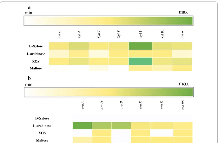
In order to investigate which genes are expressed when L. rossiae DSM 15814T is grown in presence of XOS, d-xylose, l-arabinose or maltose (as a reference) as the sole carbon source, global gene expression was deter-mined by RNAseq analysis. Compared to growth on maltose as the sole carbon source, various adjacent genes (designated xylE, xylA, xynT, xylT, xylI, xylK and xylR; and here referred to as the xyl cluster) were shown to exhibit transcriptional increases that ranged from 8.6 to 250 fold, or from 11.4 to 259.3 fold, when the strain was grown on XOS (Fig. 1a) or d-xylose, respectively. Furthermore, the co-located araA, araD, araB, araR and araRS genes, which encompass the ara gene cluster, pre-dicted to encode enzymes for l-arabinose utilization,
exhibited an increase in their transcription from 0.9 to 156 fold when l-arabinose was used as the only car-bon source (Fig. 1b) (see below for details on putative functions).
Genetic organization of the ara and xyl custers, and comparative analysis of the xylA‑encoded β‑d‑xylosidase
As mentioned above, transcriptome analysis of L. rossiae DSM 15814T revealed the presence of two gene clusters, xyl and ara, that are linked to XOS/d-xylose and l-ara-binose metabolism, respectively (Fig. 2). The xyl clus-ter encompasses seven genes, which are schematically outlined in Fig. 2a, and which are predicted (based on BLAST-mediated similarity searches, Additional file 1: Table S1) to encode a xylose isomerase (E.C. 5.3.1.5; LROS_1111, designated here as xylI), a xylulose kinase (E.C 2.7.1.17; LROS_1112, designated here as xylK) and a ROK family transcriptional regulator (LROS_1113, des-ignated here as xylR). Upstream of, but divergently ori-ented from the three genes mentioned above are genes encoding a predicted aldose 1-epimerase (E.C. 5.1.3.3;
min
a
max
b
ara A ara D ara B ara R ara E ara RS D-Xylose L-arabinose XOS Maltose xyl E xy l A Xyn T Xyl T xy lI xy l K xyl R D-Xylose L-arabinose XOS Maltose minmax
Fig. 1 Heatmap based on the transcriptome analysis of Lactobacillus rossiae DSM15814T grown on d-xylose, l-arabinose, XOS and maltose as the sole carbon source. XOS hydrolysis and utilization of the end product d-xylose (a) and the utilization of the l-arabinose (b) operon. The gene expres-sion is expressed as RPKM (Reads per kilobase per million)
LROS_1107, designated here as xylE) and a putative β-d-xylosidase (EC3.2.1.37; LROS_1108, designated as xylA), followed by xynT (a putative xyloside transporter-encod-ing gene; LROS_1109) and xylT (predicted to specify a d-xylose proton symporter; LROS_1110). Thus, the genes in this xyl cluster represent proteins that may be involved in XOS metabolism (Kyoto Encyclopedia of Genes and Genomes database; Fig. 2). Among the genes of the xyl cluster, xylA may have a key role in XOS hydrolysis as its encoded protein product is predicted to convert XOS into d-xylose. Alignment of the L. rossiae DSM 15814T xylA-encoded β-d-xylosidase with similar protein sequences from other lactic acid bacteria (NCBI website) produced the phylogenetic tree displayed in Fig. 3. This analysis shows that the β-d-xylosidase from L. rossiae DSM 15814T belongs to a cluster consisting of mostly predicted β-d-xylosidases encoded by Leuconostoc, Weis-sella and Pediococcus species, and Lc. lactis.
The ara cluster is predicted to encompasses genes required for L-arabinose metabolism (Fig. 2b). In particu-lar, based on BLAST similarity searches (Additional file 1: Table S1), it is predicetd that LROS_1243 encodes an l-arabinose isomerase (araA; E.C. 5.3.1.4), LROS_1244 specifies an l-ribulose 5P-4-epimerase (araD; E.C. 5.1.3.4), LROS_1245 encodes a ribulose kinase (araB; E.C 2.7.1.16), LROS_1248 a transcriptional repressor GntR family (araR;), LROS_1249 an aldose-1-epimerase (araE; E.C. 5.1.3.3) and LROS_1250 a transcriptional
regulator 2C ArsR family (araRS). The complete conver-sion of l-arabinose into d-xylulose-5P is allowed by the sequential activities of AraA, AraB and AraD, respec-tively. Divergently orientated are three hypothetical genes with unknown function (LROS_1246, LROS_1247 and LROS_1249).
XylA expression in Lc. lactis subsp. cremoris NZ9000 and characterization of β‑d‑xylosidase activity
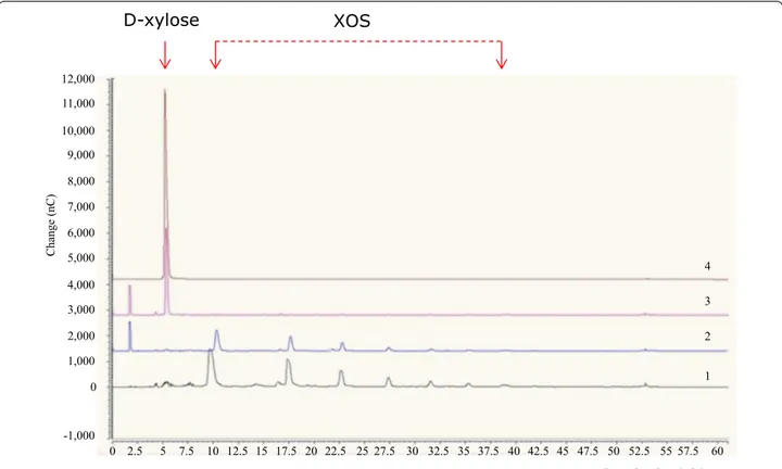
To demonstrate if xylA encodes, as predicted, a β-d-xylosidase capable of XOS hydrolysis, this gene was cloned into the protein expression vector pNZ8048, and placed under the transcriptional control of the induc-ible PnisA promoter (see “Methods” section). In order to verify the hydrolytic activity of XylA, purified protein and crude cell extract (CCE) from Nisaplin-induced Lc. lactis subsp. cremoris, harbouring the pNZ8048-xylA were individually incubated for 24 h. Following incuba-tion, CCE from Lc. lactis, harbouring the pNZ8048-xylA, showed hydrolytic activity towards XOS (Fig. 4). As expected, using identical experimental conditions, CCE from Lc. lactis, harbouring the empty pNZ8048 (nega-tive control) did not exhibit measurable activity. In agree-ment with the comparative genome sequence analysis of L. rossiae, these results demonstrate that xylA speci-fies a β-d-xylosidase responsible for the observed XOS-degrading activity. Unfortunately, for unknown reasons the XylA protein completely lost its hydrolytic activity XylA, betaxylosidase
Hypothetical protein
XynT, xyloside transporter
XylT, xylose (H+) symporter
XylE: aldose 1 epimerase XylI, xylose isomerase
XylK, xylose kinase XylR, transcriptional regulator , ROK family L. rossiaeDSM 15814 2000 4000 6000 8000 10000 LROS_1106 LROS_1113 L. rossiaeDSM 15814 5000 10000 LROS_1250 LROS_1243
araD, L-ribulose 5P-4-epimerase
araB, ribulokinase
araR, transcriptional repressor, GntR family
Transcriptional regulator ArsR family
Hypothetical protein
araA, L-arabinose isomerase
a
b
Fig. 2 Schematic representation of the genetic organization (ca. 11 kb region) in Lactobacillus rossiae DSM 15814T. XOS hydrolysis and utilization of the end product d-xylose (a) and the utilization of the l-arabinose (b) operon. The size and orientation of each of the genes were deduced from their DNA sequences. The map was derived by the use of CloneManager Professional software (Scientific and Educational Software; USA)
upon purification from the CCE (results not shown), and therefore further characterization of this enzyme was performed using XylA-containing CCE.
The effect of the pH on the activity of the recombi-nant His-tagged XylA was determined in Na-acetate (pH 3.0–6.0), phosphate (pH 6.0–7.0) and Tris–HCl (pH 7.0–9.0) buffer (Additional file 1: Figure S1). The enzyme was highly active (>80 %) in the range of pH of 5.0–7.0, displaying an optimum at pH 5.7. Above pH 7.0, enzyme activity rapidly decreased, being completely lost at pH 9.0. XylA enzyme activity was optimal at 40 °C, and it decreased thereafter (Additional file 1: Figure S1).
Using L. rossiae DSM 15814T cells, enzyme activity was also determined under conditions that mimicked sour-dough. After 24 h of fermentation, the xylosidase activity measured in sourdough was 4.4 ± 0.1 U, correspond-ing to 70 % of the maximum activity found in phosphate
buffer. Following growth of L. rossiae DSM 15814T in wheat flour hydrolysate medium (WFH), the CCE was shown to exhibit an activity of 5.6 ± 0.2 U.
Discussion
The relatively large genome (genome size ~ 2.8 Mb) of L. rossiae DSM 15814T is believed to reflect the meta-bolic and adaptive versatility of the species, mirroring an impressive potential to colonize diverse environ-ments [20]. L. rossiae DSM 15814T was shown to grow well on modified Rogosa medium with XOS or its con-stituent pentose sugar xylose, or arabinose as the only carbon source. Growth of lactobacilli on XOS is not widely documented: only Lactobacillus fermentum (syn. L. cellobiosus) [32] and Lactobacillus acidophilus [33] have been reported to exhibit moderate growth on XOS, though this less vigorous compared to the growth
Change (nC) -1,000 0 2,000 1,000 3,000 4,000 5,000 6,000 7,000 8,000 9,000 10,000 11,000 12,000
Retention time (min) 0 2.5 5 7.5 10 12.5 15 17.5 20 22.5 25 27.5 30 32.5 35 37.5 40 42.5 45 47.5 50 52.5 55 57.5 60 1 2 3 4
D-xylose
XOS
Fig. 4 Hydrolysis of XOS as assessed by high-performance anion-exchange chromatography (HPAEC). Lane 1, 5 mg ml−1 (wt/vol) standard of XOS; lane 2, Lactococcus lactis subsp. cremoris NZ9000 containing the empty plasmid (pNZ8048) (negative control); lane 3; Lc. lactis subsp. cremoris NZ9000 containing the pNZ8048.1108 construct; lane 4, 5 mg ml−1 (wt/vol) standard of d-xylose. Details on recombinant Lc. lactis subsp. cremoris NZ9000 are reported in “Methods” section
(See figure on previous page.)
Fig. 3 Phylogenetic tree showing the relationship between the amino acid sequences of β-d-xylosidase from Lactobacillus rossiae DSM15814T and reference of sequences of some lactic acid bacteria in GenBank. The tree was constructed using the neighbour-joining software, numbers at the node are the bootstrap values (%). GroEL of Bifidobacterium adolescentis was used as outlier
capacity on this sugar shown by certain Bifidobacterium spp. [5, 34]. Furthermore, a gut-derived strain of Lacto-bacillus paracasei was shown to be positively stimulated by XOS [35].
L. rossiae DSM 15814T is predicted to specify a num-ber of extracellular or cell wall-associated polysaccha-ride-degrading enzymes, as well as putative enzymatic pathways for the metabolism of arabinose and xylose-containing poly- and/or oligosaccharides [20]. In this study a β-d-xylosidase was identified in L. rossiae DSM 15814T and successfully expressed in Lc. lactis subsp. cre-moris NZ9000. β-d-xylosidase represents one of the key xylanolytic enzymes in supplying carbon and energy to a variety of organisms [36, 37]. Sequential hydrolysis of xylans leads to the release of xylose, which is then trans-formed to the common metabolic intermediate xylulose 5-phosphate [38]. In L. rossiae DSM 15814T the genes involved in the xylose and xylo-oligosaccharides utiliza-tion pathway appear to be clustered in a single operon designated here as xyl. The deduced amino acid sequence of the LROS_1108 gene belonging to the xyl cluster and designated here as xylA, showed high similarity to (pre-dicted) β-d-xylosidases encoded by various lactic acid bacteria, a glycosyl hydrolase family 43 enzyme [14]. Recombinantly produced XylA was shown to catalyse the complete hydrolysis of XOS to xylose. The presence of a highly specific xylosidase is expected to play a cru-cial role in facilitating the utilization of XOS. As shown by transcriptome analysis, all xyl cluster genes were variously up-regulated when L. rossiae DSM 15814T was grown in the presence of XOS and D-xylose, compared to cells grown when maltose was the sole carbon source. Furthermore, the up-regulation of the ara gene cluster, predicted to encode enzymes for l-arabinose utilization, was observed when, as expected, this sugar was pre-sent as the sole carbon source. Although these findings provide insights into the genes required for growth on XOS, our data are insufficient to reconstruct the xylano-lytic machinery of L. rossiae DSM 15814T. Nonetheless, β-xylosidase is generally associated with other xylano-lytic enzymes in order to impart an efficient conversion of xylo-oligosaccharides into xylose [36], and the involve-ment of other, perhaps secreted xylosidases cannot be ruled out.
Many bacterial β-d-xylosidases represent large dimeric or trimeric proteins with molecular masses in excess of 100 kDa [9, 39]. Monomeric β-d-xylosidases with a molecular mass less than 100 kDa have also been described [40]. The apparent molecular weight of the β-d-xylosidases of L. rossiae is higher than those encoded by Bifidobacterium breve K110 (49 kDa) [40] and Clostridium cellulolyticum (43 kDa) [41], and comparable to that of Lactobacillus brevis NCDC01 (58 kDa) [9].
The β-xylosidase of L. rossiae DSM 15814T was shown to exhibit pH and temperature optima of ca. 6.0 and 40 °C, respectively. It was highly active (>80 %) in the range of pH of 5.0–7.0. β-xylosidases from other bacte-ria are usually active and stable at neutral pH [42] and a temperature range of 35–45 °C. The β-xylosidase puri-fied from L. brevis NCDC01 was shown to exhibit pH and temperature optima of 6.0 and 40 °C [9], while the β-xylosidases of Bacillus thermantarcticus [43] and Bifi-dobacterium adolescentis were characterized by optimal activity at 5.0–7.0 and 50–70 °C [44]. Based on enzyme properties, the β-xylosidase of L. rossiae was found to be similar to that purified from L. brevis. L. rossiae is a novel species [23] that is able to adapt to different envi-ronments [21, 23–25, 28], and therefore further insights are desirable to exploit its biotechnological features and understand its metabolic versatility.
β-xylosidase activity appears to be produced by L. ros-siae DSM 15814T during sourdough fermentation, even though at a level slightly lower than when the strain is cultivated in WFH.
Cocktails of xylanases including β-d-xylosidase are used to hydrolyze glycosidic linkages from xylan and/ or arabinoxylan liberating XOS in a first step, which are subsequently degraded releasing smaller water-soluble fragments, positively correlated with bread properties such as oven spring and volume [45]. Although L. ros-siae was shown to be incapable of utilizing xylan and/or arabinoxylan, its β-xylosidase activity might contribute to the subsequent degradation of the XOS released by other xylanases.
Conclusions
The activity of the β-d-xylosidase encoded by L. rossiae DSM 15814T allows this bacterium to effectively fulfil the role of a xylo-oligosaccharide (XOS)-metabolizing cell factory. This study highlights some biochemical traits of L. rossiae that may be exploited for biotechnological pur-poses such as its use as a starter for the sourdough pro-cess or as a potential probiotic. The XOS metabolic trait has no doubt implications for the environmental adapta-tion by L. rossiae DSM 15814T, and functional genomic studies are underway to better understand XOS metabo-lism by L. rossiae.
Methods
Bacterial strains, plasmids, media and growth conditions L. rossiae DSM15814T, previously isolated from sour-dough, was routinely cultivated at 30 °C for 24 h on Sourdough Bacteria Medium (SDB). To assay growth of this strain on xylo-oligosaccharides (XOS) (Shandong Longlive Bio-Technology Co., China), xylan (Sigma-Aldrich, Ireland), d-xylose or l-arabinose (Oxoid,
Basingstoke, Hampshire, United Kingdom), xylan, rye arabinoxylan, wheat arabinoxylan, arabinan, arabinoga-lactan, xyloglucan, as the sole carbon source, an L. ros-siae DSM15814T culture grown on SDB broth for 24 h at 30 °C was harvested by centrifugation (10,000×g; 10 min at 4 °C), washed twice in 50 mM sterile potas-sium phosphate buffer (pH 7.0), resuspended in ster-ile distilled water to a final optical density at 620 nm (OD620) of 2.5 (final cell number corresponding to ca. log 9.0 CFU ml−1), and then used to inoculate (2 %, [vol/vol]; initial cell number corresponding to ca. log 7.0 CFU ml−1) the modified Rogosa medium containing 0.5 % (wt/vol) of each of the above mentioned carbohy-drate. Modified Rogosa medium containing the same amount [0.5 % (wt/vol)] of maltose was used as control.
Fermentations were carried out at 30 °C for 24 h. Growth kinetic data was determined by plating on SDB agar, and data were modeled according to the Gompertz equation as modified by Zwietering et al. [46]: y = k + A exp{−exp[(μmax e/A)(λ−t) + 1]}, where k is the ini-tial level of the dependent variable to be modeled (log CFU ml−1 h−1), A is the difference in cell density between inoculation and the stationary phase, μmax is the maxi-mum growth rate (expressed as Δ log CFU ml−1 h−1), λ is the length of the lag phase (expressed in hours), and t is the time. The experimental data was modeled through
the non-linear regression procedure of the statistics package Statistic for Windows (Statsoft, Tulsa, Okla-homa, USA).
Lactococcus lactis subsp. cremoris NZ9000 [47] was cultivated at 30 °C for 24 h in M17 broth (Oxoid), which was supplemented with 0.5 % (wt/vol) d-glucose (GM17 broth). Recombinant Lc. lactis cells containing pNZ8048 or derivatives were selected on GM17 agar plates supple-mented with 5 µg ml−1 of chloramphenicol.
DNA manipulations and plasmid construction
Chromosomal DNA was isolated from L. rossiae DSM15814T as previously described [48]. Plasmid DNA was isolated from Escherichia coli with the Roche High Pure plasmid isolation kit (Roche Diagnostics, Basel, Switzerland). An initial lysis step was performed with 30 mg ml−1 of lysozyme for 30 min at 37 °C prior to plas-mid isolation from E. coli. The single-stranded oligonu-cleotide primers used in this study were synthesized by Eurofins (Ebersberg, Germany) (Table 1). Gene func-tional annotations were made by BLAST. The primers used for cloning (1108CHisPstF/1108CHisXbaR) were designed so as to incorporate two different endonuclease enzyme sites (PstI and XbaI) in order to facilitate subse-quent cloning in the expression vector pNZ8048 and an in-frame His6-encoding sequence into reverse primer Table 1 Oligonucleotide primers used in this study
Primer Sequence (5′–3′) Comments
Xyl.a (F) CGCGGCTAAGATAGGTTCC Hypotetical protein/Aldoese 1-epimerase (EC 5.1.3.3) forward Xyl.a (R) CTGTCGTGGTCAACGTGTTC Hypotetical protein/Aldoese 1-epimerase (EC 5.1.3.3) reverse Xyl.b (F) GGAGAACTCGCATGACAATG β-d-xylosidase (EC 3.2.1.37)/Xyloside transporter (XynT) forward Xyl.b (R) GGTTGTTCATAGCCAGCATAATC β-d-xylosidase (EC 3.2.1.37)/Xyloside transporter (XynT) reverse Xyl.c (F) GTTGTGTCAGTGGCTGCTG d-xylose proton symporter (XylT)/Xylose isomerase (EC 5.3.1.5)
forward
Xyl.c (R) GTGTCAACGATGTAGTGGTTG d-xylose proton symporter (XylT)/Xylose isomerase (EC 5.3.1.5) reverse Xyl.d (F) GAGTTATGTATTGGGTGTGGAC Xylulose kinase (EC 2.7.1.17)/Transcriptional regulator forward Xyl.d (R) GAACGCGATGCGTAATAAGAG Xylulose kinase (EC 2.7.1.17)/Transcriptional regulator reverse 1108CHisPstF aaaaaaCTGCAGatgaaaattcaaaatcctgtactg Restriction site (PstI) flanked by homologous sequence LROS_1108,
forward
1108CHisXbaR aaaaaaTCTAGACATCACCATCACCATCACttattttgtttctggcaattctttg Restriction site (XbaI) flanked by 6 x His tag and homologous sequence LROS_1108, reverse
araRS (F) GTCTAATGAATCCCTGCTG Transcriptional regulator ArsR family reverse araRS (R) CCAAAAATCGTGCAGCCG Transcriptional regulator ArsR family forward araR (R) AACAGTAGCATCAGCAGGTT Transcriptional repressor 2C Gnt family (araR) reverse araR (F) AGTAAAATGATTGGCGTCAT Transcriptional repressor 2C Gnt family (araR) forward araB (R) GTAATTGGCGTATTCAAAGC Ribulokinase (araB) reverse
araB (F) CTAGAACAGGTTTGGACTGG Ribulokinase (araB) forward
araD (R) GACTTCGCATTATTTTGACC l-ribulose-5-phosphate-4-epimerase (araD) reverse araD (F) GAAAAGGGGTTATTCGTCAT l-ribulose-5-phosphate-4-epimerase (araD) forward araA (R) CGTTGATTTGTTCTTCGTCC l-arabinose isomerase (araA) reverse
1108CHisXbaR to facilitate downstream protein purifi-cation. Standard PCRs were performed with Taq master mix (Qiagen GmbH, Hilden, Germany), while high-fidel-ity hot start KOD DNA polymerase (Millipore, Ireland) was used to generate a DNA fragment, encompassing the predicted β-d-xylosidase-encoding gene, xylA. L. rossiae colony PCRs were carried out according to O’Connell Motherway et al. [49]. PCR fragments were purified employing the Roche High Pure PCR purification kit (Roche Diagnostics). Introduction of plasmid DNA into competent Lc. lactis by electroporation was carried out as previously reported [50]. The fragment encompassing xylA and the nisin-inducible translational fusion plasmid pNZ8048 were both digested with PstI and XbaI, and then ligated [51]. The ligation mixtures were introduced into Lc. lactis subsp. cremoris NZ9000 by electropora-tion, and transformants were selected based on chloram-phenicol resistance. The plasmid contents of a number of chloramphenicol-resistant transformants were screened by restriction analysis, and the integrity of the plasmid, designated here as pNZ8048-xylA, retrieved from posi-tively identified clones was verified by sequencing (MWG Biotech AG, Ebersberg, Germany).
The β-D-xylosidase sequence of XylA from L. rossiae DSM 15814T, as well as deduced β-d-xylosidase sequences from lactic acid bacteria belonging to Lactobacillus, Leu-conostoc, Weissella and Lactococcus genera (GenBank database), were used for comparative analysis. Sequence alignments were carried out using the MultiAlign pro-gram and Clustal W [52]. Phylogenetic tree construction was performed using the neighbor-joining program from the Clustal X software (National Center for Biotechnology Information) and visualized with the TreeView program. Heterologous XylA production
Nisin-inducible gene expression was performed as described previously [53]. When a gene is placed down-stream of the inducible promoter PnisA of plasmid pNZ8048 [47], transcription of that gene will be induced following the addition of sub-inhibitory concentra-tions of nisin [54]. In brief, 400 ml of GM17 broth sup-plemented with 200 μl of chloramphenicol (10 mg ml−1) was inoculated with a 2 % inoculum of Lc. lactis strains harboring pNZ8048.xylA, followed by incubation at 30 °C until an OD600 of 0.2 was reached. At this point, protein expression was induced by the addition of nisin (Nisap-lin™, DuPont; 40 ng ml−1), followed by continued incuba-tion for a further 4 h. Un-induced control was incubated as above without the addition of Nisaplin™. Cells were harvested by centrifugation (7000×g, 10 min), resus-pended into buffer (50 mM Tris–HCl pH 8, 300 mM NaCl and 20 mM CaCl2), and stored at −20 °C over-night until further use. The thawed cell suspension was
incubated in lysis buffer 50 mM Tris–HCl pH 8, 300 mM NaCl, 50 mM CaCl2 and 25 mg ml−1 lysozyme (Sigma-Aldrich, Ireland) at room temperature for 30 min, then sonicated in an ice bath at 200 W for 2.5 min (alternat-ing between 30 s of sonication and 10 s cool(alternat-ing), and cen-trifuged (14,000×g for 25 min at 4 °C). The supernatant, containing the soluble protein fraction, and pellet (insol-uble protein fraction and cell debris) were stored at 4 °C until further analysis. Aforementioned fractions were analyzed by SDS–polyacrylamide gel electrophoresis, fol-lowed by fixation and staining with Coomassie brilliant blue R-250. The apparent molecular weight of the protein was estimated by comparison with rainbow-pre-stained, low-molecular-weight protein markers (New England Biolabs, Herefordshire, United Kingdom). Protein con-centration was determined by the Bradford method [55]. Protein purification of the crude cell extract (CCE) from Nisaplin-induced Lc. lactis cells containing pNZ8048-xylA was performed using Ni–NTA matrices according to the manufacturers’ instructions (Qiagen).
Assessment of XOS hydrolysis by means
of high‑performance anion exchange chromatography‑ pulsed amperometric detection (HPAEC‑PAD)
To assess the β-d-xylosidase activity of XylA, 50 μl of purified XylA protein or CCE (6.27 mg ml−1) was incu-bated at 37 °C for 24 h with 0.5 % (wt/vol) XOS in 0.1 M 3-(N-morpholino) propanesulfonic acid 4-morpholine-propanesulfonic acid (MOPS) buffer (pH 7.0). The reac-tion was stopped by heat treatment at 99 °C for 2 min and the mixture was centrifuged at 12,000×g for 5 min. The hydrolysis of XOS was assessed by HPAEC-PAD analysis employing a Dionex ICS-3000 system (Dionex, Sunny-vale, CA, USA) as described by Watson et al. [56]. The end-product d-xylose was identified by comparison with the retention time of the standard (5 min).
β‑d‑xylosidase assay under in vitro sourdough conditions The enzyme properties of His-tagged XylA were deter-mined by assaying the amount of p-nitrophenol (pNP) released from p-nitrophenyl-β-d-xylopyranoside (pNPX) substrate at 37.5 ± 1 °C, as described by Lasrado and Gudi-pati [9]. The reaction mixture, consisting of 5 mM pNPX in 50 mM phosphate buffer (900 µl, pH 5.7), was incubated with 50 µl (6.27 mg ml−1) XylA-containing CCE for 10 min. The reaction was stopped by the addition of 100 µl of satu-rated solution of sodium tetraborate and the amount of pNP released was determined by measuring the absorb-ance at 410 nm. Since L. rossiae was isolated from sour-dough, enzyme activity was also assayed in sourdough and in WFH as follows. First, 300 g of dough (DY, 160) was pre-pared with 187.5 g of flour and 112.5 g of water, contain-ing L. rossiae cells at a final cell density of approximately
log 7 CFU g−1. The dough was supplemented with pNPX at a final concentration of 5 mM and incubated at 30 °C for 24 h. The amount of pNP released was determined by measuring the absorbance at 410 nm prior and after the incubation. The pNP was extracted using phosphate buffer (pH 6.0). Four milliliters of buffer was added to 1.6 g of dough, homogenized for 180 s and centrifuged for 10 min at 10,000×g at 4 °C. The activity was expressed as the increase of absorbance per min per mg of protein. WFH was prepared as described by Gobbetti [57]. The medium was inoculated with L. rossiae at a final cell density of approximately log 7 CFU ml−1. After 24 h of fermentation at 30 °C, the CCE was obtained as described for Nisaplin-induced cells and the enzyme activity was determined as described by Lasrado and Gudipati [9].
Effect of pH and temperature on β‑d‑xylosidase activity The optimum pH for the XylA-mediated β-d-xylosidase activity was determined by incubating 50 μl CCE (6.2 mg ml−1) from Nisaplin™-induced Lc. lactis NZ9000 cells containing plasmid pNZ8048-xylA at 37 °C for 10 min with pNPX. The different values of pH were obtained using 0.05 M Na-acetate (3.0–6.0), phosphate (6.0–7.0) and Tris–HCl (7.0–9.0) buffer. The optimum temperature for XylA activity was determined at pH 6.0 (50 mM phosphate buffer), in the range of 20–70 °C using pNPX as a substrate (see previous paragraph for a detailed description of the employed assay).
RNA‑seq analysis and phylogenetic study
Approximately 10 μg of total RNA was obtained from L. rossiae DSM15814T cells grown in modified Rogosa medium, containing XOS, d-xylose, l-arabinose or maltose as the only carbon source. Ribosomal RNA was removed using the Ribo-Zero rRNA removal kit according to suppli-er’s instructions (Epicentre, Madison, WI, USA). The rRNA depletion step was verified by a 2200 TapeStation (Agilent technologies, USA). Then, 100 ng of rRNA-depleted RNA was fragmented using RNaseIII (Life Technologies, USA) followed by size evaluation using a 2200 TapeStation (Agi-lent technologies, USA). A whole transcriptome library was constructed using the Ion Total-RNA Seq Kit v2 (Life Tech-nologies, USA). Barcoded libraries were quantified by qRT-PCR and each library template was amplified on Ion Sphere Particles using Ion One Touch 200 Template Kit v2 (Life Technologies, USA). Samples were loaded into 316 Chips and sequenced on the PGM (Life Technologies, USA). Sequencing reads were depleted of adapters, quality filtered (with overall quality, quality window and length filters) and aligned to the L. rossiae DSM 15814T reference genome through BWA [58]. The sequence data have been submit-ted to the Sequence Read Archive database of the National Center for Biotechnology Information under accession no.
SRP072553. Counts of reads that corresponded to ORFs were performed using HTSeq (http://www-huber.embl.de/ users/anders/HTSeq/doc/overview.html) and analysis of the count data was performed using the R package DESeq [59].
Nucleotide sequence analysis
Sequence data were obtained from the Artemis-mediated genome annotations of L. rossiae DSM15814T [20]. Data-base searches were performed using the non-redundant sequence database accessible at the National Center for Biotechnology Information website (http://www.ncbi.nlm. nih.gov) with BLAST [60]. Sequence analysis was per-formed employing Seqbuilder and Seqman programs of the DNASTAR software package (DNASTAR, Madison, WI).
Authors’ contributions
EP performed kinetics of growth, microbiological determination, participated in the primer design, molecular genetic studies, chromatographic analyses, protein purification and characterization; JM participated in the primers design, molecular genetic studies and protein purification; RDC performed the elaboration of all the data and drafted the manuscript; MOCM participated in the chromatographic analyses and enzymatic assay; GAL participated in the RNAseq analysis; AOC participated in the chromatographic analyses and enzy-matic assay; MDG performed the RNAseq data elaboration; MV performed the RNAseq analysis and statistical elaboration of data; MG conceived the study and critically revised the manuscript, DVS conceived and supervised the experimental work, and was involved in critically revising the manuscript. All authors read and approved the final manuscript.
Author details
1 Department of Soil, Plant and Food Science, University of Bari Aldo Moro, Via G. Amendola 165/A, 70126 Bari, Italy. 2 School of Microbiology, University College Cork, Cork, Ireland. 3 Alimentary Pharmabiotic Centre, University Col-lege Cork, Cork, Ireland. 4 Laboratory of Probiogenomics, Department of Life Sciences, University of Parma, Parma, Italy.
Acknowledgements
We thank the company Shandong Longlive Bio-Technology Co., Ltd. Int’l Trade Office: 15F, Shuangfu Building, No.887 Tongan Road, Qingdao, China, P.C. 266061 Factory Add: High-tech development zone, Yucheng, Shandong, China for the supply of xylo-oligosaccharides. DvS is funded by Science Foun-dation Ireland (SFI), through the Irish Government’s National Development Plan (Grant numbers 12/RC/2273 and 13/IA/1953).
Competing interests
The authors declare that they have no competing interests. Received: 10 February 2016 Accepted: 24 April 2016
References
1. Tremaroli V, Bäckhed F. Functional interactions between the gut micro-biota and host metabolism. Nature. 2012;489:242–9.
Additional file
Additional file 1: Figure S1. Effect of pH (A) and temperature (B) on the β-xylosidase activity of Lactobacillus rossiae DSM 15814T; Table S1. Gene sequences BLAST alignment.
2. Scott KP, Gratz SW, Sheridan PO, Flint HJ, Duncan SH. The influence of diet on the gut microbiota. Pharmacol Res. 2013;69:52–60.
3. Slavin J. Fiber and prebiotics: mechanisms and health benefits. Nutrients. 2013;5:1417–35.
4. Younes H, Garleb K, Behr S, Remesy C, Demigne C. Fermentable fibers or oligosaccharides reduce urinary nitroben excretion by increasing urea disposal in the rat cecum. J Nutr. 1995;125:1010–6.
5. McLaughlin HP, Motherway MO, Lakshminarayanan B, Stanton C, Paul Ross R, Brulc J, Menon R, O’Toole PW, van Sinderen D. Carbohydrate catabolic diversity of bifidobacteria and lactobacilli of human origin. Int J Food Microbiol. 2015. doi:10.1016/j.ijfoodmicro.2015.03.008.
6. Neyrinck AM, Van Hée VF, Piront N, De Backer F, Toussaint O, Cani PD, Delzenne NM. Wheat-derived arabinoxylan oligosaccharides with prebiotic effect increase satietogenic gut peptides and reduce metabolic endotoxemia in diet-induced obese mice. Nutr Diabetes. 2012;2:1–9. 7. Vasquez MG, Alfonso JL, Dominguez H, Parajo JC. Xylooligosaccharides:
manufacture and applications. Trends Food Sci Technol. 2000;11:387–93. 8. Gobinath D, Madhu AN, Prashant G, Srinivasan K, Prapulla SG. Beneficial
effect of xylo-oligosaccharides and fructo-oligosaccharides in streptozo-tocin-induced diabetic rats. J Nutr Br J Nutr. 2010;104:40–7.
9. Lasrado LD, Gudipati M. Purification and characterization of β-d-xylosidase from Lactobacillus brevis grown on xylo-oligosaccharides. Carbohydr Polym. 2013;92:1978–83.
10. Broekaert WF, Courtin CM, Verbeke K, Van de WT, Verstraete W, Delcour JA. Prebiotic and other health-related effects of cereal-derived arabinoxylans, arabinoxylan-oligosaccharides, and xylooligosaccharides. Crit Rev Food Sci Nutr. 2011;51:178–94.
11. Neyrinck AM, Delzenne NM. Potential interest of gut microbial changes induced by non-digestible carbohydrates of wheat in the manage-ment of obesity and related disorders. Curr Opin Clin Nutr Metab Care. 2010;13:722–8.
12. Beg QK, Kapoor M, Mahajan L, Hoondal GS, Terenzi HF, Jorge JA. Xyla-nases from fungi: properties and industrial applications. Appl Microbiol Biotechnol. 2005;67:577–91.
13. Ahmed S, Riaz S, Jamil A. Molecular cloning of fungal xylanases: an over-view. Appl Microbiol Biotech. 2009;84:19–35.
14. Knob A, Terrasan CRF, Carmona EC. β-Xylosidases from filamentous fungi: an overview. World J Microbiol Biotechnol. 2010;26:389–407.
15. Lagaert S, Pollet A, Courtin CM, Volckaert G. β-Xylosidases and α-l-arabinofuranosidases: accessory enzymes for arabinoxylan degradation. Biotechnol Adv. 2014;32:316–32.
16. Marques S, Pala H, Alves L, Amaral-Collaço MT, Gama FM, Gírio FM. Characterisation and application of glycanases secreted by Aspergillus
terreus CCMI 498 and Trichoderma viride CCMI 84 for enzymatic deinking
of mixed office wastepaper. J Biotechnol. 2003;100:209–19.
17. Tsujibo H, Takada A, Kosaka M, Miyamoto K, Inamori Y. Cloning, sequenc-ing, and expression of the gene encoding an intracellular β-D-xylosidase from Streptomyces thermoviolaceus OPC-520. Biosci Biotech Biochem. 2001;65:1824–31.
18. Dornez E, Gebruers K, Cuyvers S, Delcour JA, Courtin CM. Impact of wheat flour-associated endoxylanases on arabinoxylan in dough after mixing and resting. J Agric Food Chem. 2007;55:7149–55.
19. Michlmayr H, Hell J, Lorenz C, Böhmdorfer S, Rosenau T, Kneifela W. Arabi-noxylan oligosaccharide hydrolysis by family 43 and 51 glycosidases from
Lactobacillus brevis DSM 20054. Appl Environ Microbiol. 2013;79:6747–54.
20. De Angelis M, Bottacini F, Fosso B, Kelleher P, Calasso M, Di Cagno R, Ventura M, Picardi E, van Sinderen D, Gobbeti M. Lactobacillus rossiae, a vitamin B12 producer, represents a metabolically versatile species within the genus Lactobacillus. PLoS ONE. 2014;9:e107232.
21. Di Cagno R, Rizzello CG, Gagliardi F, Ricciuti P, Ndagijimana M, Francavilla R, Guerzoni ME, Crecchio C, Gobbetti M, De Angelis M. Different fecal microbiotas and volatile organic compounds in treated and untreated children with celiac disease. Appl Environ Microbiol. 2009;75:3963–71. 22. De Angelis M, Siragusa S, Berloco M, Caputo L, Settanni L, Alfonsi G,
Amerio M, Grandi A, Ragni A, Gobbetti M. Selection of potential probiotic lactobacilli from pig feces to be used as additives in pelleted feeding. Res Microbiol. 2006;157:792–801.
23. Corsetti A, Settanni L, van Sinderen D, Felis GE, Dellaglio F, Gobbetti M.
Lactobacillus rossi sp. nov. isolated from wheat sourdough. Int J Syst Evol
Microbiol. 2005;55:35–40.
24. Siragusa S, Di Cagno R, Ercolini D, Minervini F, Gobbetti M, De Angelis M. Taxonomic structure and monitoring of the dominant population of lactic acid bacteria during wheat flour sourdough type I propaga-tion using Lactobacillus sanfranciscensis starters. Appl Environ Microbiol. 2009;75:1099–109.
25. Scheirlinck I, Van der Meulen R, Vrancken G, De Vuyst L, Settanni L, Van-damme P, Huys G. Polyphasic taxonomic characterization of Lactobacillus
rossiae isolates from Belgian and Italian sourdoughs reveals intraspecific
heterogeneity. Syst Appl Microbiol. 2009;32:151–6.
26. Rizzello CG, Calasso M, Campanella D, De Angelis M, Gobbetti M. Use of sourdough fermentation and mixture of wheat, chickpea, lentil and bean flours for enhancing the nutritional, texture and sensory characteristics of white bread. Int J Food Microbiol. 2014;180:78–87.
27. Coda R, Nionelli L, Rizzello CG, De Angelis M, Tossut P, Gobbetti M. Spelt and emmer flours: characterization of the lactic acid bacteria microbiota and selection of mixed autochthonous starters for bread making. J Appl Microbiol. 2010;108:925–35.
28. Di Cagno R, Cardinali G, Minervini G, Antonielli L, Rizzello CG, Ricciuti P, Gobbetti M. Taxonomic structure of the yeasts and lactic acid bacteria microbiota of pineapple (Ananas comosus L. Merr.) and use of autochtho-nous starters for minimally processing. Food Microbiol. 2010;27:381–9. 29. Vitali B, Minervini G, Rizzello CG, Spisni E, Maccaferri S, Brigidi P, Gobbetti
M, Di Cagno R. Novel probiotic candidates for humans isolated from raw fruits and vegetables. Food Microbiol. 2012;31:116–25.
30. Dunne C, O’Mahony L, Murphy L, Thornton G, Morrissey D, O’Halloran S, Feeney M, Flynn S, Fitzgerald G, Daly C, Kiely B, O’Sullivan GC, Shanahan F, Collins JK. In vitro selection criteria for probiotic bacteria of human origin: correlation with in vivo findings. Am J Clin Nutr. 2001;73:386S–92S. 31. Liong MT, Shah NP. Acid and bile tolerance and cholesterol removal
abil-ity of lactobacilli strains. J Dairy Sci. 2005;88:55–66.
32. Moura P, Barata R, Carvalheiro F, Gírio F, Loureiro-Dias MC, Esteves MP. In
vitro fermentation of xylo-oligosaccharides from corn cobs
autohydroly-sis by Bifidobacterium and Lactobacillus strains. LWT—Food. Sci Technol. 2007;40:963–72.
33. Van Laere KMJ, Hartemink R, Bosveld M, Schols HA, Voragen AGJ. Fermentation of plant cell wall derived polysaccharides and their cor-responding oligosaccharides by intestinal bacteria. J Agric Food Chem. 2000;48:1644–52.
34. Chapla D, Pandit P, Shah A. Production of xylooligosaccharides from corn-cob xylan by fungal xylanase and their utilization by probiotics. Bioresour Technol. 2012;115:215–21.
35. Kontula P, Suihko ML, Suortti T, Tenkanen M, Mattila-Sandholm T, Von Wright A. The isolation of lactic acid bacteria from human colonic biopsies after enrichment on lactose derivatives and rye arabinoxylo-oligosaccharides. Food Microbiol. 2000;17:13–22.
36. Prade RA. Xylanases: from biology to biotechnology. Biotechnol Genet Eng Rev. 1996;13:101–31.
37. Moracci M, Cobucci Ponzano B, Trincone A, Fusco S, De Rosa M, van Der Oost J, Sensen CW, Charlebois RL, Rossi M. Identification and molecular characterization of the first a-xylosidase from an archaeon. J Biol Chem. 2000;275:22082–9.
38. Sunna A, Antranikian G. Xylanolytic enzymes from fungi and bacteria. Crit Rev Biotechnol. 1997;17:39–67.
39. Shao W, Xue Y, Wu A, Kataeva I, Pei J, Wu H, Wiegel J. Characterization of a novel β-xylosidase, XylC, from Thermoanaerobacterium saccharolyticum JW/SLYS485. Appl Environ Microbiol. 2011;77:719–26.
40. Shin HY, Lee JH, Lee JY, Han YO, Han MJ, Kim DH. Purification and charac-terization of ginsenoside ra-hydrolyzing β-d-Xylosidase from
Bifidobac-terium breve K-110, a human intestinal anaerobic bacBifidobac-terium. Biol Pharm
Bull. 2003;26:1170–3.
41. Saxena S, Fierobe HP, Gaudin C, Guerlesquin F, Belaich JP. Biochemical properties of a beta-xylosidase from Clostridium cellulolyticum. Appl Environ Microbiol. 1995;61:3509–12.
42. Sørensen HR, Jørgensen Hansen CTCH, Jørgensen Pedersen CIS, Meyer AS. A novel GH43 α-l-arabinofuranosidase from Humicola insolens: mode of action and synergy with GH51 α-l-arabinofuranosidases on wheat arabinoxylan. Appl Microbiol Biotechnol. 2006;73:850–61.
43. Lama L, Calandrelli V, Gambacorta A, Nicolaus B. Purification and charac-terization of thermostable xylanase and β-xylosidase by the thermophilic bacterium Bacillus thermantarcticus. Res Microbiol. 2004;155:283–9.
• We accept pre-submission inquiries
• Our selector tool helps you to find the most relevant journal • We provide round the clock customer support
• Convenient online submission • Thorough peer review
• Inclusion in PubMed and all major indexing services • Maximum visibility for your research
Submit your manuscript at www.biomedcentral.com/submit
Submit your next manuscript to BioMed Central
and we will help you at every step:
44. Lagaert S, Pollet A, Delcour JA, Lavigne R, Courtin CM, Volckaert G. Characterization of two β-xylosidases from Bifidobacterium adolescentis and their contribution to the hydrolysis of prebiotic xylooligosaccharides. Appl Microbiol Biotechnol. 2011;92:1179–85.
45. Kim SK, D’Appolonia BL. Effect of pentosans on the retrogradation of wheat starch gels. Cereal Chem. 1977;54:150–60.
46. Zwietering MH, Jongenburger I, Rombouts FM, ‘T Riet KV. Modelling of the bacterial growth curve. Appl Environ Microbiol. 1990;56:1875–81. 47. Kuipers OP, de Ruyter PGGA, Kleerebezem M, de Vos WM. Quorum
sensing-controlled gene expression in lactic acid bacteria. J Biotechnol. 1998;64:15–21.
48. Riordan O. Studies on antimicrobial activity and genetic diversity of
Bifi-dobacterium species: molecular characterization of a 5.75 kb plasmid and
a chromosomally encoded recA gene homologue from Bifidobacterium
breve. National University of Ireland, Cork; 1998.
49. O’Connell Motherway M, O’Driscoll J, Fitzgerald GF, Van Sinderen D. Overcoming the restriction barrier to plasmid transformation and tar-geted mutagenesis in Bifidobacterium breve UCC2003. Microb Biotechnol. 2009;2:321–32.
50. Maze A, O’Connell-Motherway M, Fitzgerald G, Deutscher J, van Sinderen D. Identification and characterization of a fructose phosphotransferase system in Bifidobacterium breve UCC2003. Appl Environ Microbiol. 2007;73:545–53.
51. Mierau I, Kleerebezem M. 10 years of the nisin-controlled gene expres-sion system (NICE) in Lactococcus lactis. Appl Microbiol Biotechnol. 2005;68:705–17.
52. Thompson JD, Higgins DG, Gibson TJ. CLUSTAL W: improving the sen-sitivity of progressive multiple sequence alignment through sequence weighting, position-specific gap penalties and weight matrix choice. Nucleic Acids Res. 1994;22:4673–80.
53. O’Connell Motherway M, Fitzgerald GF, van Sinderen D. Metabolism of a plant derived galactose-containing polysaccharide by Bifidobacterium
breve UCC2003. Microb Biotechnol. 2011;4:403–16.
54. de Ruyter PG, Kuipers OP, Beerthuyzen MM, van Alen-Boerrigter I, de Vos WM. Functional analysis of promoters in the nisin gene cluster of
Lacto-coccus lactis. J Bacteriol. 1996;178:3434–9.
55. Bradford MM. A rapid and sensitive method for the quantitation of micro-gram quantities of protein utilizing the principle of protein-dye binding. Anal Biochem. 1976;7:248–54.
56. Watson D, O’Connell Motherway M, Schoterman MHC, van Neerven RJJ, Nauta A, van Sinderen D. Selective carbohydrate utilization by lactobacilli and bifidobacteria. J Appl Microbiol. 2012;114:1132–46.
57. Gobbetti M. The sourdough microflora: interactions between lactic acid bacteria and yeasts. Trends Food Sci Technol. 1998;9:267–74.
58. Li H, Durbin R. Fast and accurate short read alignment with Burrows-Wheeler transform. Bioinformatics. 2009;25:1754–60.
59. Anders S, Huber W. Differential expression analysis for sequence count data. Genome Biol. 2010;11:R106.
60. Altschul SF, Gish W, Miller W, Myers EW, Lipman DJ. Basic local alignment search tool. J Mol Biol. 1990;215:403–10.