Fat grafts are easy to harvest and very useful in supplying the volume required for the bony prominences. Our use of coenzyme Q10 tablets as a supplement to fat grafting may be proposed as a routine practice for treating patients with lipoatrophy as a useful adjunct.
REFERENCES
1. Rohrich RJ, Pessa JE. The fat compartments of the face: anatomy and clinical implications for cosmetic surgery. Plast Reconstr Surg 2007;119:2219Y2227
2. Wojcicki P, Zachara M. Surgical treatment of patients with ParryYRomberg syndrome. Ann Plast Surg 2011;66:267Y272 3. van der Meulen JJ, Willemsen J, van der Vlugt J, et al. On the origin
of bitemporal hollowing. J Craniofac Surg 2009;20:752Y756 4. Dupere A, Poulin Y. Facial lipoatrophy following systemic lupus
erythematosus. J Cutan Med Surg 2003;7:232Y235
5. Ishiguro N, Kanazawa H, Ishibashi M, et al. Partial lipodystrophy in a patient with systemic lupus erythematosus. Dermatology 2002;204:298Y300
6. Font J, Herrero C, Bosch X, et al. Systemic lupus erythematosus in a patient with partial lipodystrophy. J Am Acad Dermatol 1990;22:337Y340
7. Jasin HE. Systemic lupus erythematosus, partial lipodystrophy and hypocomplementemia. J Rheumatol 1979;6:43Y50 8. Ascher B, Coleman S, Alster T, et al. Full scope of effect of
facial lipoatrophy: a framework of disease understanding. Dermatol Surg 2006;32:1058Y1069
9. Misra A, Peethambaram A, Garg A. Clinical features and metabolic and autoimmune derangements in acquired partial lipodystrophy. Medicine (Baltimore) 2004;83:18Y34
10. Longaker MT, Flynn A, Siebert JW. Microsurgical correction of bilateral facial contour deformities. Plast Reconstr Surg 1996;98:951Y957
11. Witort EJ, Pattarino J, Papucci L, et al. Autologous lipofilling: coenzyme Q10 can rescue adipocytes from stress-induced apoptotic death. Plast Reconstr Surg 2007;119:1191Y1199
12. Moscona R, Ullman Y, Har-Shai Y, et al. Free-fat injection for the correction of hemifacial atrophy. Plast Reconstr Surg 1989;84:501Y507
13. Carruthers A. Facial lipoatrophy. J Cutan Med Surg 2001;5:33
Intraoral Transposition of
Pedicled Temporalis Muscle Flap
Followed by Zygomatic
Implant Placement
Francesco Pia, MD,* Paolo Aluffi, MD,*Maria Cristina Crespi, MD,* Francesco Arcuri, MD,Þ Matteo Brucoli, MD,Þ Arnaldo Benech, MDÞ
Abstract: Despite the recent advances of sophisticated reconstruc-tive surgical techniques, management of maxillectomy defects con-tinues to be challenging. For a selected group of patients, who cannot sustain a sophisticated microsurgical reconstructive procedure, a prosthetic obturator is indicated to separate the oral cavity from the sinonasal cavities. After the development of the osseointegration concept, dental implants have proven to be indicated for the rehabili-tation of patients who underwent maxillectomy. Recently, surgeons can use a computer-assisted software package, which enables them to insert implants after a detailed analysis of the residual bone. For some patients with limited amount of residual maxillary bone, un-usual surgical sites such as the zygomatic complex have been tested. We introduce a successful 2-step surgical procedure using a pedicled temporalis muscle flap and zygomatic implant placement to recon-struct a maxillary defect after oncological resection.
Key Words: Temporalis muscle flap, hemimaxillectomy, zygomatic implants
M
inor salivary gland tumors are uncommon and represent less than 25% of all salivary neoplasm. About one half of the tumors that arise in these glands are malignant. The peak incident is during the sixth decade of life, and it is rare during childhood (G10 y).1The oral cavity is the most common site, and the hard palate is the most common subsite.2The growth of these neoplasms is slow and insidious; usually, the intraoral neoplasm cause a painless sub-mucosal swelling; the sub-mucosal layer is frequently adherent to the mass with a small ulcer.3
At clinical presentation, these tumors are often at T1 stage (42.6%) with no lymphonodal metastasis N0 (93.4%).4 Cervical lymph nodes are associated with decreased survival in minor sali-vary gland cancer; it is known that certain types of these tumors, such as adenoid cystic and acinic cell carcinomas, are associated with less risk of neck metastasis; however, adenocarcinomas and mucoepidermoid carcinomas are more likely to present with lymph node metastasis when they are of high grade. High tumor grade is correlated with occult metastasis, and T and N stages emerged as significant predictors of overall survival.5
Physical examination is the most important tool for the diagno-sis; computed tomography (CT) or magnetic resonance imaging may be useful. Magnetic resonance imaging is particularly recommended for the study of oral neoplasm because of the elimination of dental artifacts; moreover, it is possible to study the full extension of those neoplasm that cannot be precisely defined using a clinical exami-nation alone.6
According to the National Comprehensive Cancer Network guidelines, the standard treatment of resectable carcinomas of the minor salivary glands is the surgical excision; however, after the surgical approach, mostly for the demolitions of oral cavity (hard palate and maxilla), a surgical reconstruction is often necessary because surgical excision leads to inevitable problems related to speech, mastication, swallowing, and aesthetics. According to the scientific literature, many reconstructive options are described.7,8
Although with the advent of the recent advances of sophisti-cated surgical techniques, management of maxillectomy defects con-tinues to be challenging; moreover, surgical reconstruction is not always possible owing to local and/or general factors.9,10
For a selected group of patients, who cannot sustain a recon-structive procedure, a prosthetic obturator is indicated to separate the oral cavity from the sinonasal cavities. For dentate patients, the
From the Departments of *Otorhinolaryngology and †Maxillo-Facial Sur-gery, Azienda Ospedaliera, Carita` University of Piemonte Orientale BAmedeo Avogadro,[ Novara, Italy.
Received February 3, 2012.
Accepted for publication April 18, 2012.
Address correspondence and reprint requests to Dr Francesco Arcuri, SCDU di Chirurgia Maxillo-Facciale, Ospedale Maggiore della Carita`, Corso Mazzini 18, 28100 Novara, Italy; E-mail: [email protected] The authors report no conflicts of interest.
Copyright* 2012 by Mutaz B. Habal, MD ISSN: 1049-2275
DOI: 10.1097/SCS.0b013e31825b34f6
The Journal of Craniofacial Surgery
&
Volume 23, Number 5, September 2012 Brief Clinical Studies* 2012 Mutaz B. Habal, MD
e463
support of the prosthesis is secured by the residual teeth; nevertheless, the larger the oncological resection, the less the dental support.11
After the development of the osseointegration concept, dental implants have proven to be indicated for the rehabilitation of patients who underwent maxillectomy. Recently, surgeons can use a computer-assisted software package, which enables them to insert implants after a digital analysis of the residual alveolar and basal bones that makes for greater implant osseointegration.12
For some patients with limited amount of residual maxillary bone, unusual surgical sites such as the zygomatic complex have been experimentally and clinically tested; different approaches and techniques have been proposed based on different patients to reha-bilitate the oral cavity.13,14
CLINICAL REPORT
We introduce the case of a 77-year-old white woman who underwent (Ear, Nose, and Throat Department of Novara University Hospital, Italy) on July 2003 a left hemimaxillectomy with resection of the hard and soft palate, amputation of pterygoidal process and exposure of Bichat space for a minor salivary gland malignancy (low-grade polymorphic adenocarcinoma cT2 N0, pT1 N0 Mx), who hesitated to a bucconasal communication. This functional defect of the palate was temporarily corrected with a palatal obturator prosthesis.
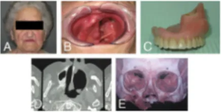
On January 2010, the patient came to the Department of Maxillo-Facial Surgery as referred by her general practitioner for aesthetic and functional oral rehabilitation; during the first visit, clinical and radiographic examinations were performed, revealing that the patient was edentulous and with severe atrophy of the maxillary and mandibular arches with collapse of the soft tissues of the left midface. The defect comprised the left side of the hard palate, extending posteriorly onto the soft palate, resulting in an opening to the nasal cavity; the resection site was free of inflam-mation and covered with mucosa (Figs. 1A, B).
The patient required a definitive rehabilitation to improve speech, mastication, swallowing, and aesthetics; for these reasons, the mi-crosurgical rehabilitation followed by implant placement was not indicated due to the poor general status of the patient and the com-promised vascular supply of the area after radiotherapy, so we decided to treat the patient using a protocol that scheduled a pedicled tem-poralis muscle flap and delayed zygomatic implant placement.
The patient underwent the first step of the treatment planning; through a left hemicoronal incision, a temporal myofascial flap was dissected and transposed into the oral cavity, filling the maxillary
loss of substance and separating the oral cavity from the left nasal cavity (Fig. 2).
Six months later, after a satisfactory healing of the temporalis muscle flap (Fig. 2C), the second step of the definitive treatment, the implant placement, was planned using a computer-assisted sur-gery software package (NobelGuide; Nobel Biocare Services AG, Gothenburg, Sweden); this system is commonly used to plan the position of the implants after a three-dimensional CT scan permit-ting sagittal, coronal, and axial views.
Impressions of both arches were obtained with silicone; the patient was referred to a radiologic center for a CT scan (spiral CT, gantry tilt at 0-, slice thickness of 0.5 mm, and slice increment of 0.3 mm). The CT scan data were processed using a specific sur-gical planning software package to create a three-dimensional image of the maxilla, evaluating the anatomic structures (Procera; Nobel Biocare).
The procedure began with the insertion of 1 implant (Nobel Speedy Replace; Nobel Biocare) on the right side of the max-illary arch (sites: 1.2); consequently, the upper right side of the maxilla was reached through a crestal incision, whereas the left zygomatic bone was reached through an incision along the previ-ous transposed temporalis muscle.
After raising the mucoperiosteal flaps, soft tissue dissection was performed along the inferior and frontal lateral surfaces of the zy-gomatic bones. The right maxillary sinus was fenestrated while keeping the Schneider membrane intact. This window allowed the visualization of the implant placement. The drilling sequence started allowing visual control of the insertion of 2 zygomatic im-plants: 1 for each side (Bra˚nemark System Zygoma; Nobel Biocare). Simple absorbable sutures were placed to close the flap (Fig. 3).
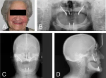
After 2 weeks, the prosthesis (Molinaro & Massaro Snc, Turin, Italy) supported by a titanium bar (Procera; Nobel Biocare) was positioned to load all the 3 implants. The patient was instructed to follow a proper oral hygiene and was prescribed a soft diet for 30 days. Postoperative plain radiographs demonstrated the correct insertion and angulation of the implants in the zygomatic bone. At 12-month follow-up examination, all the implants were correctly osseointegrated and the prosthesis appeared to be perfectly func-tional with a satisfactory occlusion and facial balance except for a left temporal hollowing, without signs or symptoms of local com-plications such as sinusitis, peri-implant mucositis, or implant mo-bility (Figs. 4A, B).
DISCUSSION
According to the National Comprehensive Cancer Network guide-lines, the standard treatment of resectable carcinomas of the minor salivary glands is the surgical excision aimed at achieving complete clearance with 1-cm margin (mucoepidermoid and adenocarcinoma) or more (2Y3 cm for adenoid cystic carcinoma); in case of N0, a
FIGURE 1. Preoperative clinical and radiologic conditions. The defect comprised the left side of the hard palate, extending posteriorly onto the soft palate, resulting in an opening to the nasal cavity; the resection site was free of inflammation and covered with mucosa (Figs. 1A-E).
FIGURE 2. Intraoperative sequence (A, B) and postoperative healing (C) of the pedicled temporalis muscle flap.
FIGURE 3. Intraoperative views of the zygomatic implant placement (AYC); the titanium bar positioned to load all the 3 implants (DYF).
Brief Clinical Studies The Journal of Craniofacial Surgery
&
Volume 23, Number 5, September 2012e464
* 2012 Mutaz B. Habal, MDroutine prophylactic neck dissection is not recommended. Neck dis-section is usually performed in the presence of clinically or radio-graphically positive lymphadenopathy. Postoperative radiotherapy is recommended in patients with advanced disease, positive or closed resection margins, histologically high-grade tumors, and evidence of perineural, intravascular, or intralymphatic spreading.3,15,16
Reconstruction after surgery is required for most patientsV mostly for patients who underwent a wide demolition of the oral cavity because local and systemic recurrences occur late and the long-term survival outcome is determined mainly by T stage, re-section margins status, N stage (which are the most powerful pre-dictors of survival) and by the systemic spread of disease.4
According to the scientific literature, many reconstructive options are described such as (1) prosthetic obturator; (2) locoregional flaps, (3) nonvascularized grafts, and (4) microvascular free flaps.7,8
In some motivated patients without recurrences of the primary tumor (in this report, the interval of the absence of disease is al-most 7 y), it is possible to complete the surgical reconstruction with implantology surgery, obviously only to improve the patient’s quality of life: physical well-being, familiar relationship, emotional status, and functional activities.
The 2 major indications for the zygomatic implant placement are defects after maxillectomy and edentulous atrophic maxilla. The zygomatic implants are usually placed at 30- to 60- in relation to the occlusal plane. Preoperative planning using computer-assisted software package is mandatory because of the anatomic complexity of the zygomatic bone and the limited intraoperative visibility.17,18
For patient who underwent maxillectomy, it is frequently difficult to obtain an ‘‘all-on-four’’ protocol because of the frequent lack of the residual maxillary bone. Afterward, it is necessary to adapt the protocol to the case, as demonstrated by our operative choice.19Y21
It was clear that it was not technically possible to insert 2 con-ventional implants in the anterior maxilla because of the lack of residual bone, thus rehabilitating the patient with 2 zygomatic implants and only 1 implant in the right anterior hemimaxilla.
The maxillectomy condition is characterized by the absence of bone for implant therapy; the loss of bone leads to less volume available to accept the placement of implants with a high rate of surgical failure. Simultaneously, the oral environment become un-suitable for an adequate denture retention.7Y11
With the previous statements in mind and having considered the published technical strategies of experienced surgeons, we have managed this case by performing an intraoral transposition of a pedicled temporalis muscle flap without any bone grafts, followed by a delayed zygomatic implant placement; despite the complex-ity of this procedure, this approach enabled us to solve a double problem: the maxillary defect with bucconasal communication and the atrophic/edentulous maxilla.
Our clinical report demonstrates a successful 2-step treatment for a patient who underwent hemimaxillectomy. Although there are scarce data on the long-term survival of the zygomatic implants,
this method permits us to solve this case, avoiding the morbidity of a fibula free flap or the inconvenience of a removable prosthetic obturator.
REFERENCES
1. Mucke T, Robitzky LK, Kesting MR, et al. Advanced malignant minor salivary glands tumors of the oral cavity. Oral Surg Oral Med Oral Pathol Oral Radiol Endod 2009;108:81Y89
2. Buchner A, Merrell PW, Carpenter WM. Relative frequency of intraoral minor salivary gland tumors: a study of 380 cases from northern California and comparison to reports from other parts of the world. J Oral Pathol Med 2007;36:207Y214
3. Guzzo M, Locati L, Prott FJ, et al. Major and minor salivary gland tumors. Crit Rev Oncol Hematol 2010;74:134Y148
4. Kakarala K, Bhattacharyya N. Survival in oral cavity minor salivary gland carcinoma. Otolaryngol Head and Neck Surg 2010;143:122Y126 5. Lloyd S, Yu JB, Ross DA, et al. A prognostic index for predicting
lymph node metastasis in minor salivary gland cancer. Int J Radiat Oncol Biol Phys 2010;76:169Y175
6. Weber AL. Imaging of the salivary glands. Curr Opin Radiol 1992;4:117Y122
7. Davison SP, Sherris DA, Meland NB. An algorithm for maxillectomy defect reconstruction. Laryngoscope 1998;108:215Y219
8. Kornblith AB, Zlotolow IM, Gooen J, et al. Quality of life of maxillectomy patients using an obturator prosthesis. Head Neck 1996;18:323Y334
9. Rogers SN, Lowe D, McNally D, et al. Health-related quality of life after maxillectomy: a comparison between prosthetic obturation and free flap. J Oral Maxillofac Surg 2003;61:174Y181
10. Rogers SN, Lakshmiah SR, Narayan B, et al. A comparison of the long-term morbidity following deep circumflex iliac and fibula free flaps for reconstruction following head and neck cancer. Plast Reconstr Surg 2003;112:1517Y1525
11. Brown JS, Jones DC, Summerwill A, et al. Vascularized iliac crest with internal oblique muscle for immediate reconstruction after maxillectomy. Br J Oral Maxillofac Surg 2002;40: 183Y190
12. Malo P, de Araujo Nobre M, Lopes A. The use of computer-guided flapless implant surgery and four implants placed in immediate function to support a fixed denture: preliminary results after a mean follow-up period of thirteen months. J Prosthet Dent 2007;97:S26YS34
13. Adell R, Lekbolm U, Rockler B, et al. A 15-year study of osseointegrated implants in the treatment of the edentulous jaw. Int J Oral Surg 1981;10:387Y416
14. Branemark PI, Adell R, Albrektsson T, et al. An experimental and clinical study of osseointegrated implants penetrating the nasal cavity and maxillary sinus. J Oral Maxillofac Surg 1984;42:497Y505 15. Strick MJ, Kelly C, Soames JV, et al. Malignant tumors of the
minor salivary glandVa 20 year review. Br J Plast Surg 2004;57:624Y631
16. Copelli C, Bianchi B, Ferrari S, et al. Malignant tumors of intraoral minor salivary glands. Oral Oncol 2008;44:658Y663
17. Albrektsson T. A multicenter report on osseointegrated oral implants. J Prosthet Dent 1988;60:75Y84
18. Johansson B, Friberg B, Nilson H. Digitally planned, immediately loaded dental implants with prefabricated prostheses in the
reconstruction of edentulous maxillae: a 1-year prospective, multicenter study. Clin Implant Dent Relat Res 2009;11:194Y200
19. Schmidt BL, Pogrel MA, Young CW, et al. Reconstruction of extensive maxillary defects using zygomaticus implants. J Oral Maxillofac Surg 2004;62:82Y89
20. Chow J, Hui E, Lee PK, et al. Zygomatic implantsVprotocol for immediate occlusal loading: a preliminary report. J Oral Maxillofac Surg 2006;64:804Y811
21. Uckan S, Oguz Y, Uyar Y, et al. Reconstruction of a total maxillectomy defect with a zygomatic implant-retained obturator. J Craniofac Surg 2005;16:485Y489
FIGURE 4. Postoperative clinical (A) and radiologic (BYD) conditions.
The Journal of Craniofacial Surgery
&
Volume 23, Number 5, September 2012 Brief Clinical Studies* 2012 Mutaz B. Habal, MD