DOI 10.1007/s00404-009-1113-1
O R I G I N A L A R T I C L E
Minilaparotomy in spinal anaesthesia: a surgical choice
in treatment of benign gynaecologic disease
Daniela Surico · Luca Mencaglia · Francesca Riboni · Alessandro Vigone · Livio Leo · Nicola Surico
Received: 1 March 2009 / Accepted: 23 April 2009 / Published online: 12 May 2009 © Springer-Verlag 2009
Abstract
Purpose Minilaparotomic access in spinal anaesthesia represents an example of mininvasive surgery and could be a valid cost–beneWt alternative in the surgical treatment of benign gynaecologic diseases.
Methods The study is a randomized study. We analyzed a consecutive series of 80 patients treated for benign gynae-cological diseases with spinal (group A) or with general anaesthesia (group B).
Results The median length of incision was 5 cm. The average operating time was 40.5 § 9.39 min, without diVerences between groups. The average hospital stay was 0.71 days shorter (p · 0.0001) and the postoperative pain was lower at 2 and 6 h from the surgery and at 10 p.m. in the group A (p · 0.0001).
Conclusions Minilaparotomy in spinal anaesthesia carries advantages from economic point of view with reduction of length of stay in hospital which is an important parameter for the evaluation of the quality of surgical treatments. Keywords Minilaparotomy · Postoperative pain · Spinal anaesthesia · Mininvasive surgery
Introduction
During the last century, laparotomic surgery techniques have been improved in order to reduce the postoperative course. In this perspective, “the length of stay in hospital” becomes an important parameter to evaluate the quality of surgical treatments, thanks to the recent attention to cost reductions in the public health system. The minilaparo-tomic access in spinal anaesthesia represents an example of mininvasive surgery and could be a valid cost–beneWt alter-native in the surgical treatment of benign gynaecologic dis-eases [1–13]. The minilaparotomic technique may be performed by a transverse suprapubic incision (4–8 cm skin incision) for gynaecological disease. This minimally inva-sive surgery could represent a valid alternative to laparo-tomic and laparoscopic gynaecologic surgery in the management of benign disease [1–13]. In this study, we describe the advantages of this approach.
Materials and methods
From January 2007 to December 2007, a consecutive series of 80 patients, who underwent minilaparotomy for benign gynaecological diseases in our Hospital, were randomized into two groups.
We randomized the patients in, patients with spinal (group A) or general anaesthesia (group B) (Canadian Task Force classiWcation I).
In this study, we included patients aVected by benign gynaecologic diseases like simple ovarian cysts or uterine leiomyomatosis.
The exclusion criteria was (1) body mass index (BMI) greater than 25 Kg/m2, (2) previous longitudinal laparot-omy or pelvic surgery, (3) indication for a concomitant D. Surico · F. Riboni (&) · A. Vigone · L. Leo · N. Surico
Department of Obstetrics and Gynecology, University “A. Avogadro”, Via Solaroli 17, 28100 Novara, Italy
e-mail: [email protected]
L. Mencaglia
Florence Centre of Ambulatory Surgery and Infertility, Florence, Italy
vaginal surgery, (4) large uterus ¸12 weeks of gestational size, (5) Wxed uterus with preoperative suspected of severe endometriosis, (6) serious renal or cardiopulmonary pathol-ogies, (7) coagulopathies, (8) corticosteroid therapy and (9) spinal malformations hindering spinal anaesthesia.
Informed consent was obtained from each patient. A preoperative evaluation included physical and gynae-cological examination, chest X-ray, CA125 serum levels (when indicated) and ultrasonographic scan with colour Doppler. Clinical evaluation and surgical procedure were performed by one senior surgeon. All patients received bowel preparation with macrogol the evening before sur-gery and antibiotic prophylaxis in short term, with piperacillin + tazobactam 2.2 g i.v. (intravenously), 30 min before the surgery. The antithrombotic prophylaxis was performed in all patients according to thrombosis risk factor.
Spinal anaesthesia was performed at L2–L3 level with hyperbaric marcain 0.5%, 16 mg with 25 G sprotte. General anaesthesia consisted in premedication with midazolam 0.15, propofol 2.5 mg/Kg to induce narcosis, cisatracurium 0.2 and remifentanil 0.25 mcg/Kg. Narcosis after intubation and assisted ventilation with 50% air–oxygen blend was maintained with sevoXurane to MAC. In both groups, post-operative analgesia was carried out with ketorolac trometa-mine and tramadol.
In group A, we administrated analgesic therapy 6 h after surgery (one giving); in group B ongoing therapy was administrated during the Wrst 24 h from the end of surgery. The post operatory pain was calculated with visual analogi-cal sanalogi-cale (VAS) immediately after surgery, at 2 and 6 h from the surgery, at 10 p.m., 8 a.m. and 10 p.m. on the fol-lowing days of stay in hospital [14–17].
For every case, we considered the following parameters: age, parity, BMI, past and present pathologies and thera-pies, weight of uterus, time of surgery, haematic loss, intra-operative complications (bowel, bladder, urethral and vascular injuries) and scar measurement at the end of sur-gery. During follow-up, we analyzed length of stay in hos-pital, postoperative morbidity, blood transfusions (for haemoglobin level ·7 g/dl in asymptomatic patients), day of bladder catheter removal and free diet onset. Postopera-tive temperature was obtained by two measurements at 8 a.m. and at 8 p.m. All women were reviewed 4 weeks after surgery.
Statistical analysis
We compared the data from the two groups of patients. Analysis was carried out with Mann–Whitney U test for the continuous variables and Fisher exact test for the fre-quency data. SigniWcance was set at a probability value of <0.05.
Surgical technique
The patient is placed in supine position with a Foley cathe-ter into bladder. The skin incision is made according to Mini-Pfannenstiel technique (photos 1–2) [1–13]. The fol-lowing subcutaneous cut is performed by electrocution, down to superWcial fascia of recti abdominis at the insertion of pyramidal muscles. The opening of the muscular fascia, longer than the laparotomic cut 2 cm/side, allows the visu-alization of the muscular plan and the median rafe of the abdominal wall muscles, that is dissected exposing the pari-etal peritoneum. The opening of paripari-etal peritoneum is per-formed with manual traction for preventing internal or vesical lesions. To widen the operating Weld, Deaver retrac-tors can be placed (Farabeuf retractor).
Myomectomy, cystectomy, salpingo-oophorectomies and total hysterectomy were performed according to our School technique [18]. At the end of the surgery, the peritoneal cavity is washed with physiological solution at 37–40°C and accurate haemostasis is obtained. The synthe-sis of the abdominal wall is performed in layers. The scar measurement is made at the end of surgery, in order to check a lengthening due to the traction on the skin.
Photo 1 Abdominal cut (4–7 cm transverse incision, 1–2 cm below
Results
A consecutive series of 80 patients (average age 36.65 § 8.59 years, range 20–68) were included into this study.
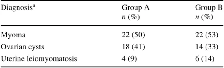
Two groups are homogenous in age, parity and BMI (Table1). Preoperative diagnosis was myoma in 44 patients (51%), ovarian cyst in 32 (37%) and uterine leiomyomatosis in 10 (12%). Intraoperative diagnoses conWrmed preoperative diagnoses (Table2). All surgical acts were performed by the same senior surgeon, as reported in Table3.
The median length of incision was 5 cm, ranging from 4 to 7 cm. The average operating time was 40.5 § 9.39 min without any signiWcant diVerences between the two groups (Table4). The weight of the removed uterus, following hysterectomy, was between 70 and 700 g. The average number of removed myoma was 2.48 (range 1–7), with an average diameter of 5.36 cm (range 2–9) with no signiWcant diVerences between groups. No intraoperative complica-tions were observed and no conversion to Pfannenstiel inci-sion was necessary. The average blood loss was 60 ml (range 20–80). Postoperative analgesia was carried out 6 h after surgery in group A (ketorolac trometamine 60 mg and tramadolo 300 mg i.v.) and during the Wrst 24 h from the end of surgery (ketorolac trometamine 90 mg and tramadolo 400 mg i.v.) in group B. We did not observe post-operative
morbidity (temperature > 38°C) and any blood transfusion was necessary. In all patients, Foley catheter was removed within 10 h from surgery; deambulation was resumed the same evening and hot liquid diet was introduced at the evening of the surgery’s day. The median duration of paralytic ileus was 1.09 days in group A and 1.33 days in group B (p < 0.0001) (Table4). The average hospital stay was 1.84 § 0.51 days for group A and 2.55 § 0.5 days for group B (Table4).
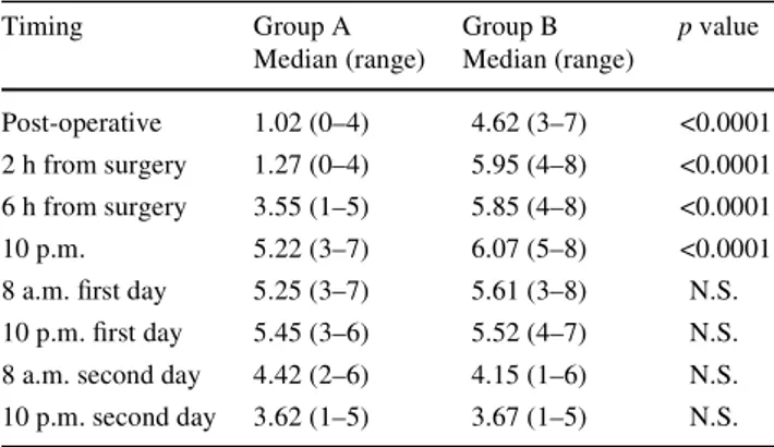
The popularity rating for spinal anaesthesia was 8 out of 10, while the rating for narcosis was 6 out of 10. Postopera-tive pain (VAS) was statistically diVerent between the two groups immediately after surgery, at 2 and 6 h from the sur-gery and at 10 p.m. (p < 0.0001), while no statistically sig-niWcant diVerences were recorded between the Wrst and the
Photo 2 Uterus in myomectomy
Table 1 Clinical characteristics of patients with spinal anaesthesia
(Group A, n = 40) or general anaesthesia (Group B, n = 40)
N.S. not signiWcant
Characteristics Group A Median (range) Group B Median (range) p value Age (years) 37.5 (29–55) 35.8 (32–68) N.S. Parity 0.4 (0–2) 0.5 (0–3) N.S BMI (kg/m2) 21.9 (18–25) 22 (19–25) N.S
Table 2 Intraoperative diagnoses in patients with spinal anaesthesia
(Group A) or general anaesthesia (Group B)
N.S. not signiWcant
a
Some patients >1 diagnosis
Diagnosisa Group A n (%) Group B n (%) Myoma 22 (50) 22 (53) Ovarian cysts 18 (41) 14 (33) Uterine leiomyomatosis 4 (9) 6 (14)
Table 3 Surgical treatment of patients with spinal anaesthesia (Group
A) or general anaesthesia (Group B)
N.S. not signiWcant
a Some patients >1 treatment
b 4 cases associated to total hysterectomy c 2 cases associated to total hysterectomy Surgical treatmenta Group A
n (%) Group B n (%) Myomectomy 22 (45.8) 22 (50) Cystectomy 14 (29.2) 8 (18.2) Salpingo-oophorectomies 8 (16.6)b 6 (13.6)c Total hysterectomy 4 (8.3) 6 (13.6) Oophorectomy 0 2 (4.5)
Table 4 Post surgical characteristics of patients with spinal
anaesthe-sia (Group A) or general anaestheanaesthe-sia (Group B)
N.S. not signiWcant
Characteristics Group A Median § D.S.
Group B Median § D.S.
p value
Surgery time (minutes) 40.25 § 10.31 40.75 § 8.51 N.S. Paralytic ileus (days) 1.09 § 0.06 1.33 § 0.13 <0.0001 Hospitalisation (days) 1.84 § 0.51 2.55 § 0.5 <0.0001
second postoperative days (Table5). All women were reviewed 4 weeks after surgery. No adverse event occurred after surgery.
Discussion
Minilaparotomy in spinal anaesthesia is an example of mininvasive surgery and it could be a valid cost–beneWt alternative in the surgical treatment of gynaecologic dis-eases with advantages from surgical, anaesthesiology and economic point of view. We analyzed advantages of minilaparotomic access in a prospective group of patients treated for benign gynaecological diseases in spinal anaesthesia.
The reduction of length of stay hospital is an important parameter for the evaluation of the quality of surgical treatments, thanks to the recent attention to cost reduc-tions in public health. As shown by our results, the aver-age hospital stay is 0.71 days shorter in the group in spinal anaesthesia.
The shorter hospitalization may be the consequence of some speciWc attentions begin given beginning during the surgery with the choice of a small abdominal incision, which implies mild trauma on soft tissue. The careful hae-mostasis, the avoidance of bowel manipulation without the use of self-retaining retractor, peritoneal washing at the end of the procedure may reduce in time the postoperative ady-namic ileus. During the postoperative course, the reduction of analgesic therapy excluding opioid drugs with early mobilization and hot liquid diet plays a crucial rule in the early discharge of our patients.
The spinal anaesthesia generally produces a motor block of shorter duration, which has advantages for earlier mobili-zation and discharge from hospital and may be particularly useful in gynaecological surgery. The regional anaesthesia and postoperative analgesia with ketorolac and tramadol appears to be the best choice for the control of post surgical
pelvic pain. The patient popularity rating about the type of anaesthesia, in fact, was better for the group A.
From economic point of view (length of stay in hospi-tal), we estimated the advantages of this technique. The cost of the postoperative stay in hospital is estimated to be in median of about 400 euro per each patient in the group in spinal anaesthesia. Naturally, there are no signiW-cant diVerences in the materials and surgery costs between the groups. With the described technique, we have a faster turnover of patients, which is an important parameter for the reduction of costs within the public health system. Moreover, most of surgeons can perform minilaparotomy which, unlike laparoscopy, dose not require a long training and expensive equipment which is not available in all Countries. Minilaparotomy can also replace laparoscopy in patients with critical physical status in which pneumoperi-toneum is not recommended or in cases with contraindica-tions to general anaesthesia.
In our opinion, the success of these results starts with the patients’ selection. The preoperative counselling should be as complete as possible and should actively involve the sur-geon, anaesthetist, nurse and patient, in order to obtain an optimal compliance before, during and after the surgery. The preoperative visit must be performed by the senior sur-geon with a great experience in physical and ultrasono-graphic examinations. This is the Wrst expedient for avoiding extensions to Pfannenstiel incision.
In conclusion, we are convinced that minilaparotomy can represent a valid alternative in gynaecological surgical approach even if diVerent surgical accesses cannot be in antagonism but must be adequate to the pathology and the patient. The surgeon must be able to carry out with ease all kinds of access and to eliminate every bias for the choice of the corrected approach in the surgical treatment of benign gynaecological disease.
ConXict of interest statement None.
References
1. Panici P, Zullo MA, Angioli R, Muzii L (2005) Minilaparotomy hysterectomy: a valid option for the treatment of benign uterine pathologies. Eur J Obstet Gynecol Reprod 119(2):228–231. doi:10.1016/j.ejogrb.2004.07.039
2. Fanfani F, Fagotti A, Longo R, Marana E, Mancuso S, Scambia G (2005) Minilaparotomy in the management of benign gynecologic disease. Eur J Obstet Gynecol Reprod 119(2):232–236. doi:10. 1016/j.ejogrb.2004.07.040
3. Flynn MK, NiloV CM (1999) Outpatient minilaparotomy for ovar-ian cysts. J Reprod Med 44:399–404
4. Cagnacci A, Pirillo D, Malmusi S, Arangino S, Alessandrini C, Volpe A (2003) Early outcome of myomectomy by laparotomy, minilaparotomy and laparoscopically assisted minilaparotomy. A randomized study. Hum Reprod 18(12):2590–2594. doi:10. 1093/humrep/deg478
Table 5 Post-operative pain (VAS scale) of patients with spinal
anaesthesia (Group A) or general anaesthesia (Group B)
N.S. not signiWcant
Timing Group A Median (range) Group B Median (range) p value Post-operative 1.02 (0–4) 4.62 (3–7) <0.0001 2 h from surgery 1.27 (0–4) 5.95 (4–8) <0.0001 6 h from surgery 3.55 (1–5) 5.85 (4–8) <0.0001 10 p.m. 5.22 (3–7) 6.07 (5–8) <0.0001 8 a.m. Wrst day 5.25 (3–7) 5.61 (3–8) N.S. 10 p.m. Wrst day 5.45 (3–6) 5.52 (4–7) N.S. 8 a.m. second day 4.42 (2–6) 4.15 (1–6) N.S. 10 p.m. second day 3.62 (1–5) 3.67 (1–5) N.S.
5. Muzii L, Basile S, Zupi E, Marconi D, Zullo MA, Manci N, Bellati F, Angioli R, Benedetti Panici P (2007) Laparoscopic-assisted vaginal hysterectomy versus minilaparotomy hysterec-tomy: a prospective, randomized, multicenter study. J Minim Invasive Gynecol 14(5):610–615. doi:10.1016/j.jmig.2007.05.012
6. Alcalde JL, GuiloV E, Ricci P, Solà V, Pardo J (2007) Minilapa-rotomy hysterectomy assisted by self-retaining elastic abdominal retractor. J Minim Invasive Gynecol 14(1):108–112. doi:10.1016/ j.jmig.2006.06.030
7. Fanfani F, Fagotti A, Ercoli A, Bifulco G, Longo R, Mancuso S, Scambia G (2004) A prospective randomized study of laparoscopy and minilaparotomy in the management of benign adnexal masses. Hum Reprod 19(10):2367–2371. doi:10.1093/humrep/deh413
8. Fanfani F, Fagotti A, Bifulco G, Ercoli A, Malzoni M, Scambia G (2005) A prospective study of laparoscopy versus minilaparotomy in the treatment of uterine myomas. J Minim Invasive Gynecol 12(6):470–474. doi:10.1016/j.jmig.2005.07.002
9. Glasser MH (2005) Minilaparotomy myomectomy: a minimally invasive alternative for the large Wbroid uterus. J Minim Invasive Gynecol 12(3):275–283. doi:10.1016/j.jmig.2005.03.009
10. Palomba S, Zupi E, Falbo A, Russo T, Marconi D, Tolino A, Manguso F, Mattei A, Zullo F (2007) A multicenter randomized, controlled study comparing laparoscopic versus minilaparotomic myomectomy: reproductive outcomes. Fertil Steril 88(4):933– 941. doi:10.1016/j.fertnstert.2006.12.047
11. Benedetti-Panici P, Maneschi F, Cutillo G, Scambia G et al (1996) Surgery by minilaparotomy in benign gynecologic disease. Obstet Gynecol 87:456–459. doi:10.1016/0029-7844(95)00441-6
12. Benedetti-Panici P, Zullo MA, Casalino B, Angioli R, Muzii L (2003) Subcutaneous drainage versus no drainage after minilapa-rotomy in gynecologic conditions: a randomized study. Am J Obstet Gynecol 188:71–75. doi:10.1067/mob.2003.103
13. HoVman MS, Lynch CM (1999) Minilaparotomy hysterectomy. Am J Obstet Gynecol 181(4):1037–1038. doi: 10.1016/S0002-9378(99)70349-2
14. Bird SB, Dickson EW (2001) Clinically signiWcant changes in pain along the visual analogy scale. Ann Emerg Med 38(6):639– 643. doi:10.1067/mem.2001.118012
15. Gracely RG, Dubner R (1987) Reliability and validity of verbal descriptor scales of painfulness. Pain 29:175–185. doi:10.1016/ 0304-3959(87)91034-7
16. Dixon JS (1986) Agreement between horizontal and vertical visual analogue scales. Br J Rheumatol 24:415–416
17. Price DD, McGrath PA, RaWi A, Buckingham B (1983) The vali-dation of visual analogy scales as ratio scale measures for chronic and experimental pain. Pain 17:45–56. doi:10.1016/0304-3959 (83)90126-4
18. Ferraris G (1986) Tecnica chirurgica. Chirurgia Ginecologica e Ostetrica. Utet