molecules
ArticleChemical Composition, Antibacterial and Antifungal
Activities of Crude Dittrichia viscosa (L.) Greuter
Leaf Extracts
Wafa Rhimi 1,2, Issam Ben Salem2,*, Davide Immediato3, Mouldi Saidi2, Abdennacer Boulila4 and Claudia Cafarchia3
1 Faculté des Sciences de Bizerte, Université de Carthage, 7021 Zarzouna, Tunisia; [email protected] 2 Laboratory of Biotechnology and Nuclear Technolog, National Centre of Nuclear Science and
Technology (CNSTN), Sidi Thabet Technopark, 2020 Ariana, Tunisia; [email protected] 3 Dipartimento di Medicina Veterinaria, Università degli Studi di Bari, 70010 Valenzano, Italy;
[email protected] (D.V.); [email protected] (C.C.)
4 Laboratory of Natural Substances LR10INRAP02, National Institute of Research and Physico-chemical Analyses, Biotechpole of Sidi Thabet, 2020 Ariana, Tunisia; [email protected]
* Correspondence: [email protected]; Tel.: +216-71-537-410 Academic Editors: Hideyuki Ito, Tsutomu Hatano and Takashi Yoshida Received: 12 May 2017; Accepted: 3 June 2017; Published: 30 June 2017
Abstract: The small amount of data regarding the antifungal activity of Dittrichia viscosa (L.) Greuter against dermatophytes, Malassezia spp. and Aspergillus spp., associated with the few comparative studies on the antimicrobial activity of methanolic, ethanolic, and butanolic extracts underpins the study herein presented. The total condensed tannin (TCT), phenol (TPC), flavonoid (TFC), and caffeoylquinic acid (CQC) content of methanol, butanol, and ethanol (80% and 100%) extracts of D. viscosa were assessed and their bactericidal and fungicidal activities were evaluated. The antibacterial, anti-Candida and anti-Malassezia activities were evaluated by using the disk diffusion method, whereas the anti-Microsporum canis and anti-Aspergillus fumigatus activities were assessed by studying the toxicity effect of the extracts on vegetative growth, sporulation and germination. The methanolic extract contained the highest TPC and CQC content. It contains several phytochemicals mainly caffeoylquinic acid derivatives as determined by liquid chromatography with photodiode array and electrospray ionisation mass spectrometric detection (LC/PDA/ESI-MS) analysis. All extracts showed an excellent inhibitory effect against bacteria and Candida spp., whereas methanolic extract exhibited the highest antifungal activities against Malassezia spp., M. canis and A. fumigatus strains. The results clearly showed that all extracts, in particular the methanolic extract, might be excellent antimicrobial drugs for treating infections that are life threatening (i.e., Malassezia) or infections that require mandatory treatments (i.e., M. canis or A. fumigatus).
Keywords:Dittrichia viscosa; antifungal activities; Candida spp.; Malassezia spp.; Microsporum canis; Aspergillus fumigates
1. Introduction
The growing worldwide concern about the alarming increase in the rate of human and animal infections caused by antibiotic-resistant microorganisms have spurred the interest of the scientific community in developing alternative methods for their control [1]. Many kinds of natural extracts from medicinal plants containing phenolic and flavonoid compounds have excellent biological properties and are used as alternative therapies. Among the large variety of Mediterranean folkloric herbs, Dittrichia viscosa belonging to the Asteraceae family, has proven to be a source of natural products forming the basis for alternative medicine and natural therapies [2–4]. Dittrichia viscosa was studied Molecules 2017, 22, 942; doi:10.3390/molecules22070942 www.mdpi.com/journal/molecules
against antibiotic-resistant microorganisms, antibacterial activity and anti-fungal activity against Candida albicans and Fusarium species [5,6]. To the best of our knowledge, reports on antifungal activity of D. viscosa against dermatophytes, Malassezia spp. and Aspergillus spp. are scant or limited to Microsporum canis. In particular, dermatophytes are a group of fungi which have the ability to invade the keratinized tissues (skin, hair, nails) causing cutaneous infections in humans and animals commonly known as dermatophytosis [7]. They are distributed worldwide and some of them are considered zoonotic, being transmitted from animals to humans [8]. The treatment of infections is mandatory due to the contagious and the zoonotic nature and usually requires long antifungal therapy with azoles [9]. In addition, these treatments are not usually performed in food producing animals since they are more expensive, and treated animals need long withholding before using in food processing industry [10].
The fungal genus Malassezia is part of the normal skin microbiota. These yeasts cause human and animal skin disorders in immune-competent hosts and systemic infections in immune- compromised patients which usually require prolonged treatment with and/or high doses of antifungal agents [11,12]. In addition, recent studies clearly show that the same species within the genus of Malassezia furfur and Malassezia pachydermatis are characterized by high minimal inhibitory concentration (MIC) values against all azole drugs commonly employed in the treatment of the infections.
Finally, Aspergillus species are found worldwide in humans and in almost all domestic animals and birds as well as in many wild species, causing a wide range of diseases from localized infections to fatal disseminated diseases, as well as allergic responses to inhaled conidia [13]. Some prevalent forms of animal aspergillosis are invasive fatal infections and are difficult to treat. In addition, the environmental diffusion of A. fumigatus strains presenting azole resistant phenomena is worldwide reported [13].
Thus, this study aimed to: (i) quantify the phenolic and flavonoids content of D. viscosa leaf extract with different solvents; (ii) evaluate their activities against gram positive and negative bacteria, and against Candida spp. (i.e., Candida albicans, Candida krusei, Candida prapsilos); and (iii) to assess their activities against Malassezia spp. (Malassezia pachydermatis and Malassezia furfur), Aspergillus fumigatus and Microsporum canis.
2. Results and Discussion 2.1. Phytochemical Screening
The total condensed tannin (CTC), phenol (TPC), flavonoid (TFC), and caffeoylquinic acid (CQC) content of different D. viscosa extracts are reported in Table1. They are expressed as mg catechin equivalent (CE), mg gallic acid equivalent, mg quercetin equivalent (QE) and mg of chlorogenic acid equivalent (ChlA E) per g dry extract, respectively.
The CTC amounts varied from 7.05± 1.6 to 27.15± 2.21 mg CE/g, being the highest in the methanolic extract (Table1). The TPC ranged from 75.34 ±1.30 to 123.39±1.22 mg GAE/g, the highest content retrieved in methanolic and 80% ethanolic extracts (Table1).
The CQC amounts of D. viscosa extracts ranged from 57.11±0.98 to 87.61±1.06 mg ChlA E/g (Table1) and the highest amount of CQC was registered in methanolic extract. The TFC varied from 30.86±1.28 to 58.03±1.85 mg QE/g and the highest content was registered in butanolic extract (Table1). The methanolic extract contains the highest CTC, TPC and CQC values while the butanolic extract contained the highest amount of TFC.
The results of this study clearly indicate that phenolic and flavonoids content of D. viscosa crude extracts vary according to the solvent extraction procedure. In particular, this study reports for the first time the presence of condensed tannins in this plant species. Indeed, no previous studies have evaluated CTC in D. viscosa leaves, but results herein indicate that the amount within methanolic extracts are in the same range as those of some Asteraceae species such as Artemisia genus [14]. On the contrary, the TPC values of Tunisian D. viscosa extracts were in same range or slightly lower than
Molecules 2017, 22, 942 3 of 13
those reported from Turkish or Moroccan samples [4,15], thus suggesting that TPC values of D. viscosa does not vary accordingly to plant origin. Accordingly, the TPC amounts depend on the polarity of the solvent, and it was highest when the solvent polarity increased. Similar results were reported by Negi and Jayaprakasha when studying methanol extracts of Punica granatum peel [16]. However, a high value of TFC in our extracts was detected in butanolic extract, suggesting that the flavonoid composition of D. viscosa might comprise of substances with a high solubility in butanol, like luteolin derivatives [17,18].
Table 1. Condensed tannins, total polyphenols, total flavonoids, and caffeoylquinic acid content of
different D. viscosa leaf extracts.
Polyphenols and Flavonoids Content Ethanolic Ethanolic 80% Butanolic Methanolic CTC(mgCAE/g extract) 14.29±1.30a 7.05±1.6b 16.86±1.62c 27.15±2.21d TPC (mgGAE/g extract) 117.58±1.29a 123.39 ±1.22b 75.34±1.30c 123.07 ±1.69b TFC (mgQE/g extract) 57.79±1.76a 49.23±1.039b 58.03±1.85a 30.86±50c CQC (mgCGAE/g extract) 71.85±0.35a 73.13±1.06a 57.11±0.98b 87.61±1.06c
Values followed by the same letter along the row are not significantly different (p < 0.05). 2.2. Phenolic Profile of D. viscosa Extracts
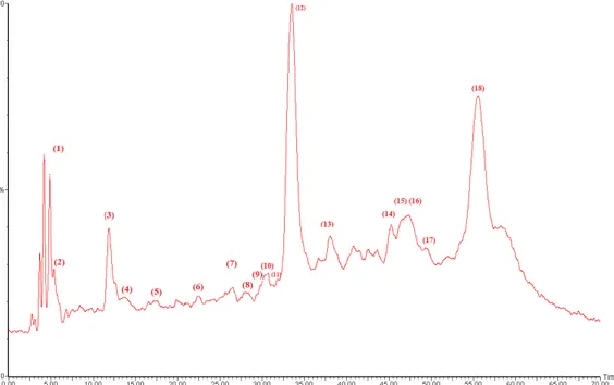
The HPLC-PDA/ESI-MS analysis allowed us to tentatively identify 18 phenolic compounds in the methanolic D. viscosa extract (Figure1). The phenolic fraction of methanolic D. viscosa extract was dominated by caffeoylquinic acid derivatives such as chlorogenic acid, dicaffeoylquinic acid isomers, and caffeoyl glucose as it shown in Table2. Other hyhydroxycinnamic acids like coumaric acid and caffeic acid derivatives were also detected.
the solvent, and it was highest when the solvent polarity increased. Similar results were reported by Negi and Jayaprakasha when studying methanol extracts of Punica granatum peel [16]. However, a high value of TFC in our extracts was detected in butanolic extract, suggesting that the flavonoid composition of D. viscosa might comprise of substances with a high solubility in butanol, like luteolin derivatives [17,18].
Table 1. Condensed tannins, total polyphenols, total flavonoids, and caffeoylquinic acid content of
different D. viscosa leaf extracts.
Polyphenols and Flavonoids Content Ethanolic Ethanolic 80% Butanolic Methanolic
CTC(mgCAE/g extract) 14.29 ± 1.30 a 7.05 ± 1.6 b 16.86 ± 1.62 c 27.15 ± 2.21 d TPC (mgGAE/g extract) 117.58 ± 1.29 a 123.39 ± 1.22 b 75.34 ± 1.30 c 123.07 ± 1.69 b
TFC (mgQE/g extract) 57.79 ± 1.76 a 49.23 ± 1.039 b 58.03 ± 1.85 a 30.86 ± 50 c CQC (mgCGAE/g extract) 71.85 ± 0.35 a 73.13 ± 1.06 a 57.11 ± 0.98 b 87.61 ± 1.06 c
Values followed by the same letter along the row are not significantly different (p < 0.05). 2.2. Phenolic Profile of D. viscosa Extracts
The HPLC-PDA/ESI-MS analysis allowed us to tentatively identify 18 phenolic compounds in the methanolic D. viscosa extract (Figure 1). The phenolic fraction of methanolic D. viscosa extract was dominated by caffeoylquinic acid derivatives such as chlorogenic acid, dicaffeoylquinic acid isomers, and caffeoyl glucose as it shown in Table 2. Other hyhydroxycinnamic acids like coumaric acid and caffeic acid derivatives were also detected.
Some flavonoid compounds were also detected. They were represented exclusively by quercetin derivatives (e.g., quercetin-O-hexoside, quercetin glucuronide, quercetin dimethyl ether isomers), and the flavonol catechin glucoside. The identification of phenolic compounds by HPLC-PDA-ESI-MS as shown in Table 2 confirmed our photochemical screening findings about the richness of D. viscosa extract in caffeoylquinic acid derivatives, and in agreement with previous reports from Israel, Turkey and Tunisia [4,15,19]. From these results, it emerges that D. viscosa might be advised as potential source of bioactive components especially caffeoylquinic acid derivatives.
Figure 1. Chemical characterization of methanolic extract of D. viscosa leaves by HPLC-PDA-ESI-S.
The peaks are numbered and assignments are given in Table 2.
Figure 1. Chemical characterization of methanolic extract ofD. viscosa leaves by HPLC-PDA-ESI-S. The peaks are numbered and assignments are given in Table2.
Some flavonoid compounds were also detected. They were represented exclusively by quercetin derivatives (e.g., quercetin-O-hexoside, quercetin glucuronide, quercetin dimethyl ether isomers), and the flavonol catechin glucoside. The identification of phenolic compounds by HPLC-PDA-ESI-MS as shown in Table2confirmed our photochemical screening findings about the richness of D. viscosa
extract in caffeoylquinic acid derivatives, and in agreement with previous reports from Israel, Turkey and Tunisia [4,15,19]. From these results, it emerges that D. viscosa might be advised as potential source of bioactive components especially caffeoylquinic acid derivatives.
Table 2. Retention time (RT), wavelengths of maximum absorption ( max), mass spectral data, relative
occurrence, and tentative identification of phenolic compounds in methanolic extract of D. viscosa leaves. Compound RT (min) max [M H] Fragment Ions Proposed Structure Occurrence
1 4.856 292sh–322 377 341 (100),179, 119 Caffeic acid hexoside ++ 2 5.346 292sh–322 341 191 (100),137, 128 3-Caffeoylquinic acid ++
3 11.876 292sh–322 353 191 (100), 161 Chlorogenic acid +++
4 13.856 292sh–322 353 161 (100)191, 4-Caffeoylquinic acidisomer +
5 17.222 293–310 467 163 (100) Coumaric acid derivative +
6 22.365 - 429 267 (100),173, 161 Feruloyl caffeolglycerol + 7 27.982 360, 262 463 301 (100),331, 255 Quercetin hexoside + 8 28.007 293sh–321 463 301(100) Hydroxyluteolin hexoside + 9 30.452 293sh–354 477 301 (100) Quercetin glucuronide + 10 30.714 294sh–354 477 301 (100), 161 Quercetin glucuronide + 11 32.888 292sh–322 353 191 (100),179, 161 5-Caffeoylquinic acid + 12 33.463 292sh–322 353 191 (100),179 (32) Dicaffeoylquinic acidisomer ++++ 13 37.98 292–322 353 179 (100),191 (78), 161 (80) Caffeoylquinic acid isomer +++ 14 44.561 290, 320 339 135 (100) Caffeoyl glucose +++ 15 47.234 253–349 329 314 (100), 299 (80), 285 (70), 271 (53), 243 (50) Quercetin-dimethyl ether isomer +++ 16 47.395 253–349 329 314 (100), 299 (85) 271 (75), 241 (40) Quercetin-dimethyl ether isomer +++ 17 49.315 253–349 329 314 (100),299 (85), 285, 243 Quercetin-dimethyl ether isomer + 18 55.566 278 493 165 (100),289 (40), 139 (80) Catechin glucoside +++ +: low in abundance; ++: moderate in abundance; +++: high in abundance); ++++: very high in abundance. 2.3. Antibacterial, Anti-Candida and Antifungal Activity of D. viscosa Extracts
Table3shows the inhibitory effects of D. viscosa extracts against Gram positive (i.e., Staphylococcus aureus, Enterococcus feacium, Streptococcus agalactiae) and Gram negative bacteria (i.e., Escherichia coli and Salmonella typhimurium) with the inhibition halo ranging from 9.5 to 34.5 mm. No statistically significant differences were recorded between different extracts. The highest antimicrobial activity was observed against Enterococcus feacium (G+) and Streptococcus agalactiae (G+) with inhibition zones of 34.5±0.7 mm and 29±1.41 mm, respectively.
Table 3. Antibacterial properties of extracts under study, expressed as diameter of inhibition halo
(in mm) versus several strains.
Bacterial spps. Concentration(mg/mL) Ethanol Ethanol 80% Butanol Methanol Eshershia coli 5010 1112±1.41a 11.5±0.70a 12.5±0.70a 12±0.70a ±1.41a 10±0.0a 10.5±0.0a 10±0.0a
Sal Salmonella typhimurium 50 10.5±0.70a 9.5±0.70a 10.5±0.70a 10±0.0a 10 9.5±0.70a 0±0.0b 9.5±0.70a 9.5±0.0a Enterococcus feacium 50 34±1.41a 28.5±0.0b 34.5±0.70a 34.5±0.7a 10 30±0.0a 25±0.0b 28±0.0c 29±0.0d Streptococcus agalactiae 50 28±1.41a 28±1.41a 29±1.41a 29±1.41a 10 18.5±0.70a 17±0.0a 21.5±1.41b 18±1.14a Staphylococus aureus 50 25±0.0a 25±0.0a 22.5±0.70b 20±0.0c 10 13.5±0.70a 10 ±0.0b 13±1.41a 11±0.0c
Values followed by the same superscript along the row are not significantly different (p < 0.05).
The results of anti-Candida and anti-Malassezia activities are reported in Table4. The diameter halo ranged from 7 to 14.5 mm according to extract concentration. No significant differences were recorded among the activity of different extracts against Candida species.
Table 4.Anti-Candida and Anti-Malassezia properties of extracts under study, expressed as diameter of inhibition halo (in mm) versus several strains.
Candida and Malassezia spp. Concentration(mg/mL) Ethanol Ethanol 80% Butanol Methanol Candida prapsilosis ATCC 22019 5010 10.25 ± 0.58a 9.66 ± 1.52a 8.75 ± 1.73a 10.75 ± 0.95a
8.66 ± 1.73a 8.5 ± 1.73a 8.66 ± 1.73a 10 ± 0.95a Candida krusei ATCC 6258 5010 10 ± 1.419.5 ± 0.7aa 10.5 ± 0.5710 ± 0.0aa 9 ± 0.8210 ± 1aa 10 ± 0.010 ± 0.0aa Candida albicans ATCC 10231 5010 13.5 ± 0. 7012 ± 0.0aa 11.5 ± 0.7013.5 ± 0.70aa 14.5 ± 0.7013 ± 0.00aa 14.5 ± 0.7012 ± 1.41aa
Candida albicans CD 1358 5010 10.5 ± 0.5710.25 ± 0.5aa 10 ± 0.5711 ± 0.00aa 10.25 ± 0.59.5 ± 0.57aa 9.5 ± 2.010 ± 2.0aa Candida albicans CD 1378 5010 10.25 ± 0.5a 11.0 ± 0b 10 ± 0.0a 10 ± 0.81a 10 ± 0.5a 10.33 ± 0.0a 10 ± 0.5a 10 ± 0.0a Candida albicans CD 140 5010 10.66 ± 010 ± 0.5aa 10.33 ± 1.898.25 ± 1.89aa 10.33 ± 0.5710.75 ± 0.5aa 9.66 ± 0.5711 ± 0.81aa Candida albicans CD 1408 5010 9.5 ± 1.917 ± 1.15aa 10.5 ± 0.5710 ± 0.57aa 10.75 ± 0.56.66 ± 0.57aa 9.66 ± 0.5711 ± 0.81aa Malassezia pachydermatis CBS1879 5010 10 ± 0.0a 10 ± 0.0a 10.33 ± 0.57a 11 ± 00a 9.33 ± 1.15a 9.33 ± 0.57a 9.66 ± 1.52a 9.33 ± 0.57a Malassezia pachydermatis CD 112 50 10.33 ± 0.57a 10.66 ± 0.57a 9.33 ± 1.15a 10.66 ± 0.57a 10 7.66 ± 0.57a 7.66 ± 0.57a 7 ± 0.0a 10.33 ± 0.57b Malassezia pachydermatis CD 90 50 10.33 ± 1.55a 10.33 ± 0.57a 9.66 ± 0.57a 9.66 ± 0.57a 10 0 ± 0.0a 7.66 ± 0.57b 7.33 ± 0.57b 9.33 ± 0.57c Malassezia furfur CBS1978 50 10.66 ± 1.54a 10.33 ± 1.52a 8.33 ± 0.57a 9.66 ± 1.15a 10 0 ± 0.0a 7 ± 0.0b 0 ± 0.0a 8 ± 1.0b Malassezia furfur CD 1006 50 9.33 ± 0.57a 9.66 ± 1.52a 8.33 ± 1.52a 9 ± 1.73a 10 0 ± 0.0a 0 ± 0.0a 0 ± 0.0a 8 ± 1.0b Malassezia furfur CD 1029 50 8 ± 1.0a 9 ± 0.0a 0 ± 0.0b 9 ± 1.0a 10 0 ± 0.0 0 ± 0.0 0 ± 0.0 0 ± 0.0 Regarding the biological activity, the results herein are not only confirmed existing data about the antibacterial activities of crude extracts of D. viscosa, but are extended our knowledge on the
antifungal activities against different Candida spp. (i.e., C. parapsilosis and C. krusei), Malassezia and A. fumigatus strains.
All the extracts investigated exhibited antibacterial and anti-Candida activities which are independent of the extraction solvent, but dependent on the extract concentrations, suggesting that both flavonoid and phenolic compounds might act as antibacterial and anti-Candida drugs [20]. It is well known, that luteolin derivatives, isorhamnetin and in particular 30-di-O-methylquercetin and 3-O-methyquercetin from Jordanian D. viscosa have an excellent inhibitory efects against B. cereus, S. typhimurium and S. aureus. Phenolic compounds such as hydroxycinnamic acids derivatives (caffeoylquinic acid and chlorogenic acid) or p-coumaric acid are also potent inhibitors of E. coli, K. pneumoniae, B. cereus and C. albicans [20,21]. Both phenolic and flavonoid compounds provoke damage in bacterial or yeast cell walls and cytoplasmic membranes [21,22]. Interestingly, the gram-positive bacteria tested were significantly more sensitive to D. viscosa extracts than gram-negative bacteria, most likely due to the presence of a lipopolysaccharide (LPS) membrane in Gram-negative bacteria, being more resistant to the foreign agents [23]. The absence of these LPS in membrane cell of Candida spp. makes them vulnerable against foreign agents.
The anti-Malassezia inhibition zone ranged from 0 to 11 mm. Among the yeast populations tested in this study, Malassezia species present a susceptibility profile varying according to the species and strain (Table4). In particular, all extracts showed good broad-spectrum action against M. pachydermatis from dog otitis/dermatitis whereas the lowest effectiveness against Malassezia furfur isolated from human blood stream infections. These results are not surprising since similar trends were observed when the susceptibility of M. pachydermatis and M. furfur to azoles was compared due to the variability of the cell wall chemical composition of Malassezia yeasts [24]. The anti-Malassezia activity of our extracts not only varied according to Malassezia species, but also to the solvent used for extraction with methanol extract most active against M. furfur (Table4). Indeed, the extracts prepared with the high polarity solvents (methanol) were more effective against Malassezia species including M. furfur than those using low polarity solvents. Similar trends have been observed using chloroformic extract of Lawsonia inermis leaves or aqueous extracts of Allium cepa and Allium sativum against Malassezia furfur [25]. The anti-Malassezia activities of D. viscosa extracts may be explained by the high TFC and CQC content identified in methanol extracts thus confirming previous results with I. paraguariensis extracts [26].
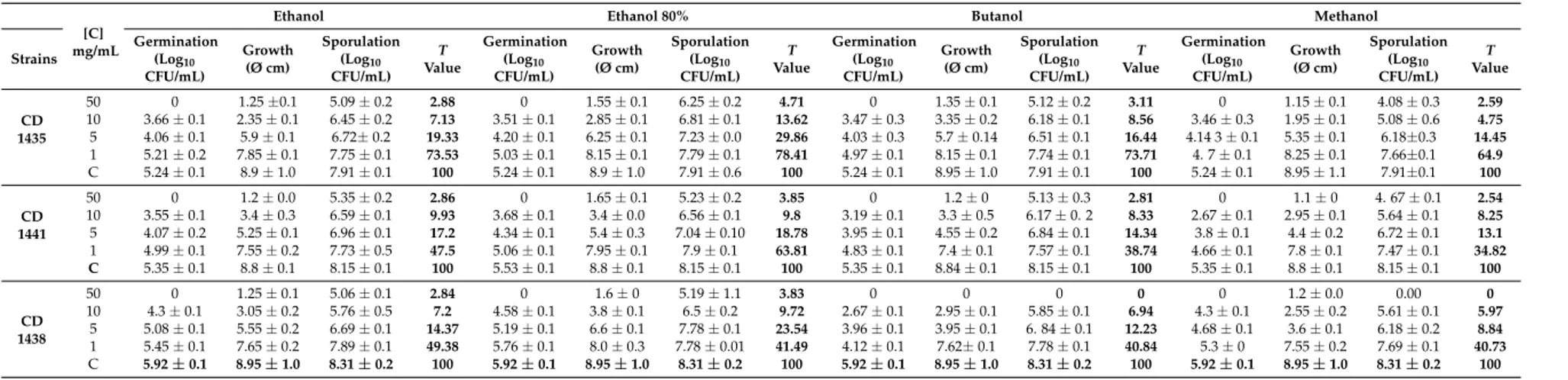
Toxicity assays and the effect on fungal germination of extracts against M. canis and A. fumigatus are reported in Tables5and6, respectively. The germination and sporulation were expressed as mean values (±standard deviation) of Log10 of colony forming units (CFU)/mL and vegetative growth as mean value (±standard deviation) of colony diameters (Ø) of three independent experiments. All D. viscosa extracts were able to completely inhibit the germination of M. canis at concentration higher than 1 mg/mL. The germination of A. fumigatus was completely inhibited at concentrations higher than 10 mg/mL. D. viscosa extracts affect both M. canis vegetative growth and sporulation, being non-toxic for M. canis CD 1279 and M. canis CD 1447 only when ethanolic and 80% ethanolic of D. viscosa extracts were used at 1 mg/mL (Table5). All D. viscosa extracts are toxic to A. fumigatus, except for the strains CD 1435 and CD 1441. In particular, all D. viscosa extracts at a concentration of 1 mg/mL are non-toxic for CD 1435, with the exception of 80% ethanolic extract which is not toxic for the A. fumigatus CD 1441 at this concentration.
Table 5. Effects ofD. viscosa extracts on conidia germination, vegetative growth and sporulation of M. canis. Degree of toxicity (T value) was also reported.
[C] mg/mL
Ethanol Ethanol 80% Butanol Methanol
Strains Germination(Log10
CFU/mL) Growth (Ø cm) Sporulation (Log10 CFU/mL) T Value Germination (Log10 CFU/mL) Growth (Ø cm) Sporulation (Log10 CFU/mL) T Value Germination (Log10 CFU/mL) Growth (Ø cm) Sporulation (Log10 CFU/mL) T Value Germination (Log10 CFU/mL) Growth (Ø cm) Sporulation (Log10 CFU/mL) T Value CD 1279 50 0 0 0 0 0 0 0 0 0 0 0 0 0 0 0 0 10 0 0 0 0 0 0 0 0 0 0 0 0 0 0 0 0 5 0 2.4 ± 0.3 3.54 ± 0.1 14.71 0 2.55 ± 0.1 4.06 ± 0.1 23.08 0 2.5 ± 0.1 3.47 ± 0.1 14.54 0 1.8 ± 0.1 2.97 ± 0.1 9.31 1 3.72 ± 0.1 3.65 ± 0.4 4.79 ± 0.1 77.78 3.87 ± 0.1 3.85 ± 0.1 4.77 ± 0.1 76.91 3.56 ± 0.2 3.5 ± 0.1 4.58 ± 0.1 53.55 3.49 ± 0.1 2.55 ± 0.1 4.43 ± 0.1 38.45 Control 4.30 ± 0.1 5.45 ± 0.1 4.91 ± 0.1 100 4.30 ± 0.2 5.45 ± 0.1 4.91 ± 0.1 100 4.30 ± 0.2 5.45 ± 0.1 4.91 ± 0.1 100 4.3 ± 0.2 5.45 ± 0.1 4.91 ± 0.1 100 CD 1243 50 0 0 0 0 0 0 0 0 0 0 0 0 0 0 0 0 10 0 0 0 0 0 0 0 0 0 0 0 0 0 0 0 0 5 0 2.1 ± 0.14 3.13 ± 0.1 10.62 0 2.25 ± 0.1 3.41 ± 0.04 13.85 0 1.85 ± 0.1 3.06 ± 0.1 9.52 0 1.85 ± 0.1 2.86 ± 0.1 8.61 1 3.73 ± 0.1 2.55 ± 0.1 4.22 ± 0.2 48.97 3.81 ± 0.1 2.85 ± 0.1 4.22 ± 0.02 45.41 3.24 ± 0.1 2.85 ± 0.1 3.79 ± 0.1 25.71 3.24 ± 0.1 2.35 ± 0.1 3.43 ± 0.1 15.11 C 4.18 ± 0.1 5.55 ± 0.1 4.51±0.1 100 4.18 ± 0.1 5.55 ± 0.1 4.51±0.1 100 4.18 ± 0.1 5.55 ± 0.1 4.51 ± 0.1 100 4.18 ± 0.1 5.55 ± 0.1 4.51 ± 0.1 100 CD 1447 50 0 0 0 0 0 0 0 0 0 0 0 0 0 0 0 0 10 0 0 0 0 0 0 0 0 0 0 0 0 0 0 0 0 5 0 2.5 ± 0.1 3.36 ± 0.2 16.58 0 2.85 ± 0.1 3.55 ± 0.2 21.12 0 2.45 ± 0.1 3.81 ± 0.1 14.77 0 1.65 ± 0.1 2.81 ± 0.1 9.16 1 3.85 ± 0.1 3.45 ± 0.1 4.43 ± 0.1 66.31 3.88 ± 0.3 3.8 ± 0.1 4.26 ± 0.1 17.19 2.52 ± 0.1 3 ± 0.1 4.29 ± 0.1 50.23 2.29 ± 0. 3 2.65 ± 0.1 4.26 ± 0.1 46.31 C 4.46 ± 0.1 4.15 ± 1 4.61 ± 0.1 100 4.46 ± 0.1 4.15 ± 0.1 4.61 ± 0.2 100 4.46 ± 0.1 4.15 ± 0.1 4.61 ± 0.1 100 4.46 ± 0.1 4.15 ± 0.1 4.61 ± 0.1 100
* T value = very toxic (0T30); toxic (31T45); moderately toxic (46T60); non-toxic (T > 60); [C]: Concentration (mg/mL); C: ControlConcentration (mg/mL); C: Control.
Table 6. Effects ofD. viscosa extracts on conidia germination, vegetative growth and sporulation of A.fumigatus. Degree of toxicity (T value) was also reported.
[C] mg/mL
Ethanol Ethanol 80% Butanol Methanol
Strains Germination(Log10
CFU/mL) Growth (Ø cm) Sporulation (Log10 CFU/mL) T Value Germination (Log10 CFU/mL) Growth (Ø cm) Sporulation (Log10 CFU/mL) T Value Germination (Log10 CFU/mL) Growth (Ø cm) Sporulation (Log10 CFU/mL) T Value Germination (Log10 CFU/mL) Growth (Ø cm) Sporulation (Log10 CFU/mL) T Value CD 1435 50 0 1.25 ±0.1 5.09 ± 0.2 2.88 0 1.55 ± 0.1 6.25 ± 0.2 4.71 0 1.35 ± 0.1 5.12 ± 0.2 3.11 0 1.15 ± 0.1 4.08 ± 0.3 2.59 10 3.66 ± 0.1 2.35 ± 0.1 6.45 ± 0.2 7.13 3.51 ± 0.1 2.85 ± 0.1 6.81 ± 0.1 13.62 3.47 ± 0.3 3.35 ± 0.2 6.18 ± 0.1 8.56 3.46 ± 0.3 1.95 ± 0.1 5.08 ± 0.6 4.75 5 4.06 ± 0.1 5.9 ± 0.1 6.72± 0.2 19.33 4.20 ± 0.1 6.25 ± 0.1 7.23 ± 0.0 29.86 4.03 ± 0.3 5.7 ± 0.14 6.51 ± 0.1 16.44 4.14 3 ± 0.1 5.35 ± 0.1 6.18±0.3 14.45 1 5.21 ± 0.2 7.85 ± 0.1 7.75 ± 0.1 73.53 5.03 ± 0.1 8.15 ± 0.1 7.79 ± 0.1 78.41 4.97 ± 0.1 8.15 ± 0.1 7.74 ± 0.1 73.71 4. 7 ± 0.1 8.25 ± 0.1 7.66±0.1 64.9 C 5.24 ± 0.1 8.9 ± 1.0 7.91 ± 0.1 100 5.24 ± 0.1 8.9 ± 1.0 7.91 ± 0.6 100 5.24 ± 0.1 8.95 ± 1.0 7.91 ± 0.1 100 5.24 ± 0.1 8.95 ± 1.1 7.91±0.1 100 CD 1441 50 0 1.2 ± 0.0 5.35 ± 0.2 2.86 0 1.65 ± 0.1 5.23 ± 0.2 3.85 0 1.2 ± 0 5.13 ± 0.3 2.81 0 1.1 ± 0 4. 67 ± 0.1 2.54 10 3.55 ± 0.1 3.4 ± 0.3 6.59 ± 0.1 9.93 3.68 ± 0.1 3.4 ± 0.0 6.56 ± 0.1 9.8 3.19 ± 0.1 3.3 ± 0.5 6.17 ± 0. 2 8.33 2.67 ± 0.1 2.95 ± 0.1 5.64 ± 0.1 8.25 5 4.07 ± 0.2 5.25 ± 0.1 6.96 ± 0.1 17.2 4.34 ± 0.1 5.4 ± 0.3 7.04 ± 0.10 18.78 3.95 ± 0.1 4.55 ± 0.2 6.84 ± 0.1 14.34 3.8 ± 0.1 4.4 ± 0.2 6.72 ± 0.1 13.1 1 4.99 ± 0.1 7.55 ± 0.2 7.73 ± 0.5 47.5 5.06 ± 0.1 7.95 ± 0.1 7.9 ± 0.1 63.81 4.83 ± 0.1 7.4 ± 0.1 7.57 ± 0.1 38.74 4.66 ± 0.1 7.8 ± 0.1 7.47 ± 0.1 34.82 C 5.35 ± 0.1 8.8 ± 0.1 8.15 ± 0.1 100 5.53 ± 0.1 8.8 ± 0.1 8.15 ± 0.1 100 5.35 ± 0.1 8.84 ± 0.1 8.15 ± 0.1 100 5.35 ± 0.1 8.8 ± 0.1 8.15 ± 0.1 100 CD 1438 50 0 1.25 ± 0.1 5.06 ± 0.1 2.84 0 1.6 ± 0 5.19 ± 1.1 3.83 0 0 0 0 0 1.2 ± 0.0 0.00 0 10 4.3 ± 0.1 3.05 ± 0.2 5.76 ± 0.5 7.2 4.58 ± 0.1 3.8 ± 0.1 6.5 ± 0.2 9.72 2.67 ± 0.1 2.95 ± 0.1 5.85 ± 0.1 6.94 4.3 ± 0.1 2.55 ± 0.2 5.61 ± 0.1 5.97 5 5.08 ± 0.1 5.55 ± 0.2 6.69 ± 0.1 14.37 5.19 ± 0.1 6.6 ± 0.1 7.78 ± 0.1 23.54 3.96 ± 0.1 3.95 ± 0.1 6. 84 ± 0.1 12.23 4.68 ± 0.1 3.6 ± 0.1 6.18 ± 0.2 8.84 1 5.45 ± 0.1 7.65 ± 0.2 7.89 ± 0.1 49.38 5.76 ± 0.1 8.0 ± 0.3 7.78 ± 0.01 41.49 4.12 ± 0.1 7.62± 0.1 7.78 ± 0.1 40.84 5.3 ± 0 7.55 ± 0.2 7.69 ± 0.1 40.73 C 5.92 ± 0.1 8.95 ± 1.0 8.31 ± 0.2 100 5.92 ± 0.1 8.95 ± 1.0 8.31 ± 0.2 100 5.92 ± 0.1 8.95 ± 1.0 8.31 ± 0.2 100 5.92 ± 0.1 8.95 ± 1.0 8.31 ± 0.2 100
The present study shows that all D. viscosa extracts significantly decrease the vegetative growth, germination, conidia production of both M. canis and A. fumigatus, thus confirming previous results against dermatophytes or other fungal species (i.e., Cladosporium cucumerinum, Botrytis cinerea, Pseudoperonospora cubensis, Phytophthora infestans, Erysiphe graminis and Puccinia helianthi [5,22]. However, all extracts evinced a concentration-dependent inhibitory activity, which varies accordingly to fungal genus. In fact, the A. fumigatus strains seems to be less susceptible than M. canis as previously reported using acetone extracts of Arctotis arctotoides [22]. Additionally the highest antifungal activity was observed with methanol extracts in both fungal species, thus suggesting the efficacy of both TPC and CQA content as antifungal drugs [21,27]. The mechanism of action of phenolic compounds against fungi was previously explained by several studies and might be due to the membrane lipid perturbation. Sung and Lee (2010) [28] demonstrated that phenolic acids might cause disruption of ion transport, whereas Teodoro et al. (2015) [20] indicated that the hydroxyl group and carboxylic acid groups of pheonlic compounds plays an important role in destabilizing the fungal cytoplasmic membrane. Even if the low toxicity values of D. viscosa methanolic extract in one strain of A. fumigatus need to be confirmed, the herein obtained results, suggested that concentrations higher than 1 mg/mL should be employed in controlling A. fumigatus strains. The antifungal activity against Malassezia yeasts, M. canis and A. fumigatus is of interest since the control of these infections is the subject of debate in the scientific community. In particular, Malassezia yeast infections in animals, mainly dogs, may be unresponsive to antifungal therapy and the animals usually have recurrences thus requiring multiple drug regimens [24]. The treatment of M. canis infections in animals is mandatory because of the zoophilic nature of this fungus, but it is not always possible in animals used for food production [29].
Finally, the high azoles resistance phenomena registered in Aspergillus spp. strains also suggests the usefulness of studies on new antifungal drugs [30]. All these findings promote the employment of drugs of plant origin.
3. Materials and Methods 3.1. Chemical and Reagents
Vanillin (C3H8O3), catechin (C15H14O6), Folin-Ciocalteu reagent, sodium carbonate (Na2CO3), gallic acid (C7H6O5), aluminum chloride (AlCl3), potassium acetate (C2H3KO2), rutin (C27H30O16), quercetin (C15H10O7), sodium molybdate dihydrate (Na2MoO4), dipotassium hydrogen phosphate (K2HPO4), potassium dihydrogen phosphate (KH2PO4), chlorogenic acid (C16H18O9) were purchased from Sigma-Aldrich®(Steinheim, Germany). Solvents of analytical and HPLC grade were purchased from Carlo Erba Reactif-CDS (Val de Reuil, France).
3.2. Plant Material
The leaves of the plants were collected in June 2015 from uncultivated land in Sidi Thabet, located in the North East of Tunisia (latitude 36 55045” N, longitude 10 06002.10” E, altitude 30 m).
3.3. Preparation of Extracts
Dried and ground leaves (10 g) were macerated in four different solvents (ethanol (80% and 100%), methanol and butanol) (10:100 w/v) for 48 h with shaking at room temperature. The extracts were filtered with Whatman No. 1 filter paper and the filtrate evaporated to dryness using a rotary evaporator. In order to test the antimicrobial activities, the samples were solubilized in dimethyl sulfoxide (DMSO) to obtain concentrations of 1, 5, 10 and 50 mg/mL.
3.4. Phytochemical Screening
3.4.1. Condensed Tannins Content (CTC)
The CTC was determined as previously described [31]. In particular, 0.5 mL of extract was condensed using 3 mL of vanillin at 4% in methanol and 1.5 mL of concentrated hydrochloric acid (HCl). The mixture was kept in the dark for 15 min at 20 C and the CTC were measured using a Jenway 6300 spectrophotometer (Cole-Parmer, Staffordshire, UK) at absorbance of 500 nm. The CTC was calculated from calibration curve using catechin (CAE) as a standard and results were expressed as milligrams of catechin equivalent per gramm (g) of dry extract (mg CAE/g).
3.4.2. Total Phenol Content (TPC)
The TPC was determined using the Folin-Ciocalteu method [32]. Briefly, 0.5 mL of each dissolved extract was mixed with 2.5 mL of Folin-Ciocalteu reagent in each test tube. After 4 min, 2 mL of saturated sodium carbonate (Na2CO3) solution (7.5%) was added to the mixture. The reaction mixtures were incubated for 2 h. Methanol was used as the blank. All assays were conducted in triplicate and the results were averaged. The TPC was calculated from a calibration curve using gallic acid (GAE) as the standard and the results were expressed as milligrams of gallic acid equivalent per gram of extract (mg GAE/g).
3.4.3. Total Flavonoid Content (TFC)
The TFC was quantified using the aluminum chloride colorimetric assay with slight modifications [33]. In brief, 0.5 mL of each solution extract was mixed with 1.5 mL methanol, 0.1 mL of 10% aluminum chloride, 0.1 mL of 1 mol/L potassium acetate solution and 2.8 mL distilled water. The mixture was allowed to stand for 15 min, and absorbance was measured at 415 nm. All assays were conducted in triplicate and the results were averaged. The TFC was calculated from a calibration curve using quercetin (QE) as the standard, and the result was expressed as mg of quercetin equivalent per gram dry extract (mg QE/g).
3.4.4. Caffeoylquinic Acid (CQC) Content
The CQC content of extracts was quantified using the molybdate colorimetric method [34]. Sodium molybdate (16.5 g), dipotassium hydrogen (8.0 g) phosphate, and potassium dihydrogen phosphate (7.9 g) were dissolved in 1 liter of deionized water to prepare the molybdate reagent. For each I. viscosa extract solution 0.3 mL was mixed with 2.7 mL of molybdate reagent. The mixture was incubated at room temperature for 10 min. Absorbance was measured at 370 nm. All assays were conducted in triplicate and the results were averaged. The CQC was calculated from a calibration curve using chlorogenic acid (ChlA) as the standard and the result was expressed as mg of ChlA equivalent per g dry extract (mg ChlA/g).
3.5. Characterization of PhenolicCcompounds by HPLC-PDA-ESI-MS
The phenolic compounds present in methonolic extract were tentatively identified using the chromatographic separation method as previously reported [4].
Chromatographic separation was performed on an Alliance e2695 HPLC system (Waters, Bedford, MA, USA) equipped with a RP-xTerra MS column (150⇥4.6 mm i.d., 3.5 µm particle size), photodiode array detector (PDA) and interfaced with a triple quadruple mass spectrometer (MSD 3100, Waters) fitted with an ESI ion source. The sample (20 µL) was eluted through the column with a gradient mobile phase consisting of A (0.1% formic acid) and B (acetonitrile acidified with formic acid 0.1%) with a flow rate of 0.5 mL/min. The following multistep linear solvent gradient was used: 0–40 min: 14–26% B; 40–60 min: 15% B; 60–75: 0% B; 75–80 min: 14% B. The HPLC-PDA-ESI-MS chromatogram spectral
data were stored and processed with Masslynx 4.1 data system. Each peak in the chromatogram was accomplished in a single chromatographic run in order to be identified [35].
3.6. Antibacterial and Antifungal Activities 3.6.1. Bacterial Strains
Five reference bacterial strains, including Gram-positive (i.e., Staphylococcus aureus ATCC 6538, Enterococcus feacium ATCC 19434, Streptococcus agalactiae ATCC 12386), and Gram-negative (i.e., Escherichia coli ATCC 8739, 29212 and Salmonella typhimurium ATCC 14028), were used to assess the antibacterial properties of the extracts.
3.6.2. Fungal Strains Candida spp.
Three references strains of Candida (i.e., Candida krusei ATCC 6258, Candida parapsilosis ATCC 22019, Candida albicans ATCC 10231), and four Candida albicans strains (i.e., CD 1358, CD 1378, CD 1407, CD 1408) isolated from cloaca of laying hens, were used to evaluate the anti-Candida activity of I. viscosa extracts. All strains were obtained from the fungal collection of the Department of Veterinary Medicine at the University of Bari (Aldo Moro, Italy).
Malassezia spp. Strains
A total of six Malassezia spp. strains (three Malassezia pachydermatis and three Malassezia furfur) were tested. Two reference strains (i.e., M. pachydermatis CBS1879 and M. fufur CBS1978), two strains isolated from dogs with dermatitis and/or otitis (i.e., M. pachydermatis CD 112 and CD 90), two M. furfur strains from human skin (i.e., M. furfur CD 1029), and one from a human blood stream infection (i.e., M. furfur CD 1006) were tested.
Aspergillus fumigatus strains
Three A. fumigatus strains (CD 1435, CD 1438 and CD 1441) were tested. All strains were isolated from the respiratory tract of critically ill human patients. All strains were obtained from the fungal collection of the Department of Veterinary Medicine at the University of Bari.
Microsporum canis strains
Three M. canis strains (CD 1243, CD 1447, and CD 1279), isolated with skin lesions from human, cat, and dog were tested, respectively. All strains were stored in the fungal collection of the Department of Veterinary Medicine at the University of Bari.
3.7. Determination of Antibacterial, Anti-Candida and Antifungal Activity of I. viscosa Extract
The antibacterial, anti-Candida and anti-Malassezia activities were evaluated by the disk diffusion method [36], whereas the antifungal activity of I. viscosa extracts against M. canis and A. fumigatus was assessed by studying the toxicity effect of the extract on vegetative growth and sporulation as well their effect on fungal germination.
3.7.1. Toxicity Assay
The antifungal activity of D. viscosa extracts against M. canis and A. fumigatus was assessed as previously reported [37]. In particular, the antifungal properties of extracts were assessed by applying the following mathematical model in order to evaluate the degree of toxicity:
where: T is the degree of toxicity useful for the classification of the product; VG is the percentage of vegetative growth with respect to the control; SR is the percentage of sporulation with respect to the control. The product was classified, based on the T value, as: very toxic (0T30); toxic (31T45) moderately toxic (46T60); non-toxic (i.e., compatible) (T > 60) [36].
The A. fumigatus and M canis strains were sub-cultured onto PDA and incubated at 25 C for 10 days before testing. Vegetative growth (VG) was measured by placing a mycelial plug (i.e., 5 mm in diameter) onto the center of a 90 mm Petri dish containing potato dextrose agar (PDA), with and without extract or DMSO (solvent control), and measuring the diameter of the colonies after incubation at 25 C for 10 days. Sporulation was evaluated by collecting the spores from surface of fungi grown on the PDA with and without the extracts after 10 incubation days at 25 C. Spores and mycelia were collected by scraping the surface of the plate with 4 mL of 20% tween 80 solution. The solution was filtered through sterile gauze to remove mycelia, and then centrifuged (3000 g⇥5 min), washed twice in 1 mL of phosphate-buffered saline solution (PBS), and re-suspended in 1 mL of PBS. Numbers of spores were determined by quantitative plate counts of (CFU)/mL on PDA after incubation at 25 C for 4 days [37].
3.7.2. Effect of D. viscosa Extract on Fungal Germination
The effect of D. viscosa extracts on M. canis and A. fumigatus germination was also measured, culturing fungi in SDA medium after 14 days at 25 C and collecting spores and mycelia as reported above. The solution obtained was diluted in PBS to obtain an inoculum concentration of 107conidia/mL which was evaluated by quantitative plate counts of CFU/mL in PDA. Finally, a total of 100 µL of the fungal spore suspensions were cultured in PDA with and without different extract concentrations. The number of germinated spores were determined by counts of CFU/mL on PDA [38]. All experiments were performed in duplicate and repeated three times on different days. 3.8. Statistical Analysis
The results of toxicity assay on vegetative growth, sporulation and fungal germination were expressed as mean values (±standard deviation (SD)) of the three independent experiments. Vegetative growth (VG) was expressed as mean value of colony diameters after incubation and the sporulation and germination as mean values of Log10CFU/mL. Results were statistically analyzed using one way analysis of variance (ANOVA). Significant differences were set at p < 0.05.
4. Conclusions
The employment of these extracts might be useful to treat infections that are life threatening (i.e., Malassezia) or infections that require a mandatory treatment (i.e., M. canis or A. fumigatus), thus providing another commercial validation of this weed and working towards reducing the hazards associated with excessive use of chemical products.
Acknowledgments: We kindly thank Bronwyn Campbell (University of Bari) for revising the English text.
This work was supported by the Tunisian Ministry of High Education and Scientific Research.
Author Contributions: Rhimi Wafa and Davide Immediato conceived and performed the experiments;
Abdennacer Boulila contributed to plant extraction and identification of phenolic compounds; Mouldi Saidi analysed and rechecked the manuscript; Claudia Cafarchia, Rhimi Wafa, Issam Ben Salem designed the experiments analyzed the data, and wrote this paper.
Conflicts of Interest: The authors declare no conflict of interest. References
1. Rossolini, G.M.; Arena, F.; Pecile, P.; Pollini, S. Update on the antibiotic resistance crisis. Curr. Opin. Pharmacol.
2014,18, 56–60. [CrossRef] [PubMed]
2. Çelik, T.A.; Aslantürk, Ö.S. Evaluation of cytotoxicity and genotoxicity of Inula viscosa leaf extracts with Allium test. J. Biomed. Biotechnol. 2010, 2010, 189252.
3. Parolin, P.; Scotta, M.I.; Bresch, I. Biology of Dittrichia viscosa, a Mediterranean ruderal plant. fyton. 2013, 83, 251–262.
4. Mahmoudi, H.; Hosni, K.; Zaouali, W.; Amri, I.; Zargouni, H.; Hamida, N.B.; Kaddour, R.; Hamrouni, L.; Nasri, M.B.; Ouerghi, Z. Comprehensive Phytochemical Analysis, Antioxidant and Antifungal Activities of Inula viscosa Aiton Leaves. J. Food Saf. 2015, 36, 77–88. [CrossRef]
5. Omezzine, F.; Daami-Remadi, M.; Rinez, A.; Ladhari, A.; Haouala, R. In vitro assessment of Inula spp. organic extracts for their antifungal activity against some pathogenic and antagonistic fungi. Afr. J. Microbiol. Res.
2011,5, 3527–3531.
6. Assaf, A.M.; Amro, B.I.; Mashallah, S.; Haddadin, R.N. Antimicrobial and anti-inflammatory potential therapy for opportunistic microorganisms. J. Infect. Dev. Ctries 2016, 10, 494–505. [CrossRef] [PubMed] 7. Duek, L.; Kaufman, G.; Ulman, Y.; Berdicevsky, I. The pathogenesis of dermatophyte infections in human
skin sections. J. Infect. 2004, 48, 175–180. [CrossRef] [PubMed]
8. Bender, J.B.; Shulman, S.A. Reports of zoonotic disease outbreaks associated with animal exhibits and availability of recommendations for preventing zoonotic disease transmission from animals to people in such settings. J. Am. Vet. Med. Assoc. 2004, 224, 1105–1109. [CrossRef] [PubMed]
9. Raad, I.I.; Graybill, J.R.; Bustamante, A.B.; Cornely, O.A.; Gaona-Flores, V.; Afif, C.; Graham, D.R.; Greenberg, R.N.; Hadley, S.; Langston, A.; et al. Safety of long-term oral posaconazole use in the treatment of refractory invasive fungal infections. Clin. Infect. Dis. 2006, 42, 1726–1734. [CrossRef] [PubMed]
10. Cafarchia, C.; Figueredo, L.A.; Otranto, D. Fungal diseases of horses. Vet. Microbiol. 2013, 167, 215–234. [CrossRef] [PubMed]
11. Cafarchia, C.; Figueredo, L.A.; Iatta, R.; Montagna, M.T.; Otranto, D. In vitro antifungal susceptibility of Malassezia pachydermatis from dogs with and without skin lesions. Vet. Microbiol. 2012, 155, 395–398. [CrossRef] [PubMed]
12. Cafarchia, C.; Figueredo, L.A.; Iatta, R.; Colao, V.; Montagna, M.T.; Otranto, D. In vitro evaluation of Malassezia pachydermatis susceptibility to azole compounds using E-test and CLSI microdilution methods. Med. Mycol. 2012, 50, 795–801. [PubMed]
13. Seyedmousavi, S.; Guillot, J.; Arn, P.; De Hoog, G.S.; Mouton, J.W.; Melchers, W.J.G.; Verweij, P.E. Aspergillus and aspergilloses in wild and domestic animals: A global health concern with parallels to human disease. Med. Mycol. 2015, 53, 765–797. [CrossRef] [PubMed]
14. Sefi, M.; Fetoui, H.; Makni, M.; Zeghal, N. Mitigating effects of antioxidant properties of Artemisia campestris leaf extract on hyperlipidemia, advanced glycation end products and oxidative stress in alloxan-induced diabetic rats. Food Chem. Toxicol. 2010, 48, 1986–1993. [CrossRef] [PubMed]
15. Trimech, I.; Weiss, E.K.; Chedea, V.S.; Marin, D.; Detsi, A.; Ioannou, E.; Roussis, V.; Kefalas, P. Evaluation of anti-oxidant and acetylcholinesterase activity and identification of polyphenolics of the invasive weed Dittrichia viscosa. Phytochem. Anal. 2014, 25, 421–428. [CrossRef] [PubMed]
16. Negi, P.S.; Jayaprakasha, G. Antioxidant and Antibacterial Activities of Punica granatum Peel Extracts. J. Food Sci. 2003, 68, 1473–1477. [CrossRef]
17. Geng, H.-M.; Zhang, D.-Q.; Zha, J.-P.; Qi, J.-L. Simultaneous HPLC Determination of Five Flavonoids in Flos Inulae. Chromatographia 2007, 66, 271–275. [CrossRef]
18. Naeem, I.; Saddiqe, Z.; Patel, A.; Hellio, C. Analysis of flavonoid and antimicrobial activity of extracts of Hypericum perforatum. Asian J. Chem. 2010, 22, 3596–3600.
19. Danino, O.; Gottlieb, H.E.; Grossman, S.; Bergman, M. Antioxidant activity of 1,3-dicaffeoylquinic acid isolated from Inula viscosa. Food Res. Int. 2009, 42, 1273–1280. [CrossRef]
20. Teodoro, G.R.; Ellepola, K.; Seneviratne, C.J.; Koga-Ito, C.Y. Potential use of phenolic acids as anti-Candida agents: A review. Front. Microbiol. 2015, 6, 1–11. [CrossRef] [PubMed]
21. Cushnie, T.P.T.; Lamb, A.J. Antimicrobial activity of flavonoids. Int. J. Antimicrob. Agents 2005, 26, 343–356. [CrossRef] [PubMed]
22. Otang, W.M.; Grierson, D.S.; Ndip, R.N. The effect of the acetone extract of Arctotis arctotoides (Asteraceae) on the growth and ultrastructure of some opportunistic fungi associated with HIV/AIDS. Int. J. Mol. Sci.
2011,12, 9226–9235. [CrossRef] [PubMed]
23. Silhavy, T.J.; Kahne, D.; Walker, S. The bacterial cell envelope. Cold Spring Harb. Perspect. Biol. 2010, 2, 1–16. [CrossRef] [PubMed]
24. Velegraki, A.; Cafarchia, C.; Gaitanis, G.; Iatta, R.; Boekhout, T. Malassezia. Infections in Humans and Animals: Pathophysiology, Detection, and Treatment. PLoS Pathog. 2015, 11, e1004523. [CrossRef] [PubMed] 25. Berenji, F.; Rakhshandeh, H.; Ebrahimipour, H. In vitro study of the effects of henna extracts (Lawsonia inermis)
on Malassezia species. Jundishapur J. Microbiol. 2010, 3, 125–128.
26. Filip, R.; Davicino, R.; Anesini, C. Antifungal activity of the aqueous extract of Ilex paraguariensis against malassezia furfur. Phyther. Res. 2010, 24, 715–719.
27. Von Nussbaum, F.; Brands, M.; Hinzen, B.; Weigand, S.; Häbich, D. Antibacterial natural products in medicinal chemistry–Exodus or revival? Angew. Chem. Int. Ed. 2006, 45, 5072–5129. [CrossRef] [PubMed] 28. Sung, W.S.; Lee, D.G. Antifungal action of chlorogenic acid against pathogenic fungi, mediated by membrane
disruption. Pure Appl. Chem. 2010, 82, 219–226. [CrossRef]
29. Magwedere, K.; Hemberger, M.Y.; Hoffman, L.C.; Dziva, F. Zoonoses: A potential obstacle to the growing wildlife industry of Namibia. Infect. Ecol. Epidemiol. 2012, 2, 1–16. [CrossRef] [PubMed]
30. Tanwar, J.; Das, S.; Fatima, Z.; Hameed, S. Multidrug Resistance: An Emerging Crisis. Interdiscip. Perspect. Infect. Dis. 2014, 2014, 541340. [CrossRef] [PubMed]
31. Chavan, U.; Shahidi, F.; Naczk, M. Extraction of condensed tannins from beach pea (Lathyrus maritimus L.) as affected by different solvents. Food Chem. 2001, 75, 509–512. [CrossRef]
32. Meda, A.; Lamien, C.E.; Romito, M.; Millogo, J.; Nacoulma, O.G. Determination of the total phenolic, flavonoid and proline contents in Burkina Fasan honey, as well as their radical scavenging activity. Food Chem.
2005,91, 571–577. [CrossRef]
33. Zheng, C.J.; Yoo, J.-S.; Lee, T.-G.; Cho, H.-Y.; Kim, Y.-H.; Kim, W.-G. Fatty acid synthesis is a target for antibacterial activity of unsaturated fatty acids. FEBS Lett. 2005, 579, 5157–5162. [CrossRef] [PubMed] 34. Chan, E.W.C.; Lim, Y.Y.; Ling, S.K.; Tan, S.P.; Lim, K.K.; Khoo, M.G.H. Caffeoylquinic acids from leaves of
Etlingera species (Zingiberaceae). LWT–Food Sci. Technol. 2009, 42, 1026–1030. [CrossRef]
35. Nyau, V.; Prakash, S.; Rodrigues, J.; Farrant, J. HPLC-PDA-ESI-MS Identification of Polyphenolic Phytochemicals in Different Market Classes of Common Beans (Phaseolus vulgaris L.). Int. J. Biochem. Res. Rev. 2015, 8, 1–11. [CrossRef]
36. Dhouioui, M.; Boulila, A.; Jemli, M.; Schiets, F.; Casabianca, H.; Zina, M.S. Fatty Acids Composition and Antibacterial Activity of Aristolochia longa L. and Bryonia dioïca Jacq. Growing Wild in Tunisia. J. Oleo Sci.
2016,65, 655–661. [CrossRef] [PubMed]
37. Immediato, D.; Aguiar, L.; Iatta, R.; Camarda, A.; Lira, R.; De Luna, N.; Giangaspero, A.; Brandão-filho, S.P.; Otranto, D.; Cafarchia, C. Veterinary Parasitology Essential oils and Beauveria. bassiana against Dermanyssus gallinae (Acari: Dermanyssidae): Towards new natural acaricides. Vet. Parasitol. 2016, 229, 159–165. [CrossRef] [PubMed]
38. Tamai, M.A.; Alves, S.B.; Lopes, R.B.; Faion, M.; Padulla, L.F.L. Toxicidade De Produtos Fitossanitários para Beauveria Bassiana (Bals.) Vuill. Arq. Inst. Biol. (Sao Paulo) 2002, 69, 65–69.
Sample Availability: Samples of the phenolic and flavonoid compounds are available from the authors.
© 2017 by the authors. Licensee MDPI, Basel, Switzerland. This article is an open access article distributed under the terms and conditions of the Creative Commons Attribution (CC BY) license (http://creativecommons.org/licenses/by/4.0/).