SCUOLA NORMALE SUPERIORE
Pisa
Classe di Scienze Matematiche, Fisiche e Naturali
Corso di Perfezionamento in Neurobiologia
Triennio 2009-2011
Tesi di Perfezionamento
EXPERIENCE-DEPENDENT EXPRESSION OF miR132
REGULATES OCULAR DOMINANCE PLASTICITY
Candidata:
Paola Tognini
Relatore:
Prof. Tommaso Pizzorusso
Index
Introduction
11. Experience-dependent plasticity 1
2. The rodent visual system 2
3. Ocular dominance plasticity and critical period in the visual cortex 4 4. The mouse as animal model for visual system studies 7
5. Dark rearing 8
6. Exploring the molecular mechanisms of OD plasticity 9 6.1 Glutamate receptors: the NR2B/NR2A switch 9
6.2 Inhibitory circuits maturation 11
6.3 Neuroactive released proteins 13
6.4 Extracellular influences 15
6.5 Environmental influences 18
6.6 Signalling pathways 19
6.7 Gene expression control 21
7. Epigenetic chromatin remodelling and neuronal plasticity 23
7.1 Epigenetic tagging of histones 25
7.2 DNA methylation 27
7.3 Chromatin remodelling influences synaptic plasticity 28 7.4 Histones PTMs and visual cortical plasticity 33
8.2 MiRNAs in the central nervous system 36 8.3 MicroRNAs 212/132 family: molecular transducer of neuronal
function and plasticity 38
Aims of this thesis
47Materials and methods
481. Chromatin immunoprecipitation (ChIP) 48
2. Quantitation of Chromatin immunoprecipitation by Real time PCR 49
3. Western blotting 50
4. Thricostatin A (TSA) treatment 52
5. RNA extraction, reverse transcription and Real time PCR 52
6. In situ hybridization 54
7. MiRNAs mimic treatment 55
8. Spine morphology assessment 56
9. In vitro miRNAs mimic treatment 57
10. Single unit recordings 58
11. VEP recordings 59
12. Statistical analysis 59
Results
601. Visual stimulation regulates histone acetylation, phosphorylation and
methylation on a CRE sequence important for transcription of the miR132/212
cluster 60
2. MiR132 levels are dynamically and bidirectionally regulated by visual
experience 63
3. Counteracting miR132 dowregulation during monocular deprivation inhibits
Discussion
83 1. Visual experience during the critical period induces epigenetic modifications on specific CRE loci on miR212/132 gene cluster 83 2. Visual experience regulates miR132 expression levels 86 3. Mir132 downregulation during MD is necessary for OD plasticity. 89Appendix
“Environmental enrichment promotes plasticity and
visual acuity recovery in adult monocular amblyopic rats.” 96
Abstract 97
Introduction 98
Materials and methods 100
1. Animal treatment and surgical procedures 100
2. In vivo electrophysiology 101
3. Behavioral assessment of visual acuity 102
4. Immunohistochemistry 103
Results 105
Discussion 109
Conclusions 112
Introduction
1. Experience-dependent plasticity
One of the most fascinating properties of the mammalian brain is its plasticity; the capacity of the neural activity generated by an experience to modify neural circuit function and thereby modify subsequent thoughts, feelings, and behaviours. Synaptic plasticity specifically refers to the activity-dependent modification of the strength or efficacy of synaptic transmission at pre-existing synapses, and for over a century has been proposed to play a central role in the capacity of the brain to incorporate transient experiences into persistent memory traces. Synaptic plasticity is also thought to play key roles in the early development of neural circuitry and evidence is accumulating that impairments in synaptic plasticity mechanisms contribute to several prominent neuropsychiatric disorders. Thus, elucidating the detailed molecular mechanisms underlying synaptic plasticity in any number of different brain regions is crucial for understanding the neural basis of many aspects of normal and pathological brain function (Citri and Malenka, 2008).
Experience-dependent regulation of gene expression is a key step in brain plasticity, and long lasting alterations in neuronal connections require complex gene expression programmes and fine regulation of protein synthesis (Greer and Greenberg, 2008).
During the early postnatal development, the brain undertakes intense morphological and functional rearrangement in response to sensory experience alterations, with visual, auditory and somatosensory systems displaying sensitive phases of enhanced plasticity that have been called “sensitive or critical periods” (Berardi, et al., 2000). Critical periods are specific windows of opportunity during
which experience provides information that is essential for normal development and permanently alters performance (Hensch, 2005). Critical periods are known to occur also during early human development, including such behaviours as language acquisition (Doupe and Kuhl, 1999) and visual function development (Levi, 2005).
Although plasticity is enhanced in malleable young brain, adult neural circuits are not completely stable and can undertake some plastic phenomena such as learning and memory. A proper understanding of the molecular mechanism regulating experience-dependent neural plasticity is a crucial point, since dysregulation of neocortical plasticity during development is a leading cause of many brain diseases from mental retardation to amblyopia (lazy eye), from psychiatric disorders to epilepsy. Therefore, investigating the molecular events underling the activation and regulation of critical period’s plasticity is of great interest in the field of Neuroscience. Manipulation of such mechanisms may potentially allow reactivation of neural circuit plasticity for clinical purposes during times when the adult brain is normally less plastic.
2. The rodent visual system
In the mammalian visual system, visual information are processed in the retina and sent to different structures of the central nervous system (CNS) through retinal ganglion cells (RGCs) axons, which represent the output of the retina. RGCs project to the visual centres of the brain that are located in the midbrain and in the thalamus. The pattern of retinal projections varies from species to species. In rodents, the vast majority of RGCs project to the superior colliculus (SC) and the pretectal nuclei, with about 30% of them sending collaterals to the dorsal-lateral geniculate nucleus (dLGN) in the thalamus (Dreher, et al., 1985). RGC axons from
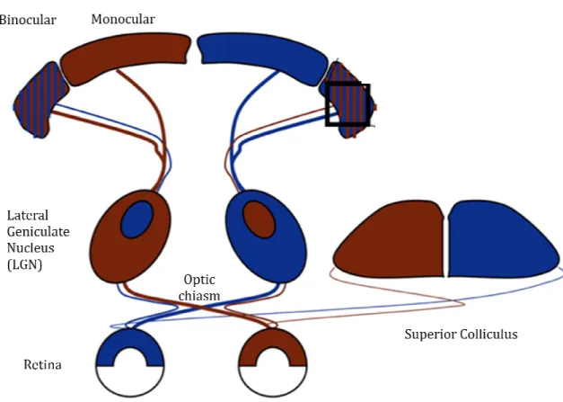
each eye project to both sides of the brain, however the major afferents to the SC and dLGN arise from the contralateral eye and only 5% of optic axons project ipsilaterally. Within the dLGN ganglion cell axons are not intermixed; in cats, ferrets and primates they terminate in a set of separate, alternate eye-specific layers (three layers in cats and six layers in primates) that are strictly monocular (Hickey and Guillery, 1974). In rodents there is not a proper lamination of the dLGN; however, ipsilateral and contralateral retinal fibers are segregated in a patchy fashion originating two eye-specific territories in the dLGN: the ipsilateral portion or inner core and the contralateral portion or outer shell. The primary visual cortex (V1), located occipitally in the brain, consists of six layers of cells between the pial surface and the underlying white matter. The dLGN projects to the visual cortex via thalamo-cortical connections that terminate in the layer IV of V1. The neurons of layer IV form synapses in the layer 2/3, that is the first site within the visual system where visual input from the two eyes converges onto single neurons. In carnivores and primates, afferents from the dLGN segregate by eye within the cortical layer IV into alternating, equal-sized stripes called ocular dominance columns (Hubel and Wiesel, 1963, Shatz and Stryker, 1978). The binocular zone is smaller in rodents than in cat and primates and it is not organized in ocular dominance columns. Finally, It is important to remember that the neurons of the rodent visual cortex are predominantly responsive to the inputs deriving from the contralateral eye, due to the high percentage of RGCs fibers crossing at the optic chiasm (Fig.1).
Figure 1. Schematic representation of the mammalian visual system.
optic chiasm to LGN and superior colliculus. Contrasting colors indicate regions receiving input from each eye. LGN eye-specific
binocular zone is smaller and more lateral in rodents than in cat and primate, whose visual cortex shows more overlap of inputs from competing eyes. (Adapted from Hooks and Chen, 2007
3. Ocular dominance plasticity and critical period in the visual cortex
Visual cortex development is strongly influenced by visual experience during a short period of postnatal develop
2003). The expression ‘‘critical period’’ was introduced for the first time by
cat. They described the physiological shift in res cortex to light stimulation when one eye was de form of plasticity is strongly
1. Schematic representation of the mammalian visual system. Retina feeds forward via the
optic chiasm to LGN and superior colliculus. Contrasting colors indicate regions receiving input from specific regions shown as in rodent. The LGN then projects to visual cortex. The binocular zone is smaller and more lateral in rodents than in cat and primate, whose visual cortex shows more overlap of inputs from competing eyes. (Adapted from Hooks and Chen, 2007
3. Ocular dominance plasticity and critical period in the visual cortex
Visual cortex development is strongly influenced by visual experience during of postnatal development, the so called critical period
The expression ‘‘critical period’’ in the context of mammalian visual system for the first time by Wiesel and Hubel (1963) in their studies in the cat. They described the physiological shift in responsiveness of neurons in the visual cortex to light stimulation when one eye was deprived of vision early in life form of plasticity is strongly robust during a specific developmental age and
Retina feeds forward via the optic chiasm to LGN and superior colliculus. Contrasting colors indicate regions receiving input from regions shown as in rodent. The LGN then projects to visual cortex. The binocular zone is smaller and more lateral in rodents than in cat and primate, whose visual cortex shows more overlap of inputs from competing eyes. (Adapted from Hooks and Chen, 2007).
3. Ocular dominance plasticity and critical period in the visual cortex
Visual cortex development is strongly influenced by visual experience during ment, the so called critical period (Berardi, et al., mammalian visual system Wiesel and Hubel (1963) in their studies in the ponsiveness of neurons in the visual prived of vision early in life. This robust during a specific developmental age and
experiments, Wiesel and Hubel proposed that there was a period of development when changes in the external visual environment can alter pre-existing neuronal connections. Proper sensory experience during this critical period is essential for shaping neuronal circuits and for the maintenance of appropriate synaptic connections. Neuronal activity evocated by sensory experience allows maturation of important properties of visual system, such as visual acuity (the spatial resolution of the visual system) and orientation preference (Fagiolini, et al., 1994, Wang, et al., 2010). Manipulations of visual experience such as dark rearing (DR) and monocular deprivation (MD) cause structural, functional and molecular changes in the neuronal network of visual cortex (Berardi, et al., 2004). MD consists in the closure of one eye resulting in an imbalance of the inputs from the two eyes with the consequent synaptic reorganization of V1 circuitry. This form of plasticity is called ocular dominance (OD) plasticity, and it represents a classic paradigm to study how experience-dependent activity models neuronal connections. MD causes a loss of visual acuity in the deprived eye resulting from a decrease in the visual inputs of the closed eye which causes an irreversible reduction of the ability of that eye to drive neuronal responses in the cortex; therefore neurons in the binocular zone of the contralateral V1, previously dominated by the deprived eye, shift their responsiveness toward the ipsilateral open eye. Behaviorally, animals MD during development lose visual acuity in the deprived eye and any subsequent experiences or visual stimulations cannot completely reverse the effects of early deprivation after the closure of critical period (Fig.2) (Berardi, et al., 2000).
Plasticity in the visual cortex declines with age. Adult visual cortex still responds to experience with plastic changes, as shown by perceptual learning (Schoups, et al., 2001), however the extent of plasticity is reduced. Markedly in
contrast to the profound effects in young animals, prolonged eye closure in adult has little or no effect (Hubel and Wiesel, 1970)
very limited once the critical period is terminated.
Figure 2. Effect of monocular deprivation during the critical period.
response to the deprived eye and a gain of open
single units from the mouse visual cortex. The ocular dominance of cells, rated o of neuronal responsiveness, indicates a typical bias toward the contralateral eye (1
(left). After 3 or more days of monocular deprivation, the distribution shifts toward the open, ipsilateral eye (4–7; right). (Adapt
Anatomically, in higher animals, such as cats and primates, MD causes a retraction of thalamocortical axons
the strengthening of projections from the open eye, which results in the expan OD columns guided by the non
eye (Hubel, et al., 1977, Stryker and Harri
carrying inputs of the closed eye become less branched and shorter. An opposite effect is observed in axons representing the open e
contrast to the profound effects in young animals, prolonged eye closure in adult has (Hubel and Wiesel, 1970), furthermore recovery from amblyopia is very limited once the critical period is terminated.
of monocular deprivation during the critical period.
response to the deprived eye and a gain of open-eye input, as measured by the neuronal discharge of single units from the mouse visual cortex. The ocular dominance of cells, rated o
of neuronal responsiveness, indicates a typical bias toward the contralateral eye (1
(left). After 3 or more days of monocular deprivation, the distribution shifts toward the open, 7; right). (Adapted from Hensch 2005).
Anatomically, in higher animals, such as cats and primates, MD causes a n of thalamocortical axons conveying the inputs from the deprived eye and the strengthening of projections from the open eye, which results in the expan
OD columns guided by the non- deprived eye, at the cost of those serving the closed (Hubel, et al., 1977, Stryker and Harris, 1986). Thalamocortical projections carrying inputs of the closed eye become less branched and shorter. An opposite effect is observed in axons representing the open eye (Tieman, 1984)
contrast to the profound effects in young animals, prolonged eye closure in adult has more recovery from amblyopia is
of monocular deprivation during the critical period. MD produces a loss of
eye input, as measured by the neuronal discharge of single units from the mouse visual cortex. The ocular dominance of cells, rated on a seven-point scale of neuronal responsiveness, indicates a typical bias toward the contralateral eye (1–3) in the rodent (left). After 3 or more days of monocular deprivation, the distribution shifts toward the open,
Anatomically, in higher animals, such as cats and primates, MD causes a conveying the inputs from the deprived eye and the strengthening of projections from the open eye, which results in the expansion of , at the cost of those serving the closed . Thalamocortical projections carrying inputs of the closed eye become less branched and shorter. An opposite
In rodents, two/three days of MD cause an initial decrease in deprived eye responses, due to the weakening of closed eye connections and a reorganization of intracortical horizontal connections in the superficial layers (II/III) of the binocular portion of V1 (Trachtenberg and Stryker, 2001). Prolonged MD (five/seven days) shows an increase in the neuronal responses to the inputs from the ipsilateral open eye (Frenkel and Bear, 2004). Architectural changes in thalamocortical arborization, terminating in layer IV, are evident only much later, more than a month in the mouse (Antonini, et al., 1999).
The anatomical changes observed in rodent visual cortex reflect those of higher mammals, although with some differences, indeed the major effect of deprivation on the contralateral projection is an arrest of growth rather than a prompt retraction of branches, as seen in the cat (Antonini and Stryker, 1996).
Furthermore, prolonged deprivation promoted the growth of the open eye's geniculocortical connections in mouse (Antonini, et al., 1999).
4. The mouse as animal model for visual sytem studies
Studies of cortical visual processing have typically used carnivores or primates, wich are considered to have a more refined visual system, including a much larger cortical region for visual processing, higher acuity, extensive visual behaviors, and orientation, ocular dominance and spatial frequencie columns. Despite the low visual acuity and relatively small region of cortex devoted to visual processing, neurons in mouse V1 show selectivity for stimulus parameters and typical response properties that are near to that found in other species (Niell and Stryker, 2008).
In contrast to the classic notion of a critical period for experience-dependent plasticity, several studies have recently reported that OD shifts in mice can also be induced in adulthood (Hofer, et al., 2006, Tagawa, et al., 2005). Nevertheless, the binocular cortical representation is still more sensitive in juvenile mice, as in adults OD shifts require longer MD durations and are generally smaller. It remains unknown whether substantial structural rearrangements that accompany functional OD shifts in juvenile animals also occur in the mature cortex during MD (Hofer, et al., 2006). Furthermore, OD shift in adult mice depends on the age of the animals and on the length of MD period. OD plasticity after 7 days of MD is present in young adult mice (90–100 days) but significantly weaker already in 109–158 days old mice. In animals older than 208 days, OD plasticity is absent even after 14 days of MD. Therefore, OD plasticity in binocular visual cortex is most pronounced in young animals, reduced but present in adolescence and absent in fully mature animals older than 110 days of age (Lehmann and Lowel, 2008). Mice are thus not basically different in OD plasticity from cats and monkeys which is an absolutely essential prerequisite for their use as valid model for visual system studies.
5. Dark rearing
DR is a form of visual manipulation consisting in the complete absence of sensory input. In animals reared in a completely dark environment from birth visual connections do not consolidate, remaining plastic well after the closure of normal critical period and visual acuity does not reach the adult level. Cortical neurons display immature properties, such as reduced orientation and direction selectivity, larger receptive field sizes and lower visual acuity. DR causes a downregulation in BDNF expression with subsequent delay in intracortical inhibition maturation.
Furthermore, neurons in V1 show change in size and density of dendritic spines (Hooks and Chen, 2007).
6. Exploring the molecular mechanisms of OD plasticity
To date, OD plasticity remains the best studied experimental model for experience-dependent refinement of neuronal circuits because of the ease of manipulating visual experience independently in the two eyes. However, a complete understanding of critical period plasticity requires linking the systems-level change in circuit function with the molecular mechanisms that make circuit changes possible. The cellular and molecular mechanisms that control the developmental plasticity of visual cortical connections and restrict experience-dependent plasticity to short critical periods are still little known, though intensely investigated.
In this paragraph I will overview recent discoveries in this field.
• 6.1 Glutamate receptors: the NR2B/NR2A switch
The first modifications induced by experience in visual cortical circuits are likely to be changes in synaptic efficacy. Plasticity is gated by the activation of N-methyl-D-aspartate (NMDA) receptors, which respond to excitatory synaptic transmission by enabling calcium (Ca2+) influx into target synapse and its neuron. NMDA receptors are both transmitter- and voltage-dependent, and their coupling via Ca2+ influx to plasticity-related intracellular signalling, has led to the notion that they might be a neural implementation of Hebbian synapses.
Involvement of NMDA receptors in developmental visual cortical plasticity has been initially suggested by the observation that block of NMDA receptors inhibits the effects of MD (Bear, et al., 1990). A difficulty with pharmacological block of NMDA
receptors can be that it significantly affects visually driven activity, but the use of different NMDA receptor antagonists (Daw, et al., 1999) or antisense oligonucleotides to reduce expression of the NMDAR1 subunit has overcome this problem, showing that it is possible to block the effects of monocular deprivation without affecting visual responses (Roberts, et al., 1998) and confirming NMDA receptor involvement in visual cortical plasticity (Berardi, et al., 2003). However, the receptor’s capacity to drive plasticity depends on its subunit composition: some receptors are built from “NR2B” subunits, which enable a high Ca2+ permeability and thus enhanced plasticity, and some are composed by “NR2A” subunits, that have a reduced Ca2+ flux, thus resulting in shortening of NMDA current. A crucial determinant of plasticity is itself regulated by the activity level of the circuit. NMDA receptors are developmentally regulated: their subunit composition varies in the visual cortex, from a dominant presence of receptors containing the subunit 2B to a high presence of receptors containing the subunit 2A, with a time course paralleling that of functional visual cortical development and the critical period. DR, which retards critical-period closure and impairs development of functional properties of the visual cortex and of visual acuity, delays the developmental shortening of NMDA-receptor currents and of subunit 2A expression; as animals are exposed to visual experience, the NR2B/NR2A ratio declines (Quinlan, et al., 1999), thus reducing the capacity for further plasticity and suggesting that the 2B-to-2A switch is related to visual cortical development (Berardi, et al., 2000). Furthermore, placing adult animals in DR for extended periods recovers the NR2B/NR2A ratio, thereby restoring the capacity for plasticity (Chen and Bear, 2007). However, the initial suggestion that developmental shortening of NMDAR currents by a subunit change from NR2B to NR2A closes the critical period (Carmignoto and Vicini, 1992) needs
revision, as animals lacking NR2A show normal sensitivity to MD during critical period, thus suggesting that expression of the 2A subunit is not essential to delineate the time course of the critical period for OD plasticity (Fagiolini, et al., 2003, Lu, et al., 2001) and might be related to other features of visual cortical plasticity.
• 6.2 Inhibitory circuits maturation
Recently it has been demonstrated that the development of inhibitory circuitry in the visual cortex plays a crucial role in controlling the onset and time course of critical periods (Hensch, 2005). Mice lacking the synaptic isoform of GABA producing enzyme, glutamic acid decarboxylase (GAD65) show no OD plasticity. This impairment can be rescued by intracortical infusion of the GABA-A receptor agonist diazepam, demonstrating that a decrease in inhibition effectively abolished critical period plasticity (Hensch, et al., 1998). Furthermore, an early enhancement of GABA-mediated inhibition by benzodiazepin treatment triggers the precocious onset of OD plasticity (Fagiolini and Hensch, 2000). Therefore, inhibition not only is a ‘brake’ for excitation but also has an important role in sculpting the pattern of electrical activity. This action contributes to the detection of imbalance of activity between the afferents to a cortical neuron. A failure of the postsynaptic neuron to evaluate the timing of arrival of its synaptic inputs is bound to be a failure in plasticity (Berardi, et al., 2003). Cortical GABAergic neurons show a high heterogeneity in morphology, protein expression and electrical properties (Huang, et al., 2007). Different interneurons subtypes play specific roles in cortical development, function and plasticity. For instance, it was demonstrated that GABA transmission mediated by the GABA-A receptor containing the α1 subunit is required for the induction of critical period plasticity (Fagiolini, et al., 2004). More recent data suggest a fundamental role
for parvalbumin positive basket interneurons maturation in the onset of critical period OD plasticity, due to the optimization of GABA-A receptors number on the soma-proximal dendritic compartment of pyramidal cells (Katagiri, et al., 2007).
The inhibitory circuits maturation strongly depends on sensory experience, since sensory deprivation significantly retards the morphological and functional development of GABAergic synapses (Chattopadhyaya, et al., 2004). Brain derived neurotrophic factor (BDNF), an activity dependent molecule, is implicated in GABAergic synapses formation. In transgenic mice with precocious BDNF expression, a marked increase in perisomatic inhibitory innervation in the visual cortex is correlated with a premature onset and closure of OD plasticity (Huang, et al., 1999). Furthermore, in adult rats OD plasticity is greatly reduced but can be enhanced by the administration of picrotoxin, an antagonist of GABA-A receptors, and mercaptopropionic acid, an inhibitor of GABA synthesis, at doses that do not disrupt cortical activity (Harauzov, et al., 2010). Furthermore, the administration of the antidepressant fluoxetine reduces GABA content in the adult visual cortex and enhances OD plasticity (Maya Vetencourt, et al., 2008). OD plasticity enhancement by fluoxetine was blocked by the administration of the GABA agonist benzodiazepines suggesting that fluoxetine effect was mediated by a reduction of GABA transmission.
Sugiyama et al (2008) proposed a novel mechanism explaining how visual input is coupled to the onset of OD plasticity. They demonstrated that a homeoprotein produced by the retina, OTX2, is transferred in the visual cortex in an activity- dependent fashion. Once in the cortex, OTX2 triggers the maturation of GABAergic inhibition and promotes critical period plasticity (Sugiyama, et al., 2008).
The correct timing of GABAergic innervation maturation is fine-tuned by inhibitory molecular mechanism that set the appropriate patterns of interneuron’s
connection and the starting of critical period plasticity. Polysialic acid (PSA), linked to the neural cell adhesion molecule (NCAM), acts as a negative signal to suppress the formation of inhibitory synapses and the onset of OD plasticity in the developing visual cortex. In the visual cortex PSA expression is developmentally and activity regulated and inversely correlated with the maturation of GABAergic circuitry. Indeed, premature enzymatic removal of PSA in the developing visual cortex results in precocious maturation of perisomatic innervation by basket interneurons, enhances inhibitory synaptic transmission and causes an earlier onset of critical period plasticity (Di Cristo, 2007).
A role for immature GABAergic innervation in promoting OD plasticity is also suggested by a study showing that transplantation of embryonic inhibitory interneurons in the visual cortex of adult mice induces OD plasticity after the closure of the critical period. These findings suggest that OD plasticity is regulated by the execution of a maturational program intrinsic to inhibitory neurons (Southwell, et al., 2010).
• 6.3 Neuroactive released proteins
Neurotrophins are a family of neurothophic factors consisting of nerve growth factor (NGF), BDNF, neurotrophin-3 (NT3) and neurotrophin-4 (NT4). Neurotrophins support neuronal survival and differentiation by binding to activating tyrosine kinase receptor of the trk family. Neuronal activity increases the synthesis and secretion of neurotrophins, indeed they are also implicated in activity-dependent neuronal plasticity (Huang and Reichardt, 2003). It was shown that exogenous supply of neurotrophins in the visual cortex strongly affects the OD plasticity induced by MD (McAllister, et al., 1999) and that these factors are crucial
regulator of normal visual cortical development and plasticity (Berardi, et al., 1994). The expression of BDNF and its receptor trkB is widespread in the visual cortex. BDNF levels are activity-dependent and regulated by visual stimulation through retinal activity (Lein, et al., 2000). BDNF increasing after eye opening during post- natal development is prevented by DR (Capsoni, et al., 1999) and MD (Bozzi, et al., 1995, Lein and Shatz, 2000). Infusion of BDNF into the rats’ visual cortex blocks the physiologiacal effect of MD (Lodovichi, et al., 2000); conversely, intracortical administration of BDNF restores OD plasticity in adult rats (Maya Vetencourt, et al., 2008). Mutant mice overexpressing BDNF in the visual cortex are characterized by a precocious maturation of the visual system and an accelerated time course of critical period plasticity (Huang, et al., 1999). These effects are mediated by a precocious maturation of intracortical inhibition. These observations suggest that BDNF may regulate the onset of plasticity influencing GABAergic interneuron circuits formation (Berardi, et al., 2000, Huang, et al., 1999).
Several studies on neurotrophin receptors expression and on the effects of neurotrophins on visual cortical neurons or afferents to the visual cortex have indicated that different neurotrophins act on different neuronal targets. Therefore, the synergy between neurotrophins and activity has to be considered to be specific for each neurotrophic factor and the neuronal populations that are its targets (Berardi, et al., 2003).
Astrocytes are capable of releasing neuroactive molecules, such as tumor necrosis factor alpha (TNFα), and thus have the potential to be not only supportive but also signaling cells in the brain. A report shows that TNFα-mediated synaptic scaling is involved in OD plasticity. After MD, neurons in the binocular region of the visual cortex decrease their response to the closed eye and increase their
responsiveness to the open eye. Using transgenic mice, this study suggests that the increase in the open eye response is a homeostatic process mediated by TNFα (Kaneko, et al., 2008). Given that astrocytes are a major source of TNFα, that they respond to visual stimulation, and that they are able to secrete permissive factors for OD plasticity, these results suggest that astrocytes have the potential to be key elements in the control of neuronal network function and plasticity in vivo (Fellin, 2009), and add TNFα to the repertoire of released proteins involved in OD plasticity regulation.
• 6.4 Extracellular influences.
Extracellular environment, and in particular the extracellular matrix (ECM), plays an important role in controlling spine dynamics and visual cortical plasticity. Past studies have shown an important role for key components of the brain ECM, the chondroitin-sulfate proteo-glycans (CSPGs), in OD plasticity of the visual cortex. During development, CSPGs condense at high concentration in lattice-like structures, called perineuronal nets (PNNs), which completely ensheath visual cortical neurons. The process of condensation of CSPGs into PNNs begins during late development and is completed after the end of the critical period (Berardi, et al., 2004). The degradation of PNN, trough intracortical injection of chondroitinase ABC, reactivates OD plasticity in the adult rat visual cortex (Pizzorusso, et al., 2002); furthermore chondroitinase ABC treatment allows a complete recovery of visual acuity and OD in adult amblyopic rats (Pizzorusso, et al., 2006). Therefore, PNNs, that condensate and entangle cortical neurons and synapses, represent an obstacle to structural and functional plasticity. The formation of PNNs is triggered by neuronal production of cartilage link protein Crtl1. Mice lacking Crtl1 have attenuated PNNs, but the overall
levels of CSPGs and their pattern of glycan sulphation are unchanged. Interestingly, Crtl1 KO animals retain juvenile levels of OD plasticity and their visual acuity remains sensitive to visual deprivation (Carulli, et al., 2010).
PNNs preferentially enwrap inhibitory interneurons, albeit CSPG-containing nets were found also around pyramidal neurons and their spines (Berardi, et al., 2004). During development, dendritic spines are highly dynamic, appear and disappear at a rapid rate. As the brain matures, spines turnover strongly decreases and MD is totally ineffective in reduce spine density in adult mice (Holtmaat and Svoboda, 2009). The inhibitory nature of the mature ECM could be one of the factors at the basis of spines remarkable stability (Berardi, et al., 2004). The extracellular protease tissue-type plasminogen activator (tPA) has been shown to be highly expressed at periods of maximal plasticity (Mataga, et al., 2004) and plays a crucial permissive role in enabling circuit remodeling during OD plasticity (Mataga, et al., 2002, Oray, et al., 2004). The released tPA increases extracellular proteolysis directly or by the activation of plasmin from the zymogen plasminogen. These proteases have a wide spectrum of targets, including CSPGs, growth factors, neurotrophins etc. Past results demonstrated tPA proteolitic activity as a key regulator of dendritic spines dynamics in the visual cortex, trough the generation of a permissive plastic environment enabling spines motility, protrusion or pruning (Berardi, et al., 2004).
Similarly, it has been demonstrated that fear memories in adult mice can be made susceptible to erasure via degradation of PNNs (Gogolla, et al., 2009).
In conclusion, ECM provides a form of “hard wiring” that can be dissolved to allow structural plasticity and to modulate circuits remodeling in the mature cortex.
In favor of this hypothesis, a recent study showed that matrix metalloproteinases (MMPs), a family of activity-dependent zinc- dependent
extracellular endopeptidases mediating extracellular matrix remodeling, play an important role in visual cortical plasticity. Indeed, the inhibition of MMPs acitivity selectively prevented the increase in the response to the nondeprived-eye stimulation after 7 days of MD, whereas no effect was present on the depression of the responses to the deprived eye both after 3 or 7 days of MD. Moreover, MMP inhibition also affects the rise in the number of spines. Therefore, the authors propose that MMPs could be essential molecular mediators of the experience-dependent potentiation process and could influence structural remodeling influencing the consolidation of plasticity events (Spolidoro, et al., 2012)
A similar restrictive role of the extracellular environment in limiting OD plasticity following the critical period has been shown using knockout animals for the Nogo-66 receptor (NgR) and Nogo-A/B. Knockout animals showed OD plasticity at adult ages (after P40 and P120) when wild-type mice do not respond to MD (McGee, et al., 2005). Therefore, NgR and Nogo-A/B are required for maturation- dependent restrictions of OD plasticity in the visual cortex. This study provides genetic evidence for the hypothesis that myelination consolidates neural circuitry by suppressing plasticity in the mature brain.
Surprisingly, a major histocompatibility complex class I (MHCI) receptor, paired-immunoglobulin–like receptor B (PirB), that is expressed in subsets of neurons at synaptic levels, is involved in the control of critical period plasticity. In mutant mice lacking functional PirB, cortical OD plasticity is more robust at all ages. Therefore, an MHCI receptor functions to limit the extent of experience-dependent plasticity in the visual cortex possibly affecting the ability of activated integrins to engage the neuronal cytoskeleton (Syken, et al., 2006).
Recently, it was discovered that cholinergic signals play a role in the regulation of critical periods plasticity. Lynx1, which is an endogenous prototoxin similar to α-bungarotoxin in snake venom and binds to the nicotinic acetylcholine receptor (nAChR), increases in V1 after the closure of the critical period, acting as a molecular “brake” for visual cortical plasticity. Adult Lynx1 knockout mice exhibites a robust OD shift during short MD. Lynx1 protein directly binds to nAChRs, to reduce their sensitivity to acetylcholine. Thus, mecamylamine, a broad- spectrum antagonist of nAChR, is sufficient to prevent OD plasticity in adult KO mice. The authors propose that modulation of cholinergic activity could affect the local excitatory-inhibitory balance, allowing maturation of cortical circuits and the reduction of juvenile brain plasticity (Morishita, et al., 2010).
• 6.5 Environmental influences
Experience is a strong determinant for the duration of critical periods: total lack of experience usually prolongs critical periods and delays development of sensory functions. The clearest example of this come from studies showing that DR prolongs the critical period for OD plasticity. Another approach to investigate the influence of experience on the brain is to manipulate the pattern of environmental stimulation to which animals are exposed. Environmental enrichment (EE) is an experimental protocol specifically devoted to investigate the influence of environment on brain and behaviour, showing that the morphology, chemistry and physiology of the brain can be artificially altered by modifying the quality and intensity of environmental stimulation (Rosenzweig and Bennett, 1969). Many studies have been performed showing that EE can elicit various plastic responses in
the brain, ranging from molecular to anatomical and functional changes (Nithianantharajah and Hannan, 2006).
Rearing animals in EE has profound effects on the development of the nervous system, leading to an acceleration of visual system development at the behavioral, electrophysiological and molecular level (Cancedda, et al., 2004, Ciucci, et al., 2007, Sale, et al., 2004). It has been shown that EE promotes a complete recovery of visual acuity and OD in adult amblyopic animals (Sale, et al., 2007). During my PhD, I have conducted some investigation trying to use EE to promote functional recovery from amblyopia in adult rats modeling a pathological condition in which amblyopia becomes extremely detrimental for patients because of the loss of the non amblyopic eye. Briefly, I studied the effect of EE on the recovery of visual acuity in adult amblyopic rats in which the normal eye had been disconnected from the brain by optic nerve section. I will show you the results of this study, recently published in PloS One, in the appendix.
• 6.6 Signalling pathways
Modification of synaptic strength is the outcome of plasticity phenomena. Electrophysiological changes are the results of intacellular molecular pathways activation that determines and gates the “dynamic status” of synapses, cells and circuits.
Recent experiments identified protein kinases implicated in OD plasticity.
ERK/MAPK pathway. Electrical activity and neurotrophins are among the
strongest activators of extracellular signal–regulated kinase 1,2 (ERK) (also called p42/44 mitogen- activated protein kinase) (Grewal, et al., 1999, Pizzorusso, et al., 2000). The activation of ERK is required for white matter LTP and for OD plasticity in
the visual cortex during the critical period (Di Cristo, et al., 2001). ERK is also implicated in activity-dependent plasticity as demonstrated by studies of learning and memory (Adams and Sweatt, 2002). ERK downstream targets include important plasticity triggers such as CREB (Impey, et al., 1998), Arc (Ying, et al., 2002), and transcription factors that regulate the expression of immediate early genes (Xia, et al., 1996). Therefore, ERK signaling cascade activation leads to the modulation of activity of crucial plasticity molecules such as synaptic proteins and ion channels, thus promoting coherent integration of inputs between single neuron and networks.
ERK phosphorylation is induced by activation of different signalling cascade. A well known upstream regulator of ERK is the protein Ras, that results extremely interesting for OD plasticity. Indeed, a constitutively active form of H-ras (H-rasG12V), expressed presynaptically at excitatory synapses in mice, accelerates and enhances multiple, mechanistically distinct forms of plasticity in the developing visual cortex. In vivo, H-rasG12V increases the rate of OD change in response to MD and accelerates the recovery from deprivation by reverse occlusion (Kaneko, et al., 2010).
dependent protein kinase cascade. Pharmacological block of
cAMP-dependent protein kinase (PKA) inhibits the OD shift induced by MD during the critical period (Beaver, et al., 2001). Investigations exploring the connection of PKA in LTD and OD have also been used to study a possible role of LTD in OD plasticity. Loss of one PKA regulatory subunit disrupts LTD, but not OD (Hensch, et al., 1998), while loss of a different subunit leaves LTD intact but disrupts OD plasticity (Rao, et al., 2004). Alternatively, a study of the predominant cortical regulatory subunit of PKA indicates that the subunit RII beta is required for OD plasticity and LTD, though LTP is not disrupted (Fischer, et al., 2004). The disparity in the cited results could be
explained by the fact that different PKA regulatory subunits are known to localize this enzyme to distinct subcellular domains and that the expression of these subunits may vary among the different types of cortical neurons (Hooks and Chen, 2007).
Calcium-Calmodulin kinase II signaling. Calcium entry at synaptic sites
leads to the activation of calcium-calmodulin kinase II (CaMKII). This kinase is spatially positioned at synaptic spines to directly capture NMDA mediated calcium fluxes (Bayer, et al., 2001) and responds by favoring the localization of AMPA receptors to synapse (Hayashi, et al., 2000). α-CaMKII has the interesting properties of autophosphorylation, which allows it to undergo long-term modification and activation. The process of autophosphotylation maintains α-CaMKII activation independently of intracellular Ca2+ concentration. In this way, the transient activation produced by the coincidence detection operated by NMDA receptors is converted into a longer-lasting molecular signal (Berardi, et al., 2003). Genetic suppression of α-CaMKII autophosphorylation, blocks the OD shift that normally follows MD (Taha, et al., 2002). Intriguingly, α-CaMKII seems to be crticial for consolidation of synaptic plasticity without impacting the architecture of sensory cortex (Gordon, et al., 1996).
Interfering with PKA, ERK or CaMKII pathways causes the same outcome: the suppression of OD plasticity during the critical period. This result is not surprising because of the crosstalk and complex overlapping interactions of these three signaling cascades, so that the blockade of a single kinase reverberate on the entire network.
Long-lasting changes in neuronal circuits require changes in gene expression and protein synthesis. This is true also for OD plasticity in the visual cortex. Thus, several groups have attempted to identify set of genes that are regulated in response to visual experience or deprivation. High-throughput analysis of mRNA expression is now being used to explore plasticity mechanisms in the cortex. Ossipow and collaborators implicated kinase-signaling pathways as key regulators of plasticity in rodent visual cortex, and this has been confirmed in subsequent studies (Ossipow, et al., 2004). Genes whose expression could be altered during the height of the critical period are good candidates for plasticity regulators, and two recent extensive studies in mouse have provided new insight: the Sur laboratory (Tropea, et al., 2006) identified the involvement of the IGF1 receptor pathway in OD plasticity using a microarray screen. Another independent screen identified five genes expressed during the height of the critical period; other visual cortical genes, such as BDNF and Fos, are regulated by visual experience at all times of development (Majdan and Shatz, 2006).
It is important noting that the pattern of kinase activation has to be translated into a pattern of gene expression, probably through the activation of transcription factors. The intensity of induction of the immediate early gene Arc in layer 4 of visual cortex has been proposed as a molecular marker for OD shifts in visual cortex (Tagawa, et al., 2005). Arc is regulated by visual experience, and can be manipulated by 4 days of visual deprivation not simply during the critical period, but also as early as P17 and as late as 13 weeks. An additional role for Arc in regulating visual cortical function was proposed by the Tonegawa group (Wang, et al., 2006), where replacement of the Arc gene with GFP restricted development of orientation selectivity in visual cortex. A recent article demonstrated that Arc knockout renders
the visual cortex impervious to the effect of sensory experience or deprivation. Arc−/−
mice did not exhibit depression of deprived-eye responses or a shift in OD after MD, suggesting that Arc is required for the experience-dependent processes that normally establish and modify synaptic connections in visual cortex (McCurry, et al., 2010).
An important hint leading to the molecular identity of the transcription factors necessary for plasticity is offered by studies demonstrating that the activation of CREB is necessary for OD plasticity (Mower, et al., 2002, Pham, et al., 1999). Activated kinases translocate to the nucleus where they phosphorylate CREB, which enhances the expression of genes under the cAMP-response-element (CRE) promoter, with the consequent production of transcripts essential for establishment and maintenance of plastic changes. Both PKA and ERK are well-characterized activators of CREB (Berardi, et al., 2003). Indeed, ERK is required for visually stimulated transcription mediated by CREB (Cancedda, et al., 2003).
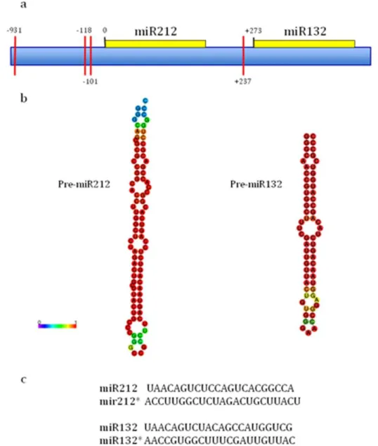
Activation of specific transcription factors might be only one among many possible ways by which visual experience can regulate gene expression. Increasing evidences show neural activity is able to induce posttranslational modifications of histones that, by rendering chromatin more or less accessible to transcription machinery, resulted in changes in gene expression (Crosio, et al., 2003, Korzus, et al., 2004). Considering that over the past decade, research into the regulation of transcriptional potential through modifications of chromatin structure has exploded, I will summarize the major findings that have linked epigenetic mechanisms to neuronal plasticity.
Most DNA in eukaryotic cells is densely packed into chromatin, where 147 base pairs (bp) are wrapped around a nucleosome core in ~1.7 super-helical turns. Nucleosomes are composed of octamers that contain four histone homodimers, one each of histones H2A, H2B, H3 and H4, with H1 binding to spans of non-nucleosomal DNA. Numerous types of post-translational modifications (PTMs) of the amino (N)-terminal tails of histones alter chromatin structure to create more “open” states (euchromatin, which is transcriptionally permissive) versus “closed” states (heterochromatin, which is transcriptionally repressive)(Robison and Nestler 2011). Structural studies indicate that the N-terminal tails of histones protrude beyond the chromosomes. The current hypothesis is that these histone tails serve as signal integration “platforms”, whereby post-translational modifications are combined in a “histone code” that ultimately directs the activity of numerous transcription factors, co-factors and the transcriptional machinery in general (Fig.3)(Levenson and Sweatt, 2005).
Histones, particularly histones H3 and H4, are subject to extensive covalent post-translational modifications, including methylation, acetylation, phosphorylation, ubiquitylation, SUMOylation, biotinylation, ADP ribosylation and proline isomerization, and probably others that have yet to be discovered, each occurring at specific sites and amino-acid residues. Most modifications localize to the N- and carboxy (C) -terminal histone tails, and a few localize to the histone globular domains (Berger, 2007). Some histone modifications act in cis to alter the local chromatin structure directly, whereas others act in trans to influence the recruitment of chromatin-modifying factors. In trans histone modifications enable specific binding partners to dock, often as part of larger multimolecular complexes that
Figure 3. Mechanisms involved in chromatin modification.
epigenetics mechanisms implicated in causing changes in chromatin structu are important for development and cell
(Adapted from Dulac, 2010
• 7.1 Epigenetic tagging of histones
Acetylation is the best characterized of the post on histones. Acetylation of lysine
side chain, which effectively neutralizes their positive charge
structure by weakening the interaction between the positively charged hi and the negatively charged DNA
acetyltransferases (HATs),
to the ε-NH+ group of a Lys residue within a histone. The process is reversible, and the enzymes that catalyze
deacetylases (HDACs). Histone methylation
acetylation, methylation of histones occurs on ε mediated by histone methyltransferases
preserves their positive charge. In addition, Lys can accept up to three methyl groups. Arginine residues within histones can also be mono
guanidine nitrogen.
. Mechanisms involved in chromatin modification. Schematic representation of some
epigenetics mechanisms implicated in causing changes in chromatin structu
are important for development and cell-fate determination of tissues, including those of the CNS. 2010).
Epigenetic tagging of histones
Acetylation is the best characterized of the post- translational
on histones. Acetylation of lysine (Lys) residues occurs on the amino group in their side chain, which effectively neutralizes their positive charge, changing nucleosome structure by weakening the interaction between the positively charged hi
and the negatively charged DNA. The reaction is catalyzed acetyltransferases (HATs), which transfer an acetyl group from acetyl
NH+ group of a Lys residue within a histone. The process is reversible, and catalyze the reversal of histone acetylation are known as histone deacetylases (HDACs).
Histone methylation is another histone-directed epigenetic tag. Similar to acetylation, methylation of histones occurs on ε-NH+ groups of Lys residues, and is istone methyltransferases. Unlike acetylation, methylation of Lys preserves their positive charge. In addition, Lys can accept up to three methyl groups. Arginine residues within histones can also be mono- or dimethylated on their
Schematic representation of some epigenetics mechanisms implicated in causing changes in chromatin structure. All of these processes fate determination of tissues, including those of the CNS.
translational modifications residues occurs on the amino group in their changing nucleosome structure by weakening the interaction between the positively charged histone tails catalyzed by histone which transfer an acetyl group from acetyl- coenzyme A NH+ group of a Lys residue within a histone. The process is reversible, and the reversal of histone acetylation are known as histone
directed epigenetic tag. Similar to NH+ groups of Lys residues, and is . Unlike acetylation, methylation of Lys preserves their positive charge. In addition, Lys can accept up to three methyl or dimethylated on their
Phosphorylation of histones H1 and H3 was first observed more than 30 years ago in the context of chromosome condensation during mitosis. H3 was the first histone whose phosphorylation was characterized in response to the activation of mitogenic signalling pathways and this PTM is correlated with active gene transcrtiption. Phosphorylation of serine 10 on H3 is mediated for example by ribosomal protein S6 kinase 2 (RSK2) and mitogen- and stress-activated protein kinase 1 (MSK1), which are both downstream of extracellular signal-regulated kinase (ERK). In order to reverse these phosphorylation events, phosphatases remove phosphate groups from histones. So far, the phosphatases PP1 and PP2A have been shown to regulate levels of phorphorylation on H3 (Levenson and Sweatt, 2005).
Acetylation, methylation and phosphorylation involve small chemical groups, whereas ubiquitylation and SUMOylation add large moieties, two-thirds the size of the histone proteins themselves,which may lead to more profound changes in chromatin structure. Another degree of complexity is that methylation can occur several times (mono-, di- or trimethylation) on one lysine side chain, and each level of modification can have different biological outcomes. Some of the functional outcomes of these modifications are clear. For example, there is abundant evidence that acetylation is activating, whereas SUMOylation seems to be repressing for gene expression, and these two types of modification may mutually interfere. By con- trast, methylation and ubiquitylation have variable effects, depending on the precise residues and contexts. For example, silenced chromatin typically has low levels of histone acetylation, together with high levels of H4K20me3 and H3K27me3, whereas hyperacetylation, H3K4me3 and H3K36me3 are recognizable marks of active transcription. Two ubiquitylation sites in the C termini of H2B and H2A correlate with active and repressed transcription, respectively (Berger, 2007).
• 7.2 DNA methylation
The methylation of cytosine nucleotides in DNA forms 5-methyl- cytosine, which in mammalian cells is mainly confined to CpG dinucleotides. Methylation of DNA is catalysed by a class of enzymes known as DNA methyltransferases (DNMTs) and it is known to have a role in the constitutive silencing of chromatin regions, the inactivation of one of the X chromosomes in females, the imprinting of parental alleles, and the silencing of retroviral genes and other individual genes. The precise mechanisms by which DNA methylation marks are set, maintained and erased, however, are the subject of much debate. DNA methylation leads to marked changes in the structure of chromatin that ultimately result in significant downregulation of transcription and it can directly interfere with the ability of transcription factors to bind to regulatory elements (Dulac, 2010).
Moreover, evidence indicates that DNA methylation may serve as a contributing mechanism in memory formation and storage (Day and Sweatt, 2010). Very recent studies demonstrated that neuronal activity modifies DNA methylation pattern of adult mouse dentate granule neurons in vivo (Guo, et al., 2011) and that 5- hydroxil-methyl cytosine, a DNA base that is derived from 5-methylcytosine and has been implicated in DNA methylation–related plasticity, is dynamically regulated during postnatal development through adulthood and ageing in mouse hippocampus and cerebellum (Szulwach, et al., 2011).
The importance of DNA methylation in assisting essential gene regulatory events that are associated with brain function is revealed by neurological disorders caused by disregulation in DNA methyation processes, such as Rett Syndrome and fragile X Syndrome.
• 7.3 Chromatin remodelling influences synaptic plasticity
Long-term modifications of chromatin may underlie some of the changes in gene expression that lead to neural plasticity. Repeated patterns of synaptic transmission lead to diverse forms of synaptic plasticity at excitatory and inhibitory synapses: long-term potentiation (LTP) or long-term depression (LTD), whereby the efficacy of synaptic transmission is up- or downre- gulated, respectively. Certain forms of LTP and LTD are long lasting and depend on changes in gene expression. Based on the critical role that chromatin remodeling plays in creating a transcription permissive or silencing state of the genome (Felsenfeld and Groudine, 2003), growing evidence suggests that histone PTMs may be involved in these processes. For example, H4 acetylation at specific promoters in Aplysia is altered after LTP and LTD (Guan, et al., 2002). Plasticity-induced epigenetic changes are also observed in mammalian models of synaptic plasticity. Several forms of LTP require the activation of NMDA receptors and engagement of the MEK–ERK/MAPK signalling cascade (English and Sweatt, 1997, Morris, et al., 1986). Direct activation of NMDA receptors in the hippocampus leads to an increase in acetylation of histone H3, which can be blocked by inhibition of the MEK–ERK/MAPK cascade. In addition, activation of dopaminergic, cholinergic and glutamatergic signalling pathways in the hippocampus induces ERK- dependent increases in the phosphorylation of histone H3. These results suggest that the induction of mammalian synaptic plasticity leads to ERK-dependent increases in histone acetylation and phosphorylation in the hippocampus (Levenson and Sweatt, 2005).
As previously mentioned, the transcription factor CREB is essential for activity- induced gene expression. At the mechanistic level, CREB-associated
transcriptional regulation has been shown to involve the recruitment of multicomponent regulator complexes, as well as the initiation of chromatin-remodelling events. Activated CREB recruits CREB-binding protein (CBP; also known as CREBBP) or its paralogue p300 (also known as EP300), which functions as both a scaffolding protein and a HAT. CBP recruitment, in turn, stimulates histone acetylation and transcriptional-complex formation at the promoters, leading to transcriptional activation of many CREB-target genes. Mutations in the gene encoding CBP are responsible for the mental-retardation syndrome Rubinstein– Taybi, the phenotype of which may result from impairment of either or both of the CREB-dependent and CREB-independent functions of CBP. The essential role of HAT activity in CBP-mediated neuronal plasticity has been genetically demonstrated by the selective long-term memory defects of a transgenic mouse line carrying a dominant-negative CBP that blocks the HAT activity of the endogenous protein (Korzus, et al., 2004). In another study, the induction of early-phase LTP and LTD — forms of plasticity that do not require transcription — was not affected in CBP+/– animals. However, the induction of late-phase LTP, which requires transcription, was significantly impaired in CBP+/– mice. Treatment of hippocampal slices from CBP+/– animals with an HDAC inhibitor significantly improved late-phase LTP induction, which indicates that inhibition of HDACs had compensated for HAT haploinsufficiency (Alarcon, et al., 2004). In other studies using hippocampal slices, induction of LTP through high-frequency stimulation was significantly enhanced by two HDAC inhibitors, trichostatin A (TSA) and sodium butyrate (Levenson, et al., 2004). In addition, LTP in the amygdala that was induced by forskolin was also enhanced by the HDAC inhibitor TSA (Yeh, et al., 2004). These discoveries indicate
that the epigenetic state of the genome affects the induction of long-term forms of mammalian synaptic plasticity.
Recent studies implicated histone PTMs in memory formation and storage. Guan et al (2009) demonstrated that neuron-specific overexpression of HDAC2, but not that of HDAC1, decreased dendritic spine density, synapse number, synaptic plasticity and memory formation. Conversely, HDAC2 deficiency resulted in increased synapse number and memory facilitation, similar to chronic treatment with HDAC inhibitors in mice. These results suggest that HDAC2 functions in modulating synaptic plasticity and long-lasting changes of neural circuits, which in turn negatively regulates learning and memory (Guan, et al., 2009). Moreover, histone methylation is actively regulated in the hippocampus and facilitates long-term memory formation. Trimethylation of histone H3 at lysine 4 (H3K4), an active mark for transcription, is upregulated in hippocampus 1 h following contextual fear conditioning, while dimethylation of histone H3 at lysine 9 (H3K9), a molecular mark associated with transcriptional silencing, is increased 1 h after fear conditioning and decreased 24 h after context exposure alone and contextual fear conditioning. Furthermore, mice deficient in the H3K4-specific histone methyltransferase, Mll, displayed deficits in contextual fear conditioning relative to wild-type animals. This suggests that histone methylation is required for proper long-term consolidation of contextual fear memories (Gupta, et al., 2010).
Drug addiction can be viewed as a form of drug-induced neural plasticity, whereby repeated exposure to drugs of abuse leads to long-lasting changes in the brain’s natural reward centers and associated memory circuits, which underlie the addiction phenotype. Multiple drugs of abuse induce changes in histone acetylation in the brain, and evidence has begun to accumulate that these modifications underlie
some of the functional abnormalities found in addiction models. First, global levels of H3 and H4 acetylation are increased in the NAc after acute or chronic exposure to cocaine (Kumar, et al., 2005, Renthal and Nestler, 2008), and gene promoters that show increased H3 or H4 acetylation have been mapped genome-wide (Renthal, et al., 2009). Despite these global increases, many genes show decreased histone acetylation after chronic cocaine, raising a key question as to what governs gene-specific acetylation changes in the face of global modifications.
Histone methylation is also directly regulated by drugs of abuse: global levels of histone 3 lysine 9 dimethylation (H3K9me2) are reduced in the NAc after chronic cocaine exposure (Maze, et al., 2010), and a genome-wide screen revealed alterations in H3K9me2 binding on the promoters of numerous genes in this brain region (Renthal, et al., 2009); both increases and decreases were observed, indicating again that epigenetic modifications at individual genes often defy global changes. The global decrease in H3K9me2 in the NAc is probably mediated by cocaine-induced downregulation of two histones methyl transferases, G9a and G9a-like protein (GLP), which catalyse the dimethylation of H3K9me2. These adaptations mediate enhanced responsiveness to cocaine, as selective knockout or pharmacological inhibition of G9a in the NAc promotes cocaine-induced behaviours, whereas G9a overexpression has the opposite effect (Maze, et al., 2010).
Accumulating data show that chromatin remodeling events may be critical for providing specificity and plasticityin circadian clock regulation. Although our under- standing of the mechanisms that direct circadian epigenetic control is still limited, emerging evidence implicates histone modifications and some chromatin remodeling proteins as particularly important in directing circadian clock entrainment and resetting (Masri and Sassone-Corsi, 2010).
Given the involvement of epigenetic mechanisms in nervous system function, it is not surprising that a growing number of disorders, in particular mental retardation and autism spectrum syndromes, have been linked to chromatin remodeling defects. The most well-studied ‘‘epigenetic disease’’ associated with altered neurological function, is Rett’s syndrome, an X-linked postnatal autism spectrum disorder characterized by stereotypical motor, learning, and social abnormalities that generally worsen over time (Moretti and Zoghbi, 2006). Candidate gene analyses identified MeCP2 as the causative gene (Amir, et al., 1999). MeCP2 was identified on the basis of binding selectively to methylated CpG dinucleotides in heterochromatic regions and it acts in a methylation-dependent repressive fashion. In Rubinstein-Taybi syndrome (RSTS), characterized by mental retardation and developmental abnormalities, the DNA-binding hook is provided by CREB. Phosphorylation of CREB leads to CBP recruitment and activation of target promoters. In RSTS, mutations in the CBP gene result in impairment of HAT activity. Mice haploinsufficient for CBP display impaired cognitive function, altered neuronal plasticity, and aberrant histone acetylation at target gene promoters. Interestingly, the behavioral symptoms can beameliorated by administration of HDAC inhibitors (Vo and Goodman, 2001).
Fragile X syndrome, the most commonly inherited form of mental retardation, is brought about by an abnormal expansion of repeated trinucleotide sequences within one of two Fragile X genes: FMR1 and FMR2 (Ashley, et al., 1993). FMR1 and FMR2 each contain a polymorphic trinucleotide repeat (CGG and CCG, respectively) in their 5’-untranslated regions that are responsible for the loss of gene expression. Expansion of these repeats results in hypermethylation of these regions and flanking
CpG islands, leading to transcriptional silencing of the FMR and surrounding genes (Levenson and Sweatt, 2005).
A striking example of developmental disruption caused by mutations in a chromatin factor gene is alpha-thalassaemia/mental retardation, X-linked syndrome, the gene for which is a helicase (spinocerebellar ataxia-7) involved in chromatin remodeling, by regulating several HAT complexes. Mutations lead to defects in psychomotor, urogenital and haematopoietic development, with maturational defects in erythroid precursors resembling those of alpha-thalassaemia (Feinberg, 2007).
These pathologies are just some examples of disorders involving alteration in epigenetic mechanisms (for a review see (Portela and Esteller, 2010).
All these observations indicate that dysfunction of the normal epigenetic status of the genome can have marked consequences on synaptic plasticity processes and normal cognitive function, indicating that drugs that target the epigenome might represent viable therapies for treating various diseases that affect cognition.
• 7.4 Histone PTMs and visual cortical plasticity
Several studies have demonstrated that regulation of gene expression through posttranslational modifications of histones is present in neuronal cells in vivo. Indeed, stimuli that reset the circadian rhythms induce phosphorylation of H3 in the suprachiasmatic nucleus (Crosio, et al., 2000) and acetylation of H3 and H4 during the transcriptional activation phase of the circadian rhythm has been described (Etchegaray, et al., 2003). Recent evidences demonstrated that experience-dependent regulation of chromatin structure could be an important mechanism of regulation of gene expression also in the developing visual cortex. Visual experience