CONTENTS
INTRODUCTION 1
MATERIALS AND METHODS
38
RESULTS 40
DISCUSSION 63
CONCLUSIONS 75
Phytochromes, cryptochromes and phototropins: the “eyes” of the
plants
The ability of animals to alter their environmental surroundings through locomotion is an option lost to the majority of plants following the dispersal of pollen and seeds. To compensate for this restriction, plants adapt through alteration of their complex chemical repertoire and modifying their development. The most remarkable difference between animal and plant development is that both growth and organogenesis can continue through most of the life of plants. New organs may be developed with a more appropriate structure if current appendages are unsuitable in an altered environment. For example, to facilitate improved light catchment in a newly shaded habitat, small and dense leaves might be replaced by broader and thinner leaves, or leaves may be dropped in their entirety to favour frost-tolerant buds as the shortening daylength indicates the approach of winter. This developmental flexibility requires the integration of multiple external signals, allowing harmonization between the growth of the plant and environmental change, thereby enabling them to compete effectively with neighbours for resources.
In addition to providing the energy for photosynthesis, light imparts crucial information regarding the surrounding environment and influences many basic physiological processes, including seed germination, seedling de-etiolation (the transition from skotomorphogenesis to photomorphogenesis), vegetative growth, organ orientation and the transition to reproductive development.
Plants have acquired the tools to precisely monitor the changing intensity and spectrum of light, its direction and, in specific cases, its plane of polarization (Kendrick and Kronenberg, 1994), through the presence of a number of photoreceptors: the red (R)/ far-red(FR) – absorbing phytochromes and the blue/UV-A – absorbing cryptochromes and phototropins (Cashmore et al. 1999; Casal 2000; Christie and Briggs 2001; Nagy and Schaefer 2002; Quail 2002a, b).
In their action, the role of the photoreceptors in light signalling is somewhat analogous to vision: they provide important information that controls plant behaviour, such as the proximity of neighbouring plants or the optimal direction of elongation, in addition to the availability of solar energy.
De-etiolation is the better understood of the light-regulated developmental transitions because it can occur reproducibly and rapidly in the lab (Kendrick and Kronenberg, 1994). This is the transformation from an “etiolated” seedling, germinating without leaves and elongating rapidly through the soil towards the surface with its apex trailing upside-down for protection, into a young plant in the light, with leaves expanding from the righted apex and chloroplasts developing as quickly as possible. A significant proportion of the transcriptome is controlled by the photoreceptors at this stage, (Quail, 2002), perhaps more than at any other time. A quantitative trace in the amount of perceived light is presented by the elongation of the hypocotyl (the seedling stem) over a period of several days since extra light reduces the amount of elongation. A “blind” plant will stand out spindly and tall above those neighbours with normal light perception, making this method of detecting photoreceptor-deficient mutants amongst the easiest of genetic screens. It also gives the fastest indication as to whether a mutant plant isolated by another phenotype, such as aberrant circadian regulation, may be deficient in light signalling. The most common molecular assay tests the activation of highly expressed, light-regulated genes such as chlorophyll a/b binding protein gene (CAB) following a brief light pulse. These, and several related approaches, have determined mutants in the genes encoding the photoreceptor proteins of the 3 families mentioned above.
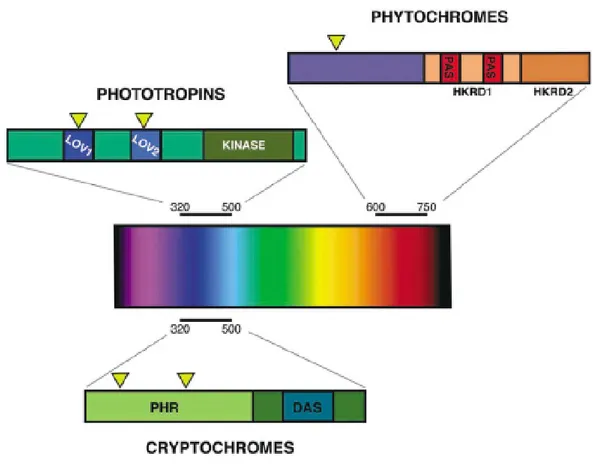
Figure 1. The plant photoreceptors.
Three classes of photoreceptors have been characterized from plants at the molecular level. (A) Phytochromes perceive red and far-red light of between 600 and 750 nm. The phytochrome apoprotein contains two histidine kinase related domains (HKRD1 and HKRD2) at the carboxyl terminus and two Per-Amt-Sim domains (PAS) within the HKRD1 domain that have been shown to function as protein–protein interaction domains and small ligand response modules. (B) Cryptochromes perceive blue and UVA light (320-500 nm); at the amino terminus is a photolyase related domain (PHR), and at the carboxyl terminus is DQXVP-acidic-STAEES (DAS) motif. (C) Phototropins also perceive blue and UVA light (320-500 nm). The phototropin apoprotein contains 2 chromophore binding domains (LOV1 and LOV2) as well as a Kinase domain at the carboxyl terminus. Yellow triangles represent the chromophore attachment sites in each of the photoreceptors.
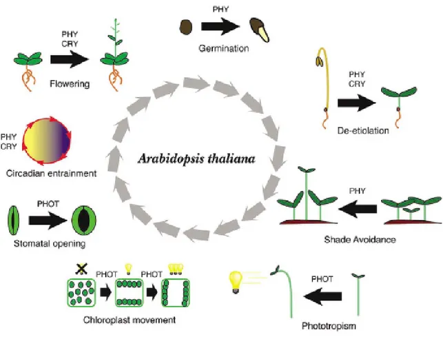
Figure 2. Light-regulated development in the model plant species Arabidopsis thaliana.
Light affects the development of Arabidopsis throughout its life cycle. Multiple aspects of development are regulated the photoreceptors phytochromes (PHY), crytochromes (CRY), or phototropins (PHOT) acting alone or in combination with each-other.
PHYTOCHROMES
The discovery of physiological responses, such the germination of lettuce seeds that is promoted by red (R) light and repressed by subsequent far-red (FR) light, led to the identification of phytochrome genes (Kendrick and Kronenberg, 1994). It has been suggested that phytochromes evolved from bacterial bilinsensory proteins, a hypothesis that is supported by the discovery of phytochrome-like proteins in photosynthetic bacteria, non photosynthetic eubacteria, and fungi (Montgomery and Lagarias, 2002). The phytochrome apoprotein is encoded by a small multigene family: in the model plant Arabidopsis thaliana this family consists of five genes (PHYA, PHYB, PHYC, PHYD and PHYE) (Sharrock and Quail, 1989). Based on their stability in the light, phytochromes have been classified into two types. Type I phytochromes (photo-labile) accumulate in etiolated seedlings and degrade rapidly upon light exposure, whereas type II phytochromes (photo-stable) are relatively stable in the light (Furuya,1992). In
Arabidopsis, PHYA is the only member of type I phytochromes; PHYB-E are type
II phytochromes (Quail, 1997; Sharrock and Clack, 2002).
Phytochrome structure
All phytochromes exist as homodimers that are composed of two 125-kDa polypeptides, each carrying a covalently linked open-chain tetrapyrrol chromophore, phytochromobilin, which is synthesized in the chloroplasts from heme (Davis et al., 1999; Kohchi et al., 2001; Nagy and Schafer, 2002; Parks and Quail, 1991; Quail, 1997).
Phytochromes are composed of two functional domains: an N-terminal light-sensing domain and a C-terminal regulatory domain (Fig. 3). The N-terminal portion is necessary and sufficient for photoperception and possesses the bilin lyase activity allowing attachment of the chromophore to the apoprotein (Terry, 1997). The minimal bilin lyase domain (BLD) is actually less than 200 amino acids
long (Wu and Lagarias, 2000). The first 70 amino acids of the protein are dispensable for chromophore binding; they constitute the N-terminal extension (NTE). The NTE is poorly conserved, possibly accounting for some functional differences among PHY. Structure function analysis has revealed that in PHYA, the NTE is composed of two subdomains (Stockhaus et al., 1992; Jordan et al., 1996).
The C-terminal signaling domain is composed of a PAS (Per/Arndt/Sim)-related domain (PRD) and a histidine kinase-related domain (HKRD) (Fig. 3) (Schneider-Poetsch et al., 1991;,Yeh and Lagarias, 1998).
Figure 3. Domain structure of Arabidopsis phytochromes.
NTE: N-terminal extension; BDL: Bilin lyase domain; PRD: Pas related domain; HKRD: Histidine kinase-related domain.
Each phytochrome can exist in two photointerconvertible conformations, denoted Pr (a red light-absorbing form) and Pfr (a far red light-absorbing form) (Fig. 4). Because sunlight is enriched in red light (compared with far red light), phytochrome is predominantly in the Pfr form in the light, and this can convert back to the Pr form during periods of darkness by a process known as dark reversion (Nagy and Schafer, 2002). Photoconversion back to Pr can also be mediated by pulses of far red light.
NTE
NTE
BDL
BDL
PRD
PRD
HKRD
HKRD
AMINO-TERMINAL DOMAIN
Figure 4. Photoconversion and dark reversion between Pr (inactive) and Pfr (active) form of phytochrome.
The dark reversion rates vary for different phytochromes. In Arabidopsis PHYB has a fast dark reversion rate, whereas PHYA is very stable in the Pfr form (Eichenberg et al., 2000; Hennig et al, 2001).
Phytochrome localization
In the dark, de novo synthesized phytochromes are accumulated within the cytoplasm, in the Pr form. Upon conversion to Pfr (due to the light) the five
Arabidopsis phytochromes translocate into the nucleus (Kircher et al., 2002;
Yamaguchi et al., 1999). The quality of the light is a very discriminating factor for the import into the nucleus. PHYA translocates to the nucleus in FR (Kircher et al., 2002; Nagy and Schafer, 2002), while the others accumulate in the nucleus in R or white light (Kircher et al., 2002). Moreover, the nuclear import of PHYA is much faster than that of PHYB,C,D and E (Kircher et al., 2002; Nagy and Schafer, 2002). In the nucleus they form discrete speckles (Nagy and Schafer, 2002), but the nature and the function of these subnuclear foci or nuclear body is still unclear.
Phytochrome functions
Phytochrome physiological responses can be divided into different groups based on the radiation energy of light, that is necessary for the response: low fluence responses (LFRs), very low fluence responses (VLFRs) and high irradiance responses (HIRs). Genetic studies of Arabidopsis phytochrome mutants demonstrate that PHYA is responsible for VLFR and FR-HIR responses, while
Pr
R(660nm)
Pfr
FR(730nm)
Dark reversion
Pr
R(660nm)
Pfr
FR(730nm)
Dark reversion
PHYB is principally involved in LFR and R-HIR responses during photomorfogenesis (Nagy and Schafer, 2002; Quail, 2002a).
The redundant and overlapping mechanisms of phytochrome action make often difficult to understand the roles of individual phytochromes in mediating plant growth. The isolation of mutants deficient in individual phytochromes and the subsequent creation of multiple mutant combinations have, therefore, been essential in the resolve of individual phytochrome functions and the dissection of functional interactions between family members.
The timing of seed germination and the consequent developmental strategy of a plant is strongly influenced by the light surroundings. Induction of Arabidopsis seed germination by R involves both PHYA and PHYB (Shinomura et al., 1994, 1996). Germination responses displaying R/FR reversibility are characteristic of the LFR response and enables buried seeds to detect proximity to the soil surface. The retention of R/FR reversible germination responses in phyAphyB double mutants implicated the participation of another phytochrome in this physiological answer, PHYE (Hennig et al., 2002).
Many seeds that have been imbibed in darkness gain acute sensitivity to light that is typical of the PHYA-mediated VLFR mode of action. It is estimated that these sensitized seeds would be induced to germinate following exposure to only a few milliseconds of daylight (Smith, 1983). Inhibition of germination following prolonged exposure to FR, most likely represents the PHYA-mediated FR-HIR response mode of phytochrome action and may be ecologically relevant as a means of delaying the germination of seeds situated under chlorophyllous vegetation or leaf litter (Casal et al., 1990).
After the induction of germination, light signals act to limit hypocotyl expansion while initiating the extension of cotyledons and the concomitant synthesis of chlorophyll. Despite showing no obvious mutant phenotype following growth under white light or R, mutants deficient in PHYA have revealed a unique role for this photoreceptor in mediating the inhibition of hypocotyl elongation growth
under FR and FR-enriched light environments (Nagatani et al., 1993; Parks and Quail, 1993; Whitelam et al., 1993). By contrast, PHYB-deficiency confers no aberrant phenotype under FR, but leads to a marked loss of seedling sensitivity to R for a wide range of de-etiolation responses (Koornneef et al., 1980; Somers et al., 1991; Reed et al., 1993). Seedlings deficient in both PHYA and PHYB display a greater insensitivity to R than monogenic PHYB seedlings (Reed et al., 1994). Thus, although PHYB plays the major role in inhibition of hypocotyl elongation in red light, PHYA can also contribute to this response. An additional minor role is performed by PHYD (Aukerman et al., 1997) whereas the contribution of PHYE to seedling de-etiolation appears insignificant (Devlin et al., 1998).
The recent identification of mutants at the PHYC locus has revealed a role for this phytochrome in the R-mediated inhibition of hypocotyl elongation (Franklin et al., 2003; Monte et al., 2003). The combined loss of PHYA and PHYC in the Ws ecotype (phyC-1) resulted in a significant increase in hypocotyl length, an effect greater than that observed in phyC-1 plants. Since loss of PHYA alone has no effect on sensitivity to R, the possibility exists that PHYA and PHYC act redundantly to regulate the R-control of hypocotyl growth (Franklin et al., 2003). The role of PHYC in this response was most pronounced at low fluence rates and not observable in the PHYB mutant background, suggesting a possible role for PHYC in modulating PHYB function (Franklin et al., 2003). No role for PHYC was identified in the inhibition of hypocotyl elongation in FR (Franklin et al., 2003; Monte et al., 2003).
The isolation and characterization of mutants deficient in cryptochromes 1 and 2 (CRY1 and CRY2) have defined roles for these photoreceptors throughout seedling development (Lin et al., 1996, 1998). Despite uncertainty over the exact nature of co-action, it is accepted that B-mediated de-etiolation involves the interaction of both phytochrome and cryptochrome signalling (Yanovsky et al., 1995; Ahmad and Cashmore, 1997; Casal and Mazzella, 1998).
A physical interaction between CRY1 and PHYA proteins has been demonstrated (Ahmad et al., 1998; Ahmad, 1999) in addition to a functional interaction between CRY2 and PHYB (Mas et al., 2000). Mutant combinations deficient in PHYC displayed elongated hypocotyls in B, an effect most evident at low fluence rates (Franklin et al., 2003). Under these conditions, it has been shown that the CRY2 function predominates in the regulation of hypocotyl elongation (Lin et al., 1998). The hyposensitivity of phyC mutants to low fluence rate of B may therefore indicate a possible functional interaction between PHYC and CRY2. There is also evidence of functional redundancy between phytochromes and cryptochromes. For example, the inhibition of hypocotyl growth by a R pulse in PHYB seedlings that have been pre-treated with white light, requires the presence of either PHYD or CRY1 (Hennig et al., 1999).
In Arabidopsis and many other plant species, lack of PHYB has a remarkable effect on the structure of the adult light-grown plant. PHYB-deficient plants show an elongated growth habit, retarded leaf development, increased apical dominance, and early flowering (Robson et al., 1993; Halliday et al., 1994; Devlin et al., 1996). This pleiotropic phenotype resembles the shade avoidance syndrome shown by wild-type plants following the perception of low R:FR ratio and suggests a predominant role for PHYB in suppressing this response under natural conditions (Whitelam and Devlin, 1997). The ability to respond to the perceived threat of shading, and therefore to execute structural changes before canopy closure, provides a crucial competitive strategy to plants growing in dense stands (Ballarè et al., 1990).
The maintenance of shade avoidance responses in phyB null mutants indicated the involvement of additional phytochromes (Robson et al., 1993; Halliday et al., 1994). Multiple mutant analyses have since revealed that the perception of low R:FR in Arabidopsis is mediated solely by PHYB, D and E, acting in a functionally redundant manner (Devlin et al., 1996, 1998, 1999;). These represent the most recently evolved members of the phytochrome family and form a distinct
subgroup (Mathews and Sharrock, 1997). It is therefore possible that competition for light may have provided the selective pressure for their evolution (Devlin et al., 1998). Adult Arabidopsis plants structure their leaves in a compact rosette phenotype. The elongated internodes observed in phyAphyBphyE triple mutant plants was the basis on which the phyE mutation was isolated and led to the proposal that maintenance of the rosette phenotype is regulated, redundantly, by PHYA, B and E (Devlin et al., 1998). The elongated appearance of
phyAphyBphyDphyE quadruple mutants grown under white light, a phenotype not
displayed in phyBphyDphyE triple mutants has supported such a proposal (Franklin et al., 2003b).
Physiological comparison of these genotypes also revealed a significant role for PHYA in the modulation of rosette leaf expansion and petiole elongation in high R:FR (Franklin et al., 2003b). Analysis of mutants deficient in PHYC revealed this phytochrome to play a similar role to PHYA in regulating rosette leaf elongation in high R:FR (Franklin et al., 2003b; Monte et al., 2003).
The phytochromes are also known to interact more directly with phototropism. For example, R, acting predominantly through PHYA is known to lead to enhancement of subsequent phototropic curvature (Parks et al., 1996; Janoudi et al., 1997).
Besides, phytochromes, together with cryptochromes, are the elements of input to to plant circadian clock. This aspect of phytochromes and cryptochromes action will be discussed in detail in the next chapter.
Tomato Phytochromes
In tomato five phytochrome genes have been discovered and analyzed so far:
PHYA, PHYB1, PHYB2, PHYE and PHYF (Hauser et al., 1995). Phylogenetic
analyses showed ortology between PHYA, PHYE and PHYC/F gene pairs in
Arabidopsis and tomato; tomato PHYB1 and PHYB2 were originated by an
independent duplication (Pratt et al., 1995).
Roles for PHYA and PHYB1 in the mediation of de-etiolation responses to R in tomato have been demonstrated previously (van Tuinen et al., 1995a; van Tuinen 1995b). In the control of anthocyanin biosynthesis under R, PHYA acts predominantly at low irradiances, and PHYB1 at higher irradiances (Kerckhoffs et al., 1997). Although the phyAphyB1 double mutant is blind to low-irradiance R, it de-etiolated normally under white light. The phenotype of phyAphyB1phyB2 mutants under natural daylight indicated an important role for PHYB2 in this residual response (Kerckhoffs et al., 1999) and it also clear that PHYB2 is also active in R-sensing (Weller et al., 2000). However, the strongly synergistic effects of phyB1 and phyB2 mutations indicate a high degree of functional redundancy between these phytochromes, as might be expected given their relatively recent divergence (Pratt et al., 1995). In seedling de-etiolation, effects of PHYB2 were only seen in the absence of PHYB1, whereas PHYB1 still retained substantial function in the absence of PHYB2.
At least one other phytochrome (PHYE or PHYF) could be active in controlling de-etiolation in tomato, but is functionally dependent on cryptochrome activity, at least in the absence of PHYA, PHYB1 and PHYB2 (Weller et al., 2000).
CRYPTOCHROMES
The cryptochromes are receptors for blue and (UV-A) light structurally related to DNA photolyases, but they don’t have photolyase activity. DNA photolyases are a group of UV-A/blue light-induced enzymes that repair UV-B-induced DNA damage by removing pyrimidine dimers from DNA (Sancar, 2003).
There are two types of DNA photolyase, which repair different types of damage: CPD photolyases repair cyclobutane pyrimidine dimers (CPDs), and 6-4 photolyases repair 6-4 pyrimidine pyrimidone photoproducts. Photolyases and cryptochromes make up a specific superfamily.
The first cryptochrome gene to be identified was Arabidopsis CRY1 (Ahmad and Cashmore, 1993), and cryptochromes were soon found by homology in other plant species, in bacteria and animals (Brudler et al., 2003; Cashmore et al., 1999).
It was initially thought that only higher eukaryotes had cryptochromes and that prokaryotes had photolyases but not cryptochromes, but further searches of the more recently available genome databases revealed the presence of a cryptochrome gene in cyanobacteria (Synechocystis) (Hitomi et al., 2000). This new type of cryptochrome was referred to as CRY-DASH, to underscore its relationship with cryptochromes found in Drosophila, Arabidopsis, Synechocystis and Homo (although CRY-DASH itself is not found in Drosophila or humans) (Brudler et al., 2003). CRY-DASH proteins have been found not only in the photosynthetic cyanobacteria but also in non-photosynthetic bacteria, fungi, plants and animals, including Arabidopsis, zebrafish and Xenopus (Kleine et al., 2003; Dayasu et al., 2004).
Cryptochrome structure and localization
Most plant cryptochromes are 70-80 kD proteins with two recognizable domains, an N-terminal PHR domain that shares sequence homology with photolyases, and a C-terminal extension that has little sequence similarity to any known protein
domain (Fig. 5). The PHR region of cryptochromes appears to bind two chromophores, cofactors that absorb light; one chromophore is flavin adenine dinucleotide (FAD) and the other 5,10-methenyltetrahydrofolate (pterin o MTHF) (Lin et al., 1995; Malhotra et al., 1995) (Fig. 5). The carboxy-terminal domains in different plant species are of variable length, but they share short stretches of homology (Lin and Shalitin, 2003). Going from tha amino-terminal to the carboxy-terminal end of this extension, one finds a DQXVP motif, a stretch of acidic residues, STAES, and finally GGXVP.
Figure 5. Domain structure of plant cryptochrome 1-2.
PHR: N-terminal photolyase related domain; DAS: C-terminal domain.
The carboxy-terminal domain of cryptochromes is generally less conserved than the PHR region (Lin and Shatilin, 2003); CRY-DASH protein has no carboxy-terminal extension and, consequently, no DAS domain (Fig. 6).
Figure 6. Domain structure of Arabidopsis cryptochrome DASH.
P: N-terminal signal peptide; PHR: Photolyase related domain
COOH NH 2
PHR
PTERIN FADDAS
DQXVP…E/D…STAES…GGXVPPlant cryptochrome 1-2
COOH NH 2PHR
PTERIN FADDAS
DQXVP…E/D…STAES…GGXVPPlant cryptochrome 1-2
Arabidopsis
cryptochrome DASH
COOH NH2 PTERIN FAD
PHR
PTERIN BINDING SITEPHR
PArabidopsis CRY1 and CRY2 are predominantly nuclear proteins that mediate
regulation of gene expression. CRY1 and CRY2 play major roles in plant photomorphogenesis; it appears that CRY1 and CRY2 control developmental changes in plants via modifications of gene expression in response to light. CRY1 and CRY2 together are responsible for blue-light-dependent changes in gene expression of up to 10-20% of the Arabidopsis genome (Ma et al., 2001).
Arabidopsis CRY-DASH protein contains a functional dual targeting signal (Fig.
6) mediating transport into chloroplast and mitochondria (Kleine et al., 2003). This is the only cryptochrome family member protein that is localized in the organelles.
Arabidopsis CRY2 is constitutively imported to the nucleus regardless of light
treatment. However, Arabidopsis CRY1 may be imported to the nucleus in the dark but may be exported or remain in the cytosol in response to light. It was found that the GUS-CCT1 (CRY1 C-terminus) fusion protein was mostly located in the nucleus in root hair cells of dark-grown transgenic plants, but the fusion protein was mostly cytosolic in the light-grown transgenic plants (Yang et al., 2000). Consistent with the notion that CRY1 may be largely cytosolic in light-grown plants, the relative amount of CRY1 detected in the nuclear extract obtained from the green tissue of light-grown Arabidopsis was significantly lower than that detected in the total protein extract (Guo et al., 1999). In contrast, the same nuclear extract was highly enriched for CRY2 (Guo et al., 1999).
Although one may expect that the PHR domain of a cryptochrome would contain the nuclear localization signal (NLS), because DNA photolyase, the presumed ancestor of the PHR domain of cryptochromes, has to move into the nucleus to carry out its DNA-repairing function, the C-terminal extension is sufficient to direct nuclear transportation for both cryptochromes in Arabidopsis (Cutler et al., 2000; Guo et al., 1999; Kleiner et al., 1999; Wang et al., 2001; Yang et al., 2000). A putative bipartite nuclear localization signal was found within the DAS domain of CRY2, and fusion proteins of β-glucuronidase (GUS) to the C-terminal extension of CRY2 are constitutively nuclear (Cutler et al., 2000; Guo et al., 1999;
Kleiner et al., 1999). Although no apparent bipartite NLS is found in CRY1, the C-terminal extension has proven sufficient for nuclear/cytoplasmic trafficking of CRY1 (Wang et al., 2001; Yang et al., 2000).
Mechanism of action
The catalytic mechanism of cryptochromes is not still clear, but a model was proposed based on the well-described light activation of CPD photolyases, where FAD plays the main catalytic role (Park et al., 1995). In a DNA-repair reaction, CPD photolyase binds to the pyrimidine dimer, to form a stable complex with the FAD-access cavity of the enzyme. The other chromophore, which is also called the “antenna” chromophore, absorbs blue-light and transfers the excited energy to the flavin of FAD. Flavin in the excited state donates an electron to the pyrimidine dimer to split the cyclobutane ring. Subsequently, the electron moves back to flavin to regenerate the catalytic active flavin, and the DNA with the two neighbouring pyrimidines restored is released from the photolyase (Sancar, 1994). Based on the homology with DNA photolyases, one might have expected that they also bind DNA. This has actually been demonstrated for Arabidopsis CRY-DASH and its Synechocystis homolog (Kleine et al., 2003; Brudler et al., 2003). In
Synechocystis, CRY-DASH is directly involved in gene regulation (Brudler et al.,
2003). A very recent report (Selby and Sancar, 2006) has shown that the CRY-DASH proteins of Vibrio cholerae, Xenopus laevis, Synechocystis and Arabidopsis have a clear single-stranded DNA photolyase activity. This protein is able to repair cyclobutane pyrimidine dimers in RNA and single but no double-stranded DNA. So the authors affirm that “these enzymes, which are found in bacteria, plants, and animals, and were previously designated CRY-DASH, because of the lack of significant photorepair activity on dsDNA, should be reclassified as ssDNA photolyases. It should be noted, however, that this classification does not necessarily exclude an additional non-repair function for ssDNA photolyases, as
indeed even some conventional photolyases have both repair and transcriptional regulatory functions ” (Selby and Sancar, 2006).
Direct DNA binding of CRY1 and CRY2 has not been reported; however, a CRY2 carboxy-terminal extension-GFP fusion is associated to chromatin (Cutler et al., 2000).
Cryptochromes are regulated by phosphorylation. It has been shown that
Arabidopsis cryptochromes are phosphorylated in response to blue light and that is
associated with the function and regulation of the photoreceptors (Shalitin et al., 2002; Shalitin et al., 2003).An additional enzymatic activity has recently been found for CRY1. The recombinant protein, expressed in insect cells, binds ATP; this binding is steichiometric and depends on FAD binding (Bouly et al., 2003). In addition, recombinant CRY1 autophosphorylates in a light-regulated manner, but no other substrate has been found (Bouly et al., 2003; Shalitin et al., 2003). Blue light triggers CRY1 and CRY2 phosphorylation at multiple sites in vivo (Bouly et al., 2003; Shalitin et al., 2003). Some of these sites are within the carboxy-terminal extension of CRY2 (Shalitin et al., 2002). This reaction is blue light specific and fluence rate dependent (Shalitin et al., 2003; Shalitin et al., 2002 ). Taken together with the in vitro characterization of CRY1, one might propose that this is the result of autophosphorylation.
An earlier report has shown that PHYA can phosphorylate the cryptochromes in vitro (Ahmad et al., 1998). However, the phosphorylation state of both CRY1 and CRY2 does not appear to depend on the phytochromes in vivo (Shalitin et al., 2003; Shalitin et al., 2002).
Given that a phyAphyBphyCphyDphyE quintuple mutant is currently not available, the role of the functional phytochromes in cryptochrome phosphorylation cannot be fully excluded. In the case of CRY2, phosphorylation is associated with proteolytic degradation (Shalitin et al., 2002). This degradation is in part mediated by the E3 ubiquitin ligase COP1. In addition, phosphorylation of both CRY1 and CRY2 appears to be closely linked to function.
Cryptochrome signal transduction
Cryptochromes are very important during de-etiolation, the transition of a dark grown seedling living from its seed reserves to a photoautotrophically competent seedling. This developmental transition includes a massive reorganization of the transcriptional program, inhibition of hypocotyl growth, promotion of cotyledon expansion, and synthesis of a number of pigments including chlorophyll and anthocyanins (Liscum et al., 2003).
Cryptochromes are involved in mediating many, if not all, of the blue light– dependent de-etiolation responses (Cashmore et al., 1999; Lin, 2002). For example, action spectra studies demonstrated that Arabidopsis CRY1 and CRY2 are the major photoreceptors mediating blue light inhibition of hypocotyl elongation (Ahmad et al., 2002; Young et al., 1992).
The function of cryptochrome in mediating de-etiolation responses has also been reported in tomato (Ninu et al., 1999; Weller et al., 2001).
A photoreceptor may trigger a developmental response by amplifying a light signal via cytosolic second messages that provoke other cellular activities including regulation of gene expression. Alternatively, a nuclear photoreceptor may directly interact with a transcription or post-transcription regulatory apparatus to alter gene expression and developmental patterns.
Based on analyses of blue light effects on plasma membrane depolarization, anion channel activity, and growth inhibition kinetics, it was proposed that cryptochromes activate anion channel activity, resulting in plasma membrane depolarization, and the inhibition of cell elongation (Parks et al., 2001; Spalding., 2000). This hypothesis may explain why Arabidopsis CRY1, which is the principal blue light receptor mediating blue light inhibition of hypocotyl elongation, is exported to the cytosol in response to light, where it may regulate cytosolic or plasma membrane proteins.
It was shown recently that the Arabidopsis cry1 and cry2 mutants were similarly impaired in the blue light–induced membrane depolarization, suggesting that these two photoreceptors play a role in the regulation of blue light activation of anion channels (Folta and Spalding., 2001). Indeed, these photoreceptors may regulate leaf expansion via light-dependent control of plasma membrane anion channels, because defects in leaf expansion have been observed in cryptochrome mutants (Lin et al., 1996; Lin et al., 1998).
The hypocotyl inhibition response as measured by hypocotyl length for seedlings grown in blue light, is significantly impaired in the cry1 mutant and slightly affected in the cry2 mutant (Ahmad and Cashmore, 1993; Lin et al., 1998). Identification of genes encoding the specific anion channels regulated by cryptochromes would help elucidate the cellular mechanisms underlying cryptochrome-dependent growth response.
In addition to calcium’s possible involvement in the phytochrome signal transduction (Bowler et al., 1994; Neuhaus et al., 1993), it may also be used as a second message for cryptochrome signal transduction (Christie and Jenkins, 1996; Guo et al., 2001; Long and Jenkins, 1998).
An Arabidopsis cell culture system has been used to study how cryptochrome mediate blue/UV-A light–induced CHS expression (Christie and Jenkins, 1996; Long and Jenkins, 1998). In this system, cryptochrome-mediated CHS expression correlates with blue light promotion of calcium efflux in the cytosol. The involvement of calcium homeostasis in cryptochrome-mediated CHS expression was indicated by the observation that compounds that inhibit voltage-gated calcium channel or Ca2C-ATPase significantly altered blue/UV-A light–induced CHS expression. A possible role of calcium homeostasis in cryptochrome signaling is consistent with a recent study of the Arabidopsis SUB1 gene, which encodes a calcium-binding protein that acts downstream from cryptochromes in the hypocotyl inhibition response (Guo et al., 2001). However, a direct demonstration of whether and how cryptochromes act through calcium channels or Ca2C-ATPase
to regulate CHS gene expression or hypocotyl inhibition depends on the identification of the specific genes encoding those proteins and the corresponding mutations.
Recent studies demonstrate that gene expression regulation is a major signaling mechanism underlying cryptochrome action. In Arabidopsis, CRY1 and CRY2 are known to regulate sets of similar genes in a partially redundant manner. A DNA microarray analysis demonstrated that the expression of about one third of
Arabidopsis genes change in response to blue light, and cryptochromes are the
major photoreceptors mediating these gene expression alterations (Ma et al., 2001). More than 71% of blue light–induced gene expressions and more than 40% of blue light–suppressed gene expressions are affected in etiolated cry1cry2 double mutants exposed to blue light, suggesting the two photoreceptors regulate expression of these genes in response to blue light (Ma et al., 2001). The rest of the blue light–dependent gene expression change is probably mediated partly or completely by PHYA (Chun et al., 2001; Thum et al., 2001). It is unclear which genes regulated by cryptochromes are directly involved in individual reactions of the de-etiolation responses and how cryptochromes regulate gene expression. One possibility is that cryptochromes regulate transcriptional or post-transcriptional processes by interacting with the respective regulatory complexes in the nucleus (Lin, 2000b).
Cryptochromes also work together with phytochromes to control photoperiodic flowering and the circadian clock. This specific role of these photoreceptors will be discussed in the next chapter.
COP1-CRY1 Interaction
Light-regulated protein degradation appears to be central to cryptochrome signaling.
Such a mechanism is well described for animal cryptochromes and also occurs for both CRY1 and CRY2 in Arabidopsis (Cashmore, 2003). Both cryptochromes
interact with the E3 ubiquitin ligase COP1 (Wang et al., 2001; Yang et al., 2001). The COP1 protein is required for the light-regulated degradation of several transcription factors involved in light-regulated transcription (Holm et al., 2002; Osterlund et al., 2000; Seo et al., 2003). In the dark, COP1 degrades these transcription factors including the bZIP protein HY5, but upon light perception this degradation is prevented (Holm et al., 2002; Osterlund et al., 2000; Seo et al., 2003). The constitutively de-etiolated phenotype of cop1 mutants is consistent with this model, since in those mutants a number of transcription factors (and presumably other COP1 targets) can accumulate in the absence of a light signal (Seo et al., 2003). Similarly, the light-hyposensitive phenotype of hy5 mutants can also be reconciled with this model (Holm et al., 2002; Osterlund et al., 2000; Seo et al., 2003). COP1 interacts with the cryptochromes both in the light and the dark, indicating that the light-driven electron-transfer reaction that was postulated to induce a conformation change in the cryptochromes does not disrupt this interaction (Wang et al., 2001; Yang et al., 2000; Yang et al., 2001). It was proposed that the light-driven conformational modification of the cryptochromes induces a structural modification of COP1 (Wang et al., 2001; Yang et al., 2001). Light-induced alteration of COP1 structure would release HY5 that was bound to COP1 in the dark. HY5 (and other COP1-regulated transcription factors) can then accumulate and bind to light-regulated promoter elements to initiate de-etiolation (Cashmore, 2003; Lin and Shalitin, 2003; Liscum et al., 2003) (Fig. 7).
Figure 7: Schematic mechanism of light activation proposed by Cashmore (Cashmore, 2003). The light signal modifies CRY1 conformation that leads to a conformational change of COP1. The new COP1 conformation causes the releasing of the transcription factor HY5 that can activate light-induced genes. LRE: Light Responsive Elements.
CRY2-PHYB interaction
Arabidopsis CRY2 directly interacts with PHYB (Mas et al., 2000). The
CRY2-PHYB interaction was shown by both yeast two-hybrid assays and coimmunoprecipitation tests. In addition, using fluorescent resonance energy transfer (FRET) microscopy, an intermolecular energy transfer was shown to occur between CRY2-RFP and PHYB-GFP fusion proteins, indicating that these two photoreceptors interact in vivo (Mas et al., 2000). Further evidence that CRY2-PHYB interaction is essential for the function of CRY2 came from a finding that CRY2-RFP, but not CRY1-RFP, was co-localized with PHYB in the nuclear speckles (Mas et al., 2000). In light of the recent discovery that PHYB could mediate light regulation of transcription via its interaction with the transcription factor PIF3 (Martinez-Garcia et al., 2000; Ni et al., 1998), the direct interaction between PHYB and CRY2 suggests that alteration of phytochrome-mediated
DARK
LIGHT
HY5 COP1 CRY1 ProteasomeLRE Light Regulated Gene
COP1
CRY1
HY5
LRE Light Regulated Gene Transcription
DARK
LIGHT
HY5 COP1 CRY1 ProteasomeLRE Light Regulated Gene
COP1
CRY1
HY5
LRE Light Regulated Gene Transcription
regulation of transcription may be an important mechanism of cryptochrome signal transduction. In addition, CRY1 has also been reported to interact, via its C-terminal domain, with PHYA in a yeast two-hybrid assay (Ahmad et al., 1998). CRY1 may also interact with PHYB, at least indirectly, because CRY1 and PHYB can each interact with COP1 (Yang et al., 2001).
The cryptochromes also interact with a number of other proteins, but the functional implications of many of these interactions are still unclear.
Tomato cryptochromes
In tomato (Solanum lycopersicum), three cryptochrome genes have been discovered so far: two CRY1 (CRY1a and CRY1b) and one CRY2 gene (Perrotta et al., 2000; Perrotta et al., 2001). The role of one of the CRY1 genes, CRY1a, has been elucidated through the use of antisense (Ninu et al., 1999) and mutant (Weller et al., 2001) plants. CRY1a controls seedling photomorphogenesis, anthocyanin accumulation, and adult plant development. No effects of CRY1a on flowering time or fruit pigmentation have been observed.
The overexpression of tomato CRY2 causes phenotypes similar to but distinct from their Arabidopsis counterparts (hypocotyls and internode shortening under both low and high fluence blue light), but also several novel ones, including a high-pigment phenotype, resulting in overproduction of anthocyanins and chlorophyll in leaves and of flavonoids and lycopene in fruits (Giliberto et al., 2005).
CIRCADIAN RHYTHMS
The day and night alternation is an environmental factor which lasts since life has appeared on the Earth. This succession of light and darkness produces in the environment deep changes to which all the creatures must adapt themselves. The organisms which are able to profit by these predictable changes, have acquired an evolutionary advantage. This benefit has promoted the development of timekeeping mechanism (endogenous clocks). Thanks to this "endogenous" time measurement, they have fitted their physiological, biochemical and behavioural functions to the day and night length. The biological clocks that generate and maintain oscillations of many physiological and molecular processes with a period length close to 24 h are also referred to as circadian clocks. ( from Latin, circa, approximately and dies, day ). When placed in constant conditions and, thus, deprived of external time cues, circadian rhythms persist and “free-run” with an endogenous period that is close to but no exactly 24 hr. In the real world, of course, organism are exposed to environmental cues such as light and temperature cycles, and these cues serve to synchronize or “entrain” the endogenous organismal clock with local solar time.
The period of a circadian rhythms remains relatively constant over the range of physiologically relevant temperatures, which is referred to as temperature compensation. This means that the circadian clock maintains its pace over a range of temperatures, but does not imply that temperature changes or cycles cannot serve as potent stimuli that can entrain the clock. These three characteristics: persistence in constant conditions with an approximately 24-hr period, entrainment and temperature compensation, are the diagnostic criteria of a circadian rhythm (Johnson et al., 1998; Sweeney, 1987).
Arabidopsis circadian clock
Our current understanding of plant circadian clock derives mostly from genetic studies in Arabidopsis and rice (Hayama and Coupland, 2004).
The circadian clock system is often divided into three general parts (Dunlap, 1999): an input pathway that entrains the clock, by transmitting light or temperature signals to the core oscillator; the central oscillator (the clock) that is the core of the system, responsible for driving 24-h rhythms; the output pathways that generates overt rhythms controlled by the core oscillator and represent a wide range of biochemical and developmental pathways.
Figure 8: Conceptual scheme showing simple linear information flow from input (entrainment) pathways through the central oscillator to output pathways.
Therefore, the information flow should proceed along these three components in a unidirectional way: from the input systems the information arrives to the oscillator centre and then to the pathways which give rise to the physiological answers (Fig. 8).
This scheme is only a simplification of the extraordinary complexity of the relationships and regulations which happen in the various parts of the clock, as shown by recent studies (Valverde et al., 2004).
This introduction will deviate from the linear order of figure 8 in its consideration of the Arabidopsis circadian system: output pathways will be considered first, the
CIRCADIAN SYSTEM
INPUT STIMULI
Light
Temperature
CENTRAL OSCILLATOR OUTPUT PATHWAYS
Gene expression Leaf movement Stomatal aperture Flowering
CIRCADIAN SYSTEM
INPUT STIMULI Light TemperatureCENTRAL OSCILLATOR OUTPUT PATHWAYS
Gene expression Leaf movement Stomatal aperture Flowering
core system will be addressed second and, finally the input pathways, in which photoreceptors are involved more directly will be analysed.
Rhythms in mRNA quantity of the output genes
In 1985 Kloppstech (Kloppstech, 1985) observed a circadian oscillation in mRNA abundance of a chlorophyll a/b binding protein gene (LHCB or CAB). This was the first example of a plant clock-controlled gene (CCG); afterwards, the list of plant
CCGs has grown to considerable length (Feyes and Nagy, 1998; Kreps et al., 2000;
McClung, 2000; McClung, 2001; Somers, 1999).
Oligo-based microarrays experiments, performed by Harmer et al, ( Harmer et al., 2000), allowed the detection of statistically significant circadian (in continuous light) oscillations in mRNA abundance of 5-6% of the 8200 genes examined. This measurement suggests that there are at least 1275-1530 Arabidopsis CCGs, based on a current estimate of ≈ 25500 Arabidopsis.
Among the CCGs genes, are present many genes associated with photosynthetic light harvesting oscillate, as genes encoding LHCA and LHCB proteins as well as photosystem I and II reaction centre proteins (Harmer et al., 2000; Scaffer et al., 2001). These genes showed a peak of mRNA abundance after subjective dawn (Harmer et al., 2000; Scaffer et al., 2001).
A number of Arabidopsis genes encoding enzymes involved in carbon metabolism and starch mobilization are clock-controlled (Harmer et al., 2000): mRNA abundance for many genes encoding enzymes of the glycolytic and oxidative pentose phosphate pathways, as well as genes encoding hexose transporters peak in the subjective afternoon; genes encoding starch kinase, β-amylase, fructose-bisphosphate aldolase and sugar transporters peak at night (Harmer et al., 2000). Plant responses to biotic and abiotic stress responses are often gated by the circadian clock (Rikin, 1992; Rikin et al., 1993). Microarray experiments identified oscillations in mRNA abundance of several genes involved in responses to stresses (Harmer et al., 2000; Schaffer et al., 2001). Particularly remarkable is the transcript
oscillation of the DREB1a/CBF3 gene encoding a transcription factor that plays a key role in cold tolerance (Harmer et al., 2000;Thomashow et al., 2001). This could suggests that the cold tolerance process underlies a circadian rhythm .
One of the most interesting observations resulting from microarray analysis was that 23 genes encoding enzymes of phenylpropanoid biosynthesis are synchronously transcripted, oscillating with mRNA peaks about 4 hours before subjective dawn (Harmer et al., 2000).
Harmer et al., found that a number of genes implicated in cell elongation are circadian-regulated and peak coordinately around the presumptive midday. These include the auxin efflux carriers PIN3 and PIN7. Auxin promotes growth in plant stems and hypocotyls, and its relocalization plays an important role in the control of cell elongation (Taiz and Zeiger, 1998). Auxin may activate expansins (enzymes that catalyze extension of cell walls), one of which was under clock control (Harmer et al., 2000). Cell expansion is also dependent on water influx, mediated by aquaporins, into plant vacuolar compartments. They found that an aquaporin gene is under clock control and peaks 8 hours after the presumptive dawn. This aquaporin, δ-tonoplast integral protein (δ-TIP), is localized to the vacuole and in young seedlings is primarily expressed in the hypocotyl and cotyledons (Daniels et al., 1996). δ -TIP may work in concert with the PINs, the expansin, and the cell wall hydrolases to effect cell elongation in young plants (Harmer et al., 2000).
Like many plants, Arabidopsis exhibits a circadian rhythm in stomatal aperture (Webb, 1998). Microarray analysis shows circadian transcript oscillations for a number of genes associated with Ca2+ signalling, including genes encoding Calmodulin and a Calmodulin-like Ca2+ -binding protein, as well as a putative Ca2+ -binding EF-hand protein and a Ca2+ -transporting ATPase (Harmer et al., 2000). Ca2+ is important in guard cell signalling (Leckie et al., 1998; Schroeder et al., 2001) and is likely to be involved in the circadian regulation of stomatal aperture and gas exchange. Ca2+ is also implicated in red and blue light signal transduction (Frohnmeyer et al., 1998; Long and Jenkins, 1998; Guo et al., 2001) and may play a
role in entrainment of the circadian oscillator. Thus, Ca2+ is likely to play multiple roles in the circadian system, but none of these roles are yet well defined.
Photoperiodism: the daylenght measurement and the output gene CONSTANS The timing of flowering in many species is photoperiodic (Lin 2000a; Simpson et al., 1999).
Several models have been proposed to explain the mechanisms by which photoperiod information is integrated into the regulation of flowering (Yanovsky and Kay, 2003). Among them, ‘the external coincidence model’ is currently the most consistent with the genetic evidence in plants (Yanovsky and Kay, 2003; Hayama and Coupland, 2004; Putteril et al., 2004). Erwin Bunning was the first to proposed this theory (Bunning, 1936); he hypothesized that circadian timekeeping was essential for photoperiodic time measurement. In this model, light plays two crucial roles. One is resetting the circadian clock, which is important for generating the daily oscillation of a key regulatory component with peak expression in the late afternoon. The other is regulating the activity of this component. Photoperiodic responses will only be triggered when regulator levels above the threshold coincide with daylight, the external signal (Fig. 9).
Figure 9: The external coincidence model. An example of the photoperiodic flowering response in long-day (LD) plants. LD plants flower only when regulator protein levels above the threshold coincide with long-daylight (From Imaizumi and Kay, 2006).
In LD plants (plants which flower only in Long Day conditions: 12-16 hours of light), the function of the key regulator is to promote flowering. Given that the circadian clock always sets peak expression of the regulator in the late afternoon, coincidence with light occurs more under LD but less under SD. Thus, the regulator is most active under LD, resulting in the acceleration of flowering. In SD plants (plants which flower only in Short Day conditions: 8 hours of light) , the clock-regulated factor functions as a suppressor of flowering.
Recent molecular-genetic studies of the flowering-time gene CONSTANS (CO) suggest that the interaction between circadian rhythms and light signalling may occur at the level of CO transcription and CO protein stability (Suarez-Lopez et al., 2001; Valverde et al., 2004). CO was isolated using a mutant that exhibits late flowering specifically under LDs (Putterill et al., 1995). The gene encodes a nuclear protein that contains a CCT motif and two B-box type zinc-finger domains, which were originally identified in several animal proteins and are believed to mediate protein-protein interaction. The transcript levels of this gene show a circadian rhythms under continuous light (Suarez-Lopez et al., 2001). However, CO overexpression does not alter the circadian rhythm in CAB gene expression in continuous light, suggesting that it does not have a general effect on circadian rhythms (Ledger et al., 2001), but it does result in dramatic early flowering (Putterill et al., 1995). This indicates that CO acts as a clock-output gene and mediates between the circadian clock and flowering (Suarez-Lopez et al., 2001). Moreover, CO directly induces the expression of FLOWERING LOCUS T (FT), which was originally isolated using a late-flowering mutant, and whose transcript is induced specifically under LDs (Samach et al., 2000).
Under the normal day-night cycle, CO transcripts show a diurnal rhythm. Under SDs, high levels of CO mRNA only occur during the night, whereas under LDs high CO levels occur at the end of the day and during the night (Suarez-Lopez et al., 2001). This observation suggested that CO mRNA level determines a light-sensitive phase and flowering is promoted specifically under LDs because only under these
conditions are plants exposed to light at times when CO is highly expressed. This is consistent with the external coincidence model, identifying CO as the clock-regulated factor and FT as the flowering gene (Fig. 9).
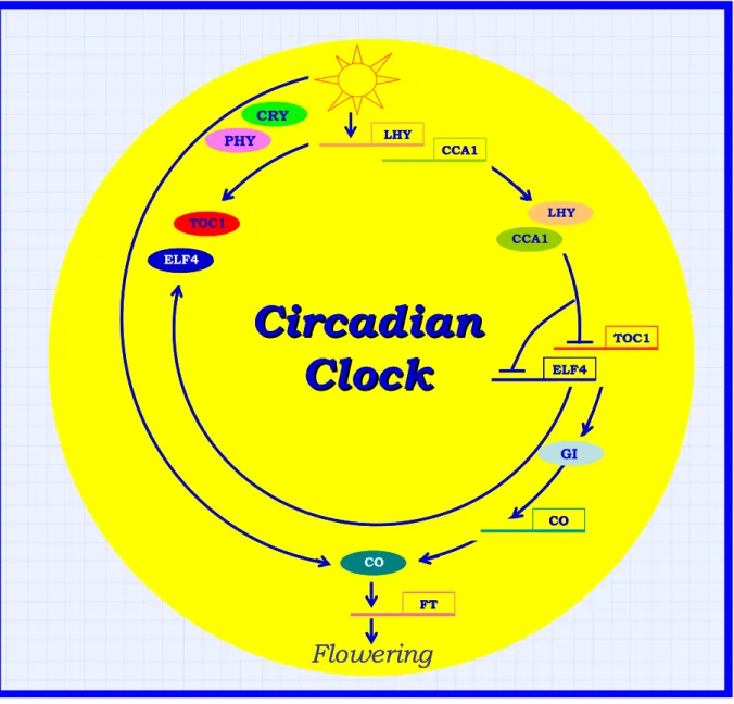
The central oscillator: the core of the circadian system
Molecular analysis of the circadian-clock in animals and cyanobacteria reveal that the core oscillator is composed of an autoregulatory transcriptional and translational negative-feedback loop (Dunlap, 1999).
In Arabidopsis, CIRCADIAN CLOCK ASSOCIATED1 (CCA1), LATE
ELONGATED HYPOCOTYL (LHY), TIMING OF CAB EXPRESSION1 (TOC1),
and EARLY FLOWERING4 (ELF4) are the candidate genes that may form the feedback loop (Fig. 10; Schaffer et al., 1998; Wang and Tobin, 1998; Strayer et al., 2000; Alabadi et al., 2001; Doyle et al., 2002). Molecular studies of these genes reveal that TOC1, whose mRNA abundance peaks in the evening, functions as a positive regulator to raise LHY and CCA1 transcript abundance in the morning (Alabadi et al., 2001). This idea is based on the observation that loss of TOC1 function severely reduces the transcript levels of LHY and CCA1. The strong reduction of these transcripts is also observed in elf4 mutants (Doyle et al., 2002). Furthermore, ELF4 transcript oscillates with a phase similar to that of TOC1, which indicates that ELF4 could act together with TOC1 to induce LHY/CCA1. TOC1 belongs to a novel family of pseudo response regulators, and has a CCT (CO, COL, and TOC1) domain that may be responsible for protein-protein interaction and nuclear localization, whereas ELF4 encodes a small nuclear protein with no similarity to other proteins.
Reciprocally, overexpression of either LHY or CCA1 strongly suppresses the expression of TOC1, and lhy-cca1 double mutants exhibit increased TOC1 mRNA levels (Alabadi et al., 2001; Mizoguchi et al., 2002). LHY and CCA1 encode MYB-related transcription factors, and suppression of TOC1 by these proteins may be mediated directly through the cis-acting evening element (AAAAATCT), which
was identified in the promoter regions of several clock-controlled genes whose transcripts peak in the evening (Harmer et al., 2000; Alabadi et al., 2001). Thus, LHY/CCA1 are proposed to act as negative regulators to generate the TOC1 rhythm, with a circadian phase opposite to that of LHY/CCA1. Therefore, as
LHY/CCA1 rise in the morning, TOC1 expression falls. This eventually causes a
reduction in expression of LHY and CCA1 leading in turn to the reactivation of
TOC1 in the evening, and the second cycle then begins with the activation of LHY
and CCA1 (Fig. 10).
Input genes
Circadian clocks, without exception, respond to light (Roenneberg and Foster, 1997) and light is the most potent and best-characterized entraining stimulus in plants (Devlin and Kay, 2001).
There is considerable experimental evidence demonstrating the roles of phytochromes and cryptochromes in providing light input to the clock (Devlin and Kay, 2001). Genetic experiments with Arabidopsis mutants have established roles for PHYA, PHYB, PHYD, PHYE, CRY1 and CRY2 in the establishment of period length (Devlin and Kay, 2000; Millar et al., 1995; Somers et al., 1998). Light-labile PHYA is the predominant photoreceptor for the clock at low intensity of red or blue light, whereas PHYB and CRY1 dominate at high intensities of red and blue light, respectively (Somers et al., 1998). Double mutant studies demonstrate a role for CRY2 in the establishment of period at intermediate intensities of blue light, although that role is redundantly specified by CRY1 (Devlin and Kay, 2000).
cry1cry2 double mutant retain rhythmicity (Devlin and Kay, 2000); moreover, the
quadruple phyAphyBcry1cry2 mutant retains both rhythmicity (leaf movement) and the ability to be entrained to a light-dark cycle, making it clear that others photoreceptors (PHYC-PHYE, or others), can provide light input to the clock (Yanovsky et al., 2000). Roles for PHYD and PHYE in clock input under high intensity red light are supported by period lengthening observed in triple
phyAphyBphyD and phyAphyBphyE mutants versus the phyAphyB double mutant
(Devlin and Kay, 2000).
A novel family of putative photoreceptors, ZEITLUPE (ZTL) and FLAVIN-BINDING KELCH REPEAT F-BOX (FKF) has recently been identified by the mutant phenotype of altered circadian rhythms (Jarillo et al., 2001; Nelson et al., 2000; Somers et al., 2000). A third family member, LOV DOMAIN KELCH PROTEIN 2 (LKP2), was recently identified (Jarillo et al., 2001; Kiyosue and Wada, 2000). FKF mRNA abundance oscillates with an evening-specific maximum, but neither ZTL nor LKP2 mRNAs oscillate (Nelson et al., 2000; Schultz et al., 2001; Somers et al., 2000). LKP2 overexpressing plants were shown to be arrhythmic by leaf movement and gene expression in constant conditions, although a rhythm could still be driven by a light-dark cycle (Schultz et al., 2001). ztl mutants show long periods in multiple rhythms and the severity of the period lengthening displays fluence rate dependence (Somers et al., 2000), whereas fkf mutants exhibit altered waveform in CCA1 and LHCB mRNA oscillations (Nelson et al., 2000). Both ztl and fkf mutants are late flowering (Nelson et al., 2000; Somers et al., 2000). There is considerable interaction among photoreceptors. PHYA and CRY1 directly interact at the molecular level, with CRY1serving as a phosphorylation substrate for PHYA in vitro (Ahmad et al., 1998). In vivo, CRY1 is phosphorylated in response to red light in a far-red reversible manner (Ahmad et al., 1998). A cry1 null mutant shows lengthened period in low intensity red or white light and there is no additivity seen in the double phyAcry1 mutant (Devlin and Kay, 2000). This suggests that CRY1 acts as a signal transduction component downstream from PHYA in the low intensity light input pathway to the clock (Devlin and Kay, 2001). ZTL has also been shown in the yeast two-hybrid assay to interact physically with the photoreceptors PHYB and CRY1 (Jarillo et al., 2001). However, it is important to recall that the compartmentalization of these photoreceptors and their downstream components is regulated (Nagy et al., 2001), so it is important to confirm the
putative interaction in vivo. For example, PHYB and CRY2 have been shown to interact in vivo by Fluorescence Resonance Energy Transfer (Màs et al., 2000).
Input pathway components may themselves be encoded by CCGs. Microarray experiments indicate that PHYB, CRY1, CRY2, and PHOT1 mRNAs oscillate (Harmer et al., 2000; Schaffer et al., 2001). PHYB transcription, as monitored with
PHYB::LUC gene fusions, is rhythmic in tobacco and Arabidopsis, although bulk
PHYB protein abundance does not oscillate (Bognàr et al., 1999). PHYA, PHYD,
PHYE, CRY1 and CRY2 show circadian oscillations both at mRNA abundance and
transcriptional levels (Tòth et al., 2001). PHYC mRNA oscillates robustly, although transcription of a PHYC::LUC fusion is only weakly rhythmic. The clear interpretation of these data is that the clock regulates its own sensitivity to entraining stimuli through regulated expression of photoreceptors.
Genetic studies have implicated two other genes, EARLY FLOWERING 3 (ELF3) and GIGANTEA (GI), in light signalling to the clock. elf3 loss of function alleles yield early flowering, hypocotyl elongation, and conditional arrhythmicity in continuous light (Covington et al., 2001; Hicks et al., 1996; McWatters et al., 2000).
ELF3 is a CCG encoding a nuclear protein; both transcript and protein
accumulation in the nucleus peak at dusk (Covington et al., 2001; Hicks et al., 2001; Liu et al., 2001). Genetic experiments suggest substantial redundancy in ELF3 and PHYB function (Reed et al., 2000). ELF3 interacts with PHYB and seems to act as a negative modulator of PHYB signalling to the clock, as ELF3 overexpression both lengthens the circadian period and attenuates the resetting effects of red light pulses whereas loss of ELF3 function renders the plant hypersensitive to light signals (Covington et al., 2001; Liu et al., 2001; McWatters et al., 2000).
The Arabidopsis GI gene acts upstream of CO. It encodes a nucleoplasmically localized protein and functions in mediating photoperiodic flowering, controlling circadian rhythms and phytochrome signalling (Araki and Komeda, 1993; Fowler et al., 1999; Park et al., 1999; Huq et al., 2000; Suarez-Lopez and al., 2001; Curtis et al., 2002). GI transcript levels oscillate with a peak of expression 8-10 hours after
dawn (Fowler et al., 1999). gi mutants are altered in leaf movement and gene expression rhythms of multiple CCGs, including GI itself (Fowler et al., 1999; Park et al., 1999). The period shortening effect of gi-1 on gene expression rhythms is less severe in extended dark than in continuous light and the extension of period length seen in light of decreasing fluence is less pronounced in gi-1 than in wild type , which indicates that GI acts in light input (Park et al., 1999).. However gi phenotypes are complicated. In the null gi-2 allele, the period of leaf movement is shortened but the period of gene expression rhythms gradually lengthens (Park et al., 1999).
A recent report has studied the relationship between the roles of GI in controlling circadian rhythms and promoting flowering (Mizoguchi et al., 2005). Plants overexpressing GI (35S:GI) and gi-3 mutant altered circadian rhythms under DD (continuous dark) as well as LL (continuous light), demonstrating that the effects of GI on the circadian system are not only due to its role in light signalling (Mizoguchi et al., 2005). Furthermore, under diurnal day/night cycles, 35S:GI delayed the phase of expression of circadian clock–controlled genes CCR2 and LHY, whereas gi-3 delayed the phase of CCR2 and reduced the amplitude of LHY expression. By contrast, 35S:GI and gi-3 cause early and late flowering, respectively, and their effects on the timing and amplitude of expression of the flowering-time genes CO and FT are much more dramatic than on the expression of other clock-controlled genes. Mizoguchi et al., proposed that GI plays a significant role in controlling at least a subset of circadian rhythms in light and dark with an effect on phase in diurnal cycles but that its effect on flowering is distinct from its function in regulating these circadian rhythms. In the regulation of flowering, GI is proposed to act downstream of the putative clock components LHY/CCA1 to promote the expression of CO and FT and probably other flowering-time genes (Mizoguchi et., 2005).
Figure 10: Model of the flowering circadian system of Arabidopsis (from Hayama and Coupland, 2004, modified).
Circadian
Circadian
Clock
Clock
FT LHY CCA1 LHY CCA1 TOC1 ELF4 ELF4 TOC1 PHY CRY GI CO COFlowering
Circadian
Circadian
Clock
Clock
FT LHY CCA1 LHY CCA1 TOC1 ELF4 ELF4 TOC1 PHY CRY GI CO COFlowering
Circadian
Circadian
Clock
Clock
FT FT LHY LHY CCA1 CCA1 LHY LHY CCA1 CCA1 TOC1 TOC1 ELF4 ELF4 ELF4 TOC1 ELF4 TOC1 PHY CRY PHY CRY GI GI CO CO CO COFlowering
Aim of PhD project
In spite of the increasing knowledge concerning the biological function of plant photoreceptors and the responses mediated by the photosensory signalling pathways which deeply impact the plant architecture, very little is known on the their potential role in entertaining the time keeping mechanisms in tomato.
We have, thus, investigated the interaction network between phytochrome (PHYA,
PHYB1, PHYB2, PHYE and PHYF) and cryptochrome (CRY1a, CRY1b, CRY2 and CRY-DASH) photoreceptors and the tomato clock machinery, by analysing their
relative expression pattern in different light conditions in wt, in a cry1a mutant (cry1a-) and in a transgenic CRY2 overexpressor (CRY2-OX).
Besides, we have isolated genomic and cDNA sequences of a putative new member of tomato cryptochrome gene family, CRY-DASH and we have evidenced that its mRNA is expressed in both seeds and adult organs showing diurnal and circadian fluctuations.
MATERIALS AND
METHODS
Standard molecular biology protocols were followed as described in Sambrook et al., (Sambrook et al., 1989).
Solanum lycopersicum (cv Moneymaker), cry1a- and CRY2-OX plants (Weller et
al., 2001; Giliberto et al., 2005) were grown in a growth chamber for 28 days in long day conditions (LD) (16 h light-25°C/8 h dark-23°C). Light intensity of about 100 µmol m-2 s-1 was provided by Osram (Munich) 11–860 daylight lamps. For continuous light (LL) experiments, plants grown as described above for 28 days were shifted to continuous light at the dawn of 29th day. The aerial parts of three plants for each genotype (wt, cry1a- and CRY2-OX) were harvested at the times shown.
Total RNA (1 µg) was retrotranscribed with oligo-dT and Superscript III (Invitrogen), according to the manufacturer’s instructions. First strand cDNA (5 ng) was used as template for quantitative real time RT-PCR (qRT-PCR). qRT-PCR assays were carried out with gene-specific primers, using an ABI PRISM 7900HT (Applied Biosystems) and the Platinum SYBR Green master mix (Invitrogen), according to manufacturer’s instructions. PCR conditions were: 50 at 95°C followed by 45 cycles at 95°C X 15’’ and at 58°C X 60’’. Quantification was performed using standard dilution curves for each studied gene fragment and the data were normalized for the quantity of the β-actin transcript.
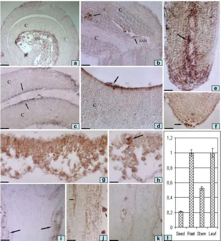
In situ hybridization was performed on seeds imbibed for 96 h and aerial parts of
wt plants grown in LD conditions for 28 days as described above and harvested 12 h after the onset of illumination. Imbibed seeds and tissues (leaves and stems) excised from adult plants were fixed, dehydrated, embedded in paraffin, cut into 8 µm sections and hybridized (55°C) to a digoxigenin-labelled antisense probe as described by Canas et al., (Canas et al., 1994). A gene-specific cDNA fragment of 265 bp was used for the synthesis of the digoxygenin-labelled probe. In parallel, RNA from seeds, leaves, stems and roots was used to monitor CRY-DASH transcription by qRT-PCR, following the procedures described above.


![Fig. 2 ) [3] . All the above mentioned residues are conserved in the corresponding positions of tomato CRY-DASH, suggest-ing that a possible DNA bindsuggest-ing activity could also occur for the tomato protein](https://thumb-eu.123doks.com/thumbv2/123dokorg/2880427.10287/44.892.166.726.780.1040/mentioned-residues-conserved-corresponding-positions-possible-bindsuggest-activity.webp)