Research Article
Prognostic Value of
18
F-Fluorocholine PET Parameters in
Metastatic Castrate-Resistant Prostate Cancer Patients
Treated with Docetaxel
E. Quaquarini ,
1,2D. D’Ambrosio ,
3F. Sottotetti ,
1F. Gallivanone ,
4M. Hodolic,
5,6P. Baiardi ,
7R. Palumbo ,
1C. Vellani ,
8C. Canevari ,
9A. Bernardo ,
1I. Castiglioni ,
4C. Porta,
10,11and G. Trifir `o
81Medical Oncology Unit, ICS Maugeri SpA SB-IRCCS, Pavia 27100, Italy
2University of Pavia, Ph.D. in Experimental Medicine, Pavia 27100, Italy
3Medical Physics Unit, ICS Maugeri SpA SB-IRCCS, Pavia 27100, Italy
4Institute of Molecular Bioimaging and Physiology, National Research Council (IBFM-CNR),
Milan 20090, Italy
5Nuclear Medicine Research Department, Iason, Graz, Austria
6Nuclear Medicine Department, Faculty of Medicine and Dentistry, Palack´y University Olomouc,
Olomouc, Czech Republic
7Scientific Direction, ICS Maugeri SpA SB-IRCCS, Pavia 27100, Italy
8Nuclear Medicine Unit, ICS Maugeri SpA SB-IRCCS, Pavia 27100, Italy
9Nuclear Medicine Unit, IRCCS San Raffaele Scientific Institute, Milan 20132, Italy
10Translational Oncology Unit, ICS Maugeri SpA SB-IRCCS, Pavia 27100, Italy
11University of Pavia, Department of Internal Medicine, Pavia 27100, Italy
Correspondence should be addressed to E. Quaquarini; [email protected] Received 7 December 2018; Accepted 19 February 2019; Published 26 March 2019 Academic Editor: Gaurav Malviya
Copyright © 2019 E. Quaquarini et al. This is an open access article distributed under the Creative Commons Attribution License, which permits unrestricted use, distribution, and reproduction in any medium, provided the original work is properly cited.
Background and Aim. The availability of new treatments for metastatic castrate-resistant prostate cancer (mCRPC) patients
increases the need for reliable biomarkers to help clinicians to choose the better sequence strategy. The aim of the present retrospective and observational work is to investigate the prognostic value of18F-fluorocholine (18F-FCH) positron emission tomography (PET) parameters in mCRPC. Materials and Methods. Between March 2013 and August 2016, 29 patients with mCRPC were included. They all received three-weekly docetaxel after androgen deprivation therapy, and they underwent18 F-FCH PET/computed tomography (CT) before and after the therapy. Semi-quantitative indices such as maximum standardized uptake value (SUVmax), mean standardized uptake value (SUVmean) with partial volume effect (PVC-SUV) correction, meta-bolically active tumour volume (MATV), and total lesion activity (TLA) with partial volume effect (PVC-TLA) correction were measured both in pre-treatment and post-treatment18F-FCH PET/CT scans for each lesion. Whole-body indices were calculated as sum of values measured for each lesion (SSUVmax, SPVC-SUV, SMATV, and STLA). Progression-free survival (PFS) and overall survival (OS) were considered as clinical endpoints. Univariate and multivariate hazard ratios for whole-body18F-FCH PET indices were performed, and p< 0.05 was considered as significant. Results. Cox regression analysis showed a statistically significant correlation between PFS, SMATV, and STLA. No correlations between OS and18F-FCH PETparameters were defined probably due to the small sample size. Conclusions. Semi-quantitative indices such as SMATV and STLA at baseline have a prognostic role in patients treated with docetaxel for mCRPC, suggesting a potential role of 18F-FCH PET/CT imaging in clinical decision-making.
Volume 2019, Article ID 4325946, 7 pages https://doi.org/10.1155/2019/4325946
1. Introduction
Prostate cancer (PC) is the first most common cancer in men worldwide, and its incidence is increasing in countries of higher socioeconomic development [1, 2].
The condition of metastatic castrate-resistant prostate cancer (mCRPC) has a poor outcome with a median overall survival (OS) of 20 months; thus, beyond palliation of symptoms and maintenance of a good quality of life, pro-longation of survival remains largely an elusive goal [3, 4]. During the last decades and particularly in the past few years, chemotherapeutic agents (docetaxel and cabazitaxel) [5], antiandrogen drugs (abiraterone and enzalutamide) [6, 7], and radium-223 dichloride have been introduced for the treatment of mCRPC [8]. The optimal timing of the different treatments has not yet been established, due to the paucity of prognostic markers for sequence and clinical management decisions.
During treatment of mCRPC, the use of PSA, a glyco-protein mainly produced by prostate tissue, as a marker of response should be carefully evaluated. PSA fluctuations are well known and described during active treatments, often not due to a disease progression but to the effects of drugs on PSA production. In addition, conventional radiology has limitation in detecting tumour biology and behaviour during treatment. Positron emission tomography/computed to-mography (PET/CT) has been widely used for the evaluation of PC by using several radiopharmaceuticals. A recent paper by Wallit and colleagues analyses the clinically available PET radiotracers for PC imaging and their mechanisms of ac-tions:18F-fluorodeoxyglucose (18F-FDG) for the evaluation of treatment response in metastatic bone disease; 18 F-sodium fluoride (18F-NaF) for bone metastasis detection;
11C-choline and 18F-choline (18F-FCH) for the staging of
high-risk patients and in presence of biochemical relapse with high PSA levels; 68Ga-labeled prostate-specific mem-brane antigen (68Ga-PSMA) highly sensitive for biochemical relapse with low PSA levels; the novel18F-fluciclovine (18 F-FACBC) appearing superior to choline in the setting of biochemical relapse [9–11].
There are several data in literature regarding the use of
11C-choline PET/CT in patients with PC treated with
docetaxel both in neadjuvant and advance setting [12, 13], but its role for the assessment of the treatment response and for predicting patient outcome still remains unclear.
In comparison to CT, 18F-FDG PET/CT, and 11 C-choline PET/CT, 18F-FCH PET/CT showed higher sensi-tivity and specificity in detection of metastatic lesions in patients with PC [14]. Its role in treatment monitoring and outcome prediction beyond PSA response of patients with mCRPC treated with abiraterone and enzalutamide has been already described [15–18].
To our knowledge, there are no data available on the use of 18F-FCH PET parameters as prognostic markers in pa-tients affected by PC and treated with docetaxel for advanced disease.
The aim of this study was to explore the prognostic role of 18F-FCH PET/CT in patients treated with three-weekly docetaxel for mCRPC by using accurate PET biomarkers corrected for partial volume effect (PVE) [19].
2. Materials and Methods
2.1. Patients and Study Design. The present study is a
ret-rospective, monocentric, observational trial that involved consecutive patients with a histological diagnosis of prostate cancer fulfilling PCWG3 criteria [20] for CRPC (baseline serum testosterone <50 ng/dl, progressive disease to an-drogen deprivation therapy).
This study was approved by the institutional ethics committee, and all patients signed written informed consent. The research was conducted according to the principles of the Declaration of Helsinki.
We considered eligible patients who had not yet received chemotherapy for advance disease, had a measurable disease according to PET Response Criteria in Solid Tumours (PERCIST) version 1.0 [21], and had an Eastern Cooperative Oncology Group (ECOG) performance status (PS) ≤ 2 and appropriate cardiac, hepatic, renal, and bone marrow function.
All patients underwent18F-FCH PET/CT before (PET1) and after (PET2) the programmed chemotherapy treatment. The study was conducted between March 2013 and August 2016. Data collection ended on 31st of December 2017 for analysis. The primary objective of this study is the identi-fication of18F-FCH PET parameters that can predict clinical outcome in patients with mCRPC. As secondary objective, we evaluated the correlations between patients’ clinical parameters and outcome.
2.1.1. Chemotherapy Protocol. Docetaxel was administered in
three-weekly schedule (75 mg/m2 day 1 every 21 days) as standard first-line chemotherapy for mCRCP according to the current guidelines [22]. The treatment was administered for a total of six cycles, and it was infused if clinical and bio-chemical parameters were permissive (conserved PS and no grade 3 or 4 adverse events according to Common Termi-nology Criteria for Adverse Events v4.03). Before the be-ginning of the therapy, patients underwent a baseline blood PSA assessment; PSA response and toxicity were evaluated before every dose administration. Dose adjustments and delays were planned to correspond with the type and grade of the observed toxicity. Concomitant medications such as antiemetic drug, granulocyte colony stimulating factors (G-CSF) for the secondary prevention of neutropenic fever, bisphosphonate treatments, and steroids were allowed.
2.1.2.18F-FCH PET/CT Procedure. PET/CT was performed
after intravenous injection of 18F-FCH (IASOcholine
®
, Graz, Austria), according to the body weight of the patient (about 3.5 MBq/kg). The mean radiopharmaceutical dose injected to the patients was 213 MBq (range: 176–346 MBq).18F-FCH PET/CT images were acquired in median 22 days
(range: 8–38) before starting the treatment with docetaxel and in median 26 days (range: 14–58) after the treatment.
Images’ acquisition was performed after an uptake time of approximately 60 min on a Discovery-690 VCT (General Electric Medical Systems, GEMS, Milwaukee, WI) scanner [23].
18F-FCH PET/CT data were acquired for 3 minutes per
bed position and reconstructed by using OSEM algorithm (3 iterations, 18 subsets, full width at half maximum (FWHM) of smoothing filter equals to 5 mm) including time of flight and resolution recovery.
2.2. 18F-FCH PET/CT Image Analysis and Interpretation.
A qualitative evaluation of 18F-FCH PET/CT images was firstly performed by two nuclear medicine physicians, and then an expert nuclear medicine physician selected the le-sions easily identifiable and accurately evaluable on18F-FCH PET/CT images before and after the chemotherapy in order to be semi-quantified. Too small lesions (volume < 1 cm) and lymph node clusters were excluded.
Semi-quantitative analysis of PET/CT lesions was per-formed by using a validated segmentation method [24].18 F-FCH PET parameters were extracted considering partial volume effect correction (PVC) in order to get accurate PET biomarkers by using a validated method developed by Gal-livanone and colleagues [25, 26]. More precisely, the following parameters were extracted from each segmented lesion on pre-treatment and post-treatment 18F-FCH PET images: maximum standardized uptake value (SUVmax), mean
stan-dardized uptake value (SUVmean) corrected for PVE
(PVC-SUV), metabolically active tumour volume (MATV), and total lesion activity (TLA). TLA was calculated as MATV multiplied by the PVC-SUV (TLA � MATV × PVC-SUV), and thus, TLA was corrected for PVE.
In order to extract whole-body indices for each patient and each 18F-FCH PET parameter (SUVmax, PVC-SUV,
MATV, and TLA), the sum of values measured for each lesion was calculated (SSUVmax, SPVC-SUV, SMATV, and
STLA, respectively).
2.3. Statistical Analysis. The distributions of the categorical
variables are described by counts and frequencies or by me-dian and range, whereas those of continuous and count variables are described by median and interquartile range. PFS was defined as the time between the date of the beginning of docetaxel and the date of progression. OS was defined as the time between the date of the starting of docetaxel and the date of death or last follow-up.
Univariate and multivariate hazard ratios for selected potential predictors of PFS and OS were performed using a Cox proportional hazards regression model. PFS and OS were estimated using the Kaplan–Meier method.
p < 0.05 was considered as significant for all analyses.
All statistical analyses were performed using SPSS version 24.
3. Results
3.1. Patients and Study Design. We enrolled 29 patients,
whose baseline characteristics are reported in Table 1. Pa-tients’ median age at the beginning of docetaxel treatment was 71 (range: 42–82). In a median follow-up period of six years, 14% (n � 4) patients obtained a complete metabolic response (CMR); 52% (n � 15) a partial metabolic response;
7% (n � 2) a stable metabolic disease; 27% (n � 8) a pro-gressive metabolic disease (PMD) as best response to treatment. Median PFS was 13.5 months (range 2.3– 37.6 months), and median OS was 37 months (range 4.7– 66 months). A PSA increase compared to baseline was seen in 5 patients (17%), whereas a PSA decline ≥50% was seen in 14 patients (47%). At the time of the analysis, 15 patients were still alive.
3.2. 18F-FCH PET/CT Image Analysis and Interpretation.
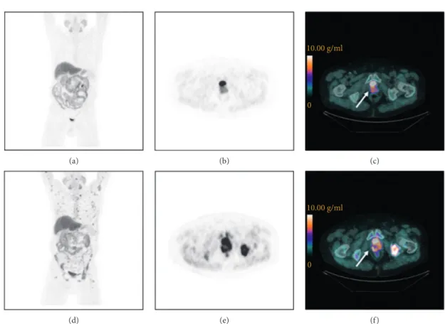
Seventy-one metastatic lesions were analysed, and semi-quantitative parameters were measured. Whole-body in-dices calculated for each patient before and after treatment are summarized in Table 2. Overall, 10% of patients had an SPVC-SUV reduction <25%, 24% had 25–74% reduction, and 10.3% had ≥75% reduction; 14% had an SSUVmax reduction <25%, 34% had 25–74% reduction, and 17% had ≥75% reduction; 17% had an SMATV reduction <25%, 38% had 25–74% reduction, and 17% had ≥75% reduction; and 21% had an STLA reduction <25%, 35% had 25–74% re-duction, and 17% had ≥75% reduction. Figures 1 and 2 represent an example of PET/CT images showing a typical
18F-FCH distribution in a patient with PMD and CMR,
respectively.
3.3. Statistical Analysis. As concerning the primary
objec-tive, at Cox regression analysis, SMATV and STLA18F-FCH PET parameters were significantly correlated with PFS (HR � 1.069, 95% CI: 1.06–1.09, p � 0.005 and HR 1.04, 95%
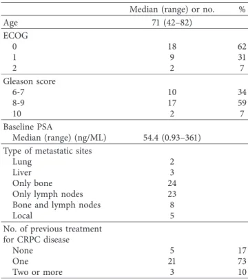
Table 1: Demographic and clinical characteristics of patients with CRPC at baseline (n � 29).
Median (range) or no. %
Age 71 (42–82) ECOG 0 18 62 1 9 31 2 2 7 Gleason score 6-7 10 34 8-9 17 59 10 2 7 Baseline PSA Median (range) (ng/ML) 54.4 (0.93–361) Type of metastatic sites
Lung 2
Liver 3
Only bone 24
Only lymph nodes 23
Bone and lymph nodes 8
Local 5
No. of previous treatment for CRPC disease
None 5 17
One 21 73
(a) (b) 10.00 g/ml 0 (c) (d) (e) 10.00 g/ml 0 (f)
Figure 1:18F-FCH PET/CT images of a mCRPC patient (83 years old) with a PMD after docetaxel treatment. Baseline (a–c) and post-treatment (d–f ) images. Maximum intensity projection images (a, d), transaxial slice of PET images (b, e), and PET/CT images (c, f ) showing the prostate (arrows): pre-treatment SUVmax�8.8 g/ml (c) and post-treatment SUVmax�12.0 g/ml (f ).
Table 2: Whole-body semi-quantitative18F-FCH PET parameters before (PET1) and after (PET2) treatment (median (IQR)).
PET1 PET2 SPVC-SUV (g/cc) 21.3 (10.8–31.2) 9.1 (4.2–26.5) SSUVmax(g/cc) 26.2 (11.2–29.9) 8.8 (3.8–22.7) SMATV (cc) 27.1 (12.1–48.0) 14.2 (1.4–32.3) STLA (g) 253.5 (94.7–469.8) 118.9 (12.3–251.4) (a) (b) 10.00 g/ml 0 (c) Figure 2: Continued.
CI: 1.02–1.08, p � 0.012, respectively). No statistical sig-nificant correlations were found with OS (Table 3).
Since there was a statistically significant association between SMATV and PFS, a ROC curve analysis was performed, and it showed that patients with an SMATV value >27 cc at PET1 have a 20% higher probability of having progression during docetaxel treatment (HR 1.19, range 0.56–2.53, p � 0.63) with a sensibility value of 73%, a specificity value of 58%, and an AUC of 0.64 (p � 0.23).
As regarding the secondary objectives, a significant correlation between PSA and PFS was found since patients with a PSA decline ≥50% had a better outcome (median PFS 12.8 months, range 9.5–15.8) than patients with a PSA de-cline <50% (median PFS 9.7 months, range 9.5–13.7) (log rank test � p < 0.001). Patients with a PSA increase of during docetaxel treatment had a poor outcome (median PFS 6.2 months, range 5.1–7.2, p � 0.036).
PSA decline was also correlated with OS since patients with a PSA decline ≥50% had a median OS of 42 months (range 34–49.53), patients with a PSA decline <50% had a median OS of 29.6 months (range 26.7–32.4), and patients with a PSA increase of had a reduced OS (median OS 26.3 months, range 25.8–26.7). The overall log-rank test, however, resulted not significant (p � 0.56) maybe due to the small number of analysed events.
Patients with an age ≥65 years at the time of the starting of docetaxel treatment had a better outcome than younger ones (PFS 10.9 months, range 7.7–14.2, vs 6.6 months, range 4.8– 8.3, respectively, p � 0.054; OS 42 months, range 33.2–50.7, vs 28.5 months, range 22.5–34.4, respectively, p � 0.076).
Considering the metastatic sites, patients with bone and lymph node lesions had a better outcome than patients with visceral involvement (PFS 11.6 months, range 9.6–13.3, vs 9.1 months, range 5.2–12.9, respectively, p � 0.04; OS 38.4 months, range 34.8–41.9, vs 29.4 months, range 5.2– 53.9, respectively, p � 0.25).
4. Discussion
To our knowledge, this is the first study that evaluated the role of 18F-FCH PET uptake before and after docetaxel treatment as a means of predicting long-term clinical out-comes in mCRPC.
Docetaxel is one of the treatment options in patients affected by metastatic PC; however, no approved biomarkers can predict the outcome to this therapy.11C-Choline and
18F-FCH PET/CTare widely used diagnostic techniques, and
recent studies have evaluated the role of18F-FCH PET in-dices in predicting treatment outcomes in CRPC patients [15–18, 27]. For the first time, Kwee and colleagues [28] assessed the potential usefulness of18F-FCH PET parameters in mCRPC patients, quantifying whole-body tumour burden on the basis of SUVmax, metabolic tumour volume (MTV),
and TLA. They found that MTV and TLA measurements proved to strongly correlate in the Kaplan–Meyer analysis. Afterwards, Caroli and colleagues retrospectively eval-uated 18F-FCH PET parameters in 94 patients treated with enzalutamide or abiraterone for mCRPC [16]. At univariate analysis, they described that the median sum of MTV (SMTV), SUVmax (SSUVmax), and TLA (STLA) resulted
significant for OS and PFS, whereas, in multivariate analysis,
Table 3: Cox regression analysis for progression-free survival and overall survival according to PET parameters. Cox regression analysis
Parameters PFS OS
HR 95% CI p HR 95% CI p
SPVC-SUV18F-FCH PET/CT 1.025 0.99–1.05 0.12 1.07 0.96–1.05 0.76
SSUVmax18F-FCH PET/CT 1.022 0.99–1.05 0.118 1.03 0.97–1.03 0.85
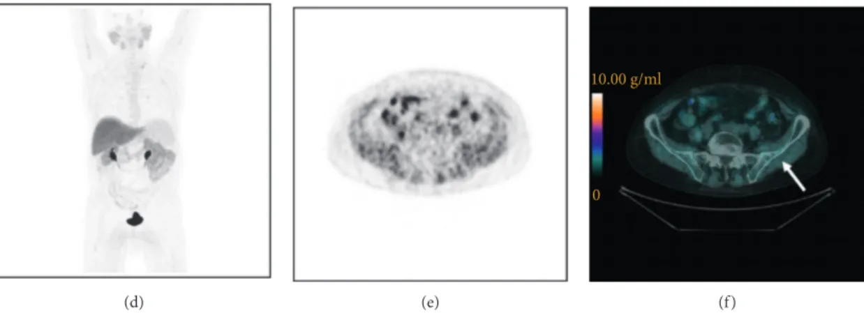
SMATV18F-FCH PET/CT 1.069 1.06–1.09 0.005 1.01 0.99–1.02 0.09 STLA18F-FCH PET/CT 1.04 1.02–1.08 0.012 1.0 0.99–1.01 0.38 (d) (e) 10.00 g/ml 0 (f )
Figure 2:18F-FCH PET/CT images of a mCRPC patient (73 years old) who underwent prostatectomy and obtained a CMR to docetaxel treatment. Baseline (a– c) and post-treatment (d–f ) images. Maximum intensity projection images (a, d), transaxial slice of PET images (b, e), and PET/CT images (c, f ) showing a metastasis in the left iliac bone (arrows) before docetaxel treatment: SUVmax�14.6 g/ml (c); after treatment, metastasis disappeared.
only STLA remained statistically significant with an HR � 1.49 for PFS (95% CI 1.24–1.78, p < 0.001) and an HR � 1.46 for OS (95% CI 1.16–1.84, p � 0.001).
Another study by Maines and colleagues enrolled 30 patients treated with enzalutamide and demonstrated, in multivariate analysis, a statistically significant correlation between baseline mean SUVmax and PFS and between
baseline mean SUVmaxand OS: patients with higher baseline
mean SUVmaxexperienced reduced PFS and OS than those
with lower values (median PFS: 4 vs 8 months; HR: 1.22; 95% CI: 1.09–1.37; p < 0.0001; median OS: 12 months vs not reached, HR: 1.21; 95% CI: 1.01–1.44; p � 0.03) [17].
De Giorgi and colleagues evaluated the utility of 18 F-FCH PET parameters to detect an early response to abir-aterone [15]. The authors concluded that a radiologic re-sponse to 18F-FCH PET/CT was associated to a better outcome compared to having obtained only a biochemical response.
Recently, Ceci and colleagues [29] have assessed the role of 11C-choline PET/CT to determine the response to docetaxel in a cohort of 61 patients with metastatic PC. The authors compared the radiologic response obtained with
11C-choline PET/CT and PSA response. The study had
demonstrated incongruent results between the two meth-odologies since a radiologic progression was observed in 44% of patients with a biochemical response.
Another study by Schwarzenb¨ock and collegues [13] evaluated the relationship between changes of SUVmaxand
SUVmean of 11C-choline PET as a predictive biomarker of
early and late response to docetaxel treatment in mCRCP. However, they did not find any significant correlation be-tween the changes in choline uptake and the objective re-sponses evaluated with RECIST and clinical criteria.
The results from our study suggest that18F-FCH PET parameters could be used to predict the clinical outcome of patients with mCRPC treated with docetaxel after pro-gressing to androgen-deprivation therapy. In particular, SMATV and STLA18F-FCH PET parameters are the most promising imaging biomarkers, taking into account the metabolic tumour volume and activity. In fact, their mean baseline values seem to predict long-term clinical outcomes, thus suggesting that metabolic imaging may be useful to select the best treatment for individual patients and open new perspectives in clinical decision-making. Imaging biomarkers may help to tailor treatments as patients with higher levels of basal metabolic activity (and therefore a poorer prognosis) may benefit from more aggressive treatments. In this context, it is still uncertain whether pre-treatment metabolic imaging may also play a predictive role. Our study benefits also from the use of a validated method to obtain tumour metabolic volume. As underlined in different publications and in particular in a work by Soret and colleagues [19], proper tumour active metabolic region assessment is paramount because it influences the mea-surement of different semi-quantitative indices to be eval-uated as imaging biomarkers. In this study, we used a segmentation procedure that was validated on lesions that reliably simulate realistic tumour conditions (non-spherical and non-homogeneous), estimating volume with 92% of
accuracy [24]. Furthermore, in order to obtain accurate quantitative indices of glucose consumption, a PVC method was applied to quantitative indices ensuring an accuracy for quantification up to 93% for lesions >1 cm as sphere-equivalent diameter.
We did not find any correlation between18F-FCH PET parameters and survival. This is possibly due to the small sample size and the few number of events at the time of data analysis; as already well known from the literature, our findings demonstrated the prognostic role of visceral metastasis involvement.
5. Conclusions
Our findings suggest that18F-FCH PET parameters, such as SMATV and STLA at baseline, have a prognostic role in patients treated with docetaxel for mCRPC and may be more useful than commonly used PET indices such as SUVmean
and SUVmax. The study has some limitations due to the
retrospective nature, the small sample size, and the single institution setting. Further investigation and larger studies are needed in order to find a significant correlation between
18F-FCH PET indices and OS.
Data Availability
The 18F-FCH PET/TC data used to support the findings of this study are available from the corresponding author upon request.
Conflicts of Interest
The authors declare that there are no conflicts of interest regarding the publication of this paper.
References
[1] L. A. Torre, F. Bray, R. L. Siegel, J. Ferlay, J. Lortet-Tieulent, and A. Jemal, “Global cancer statistics, 2012,” CA: A Cancer
Journal for Clinicians, vol. 65, no. 2, pp. 87–108, 2015.
[2] M. C. S. Wong, W. B. Goggins, H. H. X. Wang et al., “Global incidence and mortality for prostate cancer: analysis of temporal patterns and trends in 36 countries,” European
Urology, vol. 70, no. 5, pp. 862–874, 2016.
[3] M. Kirby, C. Hirst, and E. D. Crawford, “Characterising the castration-resistant prostate cancer population: a systematic review,” International Journal of Clinical Practice, vol. 65, no. 11, pp. 1180–1192, 2011.
[4] S. Halabi, C.-Y. Lin, W. K. Kelly et al., “Updated prognostic model for predicting overall survival in first-line chemo-therapy for patients with metastatic castration-resistant prostate cancer,” Journal of Clinical Oncology, vol. 32, no. 7, pp. 671–677, 2014.
[5] D. A. Loblaw, C. Walker-Dilks, E. Winquist, and S. J. Hotte, “Systemic therapy in men with metastatic castration-resistant prostate cancer: a systematic review,” Clinical Oncology, vol. 25, no. 7, pp. 406–430, 2013.
[6] A. A. Azad, B. J. Eigl, R. Leibowitz-Amit et al., “Outcomes with abiraterone acetate in metastatic castration-resistant prostate cancer patients who have poor performance sta-tus,” European Urology, vol. 67, no. 3, pp. 441–447, 2015.
[7] D. L. Suzman, B. Luber, M. T. Schweizer, R. Nadal, and E. S. Antonarakis, “Clinical activity of enzalutamide versus docetaxel in men with castration-resistant prostate cancer progressing after abiraterone,” The Prostate, vol. 74, no. 13, pp. 1278–1285, 2014.
[8] O. Sartor, R. Coleman, S. Nilsson et al., “Effect of radium-223 dichloride on symptomatic skeletal events in patients with castration-resistant prostate cancer and bone metastases: results from a phase 3, double-blind, randomised trial,” The
Lancet Oncology, vol. 15, no. 7, pp. 738–746, 2014.
[9] K. L. Wallitt, S. R. Khan, S. Dubash, H. H. Tam, S. Khan, and T. D. Barwick, “Clinical PET imaging in prostate cancer,”
RadioGraphics, vol. 37, no. 5, pp. 1512–1536, 2017.
[10] E. E. Parent and D. M. Schuster, “Update on18F-fluciclovine PET for prostate cancer imaging,” Journal of Nuclear
Medi-cine, vol. 59, no. 5, pp. 733–739, 2018.
[11] C. Nanni, L. Zanoni, C. Pultrone et al., “18F-FACBC (anti1-amino-3-18F-fluorocyclobutane-1-carboxylic acid) versus 11C-choline PET/CT in prostate cancer relapse: results of a prospective trial,” European Journal of Nuclear Medicine and
Molecular Imaging, vol. 43, no. 9, pp. 1601–1610, 2016.
[12] S. M. Schwarzenb¨ock, A. Knieling, M. Souvatzoglou et al., “[11C]choline PET/CT in therapy response assessment of a neoadjuvant therapy in locally advanced and high risk prostate cancer before radical prostatectomy,” Oncotarget, vol. 7, no. 39, pp. 63747–63757, 2016.
[13] S. M. Schwarzenb¨ock, M. Eiber, G. Kundt et al., “Prospective evaluation of [11C]choline PET/CT in therapy response as-sessment of standardized docetaxel first-line chemotherapy in patients with advanced castration refractory prostate cancer,”
European Journal of Nuclear Medicine and Molecular Imaging,
vol. 43, no. 12, pp. 2105–2113, 2016.
[14] J. Liu, Z. Chen, T. Wang et al., “Influence of four radiotracers in PET/CT on diagnostic accuracy for prostate cancer: a bi-variate random-effects meta-analysis,” Cellular Physiology
and Biochemistry, vol. 39, no. 2, pp. 467–480, 2016.
[15] U. De Giorgi, P. Caroli, S. L. Burgio et al., “Early outcome prediction on 18F-fluorocholine PET/CT in metastatic castration-resistant prostate cancer patients treated with abiraterone,” Oncotarget, vol. 5, no. 23, pp. 12448–12458, 2014.
[16] P. Caroli, U. De Giorgi, E. Scarpi et al., “Prognostic value of 18F-choline PET/CT metabolic parameters in patients with metastatic castration-resistant prostate cancer treated with abiraterone or enzalutamide,” European Journal of Nuclear
Medicine and Molecular Imaging, vol. 45, no. 3, pp. 348–354,
2018.
[17] F. Maines, O. Caffo, D. Donner et al., “Serial18F-choline-PET imaging in patients receiving enzalutamide for metastatic castration-resistant prostate cancer: response assessment and imaging biomarkers,” Future Oncology, vol. 12, no. 3, pp. 333–342, 2016.
[18] U. De Giorgi, P. Caroli, E. Scarpi et al., “18F-fluorocholine PET/CT for early response assessment in patients with metastatic castration-resistant prostate cancer treated with enzalutamide,” European Journal of Nuclear Medicine and
Molecular Imaging, vol. 42, no. 8, pp. 1276–1283, 2015.
[19] M. Soret, S. L. Bacharach, and I. Buvat, “Partial-volume effect in PET tumor imaging,” Journal of Nuclear Medicine, vol. 48, no. 6, pp. 932–945, 2007.
[20] H. I. Scher, M. J. Morris, W. M. Stadler et al., “Trial design and objectives for castration-resistant prostate cancer: updated recommendations from the prostate cancer clinical trials
working group 3,” Journal of Clinical Oncology, vol. 34, no. 12, pp. 1402–1418, 2016.
[21] J. H. O, M. A. Lodge, and R. L. Wahl, “Practical PERCIST: a simplified guide to PET response criteria in solid tumors 1.0,”
Radiology, vol. 280, no. 2, pp. 576–584, 2016.
[22] P. Cornford, J. Bellmunt, M. Bolla et al., “EAU-ESTRO-SIOG guidelines on prostate cancer. part II: treatment of relapsing, metastatic, and castration-resistant prostate cancer,”
Euro-pean Urology, vol. 71, no. 4, pp. 630–642, 2017.
[23] V. Bettinardi, L. Presotto, E. Rapisarda, M. Picchio, L. Gianolli, and M. C. Gilardi, “Physical performance of the new hybrid PET/CT discovery-690,” Medical Physics, vol. 38, no. 10, pp. 5394–5411, 2011.
[24] F. Gallivanone, M. Interlenghi, C. Canervari, and I. Castiglioni, “A fully automatic, threshold-based segmen-tation method for the estimation of the metabolic tumor volume from PET images: validation on 3D printed an-thropomorphic oncological lesions,” Journal of In-strumentation, vol. 11, no. 1, article C01022, 2016.
[25] F. Gallivanone, C. Canevari, L. Gianolli et al., “A partial volume effect correction tailored for 18F-FDG-PET onco-logical studies,” BioMed Research International, vol. 2013, Article ID 780458, 12 pages, 2013.
[26] F. Gallivanone, C. Canevari, I. Sassi et al., “Partial volume corrected 18F-FDG PET mean standardized uptake value correlates with prognostic factors in breast cancer,” Quarterly
Journal of Nuclear Medicine and Molecular Imaging, vol. 58,
no. 9, pp. 424–439, 2014.
[27] O. Caffo, F. Maines, D. Donner, A. Veccia, F. Chierichetti, and E. Galligioni, “Impact of enzalutamide administration on primary prostate cancer volume: a metabolic evaluation by choline positron emission tomography in castration-resistant prostate cancer patients,” Clinical Genitourinary Cancer, vol. 12, no. 5, pp. 312–316, 2014.
[28] S. A. Kwee, J. Lim, A. Watanabe, K. Kromer-Baker, and M. N. Coel, “Prognosis related to metastatic burden measured by18F-fluorocholine PET/CT in castration-resistant prostate cancer,” Journal of Nuclear Medicine, vol. 55, no. 6, pp. 905–910, 2014.
[29] F. Ceci, P. Castellucci, T. Graziani et al., “11C-choline PET/CT in castration-resistant prostate cancer patients treated with docetaxel,” European Journal of Nuclear Medicine and
Stem Cells
International
Hindawi www.hindawi.com Volume 2018 Hindawi www.hindawi.com Volume 2018 INFLAMMATIONEndocrinology
International Journal ofHindawi www.hindawi.com Volume 2018 Hindawi www.hindawi.com Volume 2018
Disease Markers
Hindawi www.hindawi.com Volume 2018 BioMed Research InternationalOncology
Journal of Hindawi www.hindawi.com Volume 2013 Hindawi www.hindawi.com Volume 2018Oxidative Medicine and Cellular Longevity
Hindawi
www.hindawi.com Volume 2018
PPAR Research
Hindawi Publishing Corporation
http://www.hindawi.com Volume 2013 Hindawi www.hindawi.com
The Scientific
World Journal
Volume 2018 Immunology Research Hindawi www.hindawi.com Volume 2018 Journal ofObesity
Journal of Hindawi www.hindawi.com Volume 2018 Hindawi www.hindawi.com Volume 2018 Computational and Mathematical Methods in Medicine Hindawi www.hindawi.com Volume 2018Behavioural
Neurology
Ophthalmology
Journal of Hindawi www.hindawi.com Volume 2018Diabetes Research
Journal ofHindawi
www.hindawi.com Volume 2018
Hindawi
www.hindawi.com Volume 2018 Research and Treatment
AIDS
Hindawi
www.hindawi.com Volume 2018
Gastroenterology Research and Practice
Hindawi www.hindawi.com Volume 2018